interventional vascular radiology and interventional … · interventional vascular radiology j...
TRANSCRIPT

i
A Report of the National Confidential Enquiry
into Perioperative Deaths
Data collection period 1 April 1998 to 31 March 1999
Compiled by:
K G Callum MS FRCS
F Whimster MHM
Interventional Vascular
Radiology
and
Interventional Neurovascular
Radiology

ii
Published 21 November 2000 by the National Confidential Enquiry into Perioperative Deaths
35-43 Lincoln’s Inn Fields, London WC2A 3PNTel: (020) 7831 6430Fax: (020) 7430 2958
Email: [email protected]: www.ncepod.org.uk
Requests for further information should be addressed to the Chief Executive
ISBN 0 9522069 8 6
A company limited by guarantee Company number 3019382Registered charity number 1075588
This report is printed on paper produced from wood pulp originating from managed sustainableplantations and is chlorine-free, acid-free, recyclable and biodegradable.
Additional information
This report is available for downloading from the NCEPOD website at www.ncepod.org.uk
Copies can also be purchased from the NCEPOD office.
The analysis of data from questionnaires is not included in fullin this report. A supplement containing additional data, and copies of
the questionnaires, is available free of charge from the NCEPOD office.

iii
ACKNOWLEDGEMENTS
This report could not have been achieved without the support and cooperation of a wide range ofindividuals and organisations. Our particular thanks go to the following:
• The Royal College of Radiologists and the British Society of Interventional Radiologists for supportingthe concept of this study.
• The Local Reporters, whose names are listed in Appendices D and E.
• All those radiologists whose names are listed in Appendices F and G, who contributed to the Enquiry bycompleting questionnaires.
• The Advisors whose names are listed overleaf.
• Those bodies, whose names are listed in Appendix B, who provide the funding to cover the cost of theEnquiry, together with the Department of Health who provided additional support.
• Mr Chris Macklin, Surgical Registrar, Derbyshire Royal Infirmary, for the illustrations on the front cover.
The Steering Group, Clinical Coordinators and Chief Executive would also like to record their appreciationof the hard work and tolerance of the NCEPOD administrative staff: Peter Allison, Fatima Chowdhury, PaulCoote, Sheree Cornwall, Jennifer Drummond and Dolores Jarman.
The views expressed in this publication are those of NCEPOD and not necessarily those of the NationalInstitute for Clinical Excellence, or any other funding body.

iv

v
CLINICAL CONTRIBUTORS
NCEPOD COORDINATOR
K G Callum Clinical Coordinator, NCEPODand Consultant General and VascularSurgeon, Derbyshire Royal Infirmary
SPECIALTY ADVISORS
Interventional vascular radiology
J Dyet Hull Royal Infirmary and RoyalCollege of Radiologists’ representative
on NCEPOD Steering Group
P A Gaines Northern General Hospital,Sheffield
I Gillespie Royal Infirmary of Edinburgh
L C Johnston Belfast City Hospital
M Ruttley University Hospital of Wales (retired)
Interventional neurovascular radiology
A Gholkar Newcastle General Hospital
A J Molyneux The Radcliffe Infirmary, Oxford

vi

vii
Recommendations . . . . . . . . . . . . . . . . . . . . . . . . . . . . . . . . . . . . . . . . . . . . . . . . . . . . . . . . . . . .1
Introduction . . . . . . . . . . . . . . . . . . . . . . . . . . . . . . . . . . . . . . . . . . . . . . . . . . . . . . . . . . . . . . . . . . . . . . . .3Data collection . . . . . . . . . . . . . . . . . . . . . . . . . . . . . . . . . . . . . . . . . . . . . . . . . . . . . . . . . . . . . . . . . . . . .3General data analysis . . . . . . . . . . . . . . . . . . . . . . . . . . . . . . . . . . . . . . . . . . . . . . . . . . . . . . . .4
Monthly returns of procedures performed . . . . . . . .4Reported procedures . . . . . . . . . . . . . . . . . . . . . . . . . . . . . . . . . . . . . . . . . . . . . . . . .4Reported deaths . . . . . . . . . . . . . . . . . . . . . . . . . . . . . . . . . . . . . . . . . . . . . . . . . . . . . . . . . . .5Distribution, return and analysis of questionnaires . . . . . . . . . . . . . . . . . . . . . . . . . . . . . . . . . . . . . . . . . . . . . . . . . . . . . . . .7Procedures . . . . . . . . . . . . . . . . . . . . . . . . . . . . . . . . . . . . . . . . . . . . . . . . . . . . . . . . . . . . . . . . . . . . .8
Patient profile . . . . . . . . . . . . . . . . . . . . . . . . . . . . . . . . . . . . . . . . . . . . . . . . . . . . . . . . . . . . . . . . . . .10Urgency of procedure . . . . . . . . . . . . . . . . . . . . . . . . . . . . . . . . . . . . . . . . . . . . .10Fitness of the patient . . . . . . . . . . . . . . . . . . . . . . . . . . . . . . . . . . . . . . . . . . . . . . . .11
Specialty and experience of the medical team . . . . . .13Specialty of the clinical team . . . . . . . . . . . . . . . . . . . . . . . . . . . . . . . . .13Radiologist’s assessment prior to procedure . . . . . .14Specialty and seniority of radiologist . . . . . . . . . . . . . . . . .14
Facilities, personnel and monitoring . . . . . . . . . . . . . . . . . . . . . . . .15Dedicated room . . . . . . . . . . . . . . . . . . . . . . . . . . . . . . . . . . . . . . . . . . . . . . . . . . . . . . . . .15Equipment . . . . . . . . . . . . . . . . . . . . . . . . . . . . . . . . . . . . . . . . . . . . . . . . . . . . . . . . . . . . . . . . . . .15Shortage of personnel . . . . . . . . . . . . . . . . . . . . . . . . . . . . . . . . . . . . . . . . . . . . .16Delays . . . . . . . . . . . . . . . . . . . . . . . . . . . . . . . . . . . . . . . . . . . . . . . . . . . . . . . . . . . . . . . . . . . . . . . . . . . .16Anaesthesia . . . . . . . . . . . . . . . . . . . . . . . . . . . . . . . . . . . . . . . . . . . . . . . . . . . . . . . . . . . . . . . . . .16Monitoring . . . . . . . . . . . . . . . . . . . . . . . . . . . . . . . . . . . . . . . . . . . . . . . . . . . . . . . . . . . . . . . . . .17Care following the procedure . . . . . . . . . . . . . . . . . . . . . . . . . . . . . . .17
Recovery . . . . . . . . . . . . . . . . . . . . . . . . . . . . . . . . . . . . . . . . . . . . . . . . . . . . . . . . . . . . . . . . .17Intensive and high dependency care . . . . . . . . . . .18
Complications . . . . . . . . . . . . . . . . . . . . . . . . . . . . . . . . . . . . . . . . . . . . . . . . . . . . . . . . . . . . . . . . . . .18Specific problems . . . . . . . . . . . . . . . . . . . . . . . . . . . . . . . . . . . . . . . . . . . . . . . . . . . . . .19
Level of puncture . . . . . . . . . . . . . . . . . . . . . . . . . . . . . . . . . . . . . . . . . . . . . . .19Lower limb revascularisation . . . . . . . . . . . . . . . . . . . . . . . . . .20Thrombolysis . . . . . . . . . . . . . . . . . . . . . . . . . . . . . . . . . . . . . . . . . . . . . . . . . . . . . . . .21Embolisation . . . . . . . . . . . . . . . . . . . . . . . . . . . . . . . . . . . . . . . . . . . . . . . . . . . . . . . . .22SVC stent . . . . . . . . . . . . . . . . . . . . . . . . . . . . . . . . . . . . . . . . . . . . . . . . . . . . . . . . . . . . . . .23Central venous access . . . . . . . . . . . . . . . . . . . . . . . . . . . . . . . . . . . . . . . .23Transjugular intrahepaticportosystemic shunt (TIPS) . . . . . . . . . . . . . . . . . . . . . . . . . . . . .23DVT prophylaxis . . . . . . . . . . . . . . . . . . . . . . . . . . . . . . . . . . . . . . . . . . . . . . . . .23
Postmortem examinations . . . . . . . . . . . . . . . . . . . . . . . . . . . . . . . . . . . . . . . . . . . .24
Audit and availability of patient records . . . . . . . . . . . . . . . . .25
1 Interventional Vascular Radiology
Foreword . . . . . . . . . . . . . . . . . . . . . . . . . . . . . . . . . . . . . . . . . . . . . . . . . . . . . . . . . . . . . . . . . . . . . . . . . . . . .xi
2 Interventional NeurovascularRadiology
Recommendations . . . . . . . . . . . . . . . . . . . . . . . . . . . . . . . . . . . . . . . . . . . . . . . . . . . . . . . . . .27
Introduction . . . . . . . . . . . . . . . . . . . . . . . . . . . . . . . . . . . . . . . . . . . . . . . . . . . . . . . . . . . . . . . . . . . . . .29Data collection . . . . . . . . . . . . . . . . . . . . . . . . . . . . . . . . . . . . . . . . . . . . . . . . . . . . . . . . . . . . . . . . . .30General data analysis . . . . . . . . . . . . . . . . . . . . . . . . . . . . . . . . . . . . . . . . . . . . . . . . . . . . .30
Monthly returns of procedures performed . . . . . . . . .30Reported deaths . . . . . . . . . . . . . . . . . . . . . . . . . . . . . . . . . . . . . . . . . . . . . . . . . . . . . . . .31Distribution of deaths . . . . . . . . . . . . . . . . . . . . . . . . . . . . . . . . . . . . . . . . . . . . . .32Distribution, return and analysisof questionnaires . . . . . . . . . . . . . . . . . . . . . . . . . . . . . . . . . . . . . . . . . . . . . . . . . . . . . . .33Procedures . . . . . . . . . . . . . . . . . . . . . . . . . . . . . . . . . . . . . . . . . . . . . . . . . . . . . . . . . . . . . . . . . . .34
Patient profile . . . . . . . . . . . . . . . . . . . . . . . . . . . . . . . . . . . . . . . . . . . . . . . . . . . . . . . . . . . . . . . . . . .35Urgency of procedure . . . . . . . . . . . . . . . . . . . . . . . . . . . . . . . . . . . . . . . . . . . . .35Fitness of the patient . . . . . . . . . . . . . . . . . . . . . . . . . . . . . . . . . . . . . . . . . . . . . . . .36
Facilities, personnel and monitoring . . . . . . . . . . . . . . . . . . . . . . . .38Seniority and specialty of the radiologist . . . . . . . . . .38Dedicated room . . . . . . . . . . . . . . . . . . . . . . . . . . . . . . . . . . . . . . . . . . . . . . . . . . . . . . . . .38Monitoring . . . . . . . . . . . . . . . . . . . . . . . . . . . . . . . . . . . . . . . . . . . . . . . . . . . . . . . . . . . . . . . . .38Anaesthesia . . . . . . . . . . . . . . . . . . . . . . . . . . . . . . . . . . . . . . . . . . . . . . . . . . . . . . . . . . . . . . . . . .38Care following the procedure . . . . . . . . . . . . . . . . . . . . . . . . . . . . . . .39
Intensive and high dependency care . . . . . . . . . . .39
Complications . . . . . . . . . . . . . . . . . . . . . . . . . . . . . . . . . . . . . . . . . . . . . . . . . . . . . . . . . . . . . . . . . . .39
Postmortem examinations . . . . . . . . . . . . . . . . . . . . . . . . . . . . . . . . . . . . . . . . . . . .40
Audit and quality . . . . . . . . . . . . . . . . . . . . . . . . . . . . . . . . . . . . . . . . . . . . . . . . . . . . . . . . . . . . .40Audit . . . . . . . . . . . . . . . . . . . . . . . . . . . . . . . . . . . . . . . . . . . . . . . . . . . . . . . . . . . . . . . . . . . . . . . . . . . . .40Quality of questionnaires . . . . . . . . . . . . . . . . . . . . . . . . . . . . . . . . . . . . . .40Standard of care . . . . . . . . . . . . . . . . . . . . . . . . . . . . . . . . . . . . . . . . . . . . . . . . . . . . . . . .40
REFERENCES . . . . . . . . . . . . . . . . . . . . . . . . . . . . . . . . . . . . . . . . . . . . . . . . . . . . . . . . . .41
APPENDICES
A Abbreviations . . . . . . . . . . . . . . . . . . . . . . . . . . . . . . . . . . . . . . . . . . . . . . . . . . . . . . . . . . . . . .43B NCEPOD corporate structure . . . . . . . . . . . . . . . . . . . . . . . . . . . . . .45C Data collection and review methods . . . . . . . . . . . . . . . . . . .47D Local Reporters – Interventional
Vascular Radiology . . . . . . . . . . . . . . . . . . . . . . . . . . . . . . . . . . . . . . . . . . . . . . . . . . .49E Local Reporters – Interventional
Neurovascular Radiology . . . . . . . . . . . . . . . . . . . . . . . . . . . . . . . . . . . . . . .53F Participants – Consultant
Vascular Radiologists . . . . . . . . . . . . . . . . . . . . . . . . . . . . . . . . . . . . . . . . . . . . . . .55G Participants – Consultant
Neurovascular Radiologists . . . . . . . . . . . . . . . . . . . . . . . . . . . . . . . . . . .57
CONTENTS

viii

ix
1 Interventional Vascular Radiology
General data analysis
Figure1.1: Monthly returns of total procedures performed . . . . . .4
Table 1.1: Monthly returns by region . . . . . . . . . . . . . . . . . . . . . . . . . . . . . . . .4
Figure 1.2: Total deaths reported . . . . . . . . . . . . . . . . . . . . . . . . . . . . . . . . . . . . . .5
Table 1.2: Inappropriate reports received and excluded . . . . . . . .5
Table 1.3: Deaths reported to NCEPOD by region . . . . . . . . . . . . . .5
Figure 1.3: Calendar days from procedure to death . . . . . . . . . . . . .6
Figure 1.4: Age/sex distribution of reported deaths . . . . . . . . . . . . . .6
Figure 1.5: Distribution, return and analysis of questionnaires . . . . . .7
Table 1.4: Reasons for exclusion of questionnaires
from analysis . . . . . . . . . . . . . . . . . . . . . . . . . . . . . . . . . . . . . . . . . . . . . . . .7
Figure 1.6: Reasons for non-return of questionnaires . . . . . . . . . . . . . .7
Table 1.5: Regional distribution, return and analysis rates . . . . . .8
Table 1.6: Endovascular interventions (arterial) . . . . . . . . . . . . . . . . . . . .8
Table 1.7: Endovascular interventions (venous) . . . . . . . . . . . . . . . . . . . .9
Table 1.8: Other interventions . . . . . . . . . . . . . . . . . . . . . . . . . . . . . . . . . . . . . . . . .9
Patient profile
Figure 1.7: Day of the procedure . . . . . . . . . . . . . . . . . . . . . . . . . . . . . . . . . . .10
Figure 1.8: Admission category . . . . . . . . . . . . . . . . . . . . . . . . . . . . . . . . . . . . . .10
Figure 1.9: ASA status . . . . . . . . . . . . . . . . . . . . . . . . . . . . . . . . . . . . . . . . . . . . . . . . .11
Table 1.9: Coexisting medical problems . . . . . . . . . . . . . . . . . . . . . . . . . .11
Table 1.10: Anticipated risk of death related to
the proposed procedure . . . . . . . . . . . . . . . . . . . . . . . . . . . . . . . .12
Specialty and experience of the medical team
Table 1.11: Specialty of consultant surgeon
under whose care the patient was
at the time of the procedure . . . . . . . . . . . . . . . . . . . . . . . . . . .13
Table 1.12: Specialty of consultant physician
under whose care the patient was
at the time of the procedure . . . . . . . . . . . . . . . . . . . . . . . . . . .13
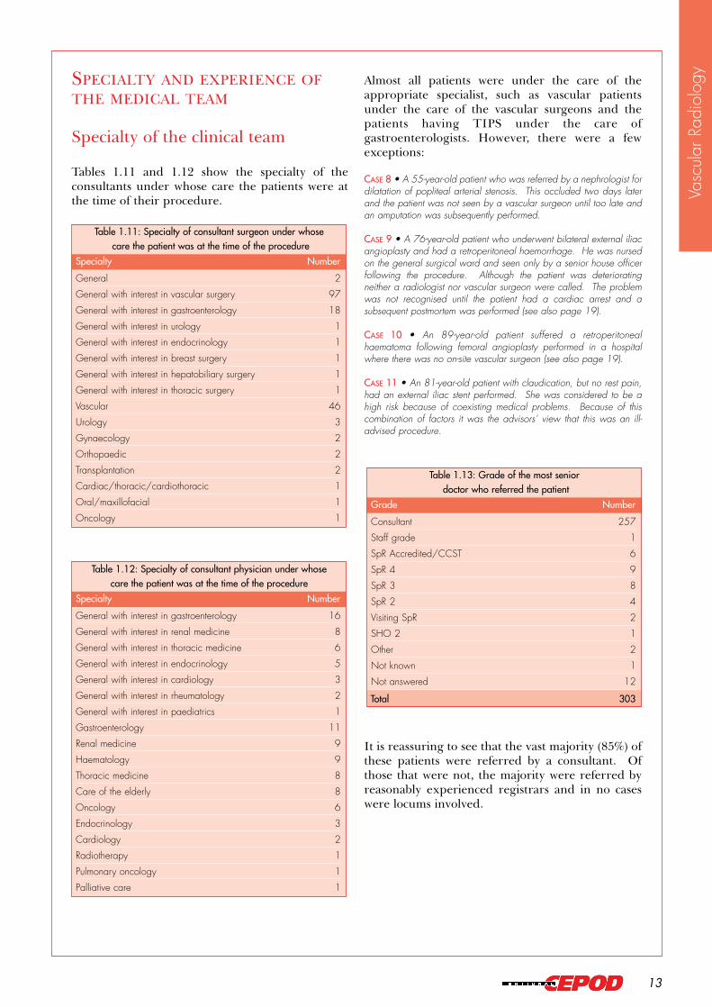
Table 1.13: Grade of the most senior doctor who
referred the patient . . . . . . . . . . . . . . . . . . . . . . . . . . . . . . . . . . . . . .13
Table 1.14: Grade of the most senior radiologist who
reviewed the patient before the procedure . . . . . . . . .14
Table 1.15: Specialty of most senior radiologist present . . . . . . . .14
Table 1.16: Grade of most senior doctor
performing the procedure . . . . . . . . . . . . . . . . . . . . . . . . . . . . . .14
Question 1.1: If the most senior operator was not a
consultant,was a more senior
doctor immediately available? . . . . . . . . . . . . . . . . . . . . . . . .14
Facilities, personnel and monitoring
Table 1.17: Location in which the procedure was performed . . . .15
Question 1.2: Was there a shortage of personnel in this case? . . . .16
Question 1.3: Were there any delays (between admission
and procedure) due to factors other
than clinical? . . . . . . . . . . . . . . . . . . . . . . . . . . . . . . . . . . . . . . . . . . . . . .16
Table 1.18: Reasons for non-clinical delay between
admission and procedure . . . . . . . . . . . . . . . . . . . . . . . . . . . . . .16
Question 1.4: Was the procedure performed solely
under local anaesthetic or sedation
administered by the operator? . . . . . . . . . . . . . . . . . . . . . . . .16
Table 1.19: Monitoring performed during or
immediately after the procedure . . . . . . . . . . . . . . . . . . . . . .17
Table 1.20: Responsibility for monitoring the patient’s
general condition during the procedure . . . . . . . . . . . . .17
Question 1.5: Was there a recovery room/area available
attached to the procedure suite? . . . . . . . . . . . . . . . . . . . . . .17
Complications
Table 1.21: Unexpected procedural complications . . . . . . . . . . . . . . . . . .18
Table 1.22: Postprocedural complications . . . . . . . . . . . . . . . . . . . . . . . . . . . . .19
Question 1.6: Were any measures taken (before, during
or after the procedure) to prevent
venous thromboembolism? . . . . . . . . . . . . . . . . . . . . . . . . . . . . . . . .23
Postmortem examinations
Question 1.7: Did the pathological information
confirm the team’s clinical impression? . . . . . . . . . . . . . . . . .24
TABLES, FIGURES AND
QUESTIONS

x
2 Interventional NeurovascularRadiology
General data analysis
Figure 2.1: Monthly returns of total procedures performed . . . . .30
Table 2.1: Monthly returns by region . . . . . . . . . . . . . . . . . . . . . . . . . . . . . . . .30
Figure 2.2: Total deaths reported . . . . . . . . . . . . . . . . . . . . . . . . . . . . . . . . . . . . . .31
Table 2.2: Deaths reported to NCEPOD by region . . . . . . . . . . . . .31
Figure 2.3: Calendar days from procedure to death . . . . . . . . . . . .32
Figure 2.4: Age/sex distribution of reported deaths . . . . . . . . . . . . .33
Figure 2.5: Distribution, return and analysis of questionnaires . . . .33
Table 2.3: Regional distribution and return rates . . . . . . . . . . . . . . . .33
Table 2.4: Neurovascular interventions . . . . . . . . . . . . . . . . . . . . . . . . . . . . .34
Patient profile
Figure 2.6: Day of the procedure . . . . . . . . . . . . . . . . . . . . . . . . . . . . . . . . . . .35
Figure 2.7: Admission category . . . . . . . . . . . . . . . . . . . . . . . . . . . . . . . . . . . . . .36
Table 2.5: Coexisting medical problems . . . . . . . . . . . . . . . . . . . . . . . . . .37
Figure 2.8: ASA status . . . . . . . . . . . . . . . . . . . . . . . . . . . . . . . . . . . . . . . . . . . . . . . . .37
Table 2.6: Modified Glasgow Coma Scale (GCS) . . . . . . . . . . . .37
Facilities, personnel and monitoring
Table 2.7: Specialty of the most senior radiologist present . . . . . .38
Question 2.1: Was there non-medical help with anaesthesia? . . . . .38
Complications
Table 2.8: Postprocedural complications . . . . . . . . . . . . . . . . . . . . . . . . . .39
Postmortem examinations
Question 2.2: Did the pathological information confirm
the team’s clinical impression? . . . . . . . . . . . . . . . . . . . . . . . .40
Table 2.9: Specialty of the pathologist who performed
the postmortem examination . . . . . . . . . . . . . . . . . . . . . . . . . . .40

xi
FOREWORD
Significant advances in interventional techniques,particularly in vascular and neurovascularradiology, in the last decade have led NCEPOD toexplore the morbidity and mortality associated withsuch procedures. It should be appreciated that thisis a new area of investigation, but in view of thefrequency with which these minimally invasivetechniques are being carried out, it is important thatthe consequences of such interventions should beinvestigated. Furthermore, this is an area of teamworking which has developed very significantly, therelationships between members of that team andthe role that each play are highlighted in aninvestigation of this type. There is a need tounderstand the potential roles that each member ofthe team can play and the responsibilities that eachshould take at different stages in the care of thepatient.
It is fundamental to the development of newtechniques that adequate facilities should beavailable. What is highlighted in this report,therefore, is the necessity for interventionalradiologists and surgeons to have not only sufficientexperience and expertise, but also the facilities andequipment with which to carry out their tasks in assafe an environment as possible.
This report not only highlights the frequency withwhich these procedures are now being carried out,but also the safety of such techniques, recognisingthat the patients in question are frequently seriouslyill, such that minor complications could havevarious serious outcomes. This is reflected in thevery low mortality rate of around 2%. The fact thatthese patients are so severely ill links with this year’sgeneral NCEPOD report "Then and Now" inemphasising the need for both high dependencyand intensive care facilities to be available wheresuch clinical activities are being performed.
The increasing demand for interventionalprocedures of this type is as yet unmet by thenumber of consultant vascular radiologists andneurovascular radiologists who are available tosatisfy that need. This report, therefore, furtherhighlights the need for an increase in resourceswhich is emphasised in our report "Then and Now"also published this year.
John Ll WilliamsChairman

xii
1
1 INTERVENTIONAL VASCULAR RADIOLOGY
RECOMMENDATIONS
● It is essential that vascular radiologists and surgeons work togetheras a team both in the decision as to what procedures to undertakeand in the management of any complications (pages 13-14, 20, 22).
● The interventional radiologist needs to have sufficient experience,facilities and equipment to perform the procedure safely and to dealwith any complications which may arise (pages 14-18, 20).
● Monitoring of pulse oximetry, blood pressure and ECG should beperformed during all interventional radiology procedures; thisshould be done by someone other than the radiologist performingthe procedure (page 17).
● Cannulation of the femoral artery should always be below theinguinal ligament to avoid the danger of retroperitonealhaematoma. Medical and nursing staff must be aware of the risksof this serious complication in order to act early when necessary(pages 19-20).
● Thrombolytic therapy should be used with caution, especially in theelderly (over 75 years) who are more prone to cerebralhaemorrhage. Patients with thrombolysis continuing after they haveleft the radiology department should be nursed in a highdependency unit so that complications may be diagnosed andtreated without delay (pages 21-22).

2
Vascular Radiology

INTRODUCTION
With the availability of new and smaller devices overthe last 20 years, interventional vascular radiologyhas become increasingly important in the treatmentof blood vessel related diseases. The interventionalprocedure is attractive to both doctors and patientsbecause it is performed under local anaesthesia andthe insertion of the device is done by direct needlepuncture through the skin without surgical incision.It may be used to treat a wide variety of conditions,but the common theme is that a fine catheter isinserted into a blood vessel and, by the use ofimaging (usually X-ray) and a guidewire, narrowareas in blood vessels may be stretched (balloonangioplasty) or held open (stent); used to deliver adrug to dissolve blood clot (thrombolysis); used toinsert a central venous catheter to deliverchemotherapy or intravenous nutrition or forhaemodialysis; used to occlude vessels with smallparticles to stop bleeding in inaccessible sites(embolisation); used to place a filter to allow bloodthrough, but not to allow blood clot to pass (inferiorvena cava filter); or used to make a new connectionbetween the portal and systemic circulation inportal hypertension (TIPS).
As these procedures can be done under localanaesthetic, usually with minimal upset to thepatient, less fit patients can be treated, who wouldnot be well enough to undergo a formal surgicaloperation. Inevitably, therefore, some of thesepatients are going to die from their underlyingdisease following, for example, an angioplastywhich in many of the patients is an incident in thecourse of their disease. However, because many ofthese patients are already very ill, otherwise minorcomplications may have very serious consequences.
DATA COLLECTION
Data were requested from all NHS hospitalsundertaking these procedures in England,Scotland, Wales, Northern Ireland, Guernsey andJersey. Participation was voluntary and somehospitals chose, for a variety of reasons, not toparticipate.
Information on the total number of patientsundergoing interventional radiology procedures ona monthly basis, together with notification of anydeaths occurring within 30 days of the procedure,were collected for the period 1 April 1998 - 31March 1999.
1. INTERVENTIONAL VASCULAR RADIOLOGY
3
Vasc
ular
Rad
iolo
gy

4
Vascular Radiology
GENERAL DATA ANALYSIS
Monthly returns of proceduresperformed
One hundred and sixty-two hospitals initiallyagreed to participate in the study, although thisreduced to 154 who subsequently contributedmonthly data. Each hospital was required to sendin a monthly return of all patients undergoinginterventional vascular radiological procedures inthe hospital. A total of 1848 forms should thereforehave been received. The return of monthly data isshown in Figure 1.1.
A regional breakdown of the number of monthlyforms received is given in Table 1.1. The returnrate of monthly forms was commendably high; therate of 72% from Scotland was a little disappointingand may have been due to similar studies beingconducted simultaneously in that country.
Reported procedures
This is the first study where NCEPOD has been ableto collect data on the total number of proceduresperformed, as well as details of those patients whodied. Just over 21 000 (21 112) of these procedureswere reported by the 154 participating hospitals inthe year from 1 April 1998 to 31 March 1999, givinga mean of 137 procedures per centre.
Figure 1.1: Monthly returns of total procedures performed
Not received115
Received1733
Total forms due1848
Included1721
Excluded12
Incomplete2
Too late10
Table 1.1: Monthly returns by region
Anglia & Oxford 12 144 144 100%
North Thames 12 132 144 92%
North West 21 243 252 96%
Northern & Yorkshire 19 213 228 93%
South & West 17 180 204 88%
South Thames 19 215 228 94%
Trent 15 165 180 92%
West Midlands 16 183 192 95%
Wales 10 120 120 100%
Northern Ireland 2 24 24 100%
Scotland 9 78 108 72%
Guernsey 1 12 12 100%
Jersey 1 12 12 100%
Region Number of Monthly forms Monthly forms Returnparticipating received expected rate
hospitals
Total 154 1721 1848 94%

5
Vasc
ular
Rad
iolo
gyReported deaths
Figure 1.2 shows that a total of 511 reports of deathswithin 30 days of a procedure were received,reducing to 476 when 35 inappropriate reportswere excluded (Table 1.2), giving a mortality rate ofjust over 2% (476/21 112; 2.3%).
A further 25 reports were received after thedeadline of 31 August 1999 and 6 remainedincomplete despite all efforts to identify missinginformation, leaving 445 cases for inclusion in thestudy.
A regional breakdown of the remaining 445 deathsis shown in Table 1.3.
Figure 1.2: Total deaths reported
Total deaths reported511
Included445
Excluded66
Incomplete6
Too late25
Inappropriate35
Table 1.2: Inappropriate reports received and excluded
More than 30 days (day of procedure to day of death) 19
Duplicate report 2
Procedure excluded by NCEPOD criteria 9
Procedure performed in non-participating independent hospital 4
Death outside study period 5
Reason for exclusion Number
Total 35
Table 1.3: Deaths reported to NCEPOD by region
Anglia & Oxford 35
North Thames 68
North West 49
Northern & Yorkshire 67
South & West 68
South Thames 29
Trent 40
West Midlands 36
Wales 13
Northern Ireland 1
Scotland 39
Guernsey 0
Jersey 0
Region Deaths reported
Total 445

6
Vascular Radiology
Figure 1.3 shows the distribution of the number ofcalendar days between procedure (day 0) anddeath. The fact that the number of days fromprocedure to death was so widely distributedsuggests that most patients died for reasonsunrelated to the procedure.
Figure 1.4 shows the distribution of age and sex.
Figure 1.3: Calendar days from procedure to death
(445 included deaths)
10
20
30
40
3029282726252423222120191817161514131211109876543210
Num
ber o
f pat
ient
s
Calendar days from procedure to death
Figure 1.4: Age/sex distribution of reported deaths
Male
Female
20
40
60
80
100
120
90-9980-8970-7960-6950-5940-4930-3920-2910-19
Num
ber o
f pat
ient
s
Age (years)

7
Vasc
ular
Rad
iolo
gyIn the majority of cases (84%) where noquestionnaire was returned, no reason was offeredfor an inability to do so. In three cases theradiologist indicated that the questionnaire hadbeen returned although it was not received in theNCEPOD offices.
Distribution, return and analysis ofquestionnaires
Questionnaires were sent to the consultantradiologist responsible for the care of each of the445 patients included. Figure 1.5 shows the returnand analysis rates of questionnaires sent.
Figure 1.5: Distribution, return and analysis of questionnaires
Not returned128
Returned317 (71%)
Total cases/questionnaires sent445
Analysed303
Not analysed14
Figure 1.6: Reasons for non-return of questionnaires
Other: 6 (5%)Notes lost: 1 (1%)
Too busy to participate: 8 (6%)Returned, but not received: 3 (2%)
No reason given: 108 (84%)
Radiologist not working at hospital: 2 (2%)
Table 1.4: Reasons for exclusion of questionnaires from analysis
Questionnaire received too late 11
Questionnaire incomplete 2
Questionnaire completed for wrong procedure 1
Total 14
Reason for exclusion Number

8
Vascular Radiology
Table 1.5 shows the return rates by region and themajority of these were in excess of 80%; of notewere the disappointingly low return rates of SouthThames (52%) and, in particular, North Thameswhere only one third (32%) of questionnaires werereturned.
Procedures
Tables 1.6, 1.7 and 1.8 summarise the total numberof procedures performed, together with deaths, forarterial, venous and other interventionsrespectively.
Table 1.5: Regional distribution, return and analysis rates
Anglia & Oxford 35 31 89% 30
North Thames 68 22 32% 20
North West 49 37 76% 37
Northern & Yorkshire 67 55 82% 55
South & West 68 55 81% 54
South Thames 29 15 52% 15
Trent 40 32 80% 29
West Midlands 36 29 81% 29
Wales 13 11 85% 11
Northern Ireland 1 1 100% 1
Scotland 39 29 74% 22
Region Questionnaires Questionnaires Return Questionnairesdistributed returned rate analysed
Total 445 317 71% 303
Table 1.6: Endovascular interventions (arterial)
Carotid 18 - 57 1 (2%) 4 - - - 29 - 3 -(includes internal &external)
Brachiocephalic 13 - 8 - 10 - - - 4 - - -
Subclavian 103 - 50 - 23 2 (9%) - - 19 - 1 -
Other 27 1 (4%) 6 - 14 2 (14%) - - 298 4 (1%) 1 -
Gut 20 1 (5%) 18 1 (6%) - - - - 149 7 (5%) 2 -
Renal 408 5 (1%) 322 5 (2%) 6 - - - 214 3 (1%) - -
Aorta 71 -........ 73 - 6 - - - 2 - 3 -
Iliac 3619 31 (1%) 1208 8 (1%) 134 5 (4%) 13 - 31 - 8 -
Pelvic 70 1 (1%) 6 - 6 - - - 236 6 (3%) 2 -
Femoral 5680 47 (1%) 76 - 550 17 (3%) 11 - 33 1 (3%) 10 -
Popliteal 1101 15 (1%) 8 - 153 8 (5%) - - 3 - 1 -
Tibial 526 9 (2%) 1 - 41 - - - 20 - 1 -
Graft 219 - 6 - 227 17 (7%) 5 - 3 - - -
Pulmonary - - 1 - 11 1 (9%) - - 31 - - -
Intervention Angioplasty Stent Thrombolysis Atherectomy Embolisation Aneurysmexclusion
Site Total Deaths Total Deaths Total Deaths Total Deaths Total Deaths Total Deaths
Total 11 875 110 (1%) 1840 15 (1%) 1185 52 (4%) 29 - 1072 21 (2%) 32 -

9
Vasc
ular
Rad
iolo
gy
The two most common venous procedures weregonadal vein embolisation, which is performed forvaricocoele and has made operation for thiscondition a rarity, and superior vena cava (SVC)stent placement, performed to relieve swelling ofthe upper half of the body due to oedema caused byobstruction of the SVC, usually due to intrathoracicmalignancy.
In 35 cases death was considered to be related to theprocedure and in 13 of these the advisors thoughtthat the risk of complications related to theprocedure was justified bearing in mind the veryserious medical condition of the patient. Thus,technical problems where the procedurecontributed to the death of the patient occurred in22 cases (0.1%) or about one in 1000 cases. Whilethis indicates a very high standard of care it stillleaves some room for improvement. Details of theseproblems will be discussed in later sections of thisreport, together with recommendations forimprovement. There were also a number ofinstances where it was felt that care could have beenbetter, even though patients did not suffer as aresult, and these too will be discussed.
Table 1.7: Endovascular interventions (venous)
Brachiocephalic 47 - 26 - 16 - - -
Subclavian 63 - 32 - 26 1 (4%) 1 -
Superior vena cava 15 1 (7%) 266 17 (6%) 17 2 (12%) 1 -
Inferior vena cava 2 - 16 2 (13%) 3 - - -
Hepatic 9 - 25 - - - 11 -
Portal 43 - 13 - 1 - 13 -
Graft 48 - 3 - 15 - - -
Renal 3 - 3 - 1 - 4 -
Iliac 10 - 14 - 15 - 19 -
Infrainguinal 7 - 1 - 3 1 (33%) 15 -
Gonadal - - - - - - 491 -
Intervention Angioplasty Stent Thrombolysis EmbolisationSite Total Deaths Total Deaths Total Deaths Total Deaths
Total 247 1 (<1%) 399 19 (5%) 97 4 (4%) 555 -
Table 1.8: Other interventions
Central venous access (not temporary) 3052 36 (1%)
Foreign body 70 1 (1%)
Inferior vena cava filters 501 17 (3%)
Transjugular intrahepatic portosystemic shunt (TIPS) 158 27 (17%)
Intervention Total Deaths
Total 3781 81 (2%)

10
Vascular Radiology
PATIENT PROFILE
The report hereafter deals only with those patientswho died.
Urgency of procedure
The majority of procedures appear to be donemidweek, with less on a Monday and very fewperformed at a weekend. It is interesting to note,however, that although the majority ofinterventional vascular procedures were performedelectively, of those who died a large number weredone urgently (63/303; 21%) or as an emergency(166/303; 55%).
Key Point
• Interventional vascular radiology procedures are generally safe with around 21 000 proceduresperformed in participating hospitals during the year and 476 deaths (2%). Many of thesepatients were extremely unwell and not fit enough for open surgery.
Figure 1.7: Day of the procedure
Num
ber o
f cas
es
10
20
30
40
50
60
70
80
SundaySaturdayFridayThursdayWednesdayTuesdayMonday
Figure 1.8: Admission category
20
40
60
80
100
120
140
160
180
Not known
Not answered
EmergencyUrgentElective
Num
ber o
f cas
es

11
Vasc
ular
Rad
iolo
gy
The majority (278/303; 92%) of those patients whodied following interventional vascular radiologyhad severe systemic disease (ASA grade 3 orhigher). Many of these patients would not havebeen considered fit enough for open surgery.
Coexisting problems (other than the maindiagnosis) existed in 268/303 (88%) of the patientsand these are shown in Table 1.9. In only 29/297(10%) cases where the question was answered didthe patient have no coexisting medical problems.
Fitness of the patient
This was assessed using the American Society ofAnesthesiologists (ASA) Classification of PhysicalStatus:
ASA 1 a normal healthy patient.ASA 2 a patient with mild systemic disease.ASA 3 a patient with severe systemic disease that
limits activity but is not incapacitating.ASA 4 a patient with incapacitating systemic
disease that is a constant threat to life.ASA 5 a moribund patient not expected to
survive for 24 hours with or without anoperation.
Figure 1.9: ASA status
ASA grade
20
40
60
80
100
120
140
160
180
Not answered
54321
Num
ber o
f pat
ient
sTable 1.9: Coexisting medical problems(268 cases; answers may be multiple)
Cardiac 153
Respiratory 93
Vascular 81
Diabetes mellitus 76
Malignancy 62
Renal 57
Gastrointestinal 48
Sepsis 43
Neurological 38
Haematological 37
Nutritional 19
Alcohol-related problems 16
Musculoskeletal 15
Obesity 13
Psychiatric 10
Other endocrine 8
Genetic abnormality 1
Other 15
Coexisting medical problem Number
Table 1.9 gives further confirmation of theconsiderable extent to which patients who diedfollowing interventional radiology had a number ofother coexisting medical problems.
12
Vascular Radiology
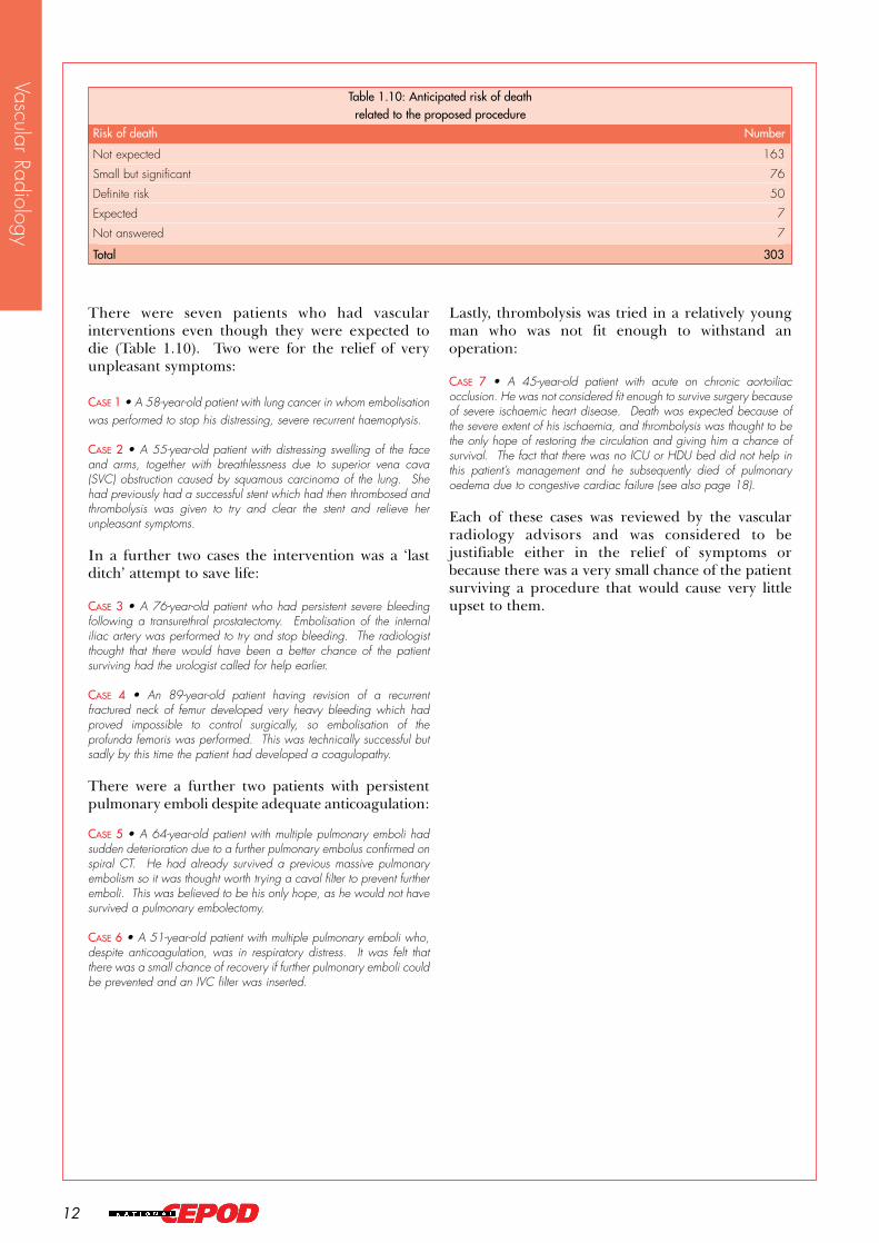
There were seven patients who had vascularinterventions even though they were expected todie (Table 1.10). Two were for the relief of veryunpleasant symptoms:
CASE 1 • A 58-year-old patient with lung cancer in whom embolisationwas performed to stop his distressing, severe recurrent haemoptysis.
CASE 2 • A 55-year-old patient with distressing swelling of the faceand arms, together with breathlessness due to superior vena cava(SVC) obstruction caused by squamous carcinoma of the lung. Shehad previously had a successful stent which had then thrombosed andthrombolysis was given to try and clear the stent and relieve herunpleasant symptoms.
In a further two cases the intervention was a ‘lastditch’ attempt to save life:
CASE 3 • A 76-year-old patient who had persistent severe bleedingfollowing a transurethral prostatectomy. Embolisation of the internaliliac artery was performed to try and stop bleeding. The radiologistthought that there would have been a better chance of the patientsurviving had the urologist called for help earlier.
CASE 4 • An 89-year-old patient having revision of a recurrentfractured neck of femur developed very heavy bleeding which hadproved impossible to control surgically, so embolisation of theprofunda femoris was performed. This was technically successful butsadly by this time the patient had developed a coagulopathy.
There were a further two patients with persistentpulmonary emboli despite adequate anticoagulation:
CASE 5 • A 64-year-old patient with multiple pulmonary emboli hadsudden deterioration due to a further pulmonary embolus confirmed onspiral CT. He had already survived a previous massive pulmonaryembolism so it was thought worth trying a caval filter to prevent furtheremboli. This was believed to be his only hope, as he would not havesurvived a pulmonary embolectomy.
CASE 6 • A 51-year-old patient with multiple pulmonary emboli who,despite anticoagulation, was in respiratory distress. It was felt thatthere was a small chance of recovery if further pulmonary emboli couldbe prevented and an IVC filter was inserted.
Lastly, thrombolysis was tried in a relatively youngman who was not fit enough to withstand anoperation:
CASE 7 • A 45-year-old patient with acute on chronic aortoiliacocclusion. He was not considered fit enough to survive surgery becauseof severe ischaemic heart disease. Death was expected because ofthe severe extent of his ischaemia, and thrombolysis was thought to bethe only hope of restoring the circulation and giving him a chance ofsurvival. The fact that there was no ICU or HDU bed did not help inthis patient’s management and he subsequently died of pulmonaryoedema due to congestive cardiac failure (see also page 18).
Each of these cases was reviewed by the vascularradiology advisors and was considered to bejustifiable either in the relief of symptoms orbecause there was a very small chance of the patientsurviving a procedure that would cause very littleupset to them.
Table 1.10: Anticipated risk of death related to the proposed procedure
Not expected 163
Small but significant 76
Definite risk 50
Expected 7
Not answered 7
Risk of death Number
Total 303
13
Vasc
ular
Rad
iolo
gySPECIALTY AND EXPERIENCE OFTHE MEDICAL TEAM
Specialty of the clinical team
Tables 1.11 and 1.12 show the specialty of theconsultants under whose care the patients were atthe time of their procedure.
Almost all patients were under the care of theappropriate specialist, such as vascular patientsunder the care of the vascular surgeons and thepatients having TIPS under the care ofgastroenterologists. However, there were a fewexceptions:
CASE 8 • A 55-year-old patient who was referred by a nephrologist fordilatation of popliteal arterial stenosis. This occluded two days laterand the patient was not seen by a vascular surgeon until too late andan amputation was subsequently performed.
CASE 9 • A 76-year-old patient who underwent bilateral external iliacangioplasty and had a retroperitoneal haemorrhage. He was nursedon the general surgical ward and seen only by a senior house officerfollowing the procedure. Although the patient was deterioratingneither a radiologist nor vascular surgeon were called. The problemwas not recognised until the patient had a cardiac arrest and asubsequent postmortem was performed (see also page 19).
CASE 10 • An 89-year-old patient suffered a retroperitonealhaematoma following femoral angioplasty performed in a hospitalwhere there was no on-site vascular surgeon (see also page 19).
CASE 11 • An 81-year-old patient with claudication, but no rest pain,had an external iliac stent performed. She was considered to be ahigh risk because of coexisting medical problems. Because of thiscombination of factors it was the advisors’ view that this was an ill-advised procedure.
It is reassuring to see that the vast majority (85%) ofthese patients were referred by a consultant. Ofthose that were not, the majority were referred byreasonably experienced registrars and in no caseswere locums involved.
Table 1.11: Specialty of consultant surgeon under whosecare the patient was at the time of the procedure
General 2
General with interest in vascular surgery 97
General with interest in gastroenterology 18
General with interest in urology 1
General with interest in endocrinology 1
General with interest in breast surgery 1
General with interest in hepatobiliary surgery 1
General with interest in thoracic surgery 1
Vascular 46
Urology 3
Gynaecology 2
Orthopaedic 2
Transplantation 2
Cardiac/thoracic/cardiothoracic 1
Oral/maxillofacial 1
Oncology 1
Specialty Number
Table 1.12: Specialty of consultant physician under whose care the patient was at the time of the procedure
General with interest in gastroenterology 16
General with interest in renal medicine 8
General with interest in thoracic medicine 6
General with interest in endocrinology 5
General with interest in cardiology 3
General with interest in rheumatology 2
General with interest in paediatrics 1
Gastroenterology 11
Renal medicine 9
Haematology 9
Thoracic medicine 8
Care of the elderly 8
Oncology 6
Endocrinology 3
Cardiology 2
Radiotherapy 1
Pulmonary oncology 1
Palliative care 1
Specialty Number
Table 1.13: Grade of the most seniordoctor who referred the patient
Consultant 257
Staff grade 1
SpR Accredited/CCST 6
SpR 4 9
SpR 3 8
SpR 2 4
Visiting SpR 2
SHO 2 1
Other 2
Not known 1
Not answered 12
Grade Number
Total 303

14
Vascular Radiology
Radiologist’s assessment prior toprocedure
Specialty and seniority of radiologist
The radiologist should not be seen as a technicianwho performs interventions when requested, butshould assess the patient prior to, and after, theprocedure and, where necessary, discuss with thereferring clinicians. It was encouraging to see that275/303 (91%) patients were assessed by aconsultant radiologist, but disconcerting that therewere 13 patients who were not reviewed by aradiologist prior to the procedure (Table 1.14).
In the majority (95%) of cases the most seniorradiologist was one with an appropriate specialinterest in interventional radiology and in only fourcases were they designated simply as a ‘generalradiologist’.
Table 1.14: Grade of the most senior radiologist who reviewed the patient before the procedure
Consultant 275
SpR Accredited/CCST 5
SpR 4 5
SpR 3 1
Other 2
None 13
Not answered 2
Grade Number
Total 303
Key Point
• The interventional radiologist should have sufficient experience to perform the procedure safelyand to deal with any complications that may arise.
Table 1.15: Specialty of most senior radiologist present
General radiologist with vascular interventional interest 187
Specialist vascular interventional radiologist 101
General radiologist 4
Other 7
Not answered 4
Total 303
Specialty Number
Table 1.16: Grade of most senior doctor performing the procedure
Consultant 283
SpR Accredited/CCST 9
SpR 4 6
SpR 3 2
SpR 2 1
Not answered 2
Total 303
Grade Number
Question 1.1: If the most senior operator was not aconsultant, was a more senior doctor immediatelyavailable?
Yes . . . . . . . . . . . . . . . . . . . . . . . . . . . . . . . . . . . . . . . . . . . . . . . . . . . . . . . . . . . . . . . . . . . . . . . . . . . . . . . . . . .12No . . . . . . . . . . . . . . . . . . . . . . . . . . . . . . . . . . . . . . . . . . . . . . . . . . . . . . . . . . . . . . . . . . . . . . . . . . . . . . . . . . . . . .4Not answered . . . . . . . . . . . . . . . . . . . . . . . . . . . . . . . . . . . . . . . . . . . . . . . . . . . . . . . . . . . . . . . .4Total . . . . . . . . . . . . . . . . . . . . . . . . . . . . . . . . . . . . . . . . . . . . . . . . . . . . . . . . . . . . . . . . . . . . . . . . . . . . . . .20
The majority of procedures were performed byconsultants (93%), with some being performed byregistrars indicating that training is in progress(Table 1.16). We do not have the evidence as towhether or not this is sufficient for the futurenumber of consultants required. In 12 of thesecases a consultant was immediately available and,whilst this is generally good, in four cases there wasno consultant available and this is a situation thatshould not occur. Some examples of problemswhich may arise are:
CASE 12 • A 69-year-old patient with lower limb ischaemia wasundergoing an iliac angioplasty and insertion of stent. The radiologisthad done only six similar procedures in the previous year. The externaliliac artery ruptured during the procedure and attempts to control thebleeding with a balloon failed. There was a delay in getting thepatient to theatre (see also page 20).
CASE 13 • A 57-year-old patient with alcoholic liver disease andoesophageal varices had a failed TIPS because of inability to puncturethe portal vein in a small cirrhotic liver. The radiologist had done onlytwo such cases in the previous three years (see also page 23).
CASE 14 • A 67-year-old patient had an attempted renal angioplastywhich failed for technical reasons. The radiologist had done only twosimilar procedures in the last year.
15
Vasc
ular
Rad
iolo
gyFACILITIES, PERSONNELAND MONITORING
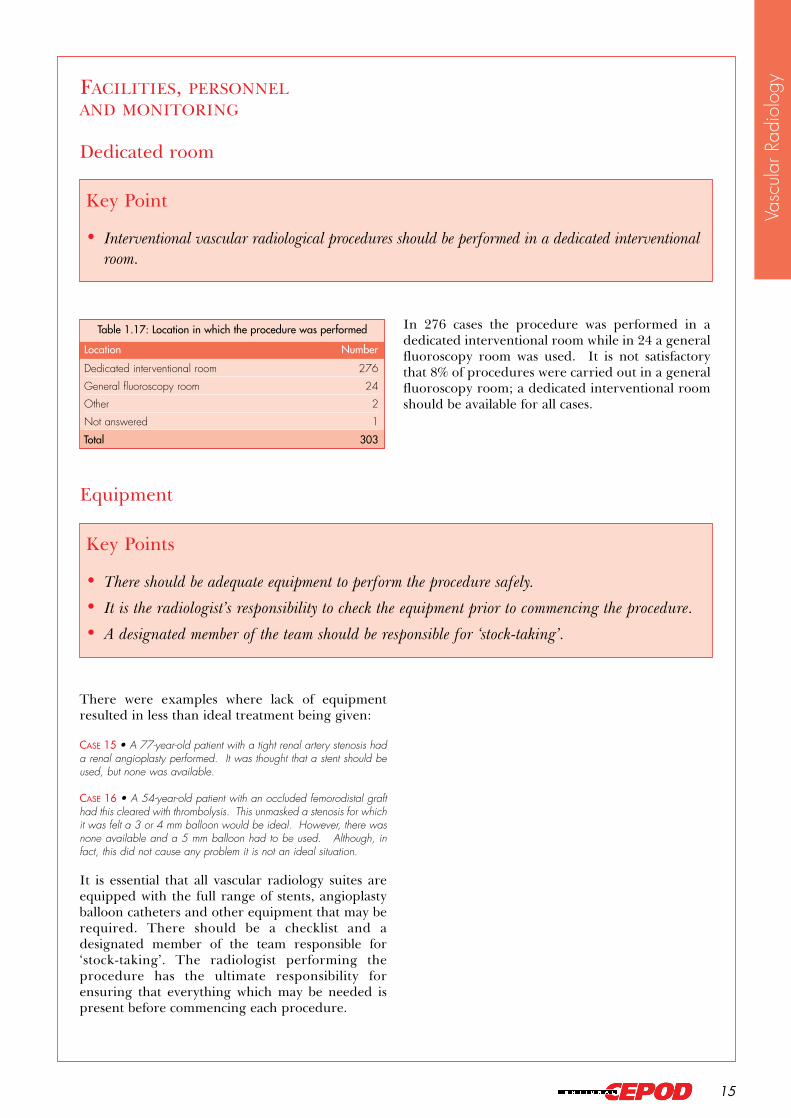
Dedicated room
Table 1.17: Location in which the procedure was performed
Dedicated interventional room 276
General fluoroscopy room 24
Other 2
Not answered 1
Total 303
Location Number
Equipment
In 276 cases the procedure was performed in adedicated interventional room while in 24 a generalfluoroscopy room was used. It is not satisfactorythat 8% of procedures were carried out in a generalfluoroscopy room; a dedicated interventional roomshould be available for all cases.
Key Points
• There should be adequate equipment to perform the procedure safely.
• It is the radiologist’s responsibility to check the equipment prior to commencing the procedure.
• A designated member of the team should be responsible for ‘stock-taking’.
Key Point
• Interventional vascular radiological procedures should be performed in a dedicated interventionalroom.
There were examples where lack of equipmentresulted in less than ideal treatment being given:
CASE 15 • A 77-year-old patient with a tight renal artery stenosis hada renal angioplasty performed. It was thought that a stent should beused, but none was available.
CASE 16 • A 54-year-old patient with an occluded femorodistal grafthad this cleared with thrombolysis. This unmasked a stenosis for whichit was felt a 3 or 4 mm balloon would be ideal. However, there wasnone available and a 5 mm balloon had to be used. Although, infact, this did not cause any problem it is not an ideal situation.
It is essential that all vascular radiology suites areequipped with the full range of stents, angioplastyballoon catheters and other equipment that may berequired. There should be a checklist and adesignated member of the team responsible for‘stock-taking’. The radiologist performing theprocedure has the ultimate responsibility forensuring that everything which may be needed ispresent before commencing each procedure.

16
Vascular Radiology
Shortage of personnel
Key Point
• There should be sufficient staff to perform the procedure safely.
Question 1.2: Was there a shortage of personnel inthis case?
Yes . . . . . . . . . . . . . . . . . . . . . . . . . . . . . . . . . . . . . . . . . . . . . . . . . . . . . . . . . . . . . . . . . . . . . . . . . . . . . . . . . . . . .4No . . . . . . . . . . . . . . . . . . . . . . . . . . . . . . . . . . . . . . . . . . . . . . . . . . . . . . . . . . . . . . . . . . . . . . . . . . . . . . . . .285Not answered . . . . . . . . . . . . . . . . . . . . . . . . . . . . . . . . . . . . . . . . . . . . . . . . . . . . . . . . . . . . . .14Total . . . . . . . . . . . . . . . . . . . . . . . . . . . . . . . . . . . . . . . . . . . . . . . . . . . . . . . . . . . . . . . . . . . . . . . . . . . . .303
If yes, which? (4 cases, answers may be multiple)Consultant radiologist . . . . . . . . . . . . . . . . . . . . . . . . . . . . . . . . . . . . . . . . . . . . . . . .1Radiographer . . . . . . . . . . . . . . . . . . . . . . . . . . . . . . . . . . . . . . . . . . . . . . . . . . . . . . . . . . . . . . . .1Consultant anaesthetist . . . . . . . . . . . . . . . . . . . . . . . . . . . . . . . . . . . . . . . . . . . . . .1Nurses . . . . . . . . . . . . . . . . . . . . . . . . . . . . . . . . . . . . . . . . . . . . . . . . . . . . . . . . . . . . . . . . . . . . . . . . . . . . . .2Porter . . . . . . . . . . . . . . . . . . . . . . . . . . . . . . . . . . . . . . . . . . . . . . . . . . . . . . . . . . . . . . . . . . . . . . . . . . . . . . .1
Whilst it is encouraging that there was a shortage ofpersonnel in so few cases it is nevertheless vital, if ahigh standard is to be maintained, to ensuresufficient staff in all cases. The interventionalradiology nurse is an important member of theteam.
Key Point
• There needs to be a sufficient number of fully-staffed interventional radiology sessions forurgent patients to receive their treatment without delay.
Question 1.3: Were there any delays (betweenadmission and procedure) due to factors other thanclinical?
Yes . . . . . . . . . . . . . . . . . . . . . . . . . . . . . . . . . . . . . . . . . . . . . . . . . . . . . . . . . . . . . . . . . . . . . . . . . . . . . . . . . . .11No . . . . . . . . . . . . . . . . . . . . . . . . . . . . . . . . . . . . . . . . . . . . . . . . . . . . . . . . . . . . . . . . . . . . . . . . . . . . . . . . .285Not answered . . . . . . . . . . . . . . . . . . . . . . . . . . . . . . . . . . . . . . . . . . . . . . . . . . . . . . . . . . . . . . . .7Total . . . . . . . . . . . . . . . . . . . . . . . . . . . . . . . . . . . . . . . . . . . . . . . . . . . . . . . . . . . . . . . . . . . . . . . . . . . . .303
Table 1.18: Reasons for non-clinical delay between admission and procedure
(11 cases; answers may be multiple)
Pressure of work 8
Poor communication 2
Shortage of beds 1
Insufficient cover at weekends 1
Vascular radiologist on leave 1
Patient refused 1
Reason Number
Delays
An anaesthetist was present for 43 of the proceduresand in 30 administered general anaesthesia. In theremainder they were there to administer localanaesthesia and sedation.
Of the 43 anaesthetists, one was an SpR 1, 16 wereSpR 3 or higher and twenty were consultants; thegrade was not known in the remaining six cases.The X-ray department is a more difficultenvironment than the operating theatre in which togive an anaesthetic. It is, therefore, inappropriatefor an inexperienced anaesthetist to be responsiblefor such cases.
In 254/303 (84%) cases the procedure wasperformed under local anaesthetic or sedationadministered solely by the operator.
AnaesthesiaQuestion 1.4: Was the procedure performed solelyunder local anaesthetic or sedation administered bythe operator?
Yes . . . . . . . . . . . . . . . . . . . . . . . . . . . . . . . . . . . . . . . . . . . . . . . . . . . . . . . . . . . . . . . . . . . . . . . . . . . . . . . .254No . . . . . . . . . . . . . . . . . . . . . . . . . . . . . . . . . . . . . . . . . . . . . . . . . . . . . . . . . . . . . . . . . . . . . . . . . . . . . . . . . . .47Not answered . . . . . . . . . . . . . . . . . . . . . . . . . . . . . . . . . . . . . . . . . . . . . . . . . . . . . . . . . . . . . . . .2Total . . . . . . . . . . . . . . . . . . . . . . . . . . . . . . . . . . . . . . . . . . . . . . . . . . . . . . . . . . . . . . . . . . . . . . . . . . . . .303

17
Vasc
ular
Rad
iolo
gyMonitoring
Of concern is the fact that in 19 cases no monitoringwas performed, as all patients having interventionalprocedures should be monitored. In addition, therewas no monitoring of the pulse in 32 patients, of theblood pressure in 40 patients, of oxygen saturationin 64 patients and the ECG in 195 patients.
In 97 cases monitoring was undertaken by theperson performing the interventional radiologicalprocedure. In 60 of these monitoring was alsobeing performed by another member of the team.However, in 37 cases the operator was the onlyperson monitoring the patient, and this is a causefor concern. Interventional procedures arefrequently technically demanding and it is notpossible for the same person to carry these out andadequately monitor the patient. Radiographers arenot trained to perform monitoring, and ideally thisshould be performed by a nurse or an operatingdepartment assistant. An oxygen supply should be
available and if any sedation in the form ofbenzodiazepines or opioid drugs is used then thespecific antagonists should be available (flumazanilor naloxone respectively)1. In all areas whereinvasive procedures are performed, there should beresuscitation equipment that is regularly checkedand core staff trained and regularly updated inresuscitation.
Care following the procedure
Recovery
It is unsatisfactory that in almost one third (29%) ofcases there was no recovery area for patients aftertheir interventional procedure. A recovery areashould be available.
Key Point
• Monitoring of pulse oximetry, blood pressure and ECG should be performed during allinterventional radiological procedures; this should be done by someone other than theradiologist performing the procedure.
Table 1.19: Monitoring performed during or immediately after the procedure
(303 cases; answers may be multiple)
Pulse 271
Blood pressure 263
Pulse oximetry 239
ECG 108
Other 30
None 19
Not answered 6
Monitoring Number
Table 1.20: Responsibility for monitoring the patient’sgeneral condition during the procedure(303 cases; answers may be multiple)
A nurse 211
The operator 97
An anaesthetist 42
Another doctor 17
A radiographer 16
Not answered 1
Person Number
Question 1.5: Was there a recovery room/areaavailable attached to the procedure suite?
Yes . . . . . . . . . . . . . . . . . . . . . . . . . . . . . . . . . . . . . . . . . . . . . . . . . . . . . . . . . . . . . . . . . . . . . . . . . . . . . . . .210No . . . . . . . . . . . . . . . . . . . . . . . . . . . . . . . . . . . . . . . . . . . . . . . . . . . . . . . . . . . . . . . . . . . . . . . . . . . . . . . . . . .88Not answered . . . . . . . . . . . . . . . . . . . . . . . . . . . . . . . . . . . . . . . . . . . . . . . . . . . . . . . . . . . . . . . .5Total . . . . . . . . . . . . . . . . . . . . . . . . . . . . . . . . . . . . . . . . . . . . . . . . . . . . . . . . . . . . . . . . . . . . . . . . . . . . .303

18
Vascular Radiology
Forty patients were admitted to an intensive careunit (ICU) and 20 to a high dependency unit(HDU) immediately after their procedure, with afurther 18 being admitted to ICU and six to HDUafter a period on a routine ward. In the remainderadmission to ICU/HDU was not felt necessary,except in three cases where the patient’s conditiondid warrant admission to ICU or HDU, but therewas no bed available within the hospital in which theprocedure took place:
CASE 7 • A 45-year-old patient with acute on chronic aortoiliacocclusion (see also page 12).
CASE 17 • A 44-year-old patient with alcoholic liver disease, togetherwith haematemesis and melaena due to oesophageal varices, wasreferred for a TIPS procedure. The patient had to be transferred backto the original hospital after the procedure, as there was no HDU bedavailable. It would have been more satisfactory to keep him at thesame hospital.
CASE 18 • A 57-year-old patient with acute myeloid leukaemia had aHickman line inserted for administration of chemotherapy. Theprocedure was uneventful. A week later he developed coagulase-negative septicaemia. A further week later he became seriously illdespite antibiotics. The Hickman line was removed. There was noICU or HDU bed available so he was nursed on the general medicalward and died three days later.
The list of complications in Table 1.21 is typical ofthose that arise during interventional procedures.The vast majority were unavoidable and dealt withappropriately. There were particular problems in afew cases which are discussed later.
In 37 cases there were unanticipated proceduralcomplications; in three of these there were twounexpected complications.
COMPLICATIONS
Intensive and high dependency care
Key Point
• These patients may be desperately ill and it is essential that ICU/HDU beds are availablewhen required.
Table 1.21: Unexpected procedural complications (37 cases; answers may be multiple)
Unable to cross the vascular lesion 6
Embolus/thrombosis of run-off vessels 6
Occlusion of artery during procedure 5
Bleeding/haematoma from groin 3
Rupture of iliac artery 3
Thrombosis of brachial artery when used to approach aortoiliac segment 2
Lack of correct-sized balloon 2
Unexpected cardiac arrest during procedure 2
Internal flap in superficial femoral artery 1
Stroke during carotid angioplasty 1
Patient’s rest pain so severe he could not lie still 1
Groin puncture site very painful 1
Extravasation of blood from femoropopliteal segment 1
Myocardial infarction during procedure 1
Severe nose bleed during thrombolysis 1
Coagulopathy during embolisation for blood loss 1
Axillary vein thrombosis during SVC stent (cleared at lysis) 1
Tumour more extensive then realised prior to SVC stent 1
Pneumothorax during insertion of Hickman line 1
Complication Number

19
Vasc
ular
Rad
iolo
gy
Specific problems
Level of puncture
With the exception of haemorrhage andpostoperative bleeding, which were more commonin patients having thrombolysis, there was noobvious correlation between any particularprocedures and subsequent complications.
CASE 19 • An 83-year-old patient with an acutely ischaemic left leg,due to a left external iliac thrombosis, had thrombolysis followed byballoon angioplasty. Four days later he died and at postmortem wasfound to have a retroperitoneal haematoma.
Table 1.22: Postprocedural complications (303 cases; answers may be multiple)
Respiratory distress 46
Low cardiac output/other cardiac problems 45
Renal failure 44
Peripheral ischaemia 34
Haemorrhage/postoperative bleeding requiring transfusion or surgical endovascular intervention 31
Cardiac arrest 29
Hepatic failure 25
Generalised sepsis 22
Stroke or other neurological problems 15
Other organ failure 11
Persistent coma 9
Pulmonary embolus 8
Problem with analgesia 6
Nutritional problems 6
Pressure sores 3
Wound infection/dehiscence 1
Endocrine system failure 1
DVT 1
Urinary retention/catheter blockage 1
Other 29
Not answered 110
None 20
Complication Number
Key Point
• Cannulation of the femoral artery should always be below the inguinal ligament to avoid thedanger of retroperitoneal haematoma. Medical and nursing staff must be aware of the risks ofthis serious complication in order to act early when necessary.
There were several cases where retroperitonealbleeding occurred:
CASE 9 • A 76-year-old patient who underwent bilateral external iliacangioplasty and had a retroperitoneal haemorrhage (see also page13).
CASE 10 • An 89-year-old patient had a high femoral puncture inorder to perform a superficial femoral artery angioplasty. Shedeveloped a massive retroperitoneal haematoma. There was no on-site vascular surgeon on the day of the angioplasty (see also page13).

20
Vascular Radiology
Great care must always be taken to keep the level ofpuncture below the inguinal ligament so thatbleeding is recognised immediately. Above theinguinal ligament a very large amount of blood maybe lost in the retroperitoneal space and may not berecognised until the patient is clinically in theadvanced stages of shock. Elderly patients withheart disease make up quite a high proportion ofpatients having angioplasties and they do nottolerate blood loss well. If the radiologist or anyother clinician is at all suspicious, an ultrasound orCT scan should be performed to confirm and
If angioplasties and stents performed in the iliac,femoral and popliteal arteries are included then atotal of 11 692 of these procedures were performed,of whom 101 patients died, giving a mortality of lessthan 1% (0.9%) (Table 1.6, page 8). Bearing in mindthat a significant number of these patients aresevere arteriopaths with critical ischaemia, on whomvascular surgeons are not keen to operate, thenthese results are generally very creditable.However, there was some cause for concern in threecases where the external iliac artery was rupturedduring angioplasty.
CASE 12 • A 69-year-old patient had an iliac angioplasty and stent.The artery ruptured, the bleeding was controlled with a balloon, butbecause of ischaemia was let down again before the patient wastaken to theatre! There was a delay of forty minutes in getting thepatient to theatre (see also page 14).
CASE 20 • A 74-year-old patient had an external iliac angioplastywhich ruptured the artery. No radiological measures were taken to tryand control the bleeding. The patient was taken to theatre urgently foriliofemoral bypass.
CASE 21 • A 63-year-old patient having an iliac stent placed had arupture of the iliac artery with the stent in situ. A covered stent wouldalmost certainly have stopped the bleeding.
These cases illustrate the need for suitableexperience and ability of the operator and theavailability of equipment. It should be possible tocontrol the bleeding of a ruptured iliac arterypermanently with a covered stent and these are
Lower limb revascularisation
perhaps monitor the amount of bleeding. It is alsoessential to warn clinical staff looking after thepatient and ensure they are aware of the possibilityand dangers of retroperitoneal bleeding. Thepatient should be nursed on a specialist vascularward where nurses and medical staff are fully awareof the risk of the possibility of development ofretroperitoneal haematoma and its seriousconsequences. If bleeding does not stop the patientmust be taken to theatre to suture the bleedingpoint before it is too late.
commercially available. If this does not control thebleeding then a balloon should give temporarycontrol until the patient can be taken to theatre,which obviously needs to be done as a matter ofgreat urgency. It is recommended that everyinterventional radiology department should have a‘rupture box’ containing all the necessaryequipment, which can be opened at a moment’snotice, to deal with this emergency.
The conclusion, therefore, is that angioplasty shouldnot be performed unless the radiologist has theexperience to recognise, and ability and equipmentto deal with, complications. The immediate help ofa vascular surgical team should be available whenneeded.
Key Points
• The interventional radiologist should have sufficient experience, facilities and equipment toperform the procedure safely and to deal with any complications that may arise.
• It should always be possible to control bleeding of a ruptured iliac artery either temporarilywith a balloon catheter or permanently with a covered stent.
• A ‘rupture box’ containing all necessary equipment should be available in every interventionalradiology department.

21
Vasc
ular
Rad
iolo
gy
Thrombolysis can be extremely useful in dissolvingclot that has formed in the blood vessel or graft, butit must be remembered that at best it can merelyrestore the vessel to the state it was in prior to thethrombus forming. The underlying cause will stillneed to be treated, for example an arterialthrombosis secondary to a stenosis will need anangioplasty once the thrombus has been cleared.Similarly, in a peripheral arterial embolus, or a deepvein thrombosis, anticoagulation will be needed toprevent further thrombosis. There is a risk ofbleeding both locally and at distance, which mayinclude spontaneous retroperitoneal haemorrhageor, more seriously, cerebral haemorrhage. Theincidence of minor haemorrhage is about 15%,major haemorrhage about 5%2 and haemorrhagicstroke is 1%. Other studies have found the risk ofstroke (both haemorrhagic and ischaemic) to be2.3%3. The risks seem to be greater in patients over75 years4. Although patients with simple emboli andnormal peripheral arteries are a rarity, neverthelesswhere the clinical features strongly suggest anembolus then surgery may give better results,especially in the elderly5. The risk of bleedingincreases with a larger dose and a longer period ofadministration. The question of dose has beendiscussed in detail5, but absolute recommendationson drugs and dosage are not possible on availabledata, although current practice has moved awayfrom the use of streptokinase to rt-PA.
Some examples of problems with thrombolysisinclude:
CASE 22 • An 87-year-old patient was given thrombolysis for athrombosed femoropopliteal graft and died due to cerebralhaemorrhage.
CASE 23 • An 83-year-old patient having thrombolysis for athrombosed iliac artery died of cerebral haemorrhage.
There were three other patients aged 84, 87 and 97years who, although they did not suffer a cerebralhaemorrhage, were considered by the advisors to betoo old to have treatment by thrombolysis.
Thrombolysis
There were two further patients in whom it was feltthe dose was inappropriate:
CASE 24 • A 77-year-old patient had an ischaemic left arm due tosubclavian thrombosis. This was treated with streptokinase 20 000units per hour and heparin 15 000 units per hour. This was continuedfor 18 hours when a check angiogram showed only partialthrombolysis. The patient subsequently died of angina and a stroke.No postmortem was performed and therefore it is unknown whetherthis was a haemorrhagic or thrombotic stroke, but either way theadvisors considered that this was too large a dose of streptokinase.
CASE 25 • A 68-year-old patient had thrombolysis for an acuteocclusion of the right superficial femoral artery due to an underlyingpopliteal aneurysm. He was given 50 mg of rt-PA in 5 mg aliquotsover a two hour period. One hour later the patient was taken totheatre for a femorodistal bypass. As far as was possible to ascertainfrom the questionnaire the surgeon was not aware of how large a dosehad been given. Certainly the advisors considered that this was toogreat a dose. The patient died 24 hours later and althoughpostmortem did show some haemorrhage around both puncture sitesdeath was due to peripheral vascular disease.
Although thrombolysis can be very effective, its usedoes carry significant risks particularly in patientsover 75 years, and even more so over the age of 80.However, one needs to bear in mind that surgery foracute ischaemia in elderly patients is also hazardouswith a mortality rate of about 30%6. Whateverchoice of treatment is given it is essential that thepatient is warned of the risks of both thrombolysisand surgery, and indeed of not undergoing either.Certainly the risks with thrombolysis are greater thehigher the dose used and the longer it is given.Although it is not possible to give absoluteguidelines, since there is insufficient data, theadvisors considered that the normal dose ofstreptokinase for continuous infusion should notexceed 5000 units per hour, but that it is probablybetter to use rt-PA with the dose not normallyexceeding 0.5-1.0 mg per hour, although it may begreater if the pulse spray infusion is used7.
Key Points
• There is a danger of cerebral haemorrhage with thrombolytic therapy particularly when used inthe elderly (over 75 years) or if too large a dose is given.
• Patients having continued thrombolytic therapy after leaving the vascular radiology suiteshould be nursed on an HDU or specialist vascular ward where there are an adequate numberof nurses to monitor them closely.

22
Vascular Radiology
Patients having thrombolysis need close observationfor early detection of any complications and formonitoring the response, so that it is given for theshortest possible time, but also so that an adequateamount has been given to clear the vessel.Therefore they should go to an HDU or specialistvascular ward with an adequate number of nurses tomonitor closely, and the easy facility for repeatangiograms as required. Case 7 (see pages 12 and
Table 1.6 (page 8) showed that embolisation wasused via the internal iliac artery for uterine fibroids,for the treatment of bleeding pelvic fractures, forrenal disease, most commonly to reduce thevascularity prior to removing a large tumour, andfor gastrointestinal bleeding. Other sites ofembolisation included the liver for treatment ofsecondaries, particularly for carcinoid tumour orAV malformations, and embolisation of the internalmammary artery for haemoptysis due to lungcancer.
When embolisation is used for persistent bleeding,early communication with the vascular radiologist isnecessary if embolisation is going to have a chanceto be effective before a serious coagulopathy hasdeveloped (see case 3 and case 4 on page 12).
Gastrointestinal bleeding can be particularlyproblematic both to diagnose where the bleeding iscoming from and to treat adequately. Embolisationcan be extremely useful, but the following illustratethe problems that can occur:
CASE 26 • A 78-year-old patient with severe colonic bleeding wastreated with selective Sterispon embolisation which successfullystopped the bleeding. The radiologist wrote in the notes: ‘ There is arisk of colonic infarction following this procedure and the patientshould be closely observed for signs of this.’ Colonic infarction didindeed develop and the patient required a laparotomy to removeischaemic bowel on the following day.
CASE 27 • A 74-year-old patient with persistent bleeding from aduodenal ulcer was treated with coil embolisation of thepancreatoduodenal arteries. This controlled the bleeding, but 13days later the duodenum perforated and it was thought that ischaemiahad played a part in this.
Embolisation
18) was an example where the advisors thought itwould have been preferable to manage the patienton an HDU or ICU. The view of the advisors wasthat wherever there is ongoing thrombolysis thepatient should be nursed on an HDU or a specialistvascular ward.
CASE 28 • A 67-year-old patient had a Whipples operation whichwas complicated by a pancreatic leak, sepsis and a subsequent falseaneurysm of the hepatic artery which bled. This was treated by coilembolisation controlling the bleeding. The patient died a few dayslater of multiple organ failure which was thought to be due to sepsis,although in fact postmortem examination showed hepatic necrosis.
CASE 29 • A 51-year-old patient had embolisation for bleeding uterinefibroids. The ischaemic uterus became infected and the patientsubsequently died.
While embolisation is an extremely usefultherapeutic measure8 it must be remembered that itcan cut off the blood supply to the affected areasufficiently to cause ischaemic necrosis. It isimportant to be aware of this possibility andrecognise and treat it early if it should occur.
Key Points
• If embolisation is to be used for severe persistent bleeding, radiologists should be called soonerrather than later in order to instigate treatment before a serious coagulopathy has developed.
• It must be remembered that embolisation to stop bleeding can occasionally cause ischaemicinfarction.

23
Vasc
ular
Rad
iolo
gySVC stent
Two hundred and sixty-six of these were performedand seventeen of the patients died (6%). Theseprocedures are performed for advancedmalignancy which has obstructed the superior venacava causing venous engorgement of the upper halfof the body. In all these cases it was thought thatdeath was due to the underlying disease, and thatSVC stenting is a useful therapeutic measure9.
Central venous access
Over 3000 of these were performed, forhaemodialysis, the administration of chemotherapyfor malignancy or for long-term intravenousfeeding. Of the 36 patients who died, one had apneumothorax, which was felt to have contributedto the patient’s death. In all the remainder it wasconsidered that death was due to the underlyingdisease. Thus, this is a very safe procedure in thehands of a radiologist.
Transjugular intrahepatic portosystemic shunt(TIPS)
Acute variceal bleeding which fails to respond tomedical management remains the primaryindication for TIPS and accounts for over 80% ofcases performed. Most of these patients aredesperately ill and operating on them is almostalways more hazardous10,11. Of the 158 casesperformed, 27 patients died (17%). Bearing inmind the very serious nature of the disease thesewere felt to be acceptable results. It has, however,been suggested that centres undertaking less thanfive cases per year are unlikely to achieve therequired expertise11; this does argue for referral ofpatients to centres with a large experience of theseprocedures. Case 13 (see page 14) reinforces thispoint.
DVT prophylaxis
Bearing in mind that most of these patients weremiddle-aged or elderly, with a significant number ofother medical problems, and the majority of themgenerally unfit as judged by the ASA grade,according to the THRIFT12 they should probably allhave had some form of DVT prophylaxis, mainlysubcutaneous heparin. However, the advisors werenot unanimous on this, particularly bearing in mindthat many diagnostic angiograms are performed asday cases.
Question 1.6: Were any measures taken (before,during or after the procedure) to prevent venousthromboembolism?
Yes . . . . . . . . . . . . . . . . . . . . . . . . . . . . . . . . . . . . . . . . . . . . . . . . . . . . . . . . . . . . . . . . . . . . . . . . . . . . . . . .115No . . . . . . . . . . . . . . . . . . . . . . . . . . . . . . . . . . . . . . . . . . . . . . . . . . . . . . . . . . . . . . . . . . . . . . . . . . . . . . . . .169Not answered . . . . . . . . . . . . . . . . . . . . . . . . . . . . . . . . . . . . . . . . . . . . . . . . . . . . . . . . . . . . . .18Not known . . . . . . . . . . . . . . . . . . . . . . . . . . . . . . . . . . . . . . . . . . . . . . . . . . . . . . . . . . . . . . . . . . . . . .1Total . . . . . . . . . . . . . . . . . . . . . . . . . . . . . . . . . . . . . . . . . . . . . . . . . . . . . . . . . . . . . . . . . . . . . . . . . . . . .303

24
Vascular Radiology
Ninety-six of the 303 deaths were reported to thecoroner and of these 58 had a coroner’spostmortem performed. Of the remaining 245cases, only 15 patients had a hospital postmortem.Of the 73 postmortems performed, in only sevendid a radiologist attend and in only four didanother clinician do so. A copy of the postmortemreport was only received by the team in 37/73 (51%)cases.
In eight out of the 64 cases on which thisinformation was available, the pathological findingat postmortem was different from that which theclinical team was expecting. The details are asfollows:
CASE 28 • A 67-year-old patient had embolisation to control bleedingfrom the hepatic artery. Postmortem showed unsuspected hepaticnecrosis (see also page 22).
CASE 30 • A 60-year-old patient had embolisation for hepaticmetastases. He was thought to have died as a result of this butpostmortem showed death was due to an unsuspected haemorrhageinto a cerebral metastasis.
CASE 31 • A 60-year-old patient was thought to have carcinoma ofthe colon and heart failure. At postmortem she was found to haveischaemic colitis and death was due to a pulmonary embolism causedby a DVT.
CASE 32 • A 69-year-old patient was admitted with a femoral veinthrombosis. She was treated with heparin which had to be stoppedwhen she had a gastrointestinal bleed. An IVC filter was inserted. Shewas thought to have died from gastrointestinal bleeding but atpostmortem no source could be found. There was a DVT of the left legand an old pulmonary embolus, but death was caused by acutemyocardial ischaemia due to coronary artery atheroma with plaquerupture.
CASE 33 • A 76-year-old patient had a stent for renal artery stenosis.Despite having heparin prophylaxis he was thought to have died frompulmonary embolism. Postmortem showed no evidence of an embolus,and death was due to congestive cardiac failure due to myocardialinfarction.
CASE 34 • A 67-year-old patient had a stent and thrombolysis for SVCthrombosis secondary to enlarged mediastinal lymph nodes thought tobe due to a bronchogenic carcinoma or possibly a lymphoma.Postmortem showed no evidence of either but death was due tometastases of intestinal origin.
CASE 35 • A 63-year-old patient had a stent for SVC obstruction dueto mediastinal lymphadenopathy. He was given dexamethasone. Twodays later he died of circulatory collapse. Postmortem showed deathwas due to peritonitis caused by a completely unsuspected perforatedduodenal ulcer.
CASE 36 • An 87-year-old patient had a popliteal angioplasty for acritically ischaemic leg. Death was thought to be due to sepsis from theischaemic limb, but postmortem showed it was caused by peritonitisdue to a perforated duodenal ulcer.
These cases illustrate why postmortemexaminations need to be performed morefrequently.
POSTMORTEM EXAMINATIONS
Key Point
• In one in eight of the postmortems performed the pathological finding was different from thatexpected by the clinical team. This surely must have educational benefit and is the reason whymore postmortems should be performed.
Question 1.7: Did the pathological informationconfirm the team’s clinical impression?
Yes . . . . . . . . . . . . . . . . . . . . . . . . . . . . . . . . . . . . . . . . . . . . . . . . . . . . . . . . . . . . . . . . . . . . . . . . . . . . . . . . . . .56No . . . . . . . . . . . . . . . . . . . . . . . . . . . . . . . . . . . . . . . . . . . . . . . . . . . . . . . . . . . . . . . . . . . . . . . . . . . . . . . . . . . . . .8None received . . . . . . . . . . . . . . . . . . . . . . . . . . . . . . . . . . . . . . . . . . . . . . . . . . . . . . . . . . . . . . .2Not answered . . . . . . . . . . . . . . . . . . . . . . . . . . . . . . . . . . . . . . . . . . . . . . . . . . . . . . . . . . . . . . . .6Not known . . . . . . . . . . . . . . . . . . . . . . . . . . . . . . . . . . . . . . . . . . . . . . . . . . . . . . . . . . . . . . . . . . . . . .1Total . . . . . . . . . . . . . . . . . . . . . . . . . . . . . . . . . . . . . . . . . . . . . . . . . . . . . . . . . . . . . . . . . . . . . . . . . . . . . . .73

25
Vasc
ular
Rad
iolo
gy
It is disappointing that in only 125/303 (41%) caseswas the patient’s death discussed at a local audit orquality control meeting. The figure should be verymuch higher. The need for radiologists to assessand audit their results is every bit as important as itis for other clinical staff.
It was also disappointing that in 82/303 (27%) casesthere was difficulty in obtaining the patient’s notesand in 64 cases (21%) either notes or images wereunavailable. The most common problem wasfinding the death certificate (43 cases) and otherproblems included obtaining a postmortem report(14 cases), postprocedure notes (12 cases) and theimages (10 cases). With the introduction of clinicalgovernance it is to be hoped that the systems will bein place for all notes and other information to bereadily available.
Key Point
• It is as important for radiologists to assess and audit their results as it is for other clinical staff.
AUDIT AND AVAILABILITY OFPATIENT RECORDS

26
Vascular Radiology

2 INTERVENTIONAL NEUROVASCULAR
RADIOLOGY
RECOMMENDATIONS
● The number of neuroradiologists and support staff needs toincrease to ensure a satisfactory on-call rota, including weekends(page 36).
● There is a need for recognised training programmes inneuroradiology to meet the demand for more consultants (page 36).
● Monitoring of the patient should be performed in all cases, andshould be the responsibility of someone other than theneuroradiologist performing the procedure (page 38).
● It is important that there are sufficient facilities for a promptemergency service, and ICU/HDU beds for subsequent care(pages 36, 39).
27

28
Neurovascular Radiology

General D
ata
Neu
rova
scul
ar R
adio
logy
29
2. INTERVENTIONAL NEUROVASCULAR RADIOLOGY
This is an important study, being the first time anaudit of deaths has been carried out for this specialty.
Over the past two decades or more, interventionalneurovascular radiology has become an importantand sophisticated specialty in its own right. It hasbenefited from the same advances in technology ascoronary angioplasty and peripheral vascularinterventional radiology. The specialty hasdeveloped with the close cooperation ofneurosurgeons and neurologists and requires thefull neuroscience support team, including otherdisciplines. In particular, access to high dependencyand intensive care units is needed every bit as muchas these facilities are required after neurosurgery.
The most common problem is the prevention offurther bleeding from subarachnoid haemorrhage(SAH) in patients with intracranial aneurysms.Arteriovenous malformations (AVMs) of the brainare a less common cause of cerebral haemorrhage.These conditions are treated by embolisation whichmay be by the use of detachable coils for aneurysms,or the tissue adhesive N butyl cyanoacrylate(Histoacryl or NBCA), or newer liquid embolicagents which are just becoming available, for AVMs.The liquid agents such as cyanoacrylates have theadvantage that they permeate into very small vesselsand reach far distally, thus enabling occlusion of thenidus of the malformation on a permanent basis. Anumber of ingenious devices have been inventedwhich enable, for example, the coils to stay attacheduntil they are in precisely the correct position andthen released. The most notable is the Guglielmidetachable platinum microcoil (GDC) which is
soldered at the end of an insulated stainless-steelguidewire and is detached by passing a currentthrough it which causes electrolysis of the exposeddetachment zone13,14.
The method of embolisation that is used depends onthe precise detail of the vasculature to be embolisedand the amount of risk there is to the surroundingnormal tissue. Embolisation may also be used forcerebral tumours.
Vasospasm of the cerebral vessels occurs in about20% of patients following aneurysmal haemorrhage.Treatment for this may take the form of eithersuper-selective injection of papaverine or ofangioplasty to the cerebral vessel in spasm, or acombination of both.
Cerebrovascular occlusive disease may be treated byangioplasty, or angioplasty and stent, andthrombotic disease may be treated by thrombolysis.
When thrombolysis is used for cerebral vascularthrombosis, it runs all the same risks as it doeselsewhere of causing haemorrhage both locallywhere it is applied and at a distance. It isparticularly likely to cause a problem if used forthromboembolic complications following treatmentfor SAH due to intracranial aneurysm and shouldbe used with great caution, particularly under thesecircumstances.
INTRODUCTION
Key Points
• In all cases the procedures were performed by appropriately experienced specialists and therewas a high standard of care.
• By far the most common condition was subarachnoid haemorrhage due to intracranialaneurysm, treated by detachable coils.
• Use of these techniques has developed from almost none in 1992, to over 800 patients a year,with very little increase in staff or facilities.
• The majority of patients are otherwise medically fit with good long-term outlook aftersuccessful treatment.
• Patients who survive an initial subarachnoid haemorrhage have an increasing risk of afurther bleed as each day passes; treatment is therefore a matter of urgency.

30
Neurovascular Radiology
DATA COLLECTION
Data was requested from all NHS hospitalsundertaking these procedures in England,Scotland, Wales and Northern Ireland.Participation was voluntary and some hospitalschose, for a variety of reasons, not to participate.
Information on the total number of patientsundergoing interventional neurovascular radiologyprocedures on a monthly basis, together withnotification of any deaths occurring within 30 daysof the procedure, were collected for the period 1April 1998 - 31 March 1999.
GENERAL DATA ANALYSIS
Monthly returns of proceduresperformed
27 hospitals agreed to participate in the study. Eachhospital was required to send in a monthly return ofall patients undergoing interventionalneurovascular radiological procedures in thehospital. A total of 324 forms should therefore havebeen received. The return rate of monthly data isshown in Figure 2.1.
A regional breakdown of the number of monthlyforms received is given in Table 2.1. The returnrate of monthly forms was excellent; only NorthThames (75%) and North West (89%) failed toachieve complete returns.
Figure 2.1: Monthly returns of total procedures performed
Not received19
Received305
Total forms due324
Included305
ExcludedNone
Table 2.1: Monthly returns by region
Anglia & Oxford 2 24 24 100%
North Thames 6 54 72 75%
North West 3 32 36 89%
Northern & Yorkshire 4 48 48 100%
South & West 2 24 24 100%
South Thames 3 36 36 100%
Trent 2 24 24 100%
West Midlands 1 12 12 100%
Wales 1 12 12 100%
Northern Ireland 1 12 12 100%
Scotland 2 24 24 100%
Region Number of Monthly forms Monthly forms Returnparticipating received expected rate
hospitals

31
General D
ata
Neu
rova
scul
ar R
adio
logy
Reported deaths
Figure 2.2 shows that a total of 41 reports of deathswithin 30 days of a procedure were received. Ofthese, two were excluded from further analysis: onewas received after the deadline of 31 August 1999and one remained incomplete despite all efforts toidentify missing information.
A regional breakdown of the 39 included deaths isshown in Table 2.2.
The differences in numbers of deaths reported byeach region reflects the total number of proceduresundertaken, together with the completeness of thedata submitted.
Figure 2.2: Total deaths reported
Excluded2
Included39
Total deaths reported41
Incomplete1
Too late1
Table 2.2: Deaths reported to NCEPOD by region
Anglia & Oxford 8
North Thames 0
North West 6
Northern & Yorkshire 7
South & West 2
South Thames 4
Trent 4
West Midlands 1
Wales 1
Northern Ireland 2
Scotland 4
Total 39
Region Deaths reported
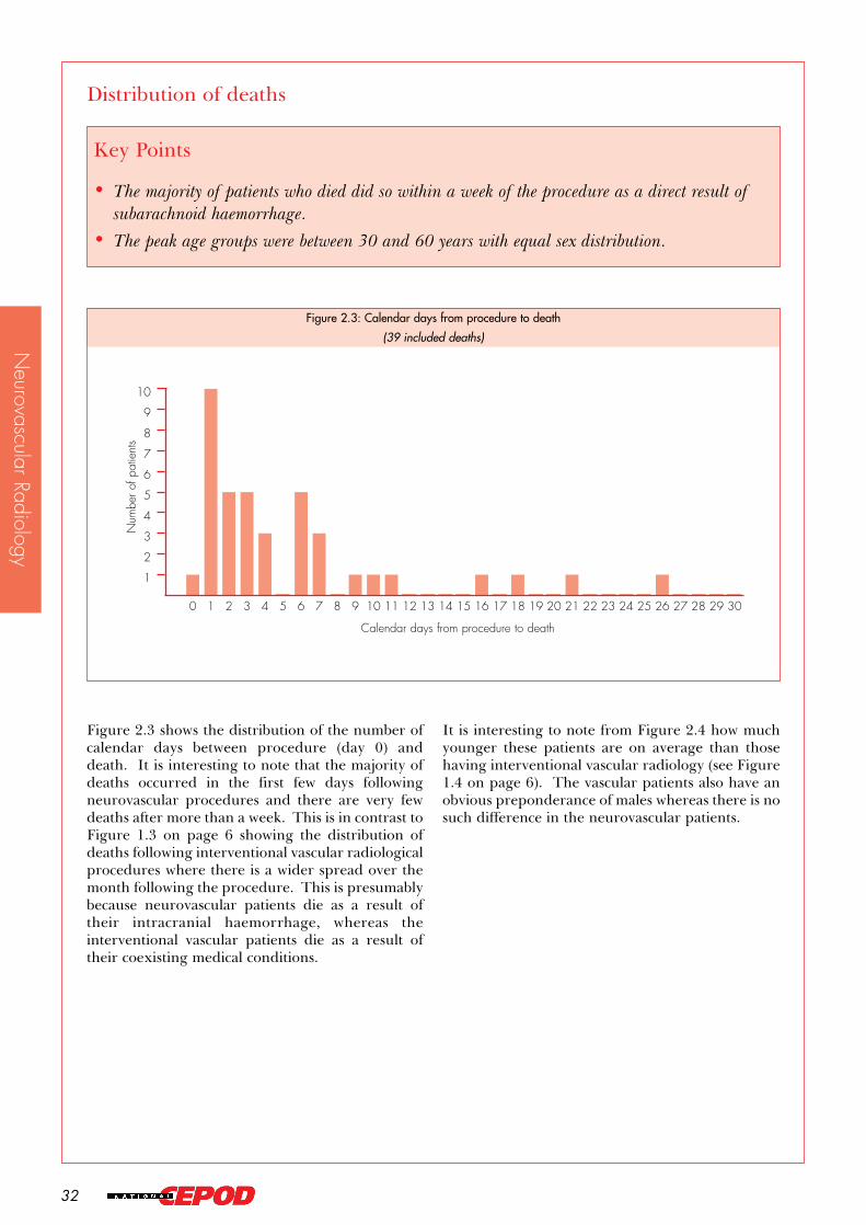
32
Neurovascular Radiology
Distribution of deaths
Figure 2.3 shows the distribution of the number ofcalendar days between procedure (day 0) anddeath. It is interesting to note that the majority ofdeaths occurred in the first few days followingneurovascular procedures and there are very fewdeaths after more than a week. This is in contrast toFigure 1.3 on page 6 showing the distribution ofdeaths following interventional vascular radiologicalprocedures where there is a wider spread over themonth following the procedure. This is presumablybecause neurovascular patients die as a result oftheir intracranial haemorrhage, whereas theinterventional vascular patients die as a result oftheir coexisting medical conditions.
It is interesting to note from Figure 2.4 how muchyounger these patients are on average than thosehaving interventional vascular radiology (see Figure1.4 on page 6). The vascular patients also have anobvious preponderance of males whereas there is nosuch difference in the neurovascular patients.
Key Points
• The majority of patients who died did so within a week of the procedure as a direct result ofsubarachnoid haemorrhage.
• The peak age groups were between 30 and 60 years with equal sex distribution.
Figure 2.3: Calendar days from procedure to death
(39 included deaths)
1
2
3
4
5
6
7
8
9
10
3029282726252423222120191817161514131211109876543210
Num
ber o
f pat
ient
s
Calendar days from procedure to death

33
General D
ata
Neu
rova
scul
ar R
adio
logy
Distribution, return and analysis ofquestionnaires
Questionnaires were sent to the consultantradiologist responsible for the care of each of the 39patients included. Figure 2.5 shows the return andanalysis rates of questionnaires sent.
The return rate of 92% was excellent; in two casesthe radiologist indicated that he/she was too busy tocomplete the questionnaire and the third gave noreason for failing to return the form. It was notnecessary to exclude any questionnaires fromsubsequent analysis.
Table 2.3 shows the returns by region and themajority achieved a 100% return rate; indeed theoverall return rate of 92% was due to a nil returnrate from South & West and West Midlands.
Figure 2.4: Age/sex distribution of reported deaths
1
2
3
4
5
6
7
8
80-8970-7960-6950-5940-4930-3920-2910-19<10
Num
bero
f pat
ient
s
Male
Female
Age (years)
Table 2.3: Regional distribution and return rates
Anglia & Oxford 8 8 100%
North Thames 0 0 -
North West 6 6 100%
Northern & Yorkshire 7 7 100%
South & West 2 0 -
South Thames 4 4 100%
Trent 4 4 100%
West Midlands 1 0 -
Wales 1 1 100%
Northern Ireland 2 2 100%
Scotland 4 4 100%
Region Questionnaires Questionnaires Return distributed returned rate
Total 39 36 92%
Figure 2.5: Distribution, return and analysis of questionnaires
Not returned3
Returned36 (92%)
Total cases/questionnaires sent39
Analysed36
Not analysedNone

34
Neurovascular Radiology
Table 2.4 shows that by far the greatest number ofprocedures performed, and subsequent deaths,were for occlusion of aneurysms. It should be notedthat AVMs usually require several treatments over aperiod of months, therefore exaggerating theirnumbers, whereas aneurysms are usually onlytreated once. The overall mortality rate is low for aserious disease where the clinical grade of thepatient is a major factor in the outcome.
Table 2.4 Neurovascular interventions
Aneurysm Endosaccular occlusion GDC 802 25 (3%)
Other coils 7 -
Parent vessel occlusion 75 5 (7%)
Spasm Papaverine 24 -
Angioplasty 0 -
Papaverine + angioplasty 1 -
Thrombolysis 7 -
Arteriovenous malformation (AVM) Brain Pial AVM 293 -
Dural AVM 40 -
CC fistula 30 1 (3%)
Vein of Galen malformation 21 -
Other 12 -
Craniofacial 23 -
Spine Dural 28 -
Other 16 -
Tumour Brain 84 -
Head & neck 42 -
Spine 20 -
Cerebrovascular disease Occlusive disease Angioplasty + stent 32 2 (6%)
Thrombotic disease Arterial thrombolysis 10 2 (20%)
Venous thrombolysis 3 1 (33%)
Other 46 -
Condition Procedure Total performed Deaths
Total 1616 36 (2%)
Procedures
Key Points
• By far the most common condition was subarachnoid haemorrhage due to intracranialaneurysm treated by detachable coils.
• A mortality rate of approximately 3% for detachable coil treatment is low considering the veryserious nature of the disease.

35
General D
ata
Neu
rova
scul
ar R
adio
logy
Following a subarachnoid haemorrhage thelikelihood of a further bleed is 4% in the first 24hours and thereafter 1% per day15. It is, therefore,very important that patients are treated urgently.
PATIENT PROFILE
The report hereafter deals only with those patientswho died.
Urgency of procedure
Key Points
• Patients who survive a subarachnoid haemorrhage have a 4% risk of a further bleed in thefirst 24 hours and a 1% risk per day thereafter.
• The majority of patients were treated as emergency or urgent cases.
• Detachable coils were introduced to the UK in 1992 and their use has risen from none to over800 patients treated in a year.
Figure 2.6: Day of the procedure
1
2
3
4
5
6
7
8
9
SundaySaturdayFridayThursdayWednesdayTuesdayMonday
Num
ber o
f cas
es

36
Neurovascular Radiology
Thirty of the patients were admitted as an inpatientfrom another acute hospital or directly from theA&E department, and the majority were admittedto a neurosurgery ward, ICU or HDU under thecare of a neurosurgeon. In no patient was therethought to be a deterioration in their conditionduring transfer, but in two patients there was adelay in referral or admission, though details arelacking. In a further two patients there were delaysbetween admission and procedure, due to factorsother than clinical ones:
CASE 1 • A 59-year-old patient with a subarachnoid haemorrhagewas delayed three days for GDC embolisation of her aneurysm dueto the heavy workload caused by other patients. The patient died fromrupture of the aneurysm during the procedure. This is a recognisedcomplication of this procedure and not related to the delay.
CASE 2 • A 56-year-old patient with a subarachnoid haemorrhagewas delayed due to a shortage of anaesthetic and nursing staff at theweekend. Death was due to rebleed because of incomplete coilocclusion, and therefore not caused by the delay.
The majority of neurovascular patients weregenerally fairly fit when compared with, forexample, the large number of comorbidities foundin the interventional vascular radiology group (seeTable 1.9, page 11) or many general surgicalpatients16.
There has been an enormous increase in theworkload of interventional neuroradiologists overthe last ten years, mainly due to the introduction ofthe Guglielmi detachable coil (GDC). Their use hasrisen from none prior to 1992, when they were firstmarketed in the UK, to over 800 in the last year, andyet there has been very little increase in the numberof neuroradiologists or support staff. Most of thiswork is urgent or emergency and there is someevidence from this study that this is having anadverse effect on the service provided, particularlyin relation to weekend work. The advisorsconsidered that this is a very real problem andconsiderably greater than the evidence in this reportwould suggest. A possible short-term solution wouldbe sharing emergency cover, particularly atweekends, with neighbouring units. In many placesthis is not possible for geographical reasons. Theonly satisfactory long-term solution is for a steadyand progressive increase in staff, which will taketime due to training implications forneuroradiologists and support staff. There is a needfor recognised training programmes inneuroradiology to be set up in order to meet theconsultant expansion which is needed.
Fitness of the patient
Figure 2.7: Admission category
5
10
15
20
25
30
EmergencyUrgentElective
Num
ber o
f cas
es
Key Points
• Comorbidities are relatively low when compared with vascular radiology or general surgicalpatients and probably represent those in the general population of equivalent age.
• The prognosis depends on the neurological grading based on the Glasgow Coma Scale (GCS)rather than the ASA grade.

37
Table 2.6 shows the World Federation ofNeurological Surgeons (WFNS) grading scale that isused following SAH; it is based on the GlasgowComa Scale.
General D
ata
Neu
rova
scul
ar R
adio
logy
Figure 2.8 shows the ASA status of the patients;details of assessment of ASA status are given onpage 11.
Thirteen patients (36%) were graded 1 or 2 andmost of those in ASA grades 4 and 5 were so gradedbecause of a poor level of consciousness rather thanbecause of cardiovascular or respiratory problems.Although assessed routinely by anaesthetists theASA grade is not normally used byneuroradiologists, who are far more concerned withthe level of consciousness as measured by theGlasgow Coma Scale (GCS).
Coexisting problems (other than the maindiagnosis) existed in 14/36 (39%) patients and theseare shown in Table 2.5.
This classification is a useful prognostic indicator asthe first two have a good prognosis, with aprocedure related mortality of 5-10%, whereas thelower three have a poor prognosis with a mortalityof 40-50% following treatment. Without treatmentthe overall mortality at six months is 50%17.
The outlook for patients undergoingneuroradiological intervention depends on theirGCS rather than their previous medical condition.Prior to their SAH most of the patients are as fit andwell as the general population, and followingsuccessful treatment return to a near-normal lifeexpectancy. This makes it particularly importantthat the facilities for their treatment are as good as itis possible to be.
Table 2.5: Coexisting medical problems (14 cases; answers may be multiple)
Respiratory 8
Cardiac 4
Vascular 4
Musculoskeletal 2
Psychiatric 2
Sepsis 2
Alcohol-related problems 1
Diabetes mellitus 1
Malignancy 1
Neurological 1
Renal 1
Coexisting medical problem Number
Figure 2.8: ASA status
ASA grade
5
10
15
20
54321
Num
ber o
f cas
es
Table 2.6: Modified Glasgow Coma Scale (GCS)
GCS 15 Grade 1 14
GCS 14-13 Grade 2 9
GCS 14-13+motor deficit Grade 3 3
GCS 12-7 Grade 4 4
GCS 6-3 Grade 5 6
Total 36
GCS WFNS grade Number

38
Neurovascular Radiology
FACILITIES, PERSONNEL ANDMONITORING
Seniority and specialty of theradiologist
Thirty-five out of the 36 cases were performed by aconsultant and one by a specialist registrar with aCCST, with a consultant present.
Table 2.7 shows that in all cases there was anappropriately experienced specialist present.
Dedicated room
All cases were performed in a dedicatedinterventional room.
Monitoring
All patients had monitoring of their pulse, bloodpressure and pulse oximetry; 34 had ECGmonitoring. Thirty-one patients had a generalanaesthetic, three received local anaesthetic withsedation and two had local anaesthetic alone,administered by the radiologist. In all 34 caseswhere an anaesthetist was present they monitoredthe patient. Of the two cases performed under localanaesthetic, in one the monitoring was done by anurse and in the other by the neuroradiologistperforming the procedure. The latter isunacceptable as monitoring should be performedby a member of the team who is not preoccupiedwith what may be the very demanding technicalitiesof the procedure.
Anaesthesia
Thirty-one out of the 36 procedures wereperformed under general anaesthetic and threeunder sedation administered by an anaesthetist. In27 of these 34 cases, the anaesthetist was aconsultant and in a further four the anaesthetist wasan accredited specialist registrar. In two cases theanaesthetist was an SpR 4 and in one an SpR 3.Thus in all cases the anaesthetist was appropriatelyexperienced.
In five cases the anaesthetist did not have a trainedassistant; one should be available for all patientshaving a general anaesthetic. This is particularlyimportant in the X-ray department being a moredifficult environment than the operating theatreand distant from other immediate anaestheticassistance.
Key Point
• All procedures were performed by appropriately experienced specialists in a dedicatedinterventional room.
Table 2.7: Specialty of the most senior radiologist present
Specialist neuroradiologist 33
Specialist interventional vascular radiologist 1
Neurosurgeon with training in radiology 1
Not stated 1
Total 36
Specialty Number
Question 2.1: Was there non-medical help withanaesthesia?
Yes . . . . . . . . . . . . . . . . . . . . . . . . . . . . . . . . . . . . . . . . . . . . . . . . . . . . . . . . . . . . . . . . . . . . . . . . . . . . . . . . . . .30No . . . . . . . . . . . . . . . . . . . . . . . . . . . . . . . . . . . . . . . . . . . . . . . . . . . . . . . . . . . . . . . . . . . . . . . . . . . . . . . . . . . . . .5Not answered . . . . . . . . . . . . . . . . . . . . . . . . . . . . . . . . . . . . . . . . . . . . . . . . . . . . . . . . . . . . . . . .1Total . . . . . . . . . . . . . . . . . . . . . . . . . . . . . . . . . . . . . . . . . . . . . . . . . . . . . . . . . . . . . . . . . . . . . . . . . . . . . . .36
If yes, who (30 cases; answers may be multiple)Radiographer . . . . . . . . . . . . . . . . . . . . . . . . . . . . . . . . . . . . . . . . . . . . . . . . . . . . . . . . . . . . . . . .1Trained anaesthetic nurse . . . . . . . . . . . . . . . . . . . . . . . . . . . . . . . . . . . . . . . . .9Trained operating department assistant . . . . . . . . . . . .13Operating department orderly . . . . . . . . . . . . . . . . . . . . . . . . . . . . . . . .7Other . . . . . . . . . . . . . . . . . . . . . . . . . . . . . . . . . . . . . . . . . . . . . . . . . . . . . . . . . . . . . . . . . . . . . . . . . . . . . . . .1

39
General D
ata
Neu
rova
scul
ar R
adio
logy
Twenty-one patients were admitted to an intensivecare unit (ICU) and nine to a high dependency unit(HDU) immediately after their procedure, with afurther four being admitted to ICU after a periodon the ward. One patient, a child of 18 months, wastransferred to another hospital which had apaediatric intensive care unit, and a further case wasmanaged on a neurosurgical ward:
CASE 3 • A 71-year-old patient had a subarachnoid haemorrhagetreated by GDCs and developed severe vasospasm. There was noICU or HDU bed available and he had to be transferred back to theneurosurgical ward.
Vasospasm is a very serious condition which mayrequire a combination of hypervolaemia,hypertension and haemodilution (Triple H therapy).Only on an HDU can this be done adequately or, ifthe patient needs ventilation, an ICU bed is required.
It is essential that ICU/HDU beds are available forthe postprocedure care of these patients in whomthere is an ever-present danger of seriouscomplications occurring and, therefore, a need forspecialist intensive care.
Many of the complications shown in Table 2.8 are,or may be, related to the initial pathology, e.g.hydrocephalus, cerebral oedema or persistent coma.The main problems were CNS thromboembolicevents and haemorrhage from an aneurysm, both ofwhich may result in brain oedema and persistentcoma, the other two common complications listed.Systemic heparin is normally given to prevent theseCNS thromboembolic events, and in 32 patients thiswas monitored using the Activated Clotting Time(ACT), while in two heparin was given with nomonitoring. In addition, three had aspirin, one hadanother antiplatelet agent and only two had notreatment aimed at preventing thromboembolicproblems. Several patients were given thrombolysisfollowing a thromboembolic complication and thensubsequently had a further fatal rebleed.
The advisors’ view was that patients shouldnormally be given systemic anticoagulation(heparin) during these procedures, and that thedose should be monitored using the ACT. Patientswho have bled from an intracranial aneurysmshould not normally be given thrombolysis, even ifthey do develop CNS thromboemboliccomplications, because of the risk of a further bleedalthough, at present, there is insufficient data to ruleout this mode of treatment completely. It was alsothought by the advisors that specific antiplatelettherapy should be given, although there is nosystematic data to support either this or themonitoring with ACT.
Of those complications described as ‘other’, six werepatients who developed cerebral vasospasm. Thiswas due to the initial disease, rather than acomplication of the procedure itself.
Care following the procedure
Intensive and high dependency care
COMPLICATIONS
Key Point
• It is essential that ICU/HDU beds are available for neurovascular radiological patients, inwhom there is an ever-present danger of the development of serious complications.
Table 2.8: Postprocedural complications (36 cases; answers may be multiple)
Cerebral oedema 10
Haemorrhage from aneurysm 8
Thromboembolic event (CNS) 8
Persistent coma 6
Respiratory distress 5
Haemorrhage from other causes 3
Migration of coils or other embolic agents 3
Hydrocephalus 2
Low cardiac output/other cardiac problems 2
Other organ failure 2
Generalised sepsis 1
Other 8
Not answered 2
None 1
Complication Number
40
Neurovascular Radiology
Twenty-eight of the 36 deaths were reported to thecoroner and of these three had a coroner’spostmortem performed. Of the remaining 33 cases,only six patients had a hospital postmortem. Of thenine postmortems performed, a radiologistattended in seven cases. The team received a copyof the postmortem report in six of the nine cases.
The postmortem rate was low, though slightlyhigher than for surgical specialties previously citedby NCEPOD18. The attendance of radiologists wascommendable. It is interesting that in two out ofseven cases the postmortem did not confirm theteam’s clinical impression:
CASE 4 • A 36-year-old patient had a GDC to a large berry aneurysmof the internal carotid artery. She developed a cerebral thrombosisfrom which she died. The neuroradiologist thought the coil hadimpinged on the lumen of the internal carotid artery, but thepostmortem showed it was in perfect position.
CASE 5 • A 57-year-old patient had an uncomplicated embolisation ofan anterior communicating aneurysm which had bled. He wasallowed home a week later but was readmitted after two days withconfusion and agitation. It was thought that the coil had prolapsedback into the parent vessel although a CT scan showed no focalischaemia. He deteriorated and died and postmortem showed deathwas caused by acute pulmonary oedema due to coronary arterydisease.
These cases illustrate the reason why postmortemsare recommended.
Specialist neuropathologists are few in number andit is not surprising that they only performed a thirdof the postmortems; ideally they should be doingthe majority.
AUDIT AND QUALITY
Audit
In 22/36 (61%) cases the death was discussed at anaudit meeting; it was felt that all should beconsidered, highlighting those to be discussed indetail.
Quality of questionnaires
A case summary was written in 34/36 (94%)questionnaires returned. In 33 the generalstandard of filling in the forms was good; a furthertwo were felt to be fair and one mediocre. Nonewere considered to be really poor.
Standard of care
In 34/36 (94%) cases the standard of care was felt tobe adequate and appropriate. In two cases therewas cause for concern. One was a patient who hadendovascular coiling for a subarachnoidhaemorrhage, developed postprocedure vasospasmand whose general condition was deteriorating;there was no ICU or HDU bed and this lack was feltto contribute to the death of the patient.
The other was an overweight patient havingembolisation of an aneurysm who developed a seriousgroin haematoma. There was a delay in recognisingthis and during the subsequent surgical repair of thefemoral artery the patient suffered hypovolaemiawhich caused an exaggeration of the cerebral state.The use of newer arterial closure devices to securehaemostasis after femoral puncture should beconsidered in patients at a high risk of serious groinhaematoma19. It is obviously important that the staffcaring for these patients are aware of the risk of groinhaematoma and should be observing carefully for it sothat appropriate timely treatment can be given.
POSTMORTEM EXAMINATIONS
Key Point
• Two out of seven postmortem examinations did not confirm the clinical team’s impression; it isfor this reason that more postmortem examinations are recommended.
Question 2.2: Did the pathological informationconfirm the team’s clinical impression?
Yes . . . . . . . . . . . . . . . . . . . . . . . . . . . . . . . . . . . . . . . . . . . . . . . . . . . . . . . . . . . . . . . . . . . . . . . . . . . . . . . . . . . . .5No . . . . . . . . . . . . . . . . . . . . . . . . . . . . . . . . . . . . . . . . . . . . . . . . . . . . . . . . . . . . . . . . . . . . . . . . . . . . . . . . . . . . . .2Not answered . . . . . . . . . . . . . . . . . . . . . . . . . . . . . . . . . . . . . . . . . . . . . . . . . . . . . . . . . . . . . . . .2Total . . . . . . . . . . . . . . . . . . . . . . . . . . . . . . . . . . . . . . . . . . . . . . . . . . . . . . . . . . . . . . . . . . . . . . . . . . . . . . . . . .9
Table 2.9: Specialty of the pathologistwho performed the postmortem examination
General histopathologist 4
Neuropathologist 3
Home Office histopathologist 1
Not answered 1
Specialty Number
Total 9

41
Refe
renc
es
1. Report of the working party on guidelines for sedationby non-anaesthetists. Royal College of Surgeons ofEngland. London, 1993.
2. Berridge DC, Niakin GS, Hopkinson BR. Locallow-dose intra-arterial thrombolytic therapy, the risksof major stroke and haemorrhage. Br J Surg, 1989;76:1230-1232.
3. Dawson K, Armon M, Braithwaite B, GallandR, Kendrick R, Downes M, Earnshaw JJ,Hamilton G. Stroke during intra-arterialthrombolysis: a survey of experience in the UK. Br JSurg, 1996; 83:568.
4. Braithwaite BD, Davies B, Birch PA, HeatherBP, Earnshaw JJ. Management of acute legischaemia in the elderly. Br J Surg, 1998; 85: 217-220.
5. Working Party on Thrombolysis in theManagement of Limb Ischaemia. Thrombolysisin the Management of Lower Limb PeripheralArterial Occlusion - A consensus document. Am JCardiol, 1998; 81: 207-218.
6. Allermand H, Westergaard-Nielsen J, NeilsenOS. Lower limb embolectomy in old age. JCardiovasc Surg, 1986; 27: 440-2.
7. Valji K, Bookstein JJ, Roberts AC, Sanchez RB.Occluded peripheral arteries and bypass grafts:stagnation as an end point for pulse-spraypharmacomechanical thrombolysis. Radiology,1993; 188: 389-394.
8. Allison D, Wallace S. In: Diagnostic Radiology: aTextbook of Medical Imaging. Eds. Grainger andAllison. Churchill Livingstone, 1997; 3rdedition. 3: 2525-2550.
9. Nicholson AA, Ettles DF, Arnold A, GreenstoneM, Dyet JF. Treatment of Malignant Superior VenaCaval Obstruction: Metal Stents or RadiationTherapy? JVIR, 1997; 8: 781-785.
10. Dick R, Patch D, Clarke D. Who’s for Tips?Clinical Radiology, 1995; 50: 590-592.
11. Tibballs JM, Patch D, Watkinson AF, Dick R.Transjugular Intrahepatic Portosystemic Shunt:Indications, Techniques, Complications andOutcomes. Interventional Radiology Monitor,1997; 1: 47-54.
12. Thromboembolic risk factors (THRIFT)consensus group. Risk of, and prophylaxis for,venous thromboembolism in hospital patients. BMJ,1992; 305: 567-574.
13. Guglielmi G, Vinuela F, Duckwiler G, et al.Endovascular treatment of posterior circulationaneurysms by electrothrombosis using electricallydetachable coils. J Neursurg, 1992; 77: 515-524.
14. Guglielmi G. Embolization of IntracranialAneurysms with Detachable Coils andElectrothrombosis. In: InterventionalNeuroradiology. Eds. Vinuela F, Halbach V, DionJ. Raven Press, 1992; 63-75.
15. Kassell NF, Torner JC, Hale EC et al. Theinternational cooperative study on the timing ofaneurysm surgery. Part 1: Overall managementresults. Journal of Neurosurgery, 1990; 73: 18-36.
16. Gallimore SC, Hoile RW, Ingram GS, SherryKM. The Report of the National ConfidentialEnquiry into Perioperative Deaths 1994/95.London, 1997.
17. Alvord EC, Loeser JD, Bailey W, Copass MK.Subarachnoid haemorrhage due to rupturedaneurysm. A simple method of estimating prognosis.Archives of Neurology, 1972; 27: 273-284.
18. Gray AJG, Hoile RW, Ingram GS, Sherry KM.The Report of the National Confidential Enquiry intoPerioperative Deaths 1996/97. London, 1998.
19. Beyer-Enke SA, Soldner J, Zeitler E. Immediatesealing of arterial puncture site followingfemoropopliteal angioplasty: a prospective randomisedtrial. Cardiovasc. Interven. Radiol, 1996; 19:406-410.
REFERENCES

References
42
43
App
endi
ces
A&E . . . . . . . . . . . . . . . . . . . . . . . . . . . . . . . . . . . . . . . . . . . . . .Accident & Emergency
ACT . . . . . . . . . . . . . . . . . . . . . . . . . . . . . . . . . . . . . . . . . . . . . . .Activated clotting time
ASA . . . . . . . . . . . . . . . . . . .American Society of Anesthesiologists
AV . . . . . . . . . . . . . . . . . . . . . . . . . . . . . . . . . . . . . . . . . . . . . . . . . . . . . . . . . . . . . . . . . . .Arteriovenous
AVM . . . . . . . . . . . . . . . . . . . . . . . . . . . . . . . . . . .Arteriovenous malformation
CC . . . . . . . . . . . . . . . . . . . . . . . . . . . . . . . . . . . . . . . . . . . . . . . . . . . . . . . . . .Caroticocavernous
CCST . . . . . . . . . . . . . . . . . . . . . . . . . . . . . . . . . . . . . . .Certificate of Completionof Specialist Training
CNS . . . . . . . . . . . . . . . . . . . . . . . . . . . . . . . . . . . . . . . . . . . . .Central nervous system
CT . . . . . . . . . . . . . . . . . . . . . . . . . . . . . . . . . . . . . . . . .Computerised tomography
DVT . . . . . . . . . . . . . . . . . . . . . . . . . . . . . . . . . . . . . . . . . . . . . . . .Deep vein thrombosis
ECG . . . . . . . . . . . . . . . . . . . . . . . . . . . . . . . . . . . . . . . . . . . . . . . . . . . . . .Electrocardiogram
GCS . . . . . . . . . . . . . . . . . . . . . . . . . . . . . . . . . . . . . . . . . . . . . . . . . . . . .Glasgow coma scale
GDC . . . . . . . . . . . . . . . . . . . . . . . . . . . . . . . . . . . . . . . . .Guglielmi detachable coil
HDU . . . . . . . . . . . . . . . . . . . . . . . . . . . . . . . . . . . . . . . . . . . . .High dependency unit
ICU . . . . . . . . . . . . . . . . . . . . . . . . . . . . . . . . . . . . . . . . . . . . . . . . . . . . . . .Intensive care unit
IVC . . . . . . . . . . . . . . . . . . . . . . . . . . . . . . . . . . . . . . . . . . . . . . . . . . . . . . . .Inferior vena cava
SAH . . . . . . . . . . . . . . . . . . . . . . . . . . . . . . . . . . . .Subarachnoid haemorrhage
SHO . . . . . . . . . . . . . . . . . . . . . . . . . . . . . . . . . . . . . . . . . . . . . . . . . . .Senior house officer
SpR . . . . . . . . . . . . . . . . . . . . . . . . . . . . . . . . . . . . . . . . . . . . . . . . . . . . . . . .Specialist registrar
SVC . . . . . . . . . . . . . . . . . . . . . . . . . . . . . . . . . . . . . . . . . . . . . . . . . . . . . .Superior vena cava
THRIFT . . . . . . . . . . . . . . . . . . . . . . . . . .Thromboembolic risk factors
TIPS . . . . . . . . . . . .Transjugular intrahepatic portosystemicshunt
WFNS . . . . . . . . . . . . . . . . . . . . .World Federation of NeurologicalSurgeons
APPENDIX A - ABBREVIATIONS

44
Appendices

45
App
endi
ces
The National Confidential Enquiry intoPerioperative Deaths (NCEPOD) is an independentbody to which a corporate commitment has beenmade by the Associations, Colleges and Facultiesrelated to its areas of activity. Each of these bodiesnominates members of the Steering Group.
Steering Group(as at 1 October 2000)
ChairmanMr John Ll Williams
MembersMrs M Beck (Royal College of Ophthalmologists)
Dr J F Dyet (Royal College of Radiologists)
Dr H H Gray (Royal College of Physicians of London)
Dr P Kishore (Faculty of Public Health Medicine)
Mr G T Layer (Association of Surgeons of Great Britainand Ireland)
Professor V J Lund (Royal College of Surgeonsof England)
Dr J M Millar (Royal College of Anaesthetists)
Dr A J Mortimer (Royal College of Anaesthetists)
Professor J H Shepherd (Royal College of Obstetriciansand Gynaecologists)
Dr P J Simpson (Royal College of Anaesthetists)
Mr M F Sullivan (Royal College of Surgeonsof England)
Professor P G Toner (Royal College of Pathologists)
Professor T Treasure (Royal College of Surgeons ofEngland)
Dr D J Wilkinson (Association of Anaesthetistsof Great Britain and Ireland)
Mr J Ll Williams (Faculty of Dental Surgery, RoyalCollege of Surgeons of England)
ObserversMr P Milligan (Institute of Health Services
Management)
Dr P A Knapman (Coroners’ Society of England andWales)
NCEPOD is a company limited by guarantee, and aregistered charity, managed by Trustees.
Trustees
Chairman Mr J Ll Williams
Treasurer Dr J N Lunn
Dr J Lumley
Dr P J Simpson
Mr M F Sullivan
Clinical Coordinators
The Steering Group appoint the Principal ClinicalCoordinators for a defined tenure. The PrincipalClinical Coordinators lead the review of the datarelating to the annual sample, advise the SteeringGroup and write the reports. They may also fromtime to time appoint Clinical Coordinators, whomust be engaged in active academic/clinical practice(in the NHS) during the full term of office.
Principal Clinical CoordinatorsAnaesthesia Dr G S IngramSurgery Mr R W Hoile
Clinical CoordinatorsAnaesthesia Dr A J G Gray
Dr K M Sherry
Surgery Mr K G CallumMr I C Martin
APPENDIX B - NCEPODCORPORATE STRUCTURE

46
Appendices
Funding
The total annual cost of NCEPOD is approximately£500,000 (1999/2000). We are pleased toacknowledge the support of the following, whocontributed to funding the Enquiry in 1999/2000.
National Institute for Clinical ExcellenceWelsh OfficeHealth and Social Services Executive (NorthernIreland)States of Guernsey Board of HealthStates of JerseyDepartment of Health and Social Security, Isle ofMan GovernmentAspen HealthcareBMI HealthcareBUPA Community Hospitals GroupNuffield HospitalsPPP/Columbia HealthcareBenenden HospitalKing Edward VII Hospital, MidhurstKing Edward VII’s Hospital for Officers, LondonSt Martin’s Hospitals The Heart Hospital The London Clinic
This funding covers the total cost of the Enquiry,including administrative salaries and reimbursementsfor Clinical Coordinators, office accommodationcharges, computer and other equipment as well astravelling and other expenses for the Coordinators,Steering Group and advisory groups.
47
App
endi
ces
The National Confidential Enquiry intoPerioperative Deaths (NCEPOD) reviews clinicalpractice and aims to identify remediable factors inthe practice of anaesthesia, all types of surgery andother invasive procedures. The Enquiry considersthe quality of the delivery of care and notspecifically causation of death. The commentary inthe reports is based on peer review of the data,questionnaires and notes submitted; it is not aresearch study based on differences against acontrol population, and does not attempt toproduce any kind of comparison between cliniciansor hospitals.
The concept of one-year studies reviewinginterventional vascular and neurovascularradiological procedures represented a uniqueopportunity for collaboration between NCEPODand the Royal College of Radiologists. The studieswere also the first by NCEPOD to specifically collectdenominator data on the total number ofprocedures performed.
The data collection and review methods common tothe two studies are described below.
Scope
All National Health Service hospitals carrying outinterventional radiology procedures in England,Scotland, Wales and Northern Ireland, and publichospitals in Guernsey and Jersey were invited toparticipate in the studies.
The period for which data was collected ran from 1April 1998 to 31 March 1999 and participation wasvoluntary, being before the introduction of clinicalgovernance and any requirement to take part in thistype of Enquiry.
Data collection and review
Hospitals were invited, via the Director ofRadiology, to participate in the study and tonominate a suitable person to take responsibility forsubmission of the necessary data. The names ofthose who agreed to undertake this task are shownin Appendices D and E, and were predominantlyconsultant radiologists.
Participating hospitals were asked to submitaggregated data on the total number of patientsundergoing radiological procedures on a monthlybasis. In addition, information was requested onany patient who died in hospital within 30 days ofthe procedure. If hospitals were aware of deathsoccurring at home, they were invited to report theseas well.
Review of deaths
For every case where NCEPOD was informed of adeath within 30 days of the procedure, aquestionnaire was sent to the relevant consultantradiologist. A copy of the full questionnaire isavailable from NCEPOD on request. Thequestionnaires were identified only by a number,allocated in the NCEPOD office. Copies ofprocedure notes, X-ray reports, postmortem notesand histology reports were also requested.
Data analysis
The NCEPOD administrative staff managed thecollection, recording and analysis of data. The datawere aggregated to produce the tables andinformation in the report.
Advisory groups
The designated NCEPOD Clinical Coordinator (KCallum), together with the advisory group memberswhose names are shown at the front of this report,reviewed the completed questionnaires and theaggregated data.
Confidentiality
NCEPOD is registered with the Data ProtectionRegistrar and abides by the Data ProtectionPrinciples. All reporting forms, questionnaires andother paper records are shredded once an individualreport is ready for publication. Similarly, allidentifiable data are removed from the computerdatabase.
Before review of questionnaires by the ClinicalCoordinators or any of the advisors, all identificationis removed from the questionnaires andaccompanying papers. The source of the informationis not revealed to any of the Coordinators or advisors.
APPENDIX C - DATA
COLLECTION AND REVIEW
METHODS

48
Appendices

49
App
endi
ces
Interventional vascularradiologyHospitals are listed according to regional divisions inplace at the publication date. It should be noted thatregional boundaries have changed since the 1998/99data collection period described in this report.
Eastern
Bedford Hospital . . . . . . . . . . . . . . . . . . . . . . . . . . . . . . . . . . . . .Dr R A Moxon
Norfolk & Norwich Hospital . . . . . . . . . . . . . . . . . . .Ms M Gracie
Peterborough District Hospital . . . . . . . . . . . .Dr R E Moshy
Queen Elizabeth Hospital . . . . . . . . . . . . . . . . . . . . . . . . .Dr M Sparks
Watford General Hospital . . . . . . . . . . . . . . . . . . . . . . .Dr N Damani
West Suffolk Hospital . . . . . . . . . . . . . . . . . . . . . . . . . . . .Dr R J Godwin
London
Chelsea & Westminster Hospital . . . . . . . . . . . . . .Dr J McCall
Edgware General Hospital . . . . . . . . . . . . . . . . . . . . . . . .Dr K Lotzof
Epsom General Hospital . . . . . . . . . . . . . . . . . . . . . .Dr C D George
Farnborough Hospital . . . . . . . . . . . . . . . . . . . . . . . . . . . . . . . .Dr R Carver
Greenwich District Hospital . . . . . . . . . . . . .Dr R Nagendran
Hammersmith Hospital . . . . . . .Professor A Hemingway
Hillingdon Hospital . . . . . . . . . . . . . . . . . . . . . . . . . . . . . . . . . . . .Dr N Chetty
Kingston Hospital . . . . . . . . . . . . . . . . . . . . . . . . . . . . . . . . . . . . . . . .Dr G Picken
Northwick Park Hospital . . . . . . . . . . . . . . . . . . . . . . . . . .Dr R Wilkins
Oldchurch Hospital . . . . . . . . . . . . . . . . . . . . . . . . . . . . .Mrs A Deterville
St Bartholomew’s Hospital . . . . . . . . . . . . . . . . . . .Dr C Blakeney
St Helier Hospital . . . . . . . . . . . . . . . . . . . . . . . . . . . . . . . . . . . . . .Dr E A North
St Mary’s Hospital . . . . . . . . . . . . . . . . . . . . . . . . . . . . . . . . . . . . . .Dr R Morgan
St Thomas’ Hospital . . . . . . . . . . . . . . . . . . . . . . . . . . . . . . . . . . . . .Dr A Irvine
The Middlesex Hospital . . . . . . . . . . . . . . . . . . . . .Dr M J Raphael
University Hospital Lewisham . . . . . . . . . . . .Dr C Kennedy
West Middlesex UniversityHospital . . . . . . . . . . . . . . . . . . . . . . . . . . . . . . . . . . . . . . . . . . . . . . . . . . . .Ms L Armistead
Whipps Cross Hospital . . . . . . . . . . . . . . . . . . . . . . . . . . .Dr N Reading
North West
Alder Hey Children’s Hospital . . . . . . . . . .Dr L Abernethy
Arrowe Park Hospital . . . . . . . . . . . . . . . . . . . . . . . . . . . . . . . .Dr M Lipton
Blackburn Royal Infirmary . . . . . . . . . . . . . . . . . .Ms P A Fenton
Chorley & South Ribble DistrictGeneral Hospital . . . . . . . . . . . . . . . . . . . . . . . . . . . . . . . . .Dr R C Stockwell
Countess of Chester Hospital . . . . . . . . . . . . . .Dr G T Abbott
Furness General Hospital . . . . . . . . . . . . .Dr P J S Crawshaw
Halton General Hospital . . . . . . . . . . . . . . . . . . . . .Dr G J Murphy
Leighton Hospital . . . . . . . . . . . . . . . . . . . . . . . . . . . . . . . . . . . . . . . . .Dr S Zaman
Macclesfield District GeneralHospital . . . . . . . . . . . . . . . . . . . . . . . . . . . . . . . . . . . . . . . . . . . . . . . . .Dr C F Loughran
Manchester Royal Infirmary . . . . . . . . . . . . . .Dr N Chalmers
North Manchester General Hospital . . . . . . .Dr A N Khan
Royal Albert Edward Infirmary . . . . . . . . . . . . . .Dr C L Poon
Royal Bolton Hospital . . . . . . . . . . . . . . . . . . . . . . . . . . . . . . . . . .Dr J S Tuck
Royal Lancaster Infirmary . . . . . . . . . . . . . . . . . . .Dr J M Lavelle
Royal Liverpool University Hospital . . . . . .Dr D Gould
Royal Preston Hospital . . . . . . . . . . . . . . . . . . . . . . . . . . . . . . .Ms R Fowles
The Royal OldhamHospital . . . . . . . . . . . . . . . . . . . . . . . . . . . . . . . . . . . . . . . .Dr L L K Lee Cheong
The Victoria Hospital . . . . . . . . . . . . . . . . . . . . . . . . . . . . . . .Dr R W Bury
University Hospital, Aintree . . . . . . . . . . . . .Dr E A O’Grady
Warrington Hospital . . . . . . . . . . . . . . . . . . . . . . . . . . . . . . . . . . .Dr J McCaig
Withington Hospital . . . . . . . . . . . . . . . . . . . . . . . . . . . . . . . .Dr R Ashleigh
APPENDIX D - LOCAL
REPORTERS

50
Northern & Yorkshire
Bradford Royal Infirmary . . . . . . . . . . . . . . .Dr S Chakraverty
Cumberland Infirmary . . . . . . . . . . . . . . . . . . . . . . . . . . . . . . . .Dr G Athey
Darlington Memorial Hospital . . . . . .Dr R G Henderson
Freeman Hospital . . . . . . . . . . . . . . . . . . . . . . . . . . . . . . . . . . .Dr R J T Owen
Friarage Hospital . . . . . . . . . . . . . . . . . . . . . . . . . . . . . . . . . . .Dr J M Randall
Harrogate District Hospital . . . . . . . . . . . . . . . . . . . . . . . . . . .Dr J Rose
Hartlepool General Hospital . . . . . . . . . . . . . . . . . .Dr G J Doyle
Huddersfield Royal Infirmary . . . . . . . . . . . . . . . . . .Dr R A Paes
Hull Royal Infirmary . . . . . . . . . . . . . . . . . . . . . . . . . . . . . . . . . . . . . . .Dr J Dyet
Leeds General Infirmary . . . . . . . . . . . . . . . . . . . . . . .Ms D Marriott
North Tees General Hospital . . . . . . . . . . . . . . . . . . . . .Dr N P Tait
North Tyneside GeneralHospital . . . . . . . . . . . . . . . . . . . . . . . . . . . . . . . . . . . . . . . . . . .Dr A W Edirisooriya
Pinderfields General Hospital . . . . . . . . .Mr G Holdsworth
Scarborough Hospital . . . . . . . . . . . . . . . . . . . . .Dr I G H Renwick
South Cleveland Hospital . . . . . . . . . . . . . . . . . . . . . . . . . . . . .Dr G Leen
South Tyneside District Hospital . . . . . . . . . . . . . . . .Dr L Cope
St James’s University Hospital . . . . . . . . . . .Dr I Robertson
Sunderland Royal Hospital . . . . . . . . . . . . . . . . . . . .Dr S England
York District Hospital . . . . . . . . . . . . . . . . . . . . . . . . . . . . . . .Mr A Magson
South East
Ashford Hospital . . . . . . . . . . . . . . . . . . . . . . . . . . . . . . . . . . . . . . . . . . .Dr R Davies
Conquest Hospital . . . . . . . . . . . . . . . . . . . . . . . . . . . . . . . . . . . . . . . . .Mrs K How
Eastbourne District GeneralHospital . . . . . . . . . . . . . . . . . . . . . . . . . . . . . . . . . . . . . . . . . . . . . . . . . . . . .Dr H Anderson
Frimley Park Hospital . . . . . . . . . . . . . . . . . . . . . . . . . . . . . . .Ms S Connor
Kent & Canterbury Hospital . . . . . . . . . . . .Dr I D Morrison
Kettering General Hospital . . . . . . . . . . . . . . . . . . . .Dr S Peterson
Medway Maritime Hospital . . . . . . . . . . . . . . . . . . . . . . .Dr K Reddy
Milton Keynes General Hospital . . . . . . . . . . . .Dr A Ul-Haq
Northampton General Hospital . . . . . . . . .Dr R Kendrick
Princess Royal Hospital . . . . . . . . . . . . . . . . . . . . . . . . . . . .Dr I J Runcie
Queen Elizabeth theQueen Mother Hospital . . . . . . . . . . . . . . . . . . . . . . . . .Dr G Giancola
Royal Berkshire Hospital . . . . . . . . . . . . . . . . . . .Dr E P H Torrie
Royal Hampshire County Hospital . . . . . . . .Dr A C Page
Royal Sussex County Hospital . . . . . . . . . . . . . .Dr T N Doyle
Southampton General Hospital . . . . . . . . . . . .Dr A Odurny
St Peter’s Hospital . . . . . . . . . . . . . . . . . . . . . . . . . . . . . . . . . .Dr M F Creagh
St Richard’s Hospital . . . . . . . . . . . . . . . . . . . . . . . . . . . .Dr N S Ashford
Stoke Mandeville Hospital . . . . . . . . . . . . . . . . . . . . . . . .Dr R Bodley
The John Radcliffe Hospital . . . . . . . . . . . . . . . .Dr P Bordman
The North Hampshire Hospital . . . . . . . . . . . . . . . .Dr G Plant
Wexham Park Hospital . . . . . . . . . . . . . . . . . . . . . . . . . . . . .Dr M Charig
William Harvey Hospital . . . . . . . . . . . . . . . . . . . . . . . . . .Mr P French
South West
Bristol Royal Infirmary . . . . . . . . . . . . . . . .Professor M R Rees
Cheltenham General Hospital . . . . . . . . . . . . . . . .Dr M Gibson
Derriford Hospital . . . . . . . . . . . . . . . . . . . . . . . . . .Dr C A Roobottom
Frenchay Hospital . . . . . . . . . . . . . . . . . . . . . . . . . . . . . . . . . . .Dr M H Morse
Gloucestershire Royal Hospital . . . . . . . . . . . . . .Dr P A Birch
Princess Margaret Hospital . . . . . . . . . . . . . . . . .Dr L K Jackson
Royal Bournemouth Hospital . . . . . . . . . . . . . . .Dr Shepherd
Royal Cornwall Hospital . . . . . . . . . . . . . . . . .Dr K S Blanshard
Royal Devon and Exeter Hospital . . . . . .Dr C R Bayliss
Royal United Hospital . . . . . . . . . . . . . . . . . . . . . . . . . . .Dr A Chalmers
Salisbury District Hospital . . . . . . . . . . . . . . . . . . . . . . . .Dr R A Frost
Taunton & Somerset Hospital . . . . . . . . . . . . . . . . . . . . . . . .Dr J Bell
Torbay District General Hospital . . . . . . . . . . . . .Dr R H Fox
Yeovil District Hospital . . . . . . . . . . . . . . . . . .Dr N C G Bathurst
Appendices

51
App
endi
ces
Trent
Barnsley District General Hospital . . . . . . . . . . . . . .Dr D Nag
Chesterfield & North DerbyshireRoyal Hospital . . . . . . . . . . . . . . . . . . . . . . . . . . . . . . . . . . . . . . . . . . . . . . .Dr A Cohen
Derbyshire Royal Infirmary . . . . . . . . . . . . . . . .Dr D Hinwood
Diana Princess of Wales Hospital . . . . . . .Dr R W J Harries
Doncaster Royal Infirmary . . . . . . . . . . . . . .Dr J Y Mackinlay
Glenfield Hospital . . . . . . . . . . . . . . . . . . . . . . . . . . . . . . . . . . . . . . . . . . . .Dr R Keal
King’s Mill Hospital . . . . . . . . . . . . . . . . . . . . . . . . . . . . . . . . . .Dr P N Panto
Leicester General Hospital . . . . . . . . . . . . . . . . . . . . .Ms M Turner
Leicester Royal Infirmary . . . . . . . . . . . . . . . . . . . . . .Dr G Fishwick
Lincoln County Hospital . . . . . . . . . . . . . . . . . . . .Dr C I Rothwell
Northern General Hospital . . . . . . . . . . . . . . . . . . .Dr P A Gaines
Nottingham City Hospital . . . . . . . . . . . . . . . . .Dr A R Manhire
Pilgrim Hospital . . . . . . . . . . . . . . . . . . . . . . . . . . . . . . . . . .Dr G M McGann
Rotherham District GeneralHospital . . . . . . . . . . . . . . . . . . . . . . . . . . . . . . . . . . . . . . . . . . . . . . . . . . . . .Dr P A Spencer
University Hospital, Nottingham . . . . . . . .Dr R Gregson
West Midlands
Alexandra Hospital . . . . . . . . . . . . . . . . . . . . . . . . . . . . . . . . . . . . .Dr C Phillips
Birmingham Children’s Hospital . . . . . . . . . . . . . . . .Dr P John
Birmingham Heartlands Hospital . . . . . . . . . . .Dr P Crowe
Dudley Road Hospital . . . . . . . . . . . . . . . . . . . . . . . . . . . . . . .Dr M S Moss
George Eliot Hospital . . . . . . . . . . . . . . . . . . . . . . . . . .Dr K A Vallance
Kidderminster General Hospital . . . . . . .Dr U L Udeshi
New Cross Hospital . . . . . . . . . . . . . . . . . . . . . . . . . . . . . . . . . . . .Dr M Collins
Princess Royal Hospital . . . . . . . . . . . . . . . . . . . . . . . . . .Dr R A Manns
Queen Elizabeth Hospital . . . . . . . . . . . . . . . . . . . . . . . . . .Dr S P Olliff
Queen’s Hospital . . . . . . . . . . . . . . . . . . . . . . . . . . . . . . . . . . . . . . . . . .Dr K Nanda
Royal Shrewsbury Hospital . . . . . . . . . . . . . . . .Dr M R E Dean
Russells Hall Hospital . . . . . . . . . . . . . . . . . . . . . . . . . .Dr A P Wolinski
Sandwell General Hospital . . . . . . . . . . . . . . . . . . . . . . . . . .Dr J Leahy
Walsgrave Hospital . . . . . . . . . . . . . . . . . . . . . . . . . . . . . . . . . . . . .Dr A Vohrah
Warwick Hospital . . . . . . . . . . . . . . . . . . . . . . . . . . . . . . . . . . . . . . . . .Dr F Millard
Worcester Royal Infirmary . . . . . . . . . . . . . . . . . . . .Dr P L Slaney
Northern Ireland
Belfast City Hospital . . . . . . . . . . . . . . . . . . . . . . . . . . . . . . .Dr L Johnston
Royal Victoria Hospital . . . . . . . . . . . . . . . . . . . . . . . . . . . . . . .Dr P K Ellis
Scotland
Falkirk & District Royal Infirmary . . . . . . . .Dr J E Barry
Gartnavel General Hospital . . . . . . . . . . . . . .Dr R D Edwards
Glasgow Royal Infirmary . . . . . . . . . . . . . . . . . . . . . . . . . .Dr A W Reid
Ninewells Hospital . . . . . . . . . . . . . . . . . . . . . . . . . . . . . . . . . . . . . .Dr J W Shaw
Queen Margaret Hospital . . . . . . . . . . . . . . . . . . . . . . .Dr H Ireland
Raigmore Hospital . . . . . . . . . . . . . . . . . . . . . . . . . . . . . . . .Dr D M Nichols
Royal Hospital for Sick Children . . . . . .Dr A Maclennan
Royal Infirmary of Edinburgh . . . . . . . .Dr G C McInnes
Victoria Infirmary . . . . . . . . . . . . . . . . . . . . . . . . . . . . . . . . . .Dr A C Downie
Wales
Morriston Hospital . . . . . . . . . . . . . . . . . . . . . . . . . . . . . . . .Dr D E Roberts
Neath General Hospital . . . . . . . . . . . . . . . . . . . . . . .Dr D E Roberts
Nevill Hall Hospital . . . . . . . . . . . . . . . . . . . . . . . . . . . . . . . . . . . . . . .Dr H Reed
Prince Philip Hospital . . . . . . . . . . . . . . . . . . . . . . . .Dr T N W Evans
Princess of Wales Hospital . . . . . . . . . . .Dr W Tudor Young
Royal Glamorgan Hospital . . . . . . . . . . . . . . . . . . . . . . . . . .Dr E Hicks
Royal Gwent Hospital . . . . . . . . . . . . . . . . . . . . . . . . . .Dr B A Sullivan
University Hospital of Wales . . . . . . . . . . . . . . . . .Dr A M Wood
Ysbyty Glan Clwyd . . . . . . . . . . . . . . . . . . . . . . . . . . . . . . . . . . . . .Dr E H Moss
Ysbyty Gwynedd . . . . . . . . . . . . . . . . . . . . . . . . . . . . . . . . . . . . . . . . .Dr P D Birch

52
Appendices
Channel Islands
Princess Elizabeth Hospital,Guernsey . . . . . . . . . . . . . . . . . . . . . . . . . . . . . . . . . . . . . . . . . . . . . . . . . . .Dr J Hanaghan
Jersey General Hospital . . . . . . . . . . . . . . . . . . . . . . . . . .Dr A P Nisbet

53
App
endi
ces
Interventional neurovascularradiologyHospitals are listed according to regional divisions inplace at the publication date. It should be noted thatregional boundaries have changed since the 1998/99data collection period described in this report.
Eastern
Addenbrooke's Hospital . . . . . . . . . . . . . . . . . . .Dr J N P Higgins
London
Atkinson Morley's Hospital . . . . . . . . . . . . . . . . . . . . . . .Dr A Clifton
Charing Cross Hospital . . . . . . . . . . . . . . . . . .Dr I R Colquhoun
Great Ormond Street Hospitalfor Sick Children . . . . . . . . . . . . . . . . . . . . . . . . . . . . . . . . . . . . . . . . . .Dr W Taylor
King's College Hospital . . . . . . . . . . . . . . . . . . . . . . . . .Dr M A Jeffree
Oldchurch Hospital . . . . . . . . . . . . . . . . . . . . . . . . . . . . .Mrs A Deterville
The National Hospital forNeurology & Neurosurgery . . . . . . . . . . . . . . . . . . . . .Dr W Taylor
The Royal Free Hospital . . . . . . . . . . . . . . . . . .Dr A R Valentine
The Royal London Hospital . . . . . . . . . . . . . . . . . . . . . .Dr P Butler
North West
Hope Hospital . . . . . . . . . . . . . . . . . . . . . . . . . . . . . . . . . . . . . . . .Dr D G Hughes
Manchester Royal Infirmary . . . . . . . . . . . . . . . . . . . .Dr R D Laitt
Walton Centre forNeurology & Neurosurgery . . . . . . . . . . . . . . . . . . . . . . .Dr T Nixon
Northern & Yorkshire
Hull Royal Infirmary . . . . . . . . . . . . . . . . . . . . .Dr C Rowland Hill
Leeds General Infirmary . . . . . . . . . . . . . . . . . . . . . . . . .Dr M Nelson
Middlesbrough General Hospital . . . . . . . . . . . .Dr J Dervin
Newcastle General Hospital . . . . . . . . . . . . . . . . . . .Dr A Gholkar
South East
Hurstwood Park Neurological Centre . . . . . . . . .Dr J Olney
Radcliffe Infirmary . . . . . . . . . . . . . . . . . . . . . . . . . . . . .Dr A J Molyneux
Southampton General Hospital . . . . . . . . . . . . . . . . .Dr J Millar
South West
Frenchay Hospital . . . . . . . . . . . . . . . . . . . . . . . . . . . . .Dr S A Renowden
Trent
Royal Hallamshire Hospital . . . . . . . . . . . . . .Dr T J Hodgson
University Hospital, Nottingham . . . . . . . . . . . .Dr T Jaspan
West Midlands
Queen Elizabeth Hospital . . . . . . . . . . . . . . . . . . . . . . . . . . . . . .Dr R West
Northern Ireland
Royal Victoria Hospital . . . . . . . . . . . . . . . . . . . . . . . . . . . . . .Dr H Kamel
Scotland
Southern General Hospital . . . . . . . . . . . .Dr J Bhattacharya
Western General Hospital . . . . . . . . . . . . . . . . . . . . . . . . . . .Dr R Sellar
Wales
University Hospital of Wales . . . . . . . . . . . . . . . . . . . . .Dr S Halpin
APPENDIX E - LOCAL
REPORTERS

54
Appendices

55
App
endi
ces
Consultant vascular radiologists
The following consultant vascular radiologistsreturned at least one questionnaire relating to theperiod 1 April 1998 to 31 March 1999.
APPENDIX F - PARTICIPANTS
Abbott G.Alcorn D.Alner M.R.Aref F.Ashleigh R.A.Banerjee A.Birch P.A.Birch P.D.Blanshard K.S.Bowker A.M.B.Bruce D.Cavanagh P.M.Chakraverty S.Chalmers A.H.Chalmers N.Charig M.Cleveland T.Cockburn J.F.Cope L.H.Corrigan N.Crowe P.D’Souza S.P.D.Dacie J.E.Denunzio M.C.Downie A.C.Doyle G.J.D.Doyle T.N.Dux A.E.W.Dyet J.F.Edwards R.England S.J.P.Ettles D.F.Fairlie N.Fawcitt R.A.Ferrando J.R.Fowler R.F.Frost R.A.Gaines P.A.George C.D.Giancola G.Gibson J.M.Girling S.D.Godwin R.J.Gould D.Hacking N.Hardman J.Hartley R.Hayward S.Hinwood D.C.Ignotus P.Isaacs J.L.Jackson L.K.
Jewell F.Jeyagopal N.S.Johnson K.J.Keane A.Kelly I.M.G.Kendrick R.Kennedy C.Kinsella D.C.Lakin K.L.H.Lamb G.H.Leahy J.F.Lee Cheong L.Leen G.Loose H.W.C.Lowe R.A.Mackie G.Malthouse S.Marsh R.L.McCall J.M.McConnell C.A.McGann G.McInnes G.C.McPherson S.J.McWilliams R.G.Mitchell A.Morgan R.A.Morrison I.D.Moss J.Moss M.S.Murphy G.J.Nagendran R.Newland C.N.Nicholson A.A.Odurny A.Olliff S.P.Owen M.Owen R.O.Page A.C.Panto P.N.Pelling M.Peterson S.Phillips-Hughes J.P.H.Plant G.Procter A.E.Raphael M.J.Redhead D.Reed D.H.Renny F.H.B.Roberts D.Roobottom C.A.R.Rose J.D.G.Runcie I.
Sampson C.Scally J.Scott-Barrett S.Seymour R.Shepherd D.F.C.Singanayagam J.K.Sissons G.Smith W.H.Sparks M.J.Spencer P.A.Stockwell R.C.Sullivan B.A.Tawn J.Thavia V.R.Thomas H.G.T.Tottle A.J.Toye R.Tuck J.S.Turner P.J.Tuson J.Vohrah A.R.Walter D.F.Watkin E.M.Wells I.P.West R.J.Williams J.G.Wood A.M.Young W.T

56
Appendices

57
App
endi
ces
Consultant neurovascularradiologists
The following consultant neurovascular radiologistsreturned at least one questionnaire relating to theperiod 1 April 1998 to 31 March 1999.
APPENDIX G - PARTICIPANTS
Bartlett R.J.V.Bhattacharya J.J.Buckenham T.Byrne J.V.Clifton A.G.Dervin J.E.Gholkar A.Halpin S.F.S.Higgins J.N.P.Hodgson T.J.Hughes D.Jaspan T.McConachie N.S.McKinstry S.Molyneux A.J.O’Sullivan M.Rowland Hill C.A.Sellar R.Teasdale E.

58
Appendices

59
App
endi
ces

60
Appendices