interventions for replacing missing teeth: augmentation ...clok.uclan.ac.uk/7038/1/esposito m (2010)...
TRANSCRIPT

Article
Interventions for replacing missing teeth: augmentation procedures of the maxillary sinus
Esposito, Marco, Grusovin, Maria Gabriella, Rees, Jonathan, Karasoulos, Dimitrios, Felice, Pietro, Alissa, Rami, Worthington, Helen V, Coulthard, Paul and Esposito, Marco
Available at http://clok.uclan.ac.uk/7038/
Esposito, Marco, Grusovin, Maria Gabriella, Rees, Jonathan, Karasoulos, Dimitrios, Felice, Pietro, Alissa, Rami, Worthington, Helen V, Coulthard, Paul and Esposito, Marco (2010) Interventions for replacing missing teeth: augmentation procedures of the maxillary sinus. Cochrane database of systematic reviews, 2010 (3). pp. 140. ISSN 1469493X
It is advisable to refer to the publisher’s version if you intend to cite from the work.http://dx.doi.org/10.1002/14651858.CD008397
For more information about UCLan’s research in this area go to http://www.uclan.ac.uk/researchgroups/ and search for <name of research Group>.
For information about Research generally at UCLan please go to http://www.uclan.ac.uk/research/
All outputs in CLoK are protected by Intellectual Property Rights law, includingCopyright law. Copyright, IPR and Moral Rights for the works on this site are retained by the individual authors and/or other copyright owners. Terms and conditions for use of this material are defined in the http://clok.uclan.ac.uk/policies/
CLoKCentral Lancashire online Knowledgewww.clok.uclan.ac.uk
Interventions for replacing missing teeth: augmentation
procedures of the maxillary sinus (Review)
Esposito M, Grusovin MG, Rees J, Karasoulos D, Felice P, Alissa R, Worthington HV,
Coulthard P
This is a reprint of a Cochrane review, prepared and maintained by The Cochrane Collaboration and published in The Cochrane Library
2010, Issue 4http://www.thecochranelibrary.com
Interventions for replacing missing teeth: augmentation procedures of the maxillary sinus (Review)
Copyright © 2010 The Cochrane Collaboration. Published by John Wiley & Sons, Ltd.
T A B L E O F C O N T E N T S
1HEADER . . . . . . . . . . . . . . . . . . . . . . . . . . . . . . . . . . . . . . .1ABSTRACT . . . . . . . . . . . . . . . . . . . . . . . . . . . . . . . . . . . . . .2PLAIN LANGUAGE SUMMARY . . . . . . . . . . . . . . . . . . . . . . . . . . . . . .2SUMMARY OF FINDINGS FOR THE MAIN COMPARISON . . . . . . . . . . . . . . . . . . .5BACKGROUND . . . . . . . . . . . . . . . . . . . . . . . . . . . . . . . . . . . .6OBJECTIVES . . . . . . . . . . . . . . . . . . . . . . . . . . . . . . . . . . . . .6METHODS . . . . . . . . . . . . . . . . . . . . . . . . . . . . . . . . . . . . . .9RESULTS . . . . . . . . . . . . . . . . . . . . . . . . . . . . . . . . . . . . . . .
Figure 1. . . . . . . . . . . . . . . . . . . . . . . . . . . . . . . . . . . . . . 1616DISCUSSION . . . . . . . . . . . . . . . . . . . . . . . . . . . . . . . . . . . . .17AUTHORS’ CONCLUSIONS . . . . . . . . . . . . . . . . . . . . . . . . . . . . . . .18ACKNOWLEDGEMENTS . . . . . . . . . . . . . . . . . . . . . . . . . . . . . . . .18REFERENCES . . . . . . . . . . . . . . . . . . . . . . . . . . . . . . . . . . . . .22CHARACTERISTICS OF STUDIES . . . . . . . . . . . . . . . . . . . . . . . . . . . . .
iInterventions for replacing missing teeth: augmentation procedures of the maxillary sinus (Review)
Copyright © 2010 The Cochrane Collaboration. Published by John Wiley & Sons, Ltd.

[Intervention Review]
Interventions for replacing missing teeth: augmentationprocedures of the maxillary sinus
Marco Esposito1 , Maria Gabriella Grusovin1 , Jonathan Rees1, Dimitrios Karasoulos1 , Pietro Felice2, Rami Alissa1, Helen V Worthing-ton3, Paul Coulthard1
1Department of Oral and Maxillofacial Surgery, School of Dentistry, The University of Manchester, Manchester, UK. 2Department ofOral and Dental Sciences, University of Bologna, Bologna, Italy. 3Cochrane Oral Health Group, School of Dentistry, The Universityof Manchester, Manchester, UK
Contact address: Marco Esposito, Department of Oral and Maxillofacial Surgery, School of Dentistry, The University of Manchester,Higher Cambridge Street, Manchester, M15 6FH, UK. [email protected]. [email protected].
Editorial group: Cochrane Oral Health Group.Publication status and date: Edited (no change to conclusions), published in Issue 4, 2010.Review content assessed as up-to-date: 6 January 2010.
Citation: Esposito M, Grusovin MG, Rees J, Karasoulos D, Felice P, Alissa R, Worthington HV, Coulthard P. Interventions forreplacing missing teeth: augmentation procedures of the maxillary sinus. Cochrane Database of Systematic Reviews 2010, Issue 3. Art.No.: CD008397. DOI: 10.1002/14651858.CD008397.
Copyright © 2010 The Cochrane Collaboration. Published by John Wiley & Sons, Ltd.
A B S T R A C T
Background
Insufficient bone volume is a common problem encountered in the rehabilitation of the edentulous posterior maxillae with implant-supported prostheses. Bone volume is limited by the presence of the maxillary sinus together with loss of alveolar bone height. Sinus liftprocedures increase bone volume by augmenting the sinus cavity with autogenous bone and/or commercially available biomaterials.
Objectives
To determine whether and when augmentation of the maxillary sinus are necessary and which are the most effective augmentationtechniques for rehabilitating patients with implant-supported prostheses.
Search methods
The Cochrane Oral Health Group’s Trials Register, CENTRAL, MEDLINE and EMBASE were searched on 7th January 2010. Severaldental journals were handsearched. The bibliographies of review articles were checked, and personal references were searched. Morethan 55 implant manufacturing companies were also contacted.
Selection criteria
Randomised controlled trials (RCTs) of different techniques and materials for augmenting the maxillary sinus for rehabilitation withdental implants reporting the outcome of implant success/failure at least to abutment connection.
Data collection and analysis
Screening of eligible studies, assessment of the methodological quality of the trials and data extraction were conducted independentlyand in duplicate. Authors were contacted for any missing information. Results were expressed as random-effects models using meandifferences for continuous outcomes and odds ratios for dichotomous outcomes with 95% confidence intervals. The statistical unit ofthe analysis was the patient.
1Interventions for replacing missing teeth: augmentation procedures of the maxillary sinus (Review)
Copyright © 2010 The Cochrane Collaboration. Published by John Wiley & Sons, Ltd.

Main results
Ten RCTs out of 29 met the inclusion criteria. One trial of 15 patients evaluated implants 5 mm long with 6 mm diameter as analternative to sinus lift in bone with a residual height of 4 to 6 mm. Nine trials with 235 patients compared different sinus lift techniques;of these four trials (114 patients) evaluated the efficacy of platelet-rich plasma (PRP). Due to the variety of techniques evaluated, meta-analysis was only possible of use of PRP for implant failure (two trials) and complications (three trials). No statistically significantdifference was observed.
Authors’ conclusions
Conclusions are based on few small trials, with short follow-up, and judged to be at high risk of bias. Therefore conclusions should beviewed as preliminary and interpreted with great caution. It is still unclear when sinus lift procedures are needed. 5 mm short implantscan be successfully loaded in maxillary bone with a residual height of 4 to 6 mm but their long-term prognosis is unknown. Elevatingthe sinus lining in presence of 1 to 5 mm of residual bone height without the addition of a bone graft may be sufficient to regeneratenew bone to allow rehabilitation with implant-supported prostheses. Bone substitutes might be successfully used as replacements forautogenous bone. If the residual alveolar bone height is 3 to 6 mm a crestal approach to lift the sinus lining, to place 8 mm implantsmay lead to fewer complications than a lateral window approach, to place implants at least 10 mm long. There is no evidence that PRPtreatment improves the clinical outcome of sinus lift procedures with autogenous bone or bone substitutes.
P L A I N L A N G U A G E S U M M A R Y
Interventions for replacing missing teeth: augmentation procedures of the maxillary sinus
Sufficient bone quantity is required for dental implant placement. Bone quantity towards the back of the upper jaw may be insufficientfor dental implant placement because of the presence of the maxillary sinus, a natural cavity within the bone. This cavity may enlargefollowing tooth loss. There are a number of techniques, termed sinus lift procedures, aimed at increasing bone quantity prior to implantplacement. These techniques utilise bone graft material, either the patients own bone (autogenous bone), a range of commerciallyavailable materials (biomaterials) or a combination of the two.
Short implants (5 to 8 mm) may be as effective and cause fewer complications than longer implants placed using a more complextechnique. It is not clear that bone graft materials are needed or whether some bone graft materials are more effective than others.Biomaterials might be used in place of autogenous bone. There is no evidence to suggest factors extracted from the patients bloodimprove bone healing.
2Interventions for replacing missing teeth: augmentation procedures of the maxillary sinus (Review)
Copyright © 2010 The Cochrane Collaboration. Published by John Wiley & Sons, Ltd.

SU
MM
AR
YO
FF
IN
DI
NG
SF
OR
TH
EM
AI
NC
OM
PA
RI
SO
N[E
xpla
nati
on]
GraftwithPRPcomparedwithgraftwithoutPRPforsinusliftprocedures
Patientorpopulation:patientswithinsufficientbonebelowmaxillarysinus
Settings:dentalpractice
Intervention:PRP
Comparison:noPRP
Outcomes
Illustrative
comparativerisks*
(95%CI)
Relativeeffect
(95%CI)
Noofparticipants
(studies)
Qualityoftheevidence
(GRADE)
Com
ments
Assumed
risk
Correspondingrisk
noPRP
PRP
Implantfailure
(at0
to2years)
Lowriskpopulationa
OR0.61
(0.09to4.27)
91 (2)
++OO
low
b
50per1000
31per1000
(5to213)
Mediumriskpopulation
100per1000
61per1000
(9to427)
Highriskpopulation
200per1000
122per1000
(18to854)
*The
basisfortheassumedrisk
(e.g.themediancontrolgroupriskacross
studies)isprovided
infootnotes.Thecorrespondingrisk(and
its95%confidence
interval)isbasedon
the
assumedriskinthecomparison
groupandtherelativeeffectoftheintervention(andits95%CI).
CI:Confidenceinterval;OR:Oddsratio.
3Interventions for replacing missing teeth: augmentation procedures of the maxillary sinus (Review)
Copyright © 2010 The Cochrane Collaboration. Published by John Wiley & Sons, Ltd.

GRADEWorkingGroupgradesofevidence
Highquality:Furtherresearchisveryunlikelytochangeourconfidenceintheestimateofeffect.
Moderatequality:Furtherresearchislikelytohaveanimportantimpactonourconfidenceintheestimateofeffectandmaychangetheestimate.
Lowquality:Furtherresearchisverylikelytohaveanimportantimpactonourconfidenceintheestimateofeffectandislikelytochangetheestimate.
Verylowquality:Weareveryuncertainabouttheestimate.
aPopulationisdefinedaspatientswho
requiredentalimplants.
bPoorstudydesignforboth(split-mouthandparallelgroup).
xxxx
xxxx
xxxx
xxxx
xxxx
xxxx
xxxx
xxxx
xxxx
xxxx
xxxx
xxxx
xxxx
xxxx
xxxx
xxxx
xxxx
xxxx
xxxx
xxxx
xxxx
xxxx
xxxx
xxxx
xxxx
xxxx
xxxx
xxxx
xxxx
xxxx
xxxx
xxxx
xxxx
xxxx
xxxx
xxxx
xxxx
xxxx
xxxx
xxxx
xxxx
xxxx
xxxx
xxxx
xxx
4Interventions for replacing missing teeth: augmentation procedures of the maxillary sinus (Review)
Copyright © 2010 The Cochrane Collaboration. Published by John Wiley & Sons, Ltd.

B A C K G R O U N D
Missing teeth may result in a functional and cosmetic deficit andhave traditionally been replaced with dentures or bridges. Dentalimplants offer an alternative, they are inserted into the jawbonesand used to support dental prostheses. Dental implants rely onthe maintenance of a direct structural and functional connectionbetween living bone and the implant surface, this is termed os-seointegration and was first described by Brånemark (Brånemark1977). Osseointegration has undoubtedly been one of the mostsignificant scientific breakthroughs in dentistry over the past 40years.
Insufficient bone volume is a common problem encountered in therehabilitation of the edentulous posterior maxilla with implant-supported prostheses. The bone available for implant placementmay be limited by the presence of the maxillary sinus togetherwith loss of alveolar bone height. Bone volume may be increasedby augmentation, commonly the sinus cavity is augmented withautogenous bone or biomaterials or both. Procedures are variouslydescribed in the literature as sinus lift, sinus augmentation, sinusfloor elevation or augmentation of atrophic maxillary sinus.
Implant placement may be combined with sinus augmentation asa ’one-stage’ technique. Alternatively sinus augmentation may becarried out at some time prior to implant placement, as a ’two-stage’ technique which requires an additional surgical episode.
Techniques of sinus augmentation (sinus lift)
Boyne described the pre-prosthetic surgical technique of retro-grade sinus augmentation, in some cases blade implants wereplaced (Boyne 1980). The technique required a window to be pre-pared in the lateral wall of the sinus via a buccal sulcus incision,the mucosal lining was elevated to create a cavity into which par-ticulate bone from the iliac crest was placed and allowed to healfor about 6 months or more before placing the implants.Tatum described five tissue incisions (crestal, palatal, split thick-ness palatal, vertical and horizontal vestibular), three types of boneaccess (crestal, buccal wall and Le Forte I), the use of autogenousbone, allograft and alloplast. In addition Tatum described sinusaugmentation and implant placement as a one-stage and a two-stage technique (Tatum 1986).The technique, known as a lateral window sinus lift, is widelyused today and is considered reliable particularly when autogenousbone is used (Wallace 2003; Del Fabbro 2004).Summers described a less invasive one-stage technique for sinusfloor elevation with simultaneous implant placement called theosteotome sinus floor elevation. Summers considered necessary atleast 6 mm of residual bone to ensure primary stability of the im-plant. Concave tipped osteotomes of increasing diameter appliedvia a crestal approach advanced a mass of bone beyond the levelof the original sinus floor, elevating the mucosal lining. Summerscombined this procedure with the addition of a bone graft material
(Summers 1994). For cases of less than 6 mm residual bone height,Summers proposed a two-stage approach. A bone plug is definedwith a trephine and displaced superiorly with the use of a broadosteotome. Hydrostatic pressure elevates the mucosal lining of thesinus. The resultant osteotomy is filled with a bone graft materialand the implant placed after a period of healing (Summers 1995).Cosci modified the crestal approach technique utilising an atrau-matic lifting drill to reduce the risk of perforation of the mucosalining the sinus using this one-stage technique with as little as 3mm of residual bone (Cosci 2000). Bone can be collected with atrephine directly from the osteotomy site to be used as graftingmaterial, a bone substitute can be used or the implant tip can holdup the sinus membrane that will work as a natural barrier for boneregeneration. While the crestal approach is less invasive and is aone-stage technique, there are some disadvantages associated withit. The amount of bone which can be gained using a crestal ap-proach is usually less than that obtained with the lateral windowtechnique, and a minimum of 3 mm crestal bone height is gen-erally recommended to stabilize the implant at placement (Cosci2000).In order to obtain simultaneous vertical bone augmentation witha sinus lift procedure, Cannizzaro proposed a technique that is acombination of a sinus lift and an onlay graft. Implants are placedin the ulna, bone blocks containing the implants are retrieved witha trephine, inserted into the sinus via a crestal approach and leftprotruding occlusally for some mm in order to obtain simultane-ous vertical bone gain (Cannizzaro 2007).
Materials used in sinus lift procedures
Autogenous bone has long been considered the gold standard(Palmer 2000). Intra-oral donor sites (chin and ramus) are conve-nient but yield limited volume. Extra-oral donor sites (iliac crest,tibia, ulna, rib and calvarium) increase surgical complexity andare associated with significant (and underreported) morbidity andscarring. Therefore alternative grafting materials (bone substitutes)have been developed.Allografts consist of ’same species’ tissue. Cadaveric bone is har-vested and various techniques (freeze drying and irradiation) re-duce antigenicity. The grafts are then sterilised and supplied byspecially licensed tissue banks.Xenografts consist of ’different species’ tissue. Anorganic bovineand equine bone predominate. Chemical removal of the organiccomponent creates a mineral scaffold.Alloplasts are synthetic bone substitutes. There are many typesclassified in terms of porosity as dense, macro-porous, micro-porous, and either crystalline or amorphous. The structure in-fluences performance. Some examples are: beta tri-calcium phos-phate, bio-active glass, calcium sulphate.All these graft materials can be delivered in various convenientforms such as bone particles or large blocks, can be mixed with
5Interventions for replacing missing teeth: augmentation procedures of the maxillary sinus (Review)
Copyright © 2010 The Cochrane Collaboration. Published by John Wiley & Sons, Ltd.

autogenous bone and can be very stable over time or highly re-sorbable, depending on their chemical characteristics.Urist discovered that cell free, decalcified bone implanted into ex-tra-skeletal sites stimulated new bone formation (Urist 1965). Thebiologically active molecules responsible belong to the growth fac-tor B family and are called bone morphogenetic proteins (BMPs)(Valentin-Opran 2002). A number have been discovered (growthfactors, platelet-rich plasma (PRP), and other molecules) and theiruse requires a delivery system that mimics the physical propertiesand release kinetics of bone.Some authors have proposed sinus augmentation without the useof a graft material, coagulated blood acting as a scaffold for boneformation. Lundgren proposed maintaining a space by suturingthe sinus lining to the lateral wall (Lundgren 2004). The implantapex may be used to support the sinus membrane (Nedir 2006;Hatano 2007; Thor 2007; Sohn 2008; Gabbert 2009; Pjetursson2009). Some bone regeneration does occur as a result of this pro-cedure though the actual clinical benefit remains in doubt sincethis method has not been evaluated against appropriate controlprocedures.
Alternative techniques to sinus lift
There are some alternative techniques to sinus augmentation,which may be possible. Onlay bone grafts may be used for hor-izontal or vertical augmentation. These procedures are evaluatedin another Cochrane systematic review (Esposito 2009).Implants can also be placed with an angulated direction in orderto avoid the maxillary sinus (Aparicio 2001). These implants arecalled ’tilted’ or ’angulated’ implants and they can only be usedwhen anatomical conditions permit.Zygomatic implants offer an alternative to sinus augmentation.Long implants pass through the sinus (Brånemark 2004) or later-ally to the sinus into the zygomatic process. Zygomatic implantsare evaluated in another Cochrane review (Esposito 2005). In somesituations angled implants may be placed into the pterygomaxilla(Graves 1994).Another alternative to sinus lift procedures is the use of shortimplants. Current research is focused on evaluating short implantsplaced without augmentation, offering the opportunity of a lesscomplex, cheaper and faster alternative to augmentation. There arefew comparative studies evaluating the efficacy of short implants(Esposito 2009). There is some variation as to the definition ofshort implants. Implants with lengths of 5 to 8 mm are currentlyused, and may be defined as short implants (Renouard 2006),though this is controversial as some authors consider implants of7 to 10 mm to be short (das Neves 2006).A review of longitudinal studies suggested a failure rate of approx-imately 10% for implants 7 mm long (das Neves 2006). Howeverthe design of the studies on which this estimate is based suggestthat this figure should be viewed with caution as it may representa gross underestimation. Nevertheless these figures suggest that
shorter implants may have a poorer prognosis than longer ones.Since it is commonly believed that shorter implants (8 mm orless) have a poorer prognosis than longer implants, clinicians placelonger implants if bone allows. When bone height is 5 to 8 mmclinicians must decide whether to augment or place short implants.It is possible that in the future new and improved implant surfacemodifications and designs, together with improved surgical tech-niques may shift the balance in favour of short implants, whenthe alternative is a more complex augmentation procedure. Noreliable evidence of the superiority of currently available surfacemodifications or designs has been documented so far (Esposito2007).Several ’systematic’ reviews have been published on the outcomeof sinus lifting procedures (Tong 1998; Wallace 2003; Del Fabbro2004; Emmerich 2005; Aghaloo 2007; Pjetursson 2008; Tan2008; Nkenke 2009), however, since those findings were not basedon the most reliable clinical studies, a systematic review basedon the most reliable evidence would be useful to summarise thecurrent scientific knowledge.
O B J E C T I V E S
General objectives
To test the null hypothesis that there is no difference in the out-comes of implant success, function, complication rate and patientsatisfaction as a result of bone augmentation, compared to no aug-mentation. Furthermore there is no difference between differentmaxillary sinus lift techniques for dental implant treatment withregard to these outcomes.
Specific objectives
(A) To test whether and when sinus lift procedures are necessary.(B) To test which is the most effective augmentation technique forsinus lift.
M E T H O D S
Criteria for considering studies for this review
Types of studies
Randomised controlled clinical trials (RCTs) including split-mouth studies.
6Interventions for replacing missing teeth: augmentation procedures of the maxillary sinus (Review)
Copyright © 2010 The Cochrane Collaboration. Published by John Wiley & Sons, Ltd.

Types of participants
Patients with missing teeth and an atrophic posterior maxilla whomay require augmentation of the maxillary sinus prior to or atplacement of dental implants.
Types of interventions
Any bone augmentation technique, active agent (such as bonemorphogenetic proteins, platelet-rich plasma) or biomaterials usedtogether with osseointegrated, root-formed dental implants. Fortrials to be considered in this review, implants have to be placedand the success/failure of the implant therapy has to be reportedat least at the endpoint of the abutment connection procedure.The following time points were considered: abutment connection,prosthetic loading, up to 1 year, 3 and 5 years after loading.
Types of outcome measures
Outcome measures included.• Prosthesis failure: planned prosthesis which could not be
placed due to implant failure(s) and loss of the prosthesissecondary to implant failure(s).
• Implant failure: implant mobility and removal of stableimplants dictated by progressive marginal bone loss or infection(biological failures). Biological failures were grouped as early(failure to establish osseointegration) and late failures (failure tomaintain the established osseointegration). Failures that occurredbefore prosthesis placement were considered early failures.Implant mobility could be assessed manually or with instrumentssuch as Periotest (Siemens AG, Benshein, Germany) or resonancefrequency (Osstell, Integration Diagnostics, Göteborg, Sweden).
• Augmentation procedure failure: failure of theaugmentation procedure not affecting the success of the implant.
• Major complications at treated sites (e.g. sinusitis,infection, haemorrhage, etc.).
• Major complications at bone donor sites (e.g. nerve injury,gait disturbance, infection, etc.).
• Patient satisfaction.• Patient preference (only in split-mouth trials).• Bone gain expressed in mm or percentage.• Duration of the treatment time starting from the first
intervention to the functional loading of the implants.• Treatment costs.
Trials evaluating only histological outcomes were not consideredin this review.
Search methods for identification of studies
For the identification of studies included or considered for thisreview, detailed search strategies were developed for each databasesearched. These were based on the search strategy developed forMEDLINE (OVID) but revised appropriately for each database.
The search strategy used a combination of controlled vocabularyand free text terms and was run with the Cochrane Highly SensitiveSearch Strategy (CHSSS) for identifying randomised trials (RCTs)in MEDLINE: sensitivity maximising version (2009 revision) asreferenced in Chapter 6.4.11.1 and detailed in box 6.4.c of theCochrane Handbook for Systematic Reviews of Interventions Version5.0.2 (updated September 2009) (Higgins 2009). Details of theMEDLINE search are provided in Appendix 1.
Searched databases
• The Cochrane Oral Health Group’s Trials Register (to 7thJanuary 2010), seeAppendix 2.
• The Cochrane Central Register of Controlled Trials(CENTRAL) (The Cochrane Library 2009, Issue 4), seeAppendix3.
• MEDLINE via OVID (1950 to 7th January 2010),seeAppendix 1.
• EMBASE via OVID (1980 to 7th January 2010),seeAppendix 4.
The most recent electronic search was undertaken on 7th January2010.
Language
There were no language restrictions.
Unpublished studies
We wrote to all the authors of the identified RCTs, we checkedthe bibliographies of all identified RCTs and relevant review ar-ticles, and we used personal contacts in an attempt to identifyunpublished or ongoing RCTs. In the first version of this reviewwe also wrote to more than 55 oral implant manufacturers andwe requested information on trials through an Internet discussiongroup ([email protected]), however we discontin-ued this due to poor yield.
Handsearching
Details of the journals being handsearched by the Cochrane OralHealth Group’s ongoing programme are given on the website:http://www.ohg.cochrane.org/.The following journals have been identified as being potentiallyimportant to be handsearched for this review: British Journal of
Oral and Maxillofacial Surgery, Clinical Implant Dentistry and Re-
lated Research, Clinical Oral Implants Research, European Journal of
Oral Implantology, Implant Dentistry, International Journal of Oral
and Maxillofacial Implants, International Journal of Oral and Max-
illofacial Surgery, International Journal of Periodontics and Restora-
tive Dentistry, International Journal of Prosthodontics, Journal of
Clinical Periodontology, Journal of Dental Research, Journal of Oral
Implantology, Journal of Oral and Maxillofacial Surgery, Journal
7Interventions for replacing missing teeth: augmentation procedures of the maxillary sinus (Review)
Copyright © 2010 The Cochrane Collaboration. Published by John Wiley & Sons, Ltd.

of Periodontology, and Journal of Prosthetic Dentistry. Where thesehave not already been searched as part of the Cochrane JournalHandsearching Programme, the journals were handsearched byone review author up to the month in which the last electronicsearch was undertaken.
Data collection and analysis
Study selection
The titles and abstracts (when available) of all reports identifiedthrough the electronic searches were scanned independently bytwo review authors. For studies appearing to meet the inclusioncriteria, or for which there were insufficient data in the title andabstract to make a clear decision, the full report was obtained. Thefull reports obtained from all the electronic and other methodsof searching were assessed independently by two review authorsto establish whether the studies met the inclusion criteria or not.Disagreements were resolved by discussion. Where resolution wasnot possible, a third review author was consulted. All studies meet-ing the inclusion criteria then underwent validity assessment anddata extraction. Studies rejected at this or subsequent stages wererecorded in the table of excluded studies, and reasons for exclusionrecorded.
Data extraction
Data were extracted independently by two review authors usingspecially designed data extraction forms. The data extraction formswere piloted on several papers and modified as required beforeuse. Any disagreement was discussed and a third review authorconsulted where necessary. All authors were contacted for clarifi-cation or missing information. Data were excluded until furtherclarification was available if agreement could not be reached.For each trial the following data were recorded.
• Year of publication, country of origin and source of studyfunding.
• Details of the participants including demographiccharacteristics, source of recruitment and criteria for inclusion.
• Details of the type of intervention.• Details of the outcomes reported, including method of
assessment, and time intervals.
Risk of bias in included studies
An assessment of the risk of bias in included studies was under-taken following the recommendations as described in Chapter 8of the Cochrane Handbook for Systematic Reviews of Interventions
5.0.2 (Higgins 2009). Two review authors independently and induplicate assessed the risk of bias of all included studies. In thecase that the paper to be assessed had one or more review authorsin the authors list, it was independently evaluated only by those
review authors not involved in the trials. Any disagreement wasdiscussed and where necessary a third review author was consultedto achieve consensus. Authors were contacted directly for clarifi-cation.A specific tool for assessing risk of bias in each included study wasadopted. This comprised a description and a judgement for eachentry in a risk of bias table, where each entry addressed a specificfeature of the study:
• Adequate sequence generation• Allocation concealment• Blinding (of outcome assessor)• Incomplete outcome data addressed• Free of selective reporting• Free of other bias.
The judgement for each entry involved answering a question, withanswers ’Yes’ indicating low risk of bias, ’No’ indicating high riskof bias, and ’Unclear’ indicating either lack of information or un-certainty over the potential for bias.After taking into account the additional information provided bythe authors of the trials, the overall risk of bias in included stud-ies was assessed using three key domains: allocation concealment,blinding of outcome assessor (where applicable) and completenessof follow-up. Studies were graded into the following categories.
• Low risk of bias (plausible bias unlikely to seriously alter theresults) if all three key domains were met.
• High risk of bias (plausible bias that seriously weakensconfidence in the results) if one or more key domains were notmet.
Further quality assessment was carried out to assess sample sizecalculations, definition of exclusion/inclusion criteria, and com-parability of control and test groups at entry. The quality assess-ment criteria were pilot tested using several articles.
Measure of treatment effect
For dichotomous outcomes, the estimate of effect of an interven-tion was expressed as odds ratios (OR) together with 95% confi-dence intervals (CIs). For continuous outcomes, mean differencesand standard deviations were used to summarise the data for eachgroup using mean differences and 95% CIs. Appropriate data wereextracted from the split-mouth studies (Lesaffre 2009) and thegeneric inverse variance method was used to enter these into Re-view Manager (RevMan).
Unit of analysis issues
In parallel group studies the statistical unit was the patient andnot the augmentation procedure or the implants. In split-mouthstudies the augmentation procedures or the prostheses within eachpair were the unit of analysis (Lesaffre 2009).
8Interventions for replacing missing teeth: augmentation procedures of the maxillary sinus (Review)
Copyright © 2010 The Cochrane Collaboration. Published by John Wiley & Sons, Ltd.

Dealing with missing data
All authors were contacted to retrieve missing data from authorsof trials. Methods for estimating missing standard deviations insection 7.7.3 of the Cochrane Handbook for Systematic Reviews of
Interventions (Higgins 2009) were used.
Assessment of heterogeneity
The significance of any variations in the estimates of the treat-ment effects from the different trials was to be assessed by meansof Cochran’s test for heterogeneity and heterogeneity would havebeen considered significant if P < 0.1. The I2 statistic, which de-scribes the percentage total variation across studies that is due toheterogeneity rather than chance, will be used to quantify het-erogeneity with I2 over 50% being considered moderate to highheterogeneity.
Assessment of reporting biases
If there had been sufficient numbers of trials (more than 10) inany meta-analysis, publication bias would have been assessed ac-cording to the recommendations on testing for funnel plot asym-metry (Egger 1997) as described in the Cochrane Handbook for
Systematic Reviews of Interventions (Higgins 2009). If asymmetrywere identified we would have examined possible causes.
Data synthesis
Meta-analysis was undertaken on where were studies of similarcomparisons reporting the same outcome measures. Odds ratioswere combined for dichotomous data, and mean differences forcontinuous data, using random-effects models provided there weremore than three studies in the meta-analysis. Data from split-mouth studies were to be combined with data from parallel grouptrials by the method outlined by Elbourne (Elbourne 2002), usingthe generic inverse variance method in RevMan.
Subgroup analysis and investigation of heterogeneity
Clinical heterogeneity was to be assessed by examining the typesof participants and interventions for all outcomes in each study. Itwas decided not to formulate any hypotheses to be investigated forsubgroup analyses since no significant meta-analysis was expected.However, this may be done in future updates of this review.
Sensitivity analyses
It was planned to undertake sensitivity analyses to examine theeffect of the study quality assessment on the overall estimates ofeffect. In addition, the effect of including unpublished literatureon the review’s findings was also to be examined. There were toofew trials to undertake these analyses.
R E S U L T S
Description of studies
See: Characteristics of included studies; Characteristics of excludedstudies.
Characteristics of the trial setting and investigators
• Of the 29 potentially eligible trials (Froum 1998; Wannfors2000; Tawil 2001; Hallman 2002; Barone 2005; Boyne 2005;Kassolis 2005; Raghoebar 2005; Steigmann 2005; Szabó 2005;Froum 2006; Suba 2006; Consolo 2007; Mangano 2007;Aimetti 2008; Barone 2008; Cordaro 2008; Froum 2008;Hallman 2008; Schaaf 2008; Bettega 2009; Cannizzaro 2009;Choi 2009; Crespi 2009; Felice 2009a; Felice 2009b; Kim 2009;Torres 2009; Triplett 2009), 19 had to be excluded for variousreasons such as: reported only histological outcomes withoutpresenting any implant related outcomes (Barone 2005; Kassolis2005; Steigmann 2005; Froum 2006; Suba 2006; Consolo 2007;Aimetti 2008; Cordaro 2008; Froum 2008; Hallman 2008; Choi2009; Crespi 2009; Kim 2009); problems with study design anddata reporting (Froum 1998; Tawil 2001; Boyne 2005; Triplett2009); too short follow-up (Barone 2008); presented data foronly 4 out of 16 treated patients (Aimetti 2008); and it was not arandomised controlled trial (RCT) (Mangano 2007).
• Of the 10 included trials, three were conducted in Italy(Cannizzaro 2009; Felice 2009a; Felice 2009b), two in Sweden(Wannfors 2000; Hallman 2002), one in Spain (Torres 2009),one in Germany (Schaaf 2008), one in France (Bettega 2009),one in The Netherlands (Raghoebar 2005), and one was amulticentre trial conducted in four European centres (Belgium,Hungary, UK and Italy) (Szabó 2005).
• Two trials had a parallel group study design (Wannfors2000; Cannizzaro 2009), six a split-mouth design (Hallman2002; Raghoebar 2005; Szabó 2005; Bettega 2009; Felice 2009a;Felice 2009b), and two a mixed split-mouth/parallel groupdesign (Schaaf 2008; Torres 2009) but only data from their split-mouth portion could be used in the present review.
• For six trials it was declared that support was received fromindustry directly involved in the product being tested also in theform of free material (Wannfors 2000; Hallman 2002;Raghoebar 2005; Szabó 2005; Felice 2009a; Felice 2009b) butfor one study (Felice 2009b) the discounted implants were notunder evaluation. The authors of four trials declared that nosupport was received from commercial parties whose productswere being tested in the trials (Schaaf 2008; Bettega 2009;Cannizzaro 2009; Torres 2009).
• Seven trials were conducted at university or specialist dentalclinics and three trials in private practices (Cannizzaro 2009;Felice 2009a; Torres 2009). One of the centres (Bruges, Belgium)of the multicentre trial (Szabó 2005) was also a private practice.
9Interventions for replacing missing teeth: augmentation procedures of the maxillary sinus (Review)
Copyright © 2010 The Cochrane Collaboration. Published by John Wiley & Sons, Ltd.

Characteristics of the interventions
The following interventions were tested.
Is sinus lift necessary? (one trial with 15 patients)
• One to three 5 mm long implants of 6 mm in diameterversus one to three 10 mm or longer implants of 4 mm indiameter placed in sinuses augmented with 100% bovineanorganic bone (Bio-Oss, Geistlich Pharmaceutical, Wolhusen,Switzerland) with their lateral windows sealed with a resorbablecollagen membrane (OsseoGuard, Biomet 3i, Palm Beach, FL,USA) 4 months prior to loading (Felice 2009a). Allaugmentation procedures were performed under localanaesthesia. All implants were left to heal submerged for 4months. Rescue implants (MegaGen Implant Co. Lld.,Gyeongbuk, South Korea) as short implants and EZ Plus(MegaGen) as long implants, with internal connection, wereused. Implant site preparation was also different since a 5 mmdiameter trephine was used initially to prepare the osteotomysites for Rescue implants. Provisional screw-retained reinforcedresin prostheses were replaced after 4 months by definitive screw-retained metal-ceramic prostheses.
Which is the most effective sinus lift procedure? (nine trials
with 235 patients)
• One-stage lateral sinus lift with monocortical iliac boneblocks fixed usually with two implants left to heal for 6 monthsversus two-stage lateral sinus lift with particulate bone from theiliac crest left to heal for 6 months and then usually two implantswere inserted into the healed graft and left to heal for anadditional 6 months (Wannfors 2000). All the augmentationprocedures were performed under general anaesthesia. Allimplants were turned titanium self tapping (Nobel Biocare,Göteborg, Sweden) and were rehabilitated with screw-retainedcross-arch implant-supported prostheses.
• Two-stage lateral sinus lift with autogenous particulate bonefrom the mandibular ramus versus two-stage lateral sinus liftwith a mixture of 80% of Bio-Oss and 20% of particulate bonefrom the mandibular ramus, left to heal for 6 months in a split-mouth trial (Hallman 2002). A fibrin glue (Tisseel Duo Quick,Immuno, Wien, Austria) was added to the grafts after thrombin(Thrombin, Immuno) for both interventions. A third treatmentgroup was composed of patients who refused to provideautogenous bone but accepted the treatment with a two-stagesinus lift with 100% Bio-Oss. For the latter group a resorbableporcine-derived collagen barrier (Bio-Gide, GeistlichPharmaceutical) was used to cover the defect of sinus and thehealing time was prolonged to an average of 8.5 months (range:8 to 9.5). Procedures were performed under local anaesthesia andoral sedation. All implants were turned titanium self tapping(Nobel Biocare, Göteborg, Sweden): Mark II implant type was
used in the former two groups and Mark III in the latter. Allpatients were rehabilitated with screw-retained metal-ceramicfixed prostheses.
• Two-stage lateral sinus lift with autogenous particulate bonefrom the iliac crest versus two-stage sinus lift with 1.5 to 2 gbeta-tricalcium phosphate (Cerasorb, Curasan AG,Kleinostheim, Germany) left to heal for 6 months (Szabó 2005).In 10 of the 20 patients the alveolar crest was also widened withcortical bone blocks fixed with microscrews. No membraneswere used to cover the bone. All the augmentation procedureswere performed under general anaesthesia. Patients wereinstructed not to wear their upper dentures for 30 days. In 16patients Ankylos (Degussa, Friadent, Germany) implants wereused, whereas in four patients Protetim (Hungary) implants wereused. The authors did not provide any explanation for using twodifferent implant systems. Two implants were placed in eachaugmented sinus.
• One-stage sinus lift using one to three 8 mm long implantsplaced in simultaneously crestally augmented sinus withautogenous particulate bone, harvested from the implant site,versus one to three 10 mm or longer implants placed insimultaneously augmented sinuses using the lateral approachwith a mixture of 50% particulate autogenous bone from thetuberosity area and 50% Bio-Oss (Cannizzaro 2009). A modified’Cosci technique’ was used to crestally augment the sinus. Inbrief implant sites were prepared with a 2.5 mm trephine drill upto about 1 mm of the sinus cortical wall, to collect autogenousbone, and with a 3.1 mm diameter atraumatic lifting drill.Resorbable barriers (Biomend Extend, Sulzer Dental Inc.,Carlsbad, CA, USA) were used to seal the lateral windows. Allaugmentation procedures were performed under localanaesthesia. All implants were left to heal submerged for 45 daysand were functionally loaded within 1 week after abutmentconnection. All implants were tapered Screw-Vent MP-1 HADual Transition Selective Surface implants (Zimmer Dental,Carlsbad, CA, USA) inserted in underprepared osteotomy siteswith a torque of at least 35 N/cm.
• Two-stage sinus lift with lateral window approach usingeither a synthetic resorbable barrier (Inion, GTR BiodegradableMembrane System, Tampere, Finland) to keep the sinusmembrane or 100% granular Bio-Oss (Felice 2009b). Inionbarriers were used to seal the lateral windows. Inion barriers aremade of a synthetic co-polymer (trimethylene carbonate l-lactidepolyglycolide) that needs to be softened in a plasticising solution,allowing the membrane to be cut and mould to fit exactly thespace. The barrier then hardens in the new position maintainingthe new shape and the space. This material should biodegrade insitu after 8-12 weeks. All augmentation procedures wereperformed under local anaesthesia. After 6 months, one to threeimplants were placed per side and submerged for 4 months. Allimplants were Way (Geass, Pozzuolo del Friuli (UD), Italy) with
10Interventions for replacing missing teeth: augmentation procedures of the maxillary sinus (Review)
Copyright © 2010 The Cochrane Collaboration. Published by John Wiley & Sons, Ltd.

a laser treated surface and internal connection. Provisional screw-retained reinforced resin prostheses were replaced after 4 monthsby definitive screw-retained metal-ceramic prostheses.
Trials evaluating the efficacy of platelet-rich plasma (PRP)
with grafts (four trials with 114 patients)
• Two-stage lateral sinus lift with autogenous blocks andparticulate bone together with buccal onlays monocortico-cancellous bone grafts, to reconstruct the width of the maxilla,fixed with titanium screws harvested from the iliac crest with orwithout PRP left to heal for 3 months in a split-mouth trial(Raghoebar 2005). Barriers were not used. PRP was made usingthe Platelet Concentration Collection System kit (PCCS kit, 3iImplant Innovations Inc. Palm Beach Gardens, FL, USA). 54 mlof blood were mixed with 6 ml of anticoagulant (citrate dextrose)and processed with the platelet concentration system. Topromote the release of growth factors from the platelets, 10%calcium chloride solution and the patient’s serum, as a source ofautologous thrombin, were added before actual reconstruction ofthe defect with the bone graft. The resulting gel was mixed withthe bone graft and some gel was applied at the closure of thewound at the side treated with PRP. Three implants wereinserted into the healed graft of each side and were left to heal foradditional 6 months. All the augmentation procedures wereperformed under general anaesthesia. Surgical templates wereused to optimise implant insertion. All implants were turnedtitanium self tapping (Nobel Biocare) and were rehabilitatedwith two implant-supported prostheses.
• Two-stage sinus lift with lateral window approach usingeither autogenous particulate bone from the iliac crest alone orthe same graft plus PRP (Schaaf 2008). All sites were alsohorizontally augmented with corticospongeous blocks from theiliac crest fixed with screws. PRP was produced at the universityInstitutes of Clinical Immunology and Transfusion Medicineunder transfusion medical standards. Autologous plateletconcentrate from PRP derived from 450 ml CPD-anticoagulatedblood. The PRP was concentrated using differentialcentrifugation, then stored for 24 h and adjusted up to 1010
platelets/ml. The concentrations obtained were 11 to 12 timesabove the baseline level of whole blood. All augmentationprocedures were performed under general anaesthesia. After 4months of healing, different implant systems (no detailsprovided) were inserted and left to heal submerged for 6 months.
• Two-stage lateral sinus lift with autogenous cortico-cancellous blocks from the iliac crest versus granules of bonewith platelet concentrates (APCs) and a biologic glue (Tissucol,Baxter SA, Maurepas, France), left to heal for 6 months in asplit-mouth trial (Bettega 2009). Plateletpheresis was made atleast 3 days before surgery on a plateletpheresis collection system(Trima Accel, Version 5.1, Gambro BCT, Lakewood, CO), asingle-needle continuous-flow separation system. It was aimed to
obtain a post-donation platelet count of more than 100 x 106per ml. Citrate (ACD-A) was used for anticoagulation. APCswere delivered by the cell-processing laboratory in a 20 mltransfer bag that was centrifuged for 15 minutes. The plasma wasremoved with a plasma extraction device to reach the targetvolume of 8 to 15 ml. 2 ml of cancellous bone was mixed withhalf of the APCs volume and 1 ml of Tissucol. The remainingAPCs were mixed with 0.5 ml Tissucol to obtain a membrane forcovering the grafted area. Sites treated with bone blocks werecovered by 1 ml of Tissucol. Implants were placed 6 months afterthe augmentation procedure.
• One trial compared one or two-stage sinus lift proceduresusing a lateral window technique and 100% granular Bio-Osswith or without PRP, left to heal for 6 months with a hybrid ofsplit-mouth parallel design trial (Torres 2009). Patients havingup to 4 mm of residual bone height were augmented first andimplant were placed after 6 months whereas patients withresidual bone more than 4 mm up to 7 mm received implantsduring the sinus lift procedures. Implants were left to healunloaded for 6 months. 10 to 20 cc of venous blood werecollected 30 minutes prior to the surgery and mixed with a 3.8%sodium citrate solution at a 5/1 ratio, achieving anticoagulationthrough calcium binding. The blood was then centrifuged intothree and separated into three layers: red blood cells (RBCs),PRP and poor plasma. Flow cytometry was used for plateletcounting. Platelets counts were 2.97 + 0.7-fold over peripheralblood. PRP was activated with 30% CaCl2 solution and a PRPgel was obtained and mixed with Bio-Oss. The entire bone of thebuccal window was removed, and, after the sinus was filled withthe bone substitute no barrier was used to seal the window.Patients were instructed not to wear their upper dentures for 2 to3 weeks after surgery. Osseotite (Biomet 3I, Palm Beach, FL,USA) implants were used.
Characteristics of outcome measures
• Prosthesis failure: Wannfors 2000; Hallman 2002;Raghoebar 2005; Szabó 2005; Cannizzaro 2009; Felice 2009a;Felice 2009b; Torres 2009.
• Implant failure by individual implant stability assessmentwith removed prostheses (with the exception for single implants):Wannfors 2000; Hallman 2002; Raghoebar 2005; Szabó 2005;Schaaf 2008; Bettega 2009; Cannizzaro 2009; Felice 2009a;Felice 2009b; Torres 2009.
• Augmentation procedure failure: Wannfors 2000; Hallman2002; Raghoebar 2005; Szabó 2005; Schaaf 2008; Bettega 2009;Cannizzaro 2009; Felice 2009a; Felice 2009b; Torres 2009.
• Major complications at treated sites: perforation of thesinus membrane only (though not a major complication):Wannfors 2000; various complications: Hallman 2002;Raghoebar 2005; Szabó 2005; Schaaf 2008; Bettega 2009;Cannizzaro 2009; Felice 2009a; Felice 2009b; Torres 2009.
11Interventions for replacing missing teeth: augmentation procedures of the maxillary sinus (Review)
Copyright © 2010 The Cochrane Collaboration. Published by John Wiley & Sons, Ltd.

• Major complications at bone donor site: Hallman 2002;Raghoebar 2005; Szabó 2005; Cannizzaro 2009. In our analyses,complications at treated and donor sites were combined whenappropriate.
• Patient satisfaction: no trial.• Patient preference (only in split-mouth trials): Felice 2009a;
Felice 2009b. Data for one trial (Felice 2009a) were reported,however they might be biased because of the study design. Allaugmentation procedures were performed first and, after 4months, test and control implants were placed bilaterally in thesame surgical session. The potential advantage of having theprostheses on the short implants loaded 4 months earlier was lostwith this study design.
• Bone gain expressed in mm or percentage: vertical bonegain was measured in mm by direct measurement in three trials(Schaaf 2008; Bettega 2009; Felice 2009b), however for twotrials (Schaaf 2008; Bettega 2009) data were presented in a waywe could not use.
• Duration of the treatment period starting from the firstintervention to the functional loading of the implants: all trials.
• Treatment costs: no trials. However, this outcome measurewas indirectly extrapolated by us for all trials.
Duration of follow-up (including unpublished data
kindly provided by the investigators)
• To the abutment connection (Szabó 2005; Schaaf 2008;Bettega 2009).
• Four-month post-loading (Felice 2009a; Felice 2009b).• One-year post-loading (Hallman 2002; Cannizzaro 2009).• Two-year post-loading (Raghoebar 2005; Torres 2009).• Three-year post-loading (Wannfors 2000).
Risk of bias in included studies
The final quality scoring after having incorporated the additionalinformation kindly provided by the authors of the trials is sum-marized in Additional Table 1. For each trial we assessed whetherit was at low or high risk of bias. Six studies were judged to be athigh risk of bias, and four at low risk of bias.
Allocation concealment
When assessing the information presented in the articles, alloca-tion concealment was scored adequate for four trials (Cannizzaro2009; Felice 2009a; Felice 2009b; Torres 2009) and unclear for allothers. When evaluating authors’ replies one trial was judged tobe properly concealed (Hallman 2002), one trial was judged notto be properly concealed (Schaaf 2008) and four trials remainedunclear (Wannfors 2000; Raghoebar 2005; Szabó 2005; Bettega2009).
Blinding
Blinding was not feasible in all of the included studies. Basedon evaluation of the trial reports, outcome assessment was scoredas blinded for three trials (Raghoebar 2005; Cannizzaro 2009;Felice 2009b), not possible for one trial (Felice 2009a), though anindependent assessor was used, and unclear for the remaining sixtrials. When evaluating authors’ replies, the outcome assessors oftwo trials were judged to be blinded (Schaaf 2008; Torres 2009)and not blinded for three trials (Wannfors 2000; Hallman 2002;Szabó 2005). Blinding of outcome assessment for non-radiologicaloutcomes in Bettega 2009 is unclear.
Completeness of follow-up
When assessing the information presented in the articles, infor-mation on drop outs was clearly presented in all trials with oneexception (Torres 2009) but the author supplied the missing in-formation.
Inclusion/exclusion criteria
For more details see the Characteristics of included studies table.
Main inclusion criteria
• Severely resorbed maxillae (classes V-VI according toCawood 1991) with maxillary sinuses having < 5 mm in heightof residual alveolar bone with reduced stability and retention ofupper dentures (Raghoebar 2005).
• 1 to 5 mm in height of residual alveolar bone in the floor ofthe edentulous sinus (Felice 2009b).
• 2 to 7 mm in height of residual alveolar bone in the floor ofthe edentulous sinus (Wannfors 2000).
• 3 to 6 mm in height of residual alveolar bone in the floor ofthe edentulous sinus (Cannizzaro 2009).
• 4 to 6 mm in height of residual alveolar bone in the floor ofthe edentulous sinus (Felice 2009a).
• Less than 5 mm in height of residual alveolar bone in thefloor of the edentulous sinus (Hallman 2002; Szabó 2005).
• Less than 8 mm in height of residual alveolar bone in thefloor of the edentulous sinus (Bettega 2009).
• Severe atrophy of the edentulous or partially edentulousposterior maxilla, and intention to treat with onlay bone blocksand sinus floor augmentation (Schaaf 2008). Residual boneheight values appear to be in the range of 1 to 12 mm accordingto the measurements kindly provided by the authors.
• 1 to 7 mm in height of residual alveolar bone in the floor ofthe edentulous sinus (Torres 2009).
Main exclusion criteria
• Smokers (Bettega 2009).• Bone metabolic diseases (Wannfors 2000).
12Interventions for replacing missing teeth: augmentation procedures of the maxillary sinus (Review)
Copyright © 2010 The Cochrane Collaboration. Published by John Wiley & Sons, Ltd.
• Medication interfering with bone metabolism (i.e.corticosteroids, bisphosphonate, etc.) (Wannfors 2000;Cannizzaro 2009; Felice 2009a; Felice 2009b).
• Sinusitis (Wannfors 2000; Bettega 2009; Cannizzaro 2009;Felice 2009a; Felice 2009b).
• History of maxillary sinusitis or sinus surgery (Bettega2009; Torres 2009).
• History of reconstructive, pre-prosthetic surgery or previousoral implantology (Raghoebar 2005).
• Edentulous period less than 1 year (Raghoebar 2005).• Severe systemic disease (ASA III and IV) (Torres 2009).• None specified (Hallman 2002; Szabó 2005).
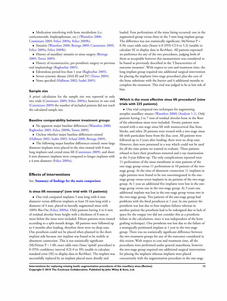
Sample size
A priori calculation for the sample size was reported in onlytwo trials (Cannizzaro 2009; Felice 2009a); however in one trial(Cannizzaro 2009) the number of included patients did not reachthe calculated sample size.
Baseline comparability between treatment groups
• No apparent major baseline differences (Wannfors 2000;Raghoebar 2005; Felice 2009b; Torres 2009).
• Unclear whether major baseline differences existed(Hallman 2002; Szabó 2005; Schaaf 2008; Bettega 2009).
• The following major baseline differences existed: more largediameter implants were placed in the sites treated with 8 mmlong implants and crestal sinus lift (Cannizzaro 2009), and short6 mm diameter implants were compared to longer implants witha 4 mm diameter (Felice 2009a).
Effects of interventions
See: Summary of findings for the main comparison
Is sinus lift necessary? (one trial with 15 patients)
• One trial compared implants 5 mm long with 6 mmdiameter versus different implants at least 10 mm long with adiameter of 4 mm, placed in laterally augmented sinus with100% Bio-Oss (Felice 2009a). Only patients having 4 to 6 mmof residual alveolar bone height with a thickness of 8 mm ormore below the sinus were included. Fifteen patients were treatedaccording to a split-mouth design. All patients were followed upto 4 months after loading, therefore there were no drop outs.One prosthesis could not be placed when planned in the shortimplant side because one implant was found to be mobile atabutment connection. This is not statistically significant(McNemar P = 1.00, exact odds ratio (Stata ’epitab’ procedure) is0 (95% confidence interval (CI) 0 to 39); unable to calculatestandard error (SE) to display data in RevMan). The implant wassuccessfully replaced by an implant placed more distally and
loaded. Four perforations of the sinus lining occurred: one in theaugmented group versus three in the 5 mm long implant group.The difference was not statistically significant, McNemar P =0.50, exact odds ratio (Stata) is 0 (95% CI 0 to 5.3) (unable tocalculate SE to display data in RevMan). All patients expressedno preference for any of the two procedures, judging both ofthem as acceptable however this measurement was considered tobe biased as previously described in the ’Characteristics ofoutcome measures’. With respect to cost and treatment time, thelong implant group required one additional surgical interventionfor placing the implants (two-stage procedure) plus the cost ofthe bone substitute with the barrier and 4 additional months tocomplete the treatment. This trial was judged to be at low risk ofbias.
Which is the most effective sinus lift procedure? (nine
trials with 235 patients)
• One trial compared two techniques for augmentingatrophic maxillary sinuses (Wannfors 2000) (Analysis 1.1). Onlypatients having 2 to 7 mm of residual alveolar bone in the floorof the edentulous sinus were included. Twenty patients weretreated with a one-stage sinus lift with monocortical iliac boneblocks, and other 20 patients were treated with a two-stage sinuslift with particulate bone from the iliac crest. All patients werefollowed up to 3 years after loading; there were no drop outs.However, data were presented in a way which could not be usedfor all the time points we wanted to evaluate. Three patientsrefused to have their prostheses removed and x-ray examinationat the 3-year follow-up. The only complications reported were11 perforations of the sinus membrane in nine patients of theone-stage group versus 11 perforations in 10 patients of the two-stage group. At the time of abutment connection 11 implants ineight patients were found to be not osseointegrated in the one-stage group versus seven implants in six patients of the two-stagegroup. At 1 year an additional five implants were lost in the one-stage group versus one in the two-stage group. At 3 years oneadditional implant was lost in the one-stage group versus two inthe two-stage group. Two patients of the one-stage group hadproblems with the fixed prostheses at 1 year. In one patient theprosthesis was lost due to four implant failures whereas inanother patient the prosthesis had to be redesigned due to lack ofspace for the tongue (we did not consider this as a prosthesisfailure in the calculations, since it was independent of the bonegrafting technique). One prosthesis was lost due to the failure ofa strategically positioned implant at 1 year in the two-stagegroup. There was no statistically significant difference betweenthe two treatment groups for any of the outcomes considered inthis review. With respect to cost and treatment time, all theprocedures were performed under general anaesthesia, howeverthe two-stage group required one additional surgical interventionfor placing the implants whereas implants were placedconcurrently with the augmentation procedure in the one-stage
13Interventions for replacing missing teeth: augmentation procedures of the maxillary sinus (Review)
Copyright © 2010 The Cochrane Collaboration. Published by John Wiley & Sons, Ltd.

group. The healing period was 6 months longer in the two-stagegroup. This trial was judged to be at high risk of bias.
• One trial compared three two-stage techniques foraugmenting atrophic maxillary sinuses (Hallman 2002) (Analysis1.2). Only patients with less than 5 mm of alveolar bone heightin the sinus floor and fixed dentition on the opposite jaw wereincluded. The trial was designed as a sort of split-mouth/parallelpreference trial. Eleven patients willing to provide autogenousbone from the mandibular ramus were treated with a split-mouth approach (autogenous bone versus 80% Bio-Oss and20% autogenous bone), whereas 10 patients who refused to havetheir bone harvested from the mandible were treated with 100%Bio-Oss. All patients were followed up to 1 year after loading;there were no drop outs. During the post-operative phase nocomplications occurred in either the augmented sites or thedonor sites. However a severe resorption of the autogenous bonegraft occurred in two patients. At abutment connection siximplants failed in five patients in the group treated withautogenous bone only and two implants failed in two patients inthe group treated with 80% Bio-Oss. No implants or prostheseswere lost at the 1-year evaluation. The author informed us thatadditional implants were lost at the 2-year follow-up in twopatients, causing the failure of the fixed prostheses. The completeinformation should be published in a future 5-year follow-upreport. There was no statistically significant difference for any ofthe outcomes considered in this review. With respect to cost andtreatment time, the only difference in cost was the use of thebone substitute. The healing period was 6 months. The trial wasjudged to be at high risk of bias.
• One trial compared two techniques for augmentingatrophic maxillary sinuses (Szabó 2005) (Analysis 1.3). Onlypatients with less than 5 mm of alveolar bone height in the sinusfloor were included. Twenty patients were treated with a split-mouth approach with a two-stage sinus lift with particulate bonefrom the iliac crest one side and with a two-stage sinus lift with100% Cerasorb (a beta-tricalcium phosphate bone substitute) onthe contralateral sinus. In 10 patients an additional autogenousonlay bone block was placed to widen the alveolar crest. Allpatients were followed up to implant loading and there were nodrop outs. No serious post-operative complications occurred atthe implant sites. Three complications occurred at the bone graftdonor sites: one permanent sensory loss of the lateral femoralcutaneous nerve and two had prolonged wound drainage (2 to 3weeks). At abutment connection two implants failed, one in eachgroup, they both had to be replaced in order to place theprosthesis and this caused a delay of 3 to 6 months (we did notconsider these as prosthesis failures in the calculations). Therewas no statistically significant difference between the twotreatments for any of the outcomes considered in this review.With respect to cost and treatment time, the only difference wasthe cost of the bone substitute. The trial was judged to be at high
risk of bias.
• One trial compared two one-stage techniques foraugmenting maxillary sinuses (Cannizzaro 2009) (Analysis 1.4).Only patients having 3 to 6 mm bone height at the sinus floorwere included. Twenty patients were treated with a sinus liftthrough a crestal approach and autogenous bone and 8 mm longimplants, and 20 patients were treated with a sinus lift through alateral window approach with a mixture of 50% particulateautogenous bone from the tuberosity area and 50% Bio-Oss andimplants at least 10 mm long. All patients were followed up to 1year after loading, and there were no drop outs. Fourcomplications occurred in four sinuses laterally augmented - oneabscess and one sinusitis, both determining the failure of thegraft and the implants, versus one peri-implant infection in theshort implant group. One implant failed in the short implantgroup at abutment connection and five implants (four in theimmediate post-operative phase and one at abutmentconnection) in three patients in the long implant group. Twoprostheses could not be placed in the long implant group versusone in the short implant group because of implant failures.There was no statistically significant difference for any of theoutcomes considered in this review. There was an additional costof the bone substitute in the group with the lateral approach. Allimplants were loaded 7 weeks after sinus lift. The trial wasjudged to be at low risk of bias.
• One trial compared two two-stage techniques foraugmenting maxillary sinuses using a lateral window approach(Felice 2009b) (Analysis 1.5; Analysis 1.6). Only patients having1 to 5 mm bilateral bone height at the sinus floor were included.Ten patients were treated with a split-mouth approach. Afterelevation of the sinus lining, one side was filled with granularBio-Oss whereas in the contra-lateral site, an Inion resorbablerigid barrier was used to maintain space to allow boneregeneration. All patients were followed up to 4 months afterloading, and there were no drop outs. After 6 months, bothinterventions gained a statistically significant amount of bone(14.4 mm for Inion versus 14.1 mm for Bio-Oss) but there wasno statistically significant difference between the procedures.There were no differences in complications between groups (twoperforations of the maxillary lining at the Inion treated sitesversus one at Bio-Oss site, Analysis 1.5), however, in one of thepatients where a perforation occurred at the Inion site, atimplant placement, the sinus was two thirds filled with softtissue. Implants were placed anyway and the site was successfullyretreated with Bio-Oss. No implant failed. The clinicianpreferred Bio-Oss because it was simpler to handle. There wereno statistically significant differences in patient preference foreither of the two techniques 1 month after surgery and 1 monthafter delivery of definitive prostheses: eight patients had nopreference while two preferred the Bio-Oss treated side. Withrespect to cost, both procedures used Inion barriers, but only one
14Interventions for replacing missing teeth: augmentation procedures of the maxillary sinus (Review)
Copyright © 2010 The Cochrane Collaboration. Published by John Wiley & Sons, Ltd.

procedure used the Bio-Oss. There was no difference in timetaken to complete the augmentation procedure (19.8 minutes forInion versus 20.5 for Bio-Oss) and all implants were loaded 11months after sinus lift. The trial was judged to be at low risk ofbias.
Trials evaluating the efficacy of platelet-rich plasma (PRP)
with grafts (four trials with 114 patients)
• One trial compared two techniques for augmentingresorbed maxillae including atrophic maxillary sinuses(Raghoebar 2005) (Analysis 1.7). Only patients with less than 5mm of alveolar bone height in the sinus floor were included. Fivepatients were treated with a split-mouth approach with two-stagesinus lift with autogenous bone together with buccal onlay grafts,harvested from the iliac crest, one side with PRP and the otherwithout. All patients were followed for 2 years after implantloading and there were no drop outs. No serious complicationsoccurred at the grafted sites: one sinus membrane was perforatedduring surgery but healing was uneventful. A small incisionbreakdown occurred in the first week at the non-PRP side of onepatient. A seroma which healed uneventfully was the onlycomplication that occurred at the donor sites. During theprosthetic phase one implant failed in the PRP side, but noprosthesis failed. There was no statistically significant differencebetween the two techniques for any of the outcomes consideredin this review. The difference in cost and treatment time was theuse of PRP. Prostheses were inserted about 10 months afteraugmentation. The trial was judged to be at high risk of bias.
• One trial compared two-stage sinus lift with lateral windowapproach using either autogenous particulate bone from the iliaccrest alone or the same graft with PRP in fully edentulouspatients (Schaaf 2008) (Analysis 1.7). All sites were alsohorizontally augmented with corticospongeous blocks and left toheal for 4 months. There were two publications for this trial.The first publication included 34 patients treated according to asplit-mouth design and 19 patients treated according to parallelgroup design but no clinical data were provided. In the secondpublication only the clinical data of the 34 patients treated witha split-mouth approach were presented and only these data arepresented in this review. All patients were followed up toabutment connection (6 months after implant insertion) andthere were no drop outs. Only complications at augmented siteswere reported: one sinusitis in two patients, one from eachgroup. Six patients experienced implant failures at abutmentconnection: one patient lost one implant at both sites, threepatients lost one implant each at the non-PRP treated sites only,and two patients lost one and three implants at the PRP side.There was no statistically significant difference between the twotechniques for any of the outcomes considered in this review.The difference in cost and treatment time was the use of PRP.The trial was judged to be at high risk of bias.
• One trial compared two two-stage techniques foraugmenting maxillary sinuses (Bettega 2009) (data not shown).Only patients with less than 8 mm of alveolar bone height in thesinus floor were included. Eighteen patients were treated with asplit-mouth approach with two-stage sinus lift with autogenousbone blocks from the iliac crest and Tissucol on one side andautologous granular bone and autologous platelet concentrate(APC) with Tissucol on the other. Patients were followed for 1year after implant placement and there were two drop outsbefore implant placement for financial reasons. There was nocomplication due to cytapheresis or surgery. All implants werestable 1 year after placement. There was no statisticallysignificant difference between the two techniques for any of theoutcomes considered in this review. The difference in cost andtreatment time was the use of APCs. The trial was judged to beat high risk of bias.
• One trial compared one or two-stage sinus lift proceduresusing a lateral window technique and 100% granular Bio-Osswith or without PRP, left to heal for 6 months with a hybrid ofsplit-mouth/parallel design trial (Torres 2009). In the originalpublication there were 87 patients included but only the datafrom the 57 patients treated according to a split-mouthprocedure are presented here (Analysis 1.7). Twenty-five patientshaving up to 4 mm of residual bone height were augmented firstand 98 implants were placed after 6 months whereas in 32patients with residual bone ranging between 4 mm to 7 mm 128implants were placed simultaneously to the sinus augmentationprocedure. Implants were left to heal unloaded for 6 months. Allpatients were followed for 2 years after loading and there were nodrop outs. Five perforations of the maxillary membrane occurredin five patients: three patients belonged to the PRP group andtwo to the non-PRP group. Partial loss of the graft occurred infive patients treated with the two-stage procedure: two patientsbelonged to the PRP group and three to the non-PRP group.According to the authors no prosthesis failed. Four implantsfailed in three patients treated according a two-stage procedure.Three implants failed in two patients at sides which were nottreated with PRP. There was no statistically significant differencebetween the group receiving PRP and the group that did notreceive PRP for any of the outcomes considered in this review.The difference in cost and treatment time was the use of PRP.The trial was judged to be at low risk of bias.
Meta-analysis was only possible for the three trials which com-pared, in split-mouth trials, particulate bone from the iliac crest(Raghoebar 2005; Schaaf 2008) or Bio-Oss (Torres 2009) withand without PRP. In two studies (Raghoebar 2005; Schaaf 2008),sites were also augmented with onlay blocks of autogenous bone.There were no statistically significant differences between groupswho received PRP and those who did not for implant failures andcomplications (Figure 1).
15Interventions for replacing missing teeth: augmentation procedures of the maxillary sinus (Review)
Copyright © 2010 The Cochrane Collaboration. Published by John Wiley & Sons, Ltd.

Figure 1. Forest plot of comparison: 1 Different sinus lift procedures, outcome: 1.7 Autogenous bone or
Bio-Oss +/- PRP.
D I S C U S S I O N
Twenty-nine potentially eligible trials were identified, but only 10met our inclusion criteria. Twelve trials had to be excluded becausepresented only histological data. The observation that the majorityof randomised clinical trials evaluating sinus lift procedures reportonly histological findings without providing any useful informa-tion on the actual clinical outcome of the sinus lift procedure andimplant rehabilitation, is both disappointing and alarming. Thisis not to say that histological information is not useful, but if notbacked up by meaningful clinical outcomes it appears that hu-man beings are used instead of animal as histological experimentalmodels and this is difficult to justify.
Sample sizes were relatively small with only two trials (Cannizzaro2009; Felice 2009a) reporting a sample size calculation. It is there-fore possible that many of these trials were underpowered todemonstrate any significant difference between groups. Neverthe-less the included trials did provide limited but indeed useful insightinto possible avenues for future clinical research and some clinicalindications which should be carefully evaluated by clinicians whendeciding whether to perform an augmentation procedure or not,or which augmentation procedure to select.
We first evaluated whether and when it may be necessary to aug-ment maxillary sinus and then which are the most effective aug-mentation procedures. This distinction is relevant since it is pos-sible that ineffective procedures which could be even potentiallydangerous are widely performed, despite no improvements oftreatment prognosis or patients’ quality of life.
Only one trial evaluated whether sinus lift procedures are indicatedin patients having a residual crestal height between 4 to 6 mm (Felice 2009a). The findings of this study are inconclusive due to thesmall sample size and the short follow-up (4 months after loading),however, they suggest that 5 mm long implants with a diameterof 6 mm can be successfully loaded 4 months after placementwithout the need of any augmentation procedure. Though the onlyimplant failure occurred in the short implant group, the implantwas successfully replaced with another short implant placed moredistally. There is the need for more trials to understand in whichclinical situations sinus lift procedures are beneficial for patients.
When evaluating which are the most effective augmentation pro-cedures we have eight trials providing some indications (Wannfors2000; Hallman 2002; Raghoebar 2005; Szabó 2005; Schaaf 2008;Bettega 2009; Cannizzaro 2009; Felice 2009b). Studies can begrouped in the following way.
When trying to answer the question whether grafting is necessaryto obtain bone regeneration, even in case of severely atrophic sinus,
16Interventions for replacing missing teeth: augmentation procedures of the maxillary sinus (Review)
Copyright © 2010 The Cochrane Collaboration. Published by John Wiley & Sons, Ltd.

the findings from the only pilot trial investigating this hypothesis(Felice 2009b) clearly indicated that a graft is not needed to obtainnew bone in the sinus cavity, if it is possible to keep sufficient spaceusing a resorbable rigid barrier. On the other hand, the operatorfound that it was technically simpler to use a bone substitute ratherthan to mould a space-maintaining barrier. The same study alsosuggested that there is not a clear correlation between the amountof newly formed bone, evaluated with histomorphometry, and theclinical success of the implants. In fact, all implants became suc-cessfully osseointegrated also in presence of an average of 24% ofnewly formed bone. In general, using surrogate outcomes, suchas histomorphometry, as the only outcome on which to predictimplant success in case of sinus augmentations with various mate-rials, is shown to be inaccurate and misleading. Clinically relevantprimary outcomes such as implant failure and complication ratesshould be used in conjunction with surrogate outcomes.
The question whether autogenous bone could be replaced by bonesubstitutes to reduce patient morbidity was addressed in two trials(Hallman 2002; Szabó 2005). One trial (Szabó 2005) is of littleuse because the follow-up was limited to abutment connection andonlay bone blocks were used in half of the patients. The findings ofother trial (Hallman 2002) suggest that 80% to 100% of Bio-Osscan be used as bone substitute. Autogenous bone grafting mightbe replaced by bone substitutes for this indication, however largertrials with longer follow-up should be conducted to validate thesepreliminary findings.
One trial compared a one-stage crestal sinus lift procedure withautogenous bone and 8 mm long implants with a lateral windowsinus lift with a mix of autogenous bone and 50% Bio-Oss toplace longer implants (Cannizzaro 2009). Though no statisticallysignificant differences were found, there were more complicationsand failures with the lateral window augmentation procedure. It isinteresting to observe that all implants were placed in bone with aresidual height of 3 to 6 mm and were loaded less than 2 monthsafter sinus lift. It is generally accepted that 2 months in humans isinsufficient time to allow for new bone formation. Therefore theoriginal bone must have been sufficient to hold the implants, withboth lifting procedures adding little or no benefit.
When comparing a one-stage monocortical bone block versus atwo-stage technique with particulate bone harvested from the iliaccrest for maxillary sinus lifting, no statistically or clinically signif-icant differences were observed (Wannfors 2000). However, theuse of autogenous bone blocks from the iliac crest in a one-stageprocedure is a technique seldom used nowadays and most of thesinus lifting procedures are now performed under local anaesthe-sia.
Four trials (Raghoebar 2005; Schaaf 2008; Bettega 2009; Torres2009) evaluated the possible advantage of using platelet-richplasma (PRP) to speed bone healing for sinus augmentation. Thereis no evidence of a clinical benefit from the use of PRP, therefore
there appears to be no reasons to justify its use in this application.
Most of the augmentation procedures evaluated in these trials wereperformed by experienced clinicians, therefore caution is recom-mended when extrapolating the results of the present review toother clinical settings, such as general practice. Clinicians shouldcarefully evaluate the potential added benefits for each individualpatient of maxillary sinus augmentation. Where a benefit is antic-ipated the more effective procedure associated with fewer risks ofcomplication/discomfort for the patient should be selected.
A U T H O R S ’ C O N C L U S I O N SImplications for practice
Conclusions are based on 10 small trials including 250 patients,sometimes with short follow-up, and generally assessed as being athigh risk of bias. Therefore these results should be interpreted withgreat caution and should be viewed as very preliminary, requiringconfirmation by large multicentre trials.
• One trial investigated whether and when it is necessary toaugment maxillary sinus.
(1) It is still unclear when sinus lift procedures are needed.
(2) Implants 5 mm long and 6 mm wide can be successfully loadedin maxillary bone with a residual height of 4 to 6 mm below thesinus without making any augmentation procedure, though thelong-term prognosis is unclear.
• Nine trials investigated which are the most effective sinuslift techniques, four of which evaluated the efficacy of platelet-rich plasma (PRP).
(1) If the residual alveolar bone height is 3 to 6 mm, a crestal ap-proach to lift the sinus lining and place 8 mm implants may pos-sibly lead to fewer complications than a lateral window approachto place implants at least 10 mm long.
(2) Keeping the sinus lining elevated by means of a rigid resorbablebarrier in presence of 1 to 5 mm of residual bone height withoutthe addition of a graft is sufficient to regenerate new bone to allowrehabilitation with implant-supported prostheses. However, it istechnically simpler to fill the sinus with a granular bone substitute.
(3) Bone substitutes such Bio-Oss and Cerasorb appear to be aseffective as autogenous bone grafts for augmenting atrophic max-illary sinuses, therefore they could be used as a replacement forautogenous bone grafting.
(4) There is no evidence that the addition of PRP treatment toautogenous bone grafts or bone substitutes improves the outcomeof sinus lift procedures for implant rehabilitation.
17Interventions for replacing missing teeth: augmentation procedures of the maxillary sinus (Review)
Copyright © 2010 The Cochrane Collaboration. Published by John Wiley & Sons, Ltd.

Implications for research
In order to understand when sinus lift procedures are needed andwhich are the most effective sinus lift techniques, larger, well de-signed trials are needed. Such trials should include long-term fol-low-up and be reported according to the Consolidated Standardsof Reporting Trials (CONSORT) guidelines (Moher 2001) (http://www.consort-statement.org/). It is difficult to provide clear in-dications with respect of which sinus lift procedures should beevaluated first, however, once established in which clinical situa-tions these procedures are actually needed, priority should be givento those interventions that are simpler, less invasive, involve lessrisk of complications, and reach their goals within the shortesttimeframe. Research efforts should be concentrated on a few im-portant clinical questions, using larger sample sizes. This mightbe obtained through collaborative efforts among various researchgroups. Among of the identified research priorities is to evaluatewhether and when one-stage lifting via a crestal approach can re-place the more invasive lateral window procedures and whetherbone substitutes can be used for replacing autogenous bone in
augmenting severely atrophic maxillary sinuses.
A C K N O W L E D G E M E N T S
We wish to thank all those people who helped us with the finalpreparation of the current review, and in particular: Anne Little-wood (Cochrane Oral Health Group) and Sylvia Bickley for theirassistance with literature searching; Luisa Fernandez Mauleffinchand Philip Riley (Cochrane Oral Health Group) for their help withthe preparation of this review; Stella Kwan as a coauthor of oneprevious version of this review; Lars Andersson, Filippo Cangini,Mats Hallman, Björn Johansson, Adriano Piattelli, Gerry Raghoe-bar, Heidrun Schaaf, Andreas Thor and Jesus Torres for kindlyproviding us with additional information on their trials. We wouldalso like to thank the following referees: Stephen Chen, MatteoChiapasco, Christer Dahlin, Mats Hallman, Jayne Harrison, JanHirsch, Anne Littlewood, Ian Needleman, Michele Nieri, GerryRaghoebar, Heidrun Schaaf, Bill Shaw and Jesus Torres.
R E F E R E N C E S
References to studies included in this review
Bettega 2009 {published data only}∗ Bettega G, Brun JP, Boutonnat J, Cracowski JL, QuesadaJL, Hegelhofer H, et al.Autologous platelet concentrates forbone graft enhancement in sinus lift procedure. Transfusion
2009;49:779–85.Bettega G, Brun JP, Cracowski JL, Vérain A, Raphael B. [Useof autologous platelet concentrates during pre-implantationmaxillary reconstruction]. Revue de Stomatologie et de
Chirurgie Maxillofaciale 2005;106(3):189–91.
Cannizzaro 2009 {published data only}
Cannizzaro G, Felice P, Leone M, Viola P, Esposito M.Early loading of hydroxyapatite coated implants in theatrophic posterior maxilla: lateral sinus lift with autogenousbone and Bio-Oss versus crestal mini-sinus lift and 8 mmimplants. A randomized controlled clinical trial. European
Journal of Oral Implantology 2009;2:25–38.
Felice 2009a {published data only}
Felice P, Checchi V, Pistilli R, Scarano A, Pellegrino G,Esposito M. Bone augmentation versus 5 mm long dentalimplants in posterior atrophic jaws. Four-month post-loading results from a randomized controlled clinical trial.European Journal of Oral Implantology 2009;2:267–81.
Felice 2009b {published data only}
Felice P, Esposito M, Scarano A, Pistilli R, Lizio G, ChecchiL. A comparison of two techniques to augment maxillarysinus with the lateral approach: no grafting procedure vsanorganic bone placement. Preliminary histological and
clinical outcomes of a randomized controlled clinical trial.Clinical Oral Implants Research 2009;20:973 (Abs 261).∗ Felice P, Scarano A, Pistilli R, Checchi L, PiattelliM, Pellegrino G, et al.A comparison of two techniquesto augment maxillary sinuses using the lateral windowapproach: rigid synthetic resorbable barriers versusanorganic bovine bone. Five-month post-loading clinicaland histological results of a pilot randomized controlledclinical trial. European Journal of Oral Implantology 2009;2:293–306.
Hallman 2002 {published and unpublished data}
Hallman M, Sennerby L, Lundgren S. A clinical andhistologic evaluation of implant integration in the posteriormaxilla after sinus floor augmentation with autogenousbone, bovine hydroxyapatite, or a 20:80 mixture. The
International Journal of Oral and Maxillofacial Implants
2002;17(5):635–43.
Raghoebar 2005 {published and unpublished data}
Raghoebar GM, Schortinghuis J, Liem RS, Ruben JL, vander Wal JE, Vissink A. Does platelet-rich plasma promoteremodeling of autologous bone grafts used for augmentationof the maxillary sinus floor?. Clinical Oral Implants Research
2005;16(3):349–56.
Schaaf 2008 {published and unpublished data}
Schaaf H, Streckbein P, Lendeckel S, Heidinger K, Görtz B,Bein G, et al.Topical use of platelet-rich plasma to influencebone volume in maxillary augmentation: a prospectiverandomized trial. Vox Sanguinis 2008;94(1):64–9.∗ Schaaf H, Streckbein P, Lendeckel S, Heidinger K,Rehmann P, Boedeker RH, et al.Sinus lift augmentationusing autogenous bone grafts and platelet-rich plasma:
18Interventions for replacing missing teeth: augmentation procedures of the maxillary sinus (Review)
Copyright © 2010 The Cochrane Collaboration. Published by John Wiley & Sons, Ltd.
radiographic results. Oral Surgery, Oral Medicine, Oral
Pathologhy, Oral Radiology and Endodontics 2008;106:673–8.
Szabó 2005 {published and unpublished data}
Szabó G, Huys L, Coulthard P, Maiorana C, GaragiolaU, Barabas J, et al.A prospective multicenter randomizedclinical trial of autogenous bone versus beta-tricalciumphosphate graft alone for bilateral sinus elevation: histologicand histomorphometric evaluation. The International
Journal of Oral and Maxillofacial Implants 2005;20(3):371–81.
Torres 2009 {published data only}
Torres J, Tamimi F, Martinez PP, Alkhraisat MH, Linares R,Hernandez G, et al.Effect of platelet-rich plasma on sinuslifting: a randomized-controlled clinical trial. Journal Of
Periodontology 2009;36:677–87.
Wannfors 2000 {published and unpublished data}
Johansson B. Bone Grafts and Dental Implants in the
Reconstruction of the Severely Atrophied, Edentulous Maxilla.Uppsala: Uppsala University, 2001.Johansson B. Bone grafts. In manuscript 2005.∗ Wannfors K, Johansson B, Hallman M, Strandkvist T. Aprospective randomized study of 1- and 2-stage sinus inlaybone grafts: 1-year follow-up. The International Journal of
Oral and Maxillofacial Implants 2000;15(5):625–32.
References to studies excluded from this review
Aimetti 2008 {published and unpublished data}
Aimetti M, Romano F, Dellavia C, De Paoli S. Sinusgrafting using autogenous bone and platelet-rich plasma:histologic outcomes in humans. The International Journal of
Periodontics and Restorative Dentistry 2008;28:585–91.
Barone 2005 {published data only}
Barone A, Crespi R, Nicoli N, Fini M, Giardino R,Covani U. Maxillary sinus augmentation: histologic andhistomorphometric analysis. The International Journal of
Oral and Maxillofacial Implants 2005;20:519–25.
Barone 2008 {published data only}
Barone A, Santini S, Marconcini S, Giacomelli L, GherloneE, Covani U. Osteotomy and membrane elevation duringthe maxillary sinus augmentation procedure. A comparativestudy: piezoelectric device vs conventional rotativeinstruments. Clinical Oral Implants Research 2008;19:511–5.
Boyne 2005 {published data only}
Boyne PJ, Lilly LC, Marx RE, Moy PK, Nevins M, SpagnoliDB, et al.De novo bone induction by recombinant humanbone morphogenetic protein-2 (rh BMP-2) in maxillarysinus floor augmentation. Journal of Oral and Maxillofacial
Surgery 2005;63(12):1693–707.
Choi 2009 {published data only}
Choi KS, Kan JY, Boyne PJ, Goodacre CJ, Lozada JL,Rungcharassaeng K. The effects of resorbable membrane onhuman maxillary sinus graft: a pilot study. The International
Journal of Oral and Maxillofacial Implants 2009;24:73–80.
Consolo 2007 {published data only}
Consolo U, Zaffe D, Bertoldi C, Ceccherelli G. Platelet-rich plasma activity on maxillary sinus floor augmentationby autologous bone. Clinical Oral Implants Research 2007;18(2):252–62.
Cordaro 2008 {published data only}
Cordaro L, Bosshardt DD, Palattella P, Rao W, Serino G,Chiapasco M. Maxillary sinus grafting with Bio-Oss orStraumann Bone Ceramic: histomorphometric results froma randomized controlled multicenter clinical trial. Clinical
Oral Implants Research 2008;19:796–803.
Crespi 2009 {published data only}
Crespi R, Mariani E, Benasciutti E, Capparé P, Cenci S,Gherlone E. Magnesium-enriched hydroxyapatite versusautologous bone in maxillary sinus grafting: combininghistomorphometry with osteoblast gene expression profilesex vivo. Journal of Periodontology 2009;80:586–93.
Froum 1998 {published data only}∗ Froum SJ, Tarnow DP, Wallace SS, Rohrer MD, ChoS-C. Sinus floor elevation using anorganic bovine bonematrix (OsteoGraf/N) with and without autogenous bone:a clinical, histologic, radiographic, and histomorphometricanalysis - Part 2 of an ongoing prospective study. The
International Journal of Periodontics and Restorative Dentistry
1998;18(6):529–43.Tarnow DP, Wallace SS, Froum SJ, Rohrer MD, Cho SC.Histologic and clinical comparison of bilateral sinus floorelevations with and without barrier membrane placement in12 patients: Part 3 of an ongoing prospective study. The
International Journal of Periodontics and Restorative Dentistry
2000;20(2):117–25.
Froum 2006 {published data only}
Froum SJ, Wallace SS, Elian N, Cho SC, Tarnow DP.Comparison of mineralized cancellous bone allograft(Puros) and anorganic bovine bone matrix (Bio-Oss) forsinus augmentation: histomorphometry at 26 to 32 weeksafter grafting. The International Journal of Periodontics and
Restorative Dentistry 2006;26(6):543–51.
Froum 2008 {published data only}
Froum SJ, Wallace SS, Cho SC, Elian N, Tarnow DP.Histomorphometric comparison of a biphasic bone ceramicto anorganic bovine bone for sinus augmentation: 6- to 8-month postsurgical assessment of vital bone formation. Apilot study. The International Journal of Periodontics and
Restorative Dentistry 2008;28:273–81.
Hallman 2008 {published data only}
Hallman M, Lindgren C, Sennerby L, Mordenfeld A,Strandkvist T. Clinical histology of SLA microimplantsplaced into different biomaterials. EAO conference,Warsaw. 2008.
Kassolis 2005 {published data only}
Kassolis JD, Reynolds MA. Evaluation of the adjunctivebenefits of platelet-rich plasma in subantral sinusaugmentation. The Journal of Craniofacial Surgery 2005;16
(2):280–7.
19Interventions for replacing missing teeth: augmentation procedures of the maxillary sinus (Review)
Copyright © 2010 The Cochrane Collaboration. Published by John Wiley & Sons, Ltd.

Kim 2009 {published data only}
Kim YK, Yun PY, Kim SG, Lim SC. Analysis of thehealing process in sinus bone grafting using various graftingmaterials. Oral Surgery, Oral Medicine, Oral Pathology, Oral
Radiology, and Endodontics 2009;107:204–11.
Mangano 2007 {published and unpublished data}
Mangano C, Scarano A, Perrotti V, Iezzi G, Piattelli A.Maxillary sinus augmentation with a porous synthetichydroxyapatite and bovine-derived hydroxyapatite: acomparative clinical and histologic study. The International
Journal of Oral and Maxillofacial Implants 2007;22(6):980–6.
Steigmann 2005 {published data only}
Steigmann M, Garg AK. A comparative study of bilateralsinus lifts performed with platelet-rich plasma alone versusalloplastic graft material reconstituted with blood. Implant
Dentistry 2005;14(3):261–6.
Suba 2006 {published data only}
Suba Z, Takács D, Matusovicz D, Fazekas A, Szabó G,Barabás J. Quantitative and qualitative comparison ofthe maxillary bone regeneration after beta-tricalciumphosphate and autogenous bone implantation [Amaxilla csontregeneraciojanak mennyisegi es minosegiosszehasonlitasa beta–tricalcium phosphate es autologcsontbeultetes utan]. Fogorvosi Szemle 2006;99(1):21–8.∗ Suba Z, Takács D, Matusovits D, Barabás J, Fazekas A,Szabó G. Maxillary sinus floor grafting with beta-tricalciumphosphate in humans: density and microarchitecture of thenewly formed bone. Clinical Oral Implants Research 2006;17(1):102–8.
Tawil 2001 {published data only}
Tawil G, Mawla M. Sinus floor elevation using a bovinebone mineral (Bio-Oss) with or without the concomitantuse of a bilayered collagen barrier (Bio-Guide): a clinicalreport of immediate and delayed implant placement. The
International Journal of Oral and Maxillofacial Implants
2001;16(5):713–21.
Triplett 2009 {published data only}
Triplett RG, Nevins M, Marx RE, Spagnoli DB, OatesTW, Moy PK, et al.Pivotal, randomized, parallel evaluationof recombinant human bone morphogenetic protein-2/absorbable collagen sponge and autogenous bone graft formaxillary sinus floor augmentation. Journal of Oral and
Maxillofacial Surgery 2009;67:1947–60.
Additional references
Aghaloo 2007
Aghaloo TL, Moy PK. Which hard tissue augmentationtechniques are the most successful in furnishing bonysupport for implant placement?. The International Journal
of Oral and Maxillofacial Implants 2007;22(Suppl):49–70.
Aparicio 2001
Aparicio C, Perales P, Rangert B. Tilted implants as analternative to maxillary sinus grafting: a clinical, radiologic,and periotest study. Clinical Implant Dentistry and Related
Research 2001;3:39–49.
Boyne 1980
Boyne PJ, James RA. Grafting of the maxillary sinus floorwith autogenous marrow and bone. Journal of Oral Surgery
1980;38:613–6.
Brånemark 1977
Brånemark PI, Hansson BO, Adell R, Breine U, LindstromJ, Hallen O, et al.Osseointegrated implants in the treatmentof the edentulous jaw. Experience from a 10-year period.Scandinavian Journal of Plastic and Reconstructive Surgery.
Supplementum 1977;16:1–132.
Brånemark 2004
Brånemark PI, Gröndahl K, Öhrnell LO, Nilsson, P,Petruson B, Svensson B, et al.Zygoma fixture in themanagement of advanced atrophy of the maxilla: techniqueand long-term results. Scandinavian Journal of Plastic and
Reconstructive Surgery 2004;38(2):70–85.
Cannizzaro 2007
Cannizzaro G, Leone M, Consolo U, Ferri V, LicitraG, Worthington H, et al.Augmentation of the posterioratrophic edentulous maxilla with implants placed in theulna: a prospective single-blind controlled clinical trial.The International Journal of Oral and Maxillofacial Implants
2007;22:280–8.
Cawood 1991
Cawood JI, Howell RA. Reconstructive preprostheticsurgery. I. Anatomical considerations. International Journal
of Oral and Maxillofacial Surgery 1991;20(2):75–82.
Cosci 2000
Cosci F, Luccioli M. A new sinus lift technique inconjunction with placement of 265 implants: a 6-yearretrospective study. Implant Dentistry 2000;9:363–8.
das Neves 2006
das Neves FD, Fones D, Bernardes SR, do Prado CJ, NetoAJ. Short implants - an analysis of longitudinal studies. The
International Journal of Oral and Maxillofacial Implants
2006;21:86–93.
Del Fabbro 2004
Del Fabbro M, Testori T, Francetti L, Weinstein R.Systematic review of survival rates for implants placed inthe grafted maxillary sinus. The International Journal of
Periodontics and Restorative Dentistry 2004;24(6):565–77.
Egger 1997
Egger M, Smith GD, Schneider M, Minder C. Bias inmeta-analysis detected by a simple, graphical test. BMJ
1997;315:629–34.
Elbourne 2002
Elbourne DR, Altman DG, Higgins JP, Curtin F,Worthington HV, Vail A. Meta-analyses involving cross-over trials: methodological issues. International Journal of
Epidemiology 2002;31(1):140–9.
Emmerich 2005
Emmerich D, Att W, Stappert C. Sinus floor elevation usingosteotomes: a systematic review and meta-analysis. Journal
of Periodontology 2005;76(8):1237–51.
20Interventions for replacing missing teeth: augmentation procedures of the maxillary sinus (Review)
Copyright © 2010 The Cochrane Collaboration. Published by John Wiley & Sons, Ltd.

Esposito 2005
Esposito M, Worthington HV, Coulthard P. Interventionsfor replacing missing teeth: dental implants in zygomaticbone for the rehabilitation of the severely deficientedentulous maxilla. Cochrane Database of Systematic Reviews
2005, Issue 4. [Art. No.: CD004151. DOI: 10.1002/14651858.CD004151.pub2]
Esposito 2007
Esposito M, Murray-Curtis L, Grusovin MG, CoulthardP, Worthington HV. Interventions for replacing missingteeth: different types of dental implants. Cochrane Database
of Systematic Reviews 2007, Issue 4. [DOI: 10.1002/14651858.CD003815.pub3]
Esposito 2009
Esposito M, Grusovin MG, Felice P, Karatzopoulos G,Worthington HV, Coulthard P. Interventions for replacingmissing teeth: horizontal and vertical bone augmentationtechniques for dental implant treatment. Cochrane Database
of Systematic Reviews 2009, Issue 4. [DOI: 10.1002/14651858.CD003607.pub4]
Gabbert 2009
Gabbert O, Koob A, Schmitter M, Rammelsberg P.Implants placed in combination with an internal sinus liftwithout graft material: an analysis of short-term failure.Journal of Clinical Periodontology 2009;36:177–83.
Graves 1994
Graves SL. The pterygoid plate implant: a solution forrestoring the posterior maxilla. The International Journal of
Periodontics and Restorative Dentistry 1994;14:513–23.
Hatano 2007
Hatano N, Sennerby L, Lundgren S. Maxillarysinus augmentation using sinus membrane elevationand peripheral venous blood for implant-supportedrehabilitation of the atrophic posterior maxilla: case series.Clinical Implant Dentistry and Related Research 2007;9:150–5.
Higgins 2009
Higgins JPT, Green S (editors). Cochrane Handbook forSystematic Reviews of Interventions version 5.0.2 (updatedSeptember 2009). The Cochrane Collaboration, 2009.Available from www.cochrane-handbook.org.
Lesaffre 2009
Lesaffre E, Philstrom B, Needleman I, Worthington H.The design and analysis of split-mouth studies: Whatstatisticians and clinicians should know. Statistics in
Medicine 2009;28(28):3470–82.
Lundgren 2004
Lundgren S, Andersson S, Gualini F, Sennerby L. Bonereformation with sinus membrane elevation: a new surgicaltechnique for maxillary sinus floor augmentation. Clinical
Implant Dentistry and Related Research 2004;6:165–73.
Moher 2001
Moher D, Schulz KF, Altman DG. The CONSORTstatement revised recommendations for improving thequality of reports of parallel-group randomised trials. The
Lancet 2001;357(9263):1191–4.
Nedir 2006
Nedir R, Bischof M, Vazquez L, Szmukler-Moncler S,Bernard J P. Osteotome sinus floor elevation withoutgrafting material: a 1-year prospective pilot study with ITIimplants. Clinical Oral Implants Research 2006;17:679–86.
Nkenke 2009
Nkenke E, Stelzle F. Clinical outcomes of sinus flooraugmentation for implant placement using autogenousbone or bone substitutes: a systematic review. Clinical Oral
implants Research 2009;20(Suppl 4):124–33.
Palmer 2000
Palmer P, Palmer R. Implant surgery to overcome anatomicaldifficulties. In: Palmer R editor(s). A Clinical Guide to
Implants in Dentistry. London: British Dental Association,2000:57–65.
Pjetursson 2008
Pjetursson BE, Tan WC, Zwahlen M, Lang NP. A systematicreview of the success of sinus floor elevation and survival ofimplants inserted in combination with sinus floor elevation.Journal of Clinical Periodontology 2008;35(8 Suppl):216–40.
Pjetursson 2009
Pjetursson BE, Ignjatovic D, Matuliene G, Brägger U,Schmidlin K, Lang NP. Transalveolar maxillary sinusfloor elevation using osteotomes with or without graftingmaterial. Part II: Radiographic tissue remodeling. Clinical
Oral Implants Research 2009;20:677–83.
Renouard 2006
Renouard F, Nisand D. Impact of implant length anddiameter on survival rates. Clinical Oral Implants Research
2006;17(Suppl 2):35–51.
Sohn 2008
Sohn DS, Lee JS, Ahn MR, Shin HI. New bone formationin the maxillary sinus without bone grafts. Implant Dentistry
2008;17:321–31.
Summers 1994
Summers RB. A new concept in maxillary implant surgery:the osteotome technique. Compendium of Continuing
Education in Dentistry 1994;15:152–8.
Summers 1995
Summers RB. The osteotome technique: Part 4 - Futuresite development. Compendium of Continuing Education in
Dentistry 1995;16:1080–92.
Tan 2008
Tan WC, Lang NP, Zwahlen M, Pjetursson BE. Asystematic review of the success of sinus floor elevation andsurvival of implants inserted in combination with sinusfloor elevation. Part II: transalveolar technique. Journal of
Clinical Periodontology 2008;35(8 Suppl):241–54.
Tatum 1986
Tatum H. Maxillary and sinus implant reconstructions.Dental Clinics of North America 1986;30(2):207–29.
Thor 2007
Thor A, Sennerby L, Hirsch JM, Rasmusson L.Bone formation at the maxillary sinus floor followingsimultaneous elevation of the mucosal lining and implant
21Interventions for replacing missing teeth: augmentation procedures of the maxillary sinus (Review)
Copyright © 2010 The Cochrane Collaboration. Published by John Wiley & Sons, Ltd.

installation without graft material: an evaluation of 20patients treated with 44 Astra Tech implants. Journal of
Oral and Maxillofacial Surgery 2007;65(7 Suppl 1):64–72.
Tong 1998
Tong DC, Rioux K, Drangsholt M, Beirne OR. A reviewof survival rates for implants placed in grafted maxillarysinuses using meta-analysis. The International Journal of
Oral and Maxillofacial Implants 1998;13(2):175–82.
Urist 1965
Urist MR. Bone: formation by autoinduction. Science
1965;150(3698):893–9.
Valentin-Opran 2002
Valentin-Opran A, Wozney J, Csimma C, Lilly L, RiedelGE. Clinical evaluation of recombinant human bonemorphogenic protein-2. Clinical Orthopaedics and Related
Research 2002;395:110–20.
Wallace 2003
Wallace SS, Froum SJ. Effect of maxillary sinusaugmentation on the survival of endosseous dental implants.A systematic review. Annals of Periodontology 2003;8(1):328–43.
References to other published versions of this review
Coulthard 2003
Coulthard P, Esposito M, Jokstad A, WorthingtonHV. Interventions for replacing missing teeth: bone
augmentation techniques for dental implant treatment.Cochrane Database of Systematic Reviews 2003, Issue 3. [Art.No.: CD003607. DOI: 10.1002/14651858.CD3607]
Esposito 2006a
Esposito M, Grusovin MG, Worthington HV,Coulthard P. Interventions for replacing missing teeth:bone augmentation techniques for dental implanttreatment. Cochrane Database of Systematic Reviews
2006, Issue 1. [Art. No.: CD003607. DOI: 10.1002/14651858.CD3607.pub2]
Esposito 2006b
Esposito M, Grusovin MG, Coulthard P, WorthingtonHV. The efficacy of various bone augmentation proceduresfor dental implants: a Cochrane systematic review ofrandomized controlled clinical trials. The International
Journal of Oral and Maxillofacial Implants 2006;21(5):696–710.
Esposito 2008
Esposito M, Grusovin MG, Kwan S, Worthington HW,Coulthard P. Interventions for replacing missing teeth: boneaugmentation techniques for dental implant treatment.Cochrane Database of Systematic Reviews 2008, Issue 3.[DOI: 10.1002/14651858.CD003607.pub3]
∗ Indicates the major publication for the study
22Interventions for replacing missing teeth: augmentation procedures of the maxillary sinus (Review)
Copyright © 2010 The Cochrane Collaboration. Published by John Wiley & Sons, Ltd.

C H A R A C T E R I S T I C S O F S T U D I E S
Characteristics of included studies [ordered by study ID]
Bettega 2009
Methods Randomised, split-mouth study, 1-year post-implant placement follow-up. Two with-drawals for financial reasons
Participants Patients having less than 8 mm of alveolar bone at the floor of the sinus. Adults treatedat the Plastic and Maxillofacial Surgery Unit, Grenoble, France. Exclusion criteria weresmokers, maxillary sinus lesion or history of sinusitis or sinus surgery, abnormal bloodcounts, ASA score 2 or more. 18 patients were treated
Interventions Two-stage sinus lift with autogenous cortico-cancellous blocks from the iliac crest versusgranules of bone with platelet concentrates (APCs) and a biologic glue (Tissucol, BaxterSA, Maurepas, France) left to heal for 6 months in a split-mouth trial. Plateletpheresis wasmade at least 3 days before surgery on a plateletpheresis collection system (Trima Accel,Version 5.1, Gambro BCT, Lakewood, CO), a single-needle continuous-flow separationsystem. It was aimed to obtain a post-donation platelet count of more than 100 x 106per ml. Citrate (ACD-A) was used for anticoagulation. APCs were delivered by the cell-processing laboratory in a 20 ml transfer bag that was centrifuged for 15 minutes. Theplasma was removed with a plasma extraction device to reach the target volume of 8 to15 ml. 2 ml of cancellous bone was mixed with half of the APCs volume and 1 ml ofTissucol. The remaining APCs were mixed with 0.5 ml Tissucol to obtain a membranefor covering the grafted area. Sites treated with the bone block were covered by 1 ml ofTissucol. Implants were placed 6 months after the augmentation procedure
Outcomes Implant failure. Complications at the augmented and donor sites. Histomorphometricaland radiographic evaluation: sinus floor height and bone density on panoramic radio-graphs and CT scans
Notes
Risk of bias
Item Authors’ judgement Description
Adequate sequence generation? Yes Article: “(Randomization) was chosenthrough a two-element randomization ta-ble”
Allocation concealment? Unclear Article: “The iliac crest graft was then har-vested, and the destination for the two tech-niques (traditional versus APC) was chosenthrough a two-element randomization ta-ble”Author did not reply to our request for clar-ification.
23Interventions for replacing missing teeth: augmentation procedures of the maxillary sinus (Review)
Copyright © 2010 The Cochrane Collaboration. Published by John Wiley & Sons, Ltd.

Bettega 2009 (Continued)
Blinding?All outcomes
Unclear Article: “All radiological images were readblindly by a radiologist”. However it wasnot specified if the other outcome measureswere assessed blindlyAuthor did not reply to our request for clar-ification.
Incomplete outcome data addressed?All outcomes
No Data partially presented.Author did not reply to our request for clar-ification.
Free of selective reporting? Yes All outcomes reported were previously de-scribed in the published research protocol
Free of other bias? Yes
Cannizzaro 2009
Methods Randomised trial of parallel group design, 1-year post-loading follow-up. No withdrawals
Participants Patients having 3 to 6 mm of alveolar bone at the floor of the sinus. Adults treated ata private dental practice, Pavia, Italy. Exclusion criteria were: general contraindicationsto implant surgery, subjected to irradiation in the head and neck area less than 1 yearago, treated or under treatment with intravenous amino-bisphosphonates, poor oralhygiene and motivation, uncontrolled diabetes, pregnant or lactating, substance abusers,psychiatric problems or unrealistic expectations, lack of opposite occluding dentition/prosthesis in the area intended for implant placement, acute infection in the area intendedfor implant placement. 20 patients were treated in each group
Interventions One-stage sinus lift using one to three 8 mm long implants placed in simultaneouslycrestally augmented sinus with autogenous particulate bone, harvested from the implantsite, versus one to three 10 mm or longer implants placed in simultaneously augmentedsinuses using the lateral approach with a mixture of 50% particulate autogenous bonefrom the tuberosity area and 50% Bio-Oss. A modified ’Cosci technique’ was used tocrestally augment the sinus. In brief implant sites were prepared with a 2.5 mm trephinedrill up to about 1 mm of the sinus cortical wall, to collect autogenous bone, andwith a 3.1 mm diameter atraumatic lifting drill. Resorbable barriers (Biomend Extend,Sulzer Dental Inc., Carlsbad, CA, USA) were used to seal the lateral windows. All theaugmentation procedures were performed under local anaesthesia. All implants were leftto heal submerged for 45 days and were functionally loaded within 1 week after abutmentconnection. All implants were tapered Screw-Vent MP-1 HA Dual Transition SelectiveSurface implants (Zimmer Dental, Carlsbad, CA, USA) inserted in under preparedosteotomy sites with a torque of at least 35 N/cm
Outcomes Prosthesis and implant failure. Complications at the augmented and donor sites. Peri-implant marginal bone levels
Notes
24Interventions for replacing missing teeth: augmentation procedures of the maxillary sinus (Review)
Copyright © 2010 The Cochrane Collaboration. Published by John Wiley & Sons, Ltd.

Cannizzaro 2009 (Continued)
Risk of bias
Item Authors’ judgement Description
Adequate sequence generation? Yes Article: “A computer generated restrictedrandomisation list was created”
Allocation concealment? Yes Article: “Only one of the investigators(Marco Esposito), not involved in the se-lection and treatment of the patients, wasaware of the randomization sequence andcould have access to the randomization liststored in his pass-word protected portablecomputer. The randomized codes were en-closed in sequentially numbered, identical,opaque, sealed envelopes. Envelopes wereopened sequentially after eligible patientswere anaesthetised, therefore, treatment al-location was concealed to the investigatorsin charge of enrolling and treating the pa-tients”
Blinding?All outcomes
Yes Article: “One dentist (Giuseppe Fontana)not involved in the treatment of the pa-tients made all the clinical assessmentswithout knowing group allocation, there-fore outcome assessor was blinded, how-ever Bio-Oss augmented sites could beidentified on radiographs because they ap-peared more radio-opaque and implantswere longer”
Incomplete outcome data addressed?All outcomes
Yes All data presented, no drop outs.
Free of selective reporting? Yes All planned outcomes were reported.
Free of other bias? Unclear The 8 mm implants were in general oflarger diameter than the longer implants,therefore it cannot be ruled out that alsothe implant diameter played a role in theclinical outcome
25Interventions for replacing missing teeth: augmentation procedures of the maxillary sinus (Review)
Copyright © 2010 The Cochrane Collaboration. Published by John Wiley & Sons, Ltd.

Felice 2009a
Methods Randomised trial of split-mouth design, 4-month post-loading follow-up. No with-drawals
Participants Patients having 4 to 6 mm of alveolar bone at the floor of the sinus. Adults treated indental hospitals/university clinics in Bologna, Roma and Chieti, Italy. Exclusion criteriawere: general contraindications to implant surgery, patients irradiated in the head andneck area, immunosuppressed or immunocompromised patients, patients who took orare taking bisphosphonates intravenously, patients with periodontitis, poor oral hygieneand motivation, uncontrolled diabetes, pregnancy or lactation, addiction to alcohol ordrugs, psychiatric problems, lack of opposite occluding dentition in the area intendedfor implant placement, patients with an acute or chronic infection inflammation in thearea intended for implant placement. 15 patients were treated
Interventions One to three 5 mm long implants of 6 mm in diameter versus one to three 10 mm orlonger implants of 4 mm in diameter placed in sinuses augmented with 100% bovineanorganic bone (Bio-Oss, Geistlich Pharmaceutical, Wolhusen, Switzerland) with theirlateral windows sealed with a resorbable collagen membrane (OsseoGuard, Biomet 3i,Palm Beach, FL, USA) 4 months before. All augmentation procedures were performedunder local anaesthesia. All implants were left to left to heal submerged for 4 months.Rescue implants (MegaGen Implant Co. Lld., Gyeongbuk, South Korea) as short im-plants and EZ Plus (MegaGen) as long implants, all with internal connection were used.Implant site preparation was also different since a 5 mm diameter trephine was usedinitially to prepare the osteotomy sites for Rescue implants. Provisional screw-retainedreinforced resin prostheses were replaced after 4 months by definitive screw-retainedmetal-ceramic prostheses
Outcomes Prosthesis and implant failure. Complications at the treated sites. Peri-implant marginalbone levels and patient preference
Notes
Risk of bias
Item Authors’ judgement Description
Adequate sequence generation? Yes Article: “A computer generated restrictedrandomisation list was created”
Allocation concealment? Yes Article: “Only one of the investigators(ME), not involved in the selection andtreatment of the patients, was aware of therandomisation sequence and could haveaccess to the randomisation list storedin his pass-word protected portable com-puter. The randomised codes were en-closed in sequentially numbered, identical,opaque, sealed envelopes. Envelopes indi-cating which site to augment were openedsequentially just prior to the augmentation
26Interventions for replacing missing teeth: augmentation procedures of the maxillary sinus (Review)
Copyright © 2010 The Cochrane Collaboration. Published by John Wiley & Sons, Ltd.

Felice 2009a (Continued)
procedure. Therefore, treatment allocationwas concealed to the investigators in chargeof enrolling and treating the patients”
Blinding?All outcomes
Yes Article: “One dentist (GP) not involved inthe treatment of the patients performed allclinical and radiographic assessments with-out knowing group allocation, thereforethe outcome assessor was blinded, howeverthe Bio-Oss augmented sites could be iden-tified both clinical when testing implantstability because of the different diametersand on radiographs because they appearedmore radio-opaque and implants were dif-ferent”
Incomplete outcome data addressed?All outcomes
Yes All data presented, no drop outs.
Free of selective reporting? Yes All planned outcomes were reported.
Free of other bias? No Implants of small diameter (4 mm) and ofdifferent design were inserted with a dif-ferent surgical technique in the augmentedgroup instead of the originally plannedidentical but longer implantsPatients had always the augmentation pro-cedure performed first and then had im-plant placement bilaterally. This may haveaffected patient preference since patientcould not experience the benefit of havingthe prosthesis 4 months earlier
Felice 2009b
Methods Randomised trial of split-mouth design, 5-month post-loading follow-up. No with-drawals
Participants Patients having 1 to 4 mm of alveolar bone at the floor of the sinus. Adults treated indental hospitals/university clinics in Bologna, Roma and Chieti, Italy. Exclusion criteriawere: general contraindications to implant surgery, patients irradiated in the head andneck area, immunosuppressed or immunocompromised patients, patients who took orare taking bisphosphonates intravenously, patients with periodontitis, poor oral hygieneand motivation, uncontrolled diabetes, pregnancy or lactation, addiction to alcohol ordrugs, psychiatric problems, lack of opposite occluding dentition in the area intendedfor implant placement, patients with an acute or chronic infection inflammation in thearea intended for implant placement. 10 patients were treated
27Interventions for replacing missing teeth: augmentation procedures of the maxillary sinus (Review)
Copyright © 2010 The Cochrane Collaboration. Published by John Wiley & Sons, Ltd.

Felice 2009b (Continued)
Interventions Two-stage sinus lift with lateral window approach using either a synthetic resorbablebarrier (Inion, GTR Biodegradable Membrane System, Tampere, Finland) to keep thesinus membrane or 100% granular Bio-Oss. Inion barriers were used to seal the lateralwindows. Inion barriers are made of a synthetic co-polymer (trimethylene carbonate l-lactide polyglycolide) that needs to be softened in a plasticising solution, allowing themembrane to be cut and mould to fit exactly the space. The barrier then harden in the newposition maintaining the new shape and the space. This material should biodegrade in situafter 8-12 weeks. All augmentation procedures were performed under local anaesthesia.After 6 months, one to three implants were placed per side and submerged for 4 months.All implants were Way (Geass, Pozzuolo del Friuli (UD), Italy) with a laser treatedsurface and internal connection. Provisional screw-retained reinforced resin prostheseswere replaced after 4 months by definitive screw-retained metal-ceramic prostheses
Outcomes Prosthesis and implant failure. Complications at the treated sites. Amount of verticallyaugmented bone (mm). Peri-implant marginal bone levels, patient and clinician prefer-ence. Time necessary to complete the augmentation procedure
Notes
Risk of bias
Item Authors’ judgement Description
Adequate sequence generation? Yes Article: “A computer generated restrictedrandomisation list was created”
Allocation concealment? Yes Article: “Only one of the investigators(Marco Esposito), not involved in the se-lection and treatment of the patients, wasaware of the randomization sequence andcould have access to the randomization liststored in his pass-word protected portablecomputer. The randomized codes were en-closed in sequentially numbered, identical,opaque, sealed envelopes. Envelopes wereopened sequentially after the sinus liningepithelium of site number 1 was lifted,therefore, treatment allocation was con-cealed to the investigators in charge of en-rolling and treating the patients”
Blinding?All outcomes
Yes Article: “One dentist (Gerardo Pellegrino), not involved in the treatment of the pa-tients, made all the clinical assessmentswithout knowing group allocation, there-fore outcome assessor was blinded, howeverBio-Oss augmented sites could be identi-fied on radiographs because they appeared
28Interventions for replacing missing teeth: augmentation procedures of the maxillary sinus (Review)
Copyright © 2010 The Cochrane Collaboration. Published by John Wiley & Sons, Ltd.

Felice 2009b (Continued)
more radio-opaque while Inion treated sitesappeared rather radiolucent”
Incomplete outcome data addressed?All outcomes
Yes All data presented, no drop outs.
Free of selective reporting? Yes All planned outcomes were reported.
Free of other bias? Yes
Hallman 2002
Methods Randomised, split-mouth study, 1-year post-loading follow-up. No withdrawals
Participants Patients having less than 5 mm of alveolar bone in the floor of the sinus. Adults treated atthe Gävla Hospital, Gävla, Sweden. No specific exclusion criteria were given (unhealthy,systemic or local contraindications such as undergoing radiation therapy). 11 patientswere treated in the split-mouth study and 10 in the preference group
Interventions Two-stage sinus lift with autogenous particulate bone from the mandibular ramus versustwo-stage sinus lift with a mixture of 80% of bovine anorganic bone (Bio-Oss) and 20%of particulate bone from the mandibular ramus left to heal for 6 months. A fibrin glue(Tisseel Duo Quick, Immuno, Wien, Austria) was added to the grafts after thrombin(Thrombin, Immuno). A third group was composed by patients who refused to provideautogenous bone but accepted the treatment with a two-stage sinus lift with 100%Bio-Oss. For the latter group a resorbable porcine-derived collagen barrier (Bio-Gide,Geistlich Pharmaceutical) was used to cover the defect of sinus, and the healing timewas prolonged to an average of 8.5 months (range: 8 to 9.5). Procedures were performedunder local anaesthesia and oral sedation. All implants were turned titanium self tapping(Nobel Biocare, Göteborg, Sweden): Mark II type implants were used in the former twogroups and Mark III in the latter. All patients were rehabilitated with screw-retainedmetal-ceramic fixed prostheses
Outcomes Prosthesis and implant failure. Complications at the augmented and donor sites. Histo-morphometrical evaluation
Notes
Risk of bias
Item Authors’ judgement Description
Adequate sequence generation? Yes Article: No information presented.Author’s reply: “The randomization wasdone by a third party as a lottery. In an en-velope there were 12 papers with either 6possibilities for the mixture to be on the leftside or the right side. Each patient was al-
29Interventions for replacing missing teeth: augmentation procedures of the maxillary sinus (Review)
Copyright © 2010 The Cochrane Collaboration. Published by John Wiley & Sons, Ltd.
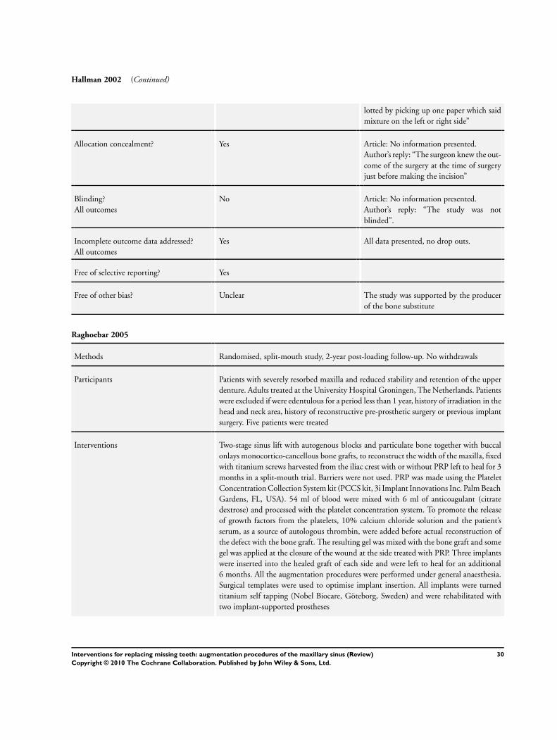
Hallman 2002 (Continued)
lotted by picking up one paper which saidmixture on the left or right side”
Allocation concealment? Yes Article: No information presented.Author’s reply: “The surgeon knew the out-come of the surgery at the time of surgeryjust before making the incision”
Blinding?All outcomes
No Article: No information presented.Author’s reply: “The study was notblinded”.
Incomplete outcome data addressed?All outcomes
Yes All data presented, no drop outs.
Free of selective reporting? Yes
Free of other bias? Unclear The study was supported by the producerof the bone substitute
Raghoebar 2005
Methods Randomised, split-mouth study, 2-year post-loading follow-up. No withdrawals
Participants Patients with severely resorbed maxilla and reduced stability and retention of the upperdenture. Adults treated at the University Hospital Groningen, The Netherlands. Patientswere excluded if were edentulous for a period less than 1 year, history of irradiation in thehead and neck area, history of reconstructive pre-prosthetic surgery or previous implantsurgery. Five patients were treated
Interventions Two-stage sinus lift with autogenous blocks and particulate bone together with buccalonlays monocortico-cancellous bone grafts, to reconstruct the width of the maxilla, fixedwith titanium screws harvested from the iliac crest with or without PRP left to heal for 3months in a split-mouth trial. Barriers were not used. PRP was made using the PlateletConcentration Collection System kit (PCCS kit, 3i Implant Innovations Inc. Palm BeachGardens, FL, USA). 54 ml of blood were mixed with 6 ml of anticoagulant (citratedextrose) and processed with the platelet concentration system. To promote the releaseof growth factors from the platelets, 10% calcium chloride solution and the patient’sserum, as a source of autologous thrombin, were added before actual reconstruction ofthe defect with the bone graft. The resulting gel was mixed with the bone graft and somegel was applied at the closure of the wound at the side treated with PRP. Three implantswere inserted into the healed graft of each side and were left to heal for an additional6 months. All the augmentation procedures were performed under general anaesthesia.Surgical templates were used to optimise implant insertion. All implants were turnedtitanium self tapping (Nobel Biocare, Göteborg, Sweden) and were rehabilitated withtwo implant-supported prostheses
30Interventions for replacing missing teeth: augmentation procedures of the maxillary sinus (Review)
Copyright © 2010 The Cochrane Collaboration. Published by John Wiley & Sons, Ltd.

Raghoebar 2005 (Continued)
Outcomes Prosthesis and implant failure. Complications at the augmented and donor sites. Histo-morphometrical evaluation
Notes
Risk of bias
Item Authors’ judgement Description
Adequate sequence generation? Yes Article: No information presented.Author’s reply: “Randomisation by lot”.
Allocation concealment? Unclear Article: No information presented.Author’s reply failed to clarify the issue.
Blinding?All outcomes
Yes Article: “The investigators were blinded forboth the clinical and laboratory investiga-tions with regard to the PRP-treated side”
Incomplete outcome data addressed?All outcomes
Yes All data presented, no drop outs.
Free of selective reporting? Yes
Free of other bias? Unclear Author: “The apparatus was gift from thecompany promoting the use of PRP”This was considered to be irrelevant.In all patients additional buccal onlayswere performed meaning that these pa-tients might have not been the ideal candi-dates for the hypothesis tested. It is there-fore more difficult to interpret the resultssince the role of the additional buccal on-lays on the final outcome cannot be quan-tified
Schaaf 2008
Methods Randomised, split-mouth study with follow-up to abutment connection. No with-drawals. Data from patients treated according to a parallel group design were not pro-vided or sought
Participants Severe atrophy of the edentulous or partially edentulous posterior maxilla and intentionto treat with onlay bone blocks and sinus floor augmentation. Residual bone heightvalues appears to be in the range of 1 to 12 mm according to the measurements kindlyprovided by the authors. Adults treated at university clinics of Giessen and Herlangen(Germany). Patients were excluded if they had blood platelet disorders, haemorrhagicdiathesis or medication with aspirin before surgery, infectious diseases such as hepatitis,
31Interventions for replacing missing teeth: augmentation procedures of the maxillary sinus (Review)
Copyright © 2010 The Cochrane Collaboration. Published by John Wiley & Sons, Ltd.
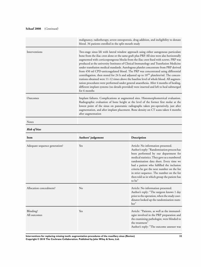
Schaaf 2008 (Continued)
malignancy, radiotherapy, severe osteoporosis, drug addition, and ineligibility to donateblood. 34 patients enrolled in the split-mouth study
Interventions Two-stage sinus lift with lateral window approach using either autogenous particulatebone from the iliac crest alone or the same graft plus PRP. All sites were also horizontallyaugmented with corticospongeous blocks from the iliac crest fixed with screws. PRP wasproduced at the university Institutes of Clinical Immunology and Transfusion Medicineunder transfusion medical standards. Autologous platelet concentrate from PRP derivedfrom 450 ml CPD-anticoagulated blood. The PRP was concentrated using differentialcentrifugation, then stored for 24 h and adjusted up to 1010 platelets/ml. The concen-trations obtained were 11-12 times above the baseline level of whole blood. All augmen-tation procedures were performed under general anaesthesia. After 4 months of healing,different implant systems (no details provided) were inserted and left to heal submergedfor 6 months
Outcomes Implant failures. Complications at augmented sites. Histomorphometrical evaluation.Radiographic evaluation of bone height at the level of the former first molar at thelowest point of the sinus on panoramic radiographs taken pre-operatively, just afteraugmentation, and after implant placement. Bone density on CT scans taken 4 monthsafter augmentation
Notes
Risk of bias
Item Authors’ judgement Description
Adequate sequence generation? Yes Article: No information presented.Author’s reply: “Randomisation process hasbeen performed by our department formedical statistics. They gave us a numberedrandomisation data sheet. Every time wehad a patient who fulfilled the inclusioncriteria he got the next number on the listin strict sequence. The number on the listthen told us in which group the patient hasto be”
Allocation concealment? No Article: No information presented.Author’s reply: “The surgeon knows 1 dayprior to the operation, when the study coor-dinator looked up the randomisation num-ber”
Blinding?All outcomes
Yes Article: “Patients, as well as the immunol-ogist involved in the PRP preparation andthe examining pathologist, were blinded tothe treatment”Author’s reply: “The outcome assessor was
32Interventions for replacing missing teeth: augmentation procedures of the maxillary sinus (Review)
Copyright © 2010 The Cochrane Collaboration. Published by John Wiley & Sons, Ltd.

Schaaf 2008 (Continued)
blinded to the belonging of the group,whether PRP was used or not”
Incomplete outcome data addressed?All outcomes
No Not complete radiographic and data pre-sented or supplied so we were unable to usepart of the data
Free of selective reporting? Yes
Free of other bias? Unclear In all patients additional buccal onlayswere performed meaning that these pa-tients might have not been the ideal candi-dates for the hypothesis tested. It is there-fore more difficult to interpret the resultssince the role of the additional buccal on-lays on the final outcome cannot be quan-tified
Szabó 2005
Methods Randomised, split-mouth study with follow-up to implant loading. No withdrawals
Participants Edentulous patients having less than 5 mm of alveolar bone in the floor of the sinus.Adults treated at university hospitals in Budapest (Hungary), Manchester (UK), Milan(Italy) and in a private practice in Bruges (Belgium). No specific exclusion criteria weregiven and patients were healthy and had no disease that might influence the treatmentoutcome. 20 patients enrolled
Interventions Two-stage sinus lift with autogenous particulate bone from the iliac crest versus two-stagesinus lift with 1.5 to 2 g beta-tricalcium phosphate (Cerasorb, Curasan AG, Kleinostheim,Germany) left healing for 6 months. In 10 of the 20 patients the alveolar crest wasalso widened with cortical bone blocks fixed with microscrews. No membranes wereused to cover the bone. All the augmentation procedures were performed under generalanaesthesia. Patients were instructed not to wear any dentures for 30 days. In 16 patientsAnkylos (Degussa, Friadent, Germany) implants were used, whereas in four patientsProtetim (Hungary) implants were used. The authors did no provide any explanationfor using two different implant systems. Two implants were placed in each augmentedsinus
Outcomes Implant failure at abutment connection. Post-operative complications at the grafted siteand at the donor site. Panoramic and computer tomographic imagines (CT images in10 patients only) to provide a qualitative description of the consolidation of the grafts.Histomorphometrical assessment of cylindrical bone biopsies
Notes
Risk of bias
33Interventions for replacing missing teeth: augmentation procedures of the maxillary sinus (Review)
Copyright © 2010 The Cochrane Collaboration. Published by John Wiley & Sons, Ltd.

Szabó 2005 (Continued)
Item Authors’ judgement Description
Adequate sequence generation? Unclear Article: “The choice of sides was random-ized using the coin-toss method”One author from one of the centres replied:“Centralised randomization with code in asealed envelope”
Allocation concealment? Unclear Article: No information presented.One author from one of the centres replied:“Sealed envelopes opened just before plac-ing the graft into the sinus”
Blinding?All outcomes
No Article: No information presented.Author’s reply: “No blinding was used”.
Incomplete outcome data addressed?All outcomes
No No radiographic data presented.
Free of other bias? No Author informed us that they received freebone substitutes.This is considered to be irrelevant.In 10 patients additional buccal onlayswere performed meaning that these pa-tients might have not been the ideal candi-dates for the hypothesis tested. It is there-fore more difficult to interpret the resultssince the role of the additional buccal on-lays on the final outcome cannot be quan-tified
Torres 2009
Methods Randomised hybrid study design combining patients with a split-mouth study designwith patients treated according to a quasi-random parallel group design with follow-upto 2 years after loading. No withdrawals
Participants Patients having less than 7 mm of alveolar bone at the floor of the sinus. Adults treatedat private clinic in Madrid, Spain. Exclusion criteria were severe systemic diseases (ASAscore 3 or more), a previous history of chronic sinusitis. 57 patients were treated
Interventions One or two-stage sinus lift procedures using a lateral window technique and 100%granular Bio-Oss with or without PRP, left to heal for 6 months. Patients having upto 4 mm of residual bone height were augmented first and implant were placed after6 months whereas patients with residual bone more than 4 mm up to 7 mm receivedimplants during the sinus lift procedures. Implants were left to heal unloaded for 6months. 10 to 20 cc of venous blood were collected 30 minutes prior to the surgeryand mixed with a 3.8% sodium citrate solution at a 5/1 ratio, achieving anticoagulation
34Interventions for replacing missing teeth: augmentation procedures of the maxillary sinus (Review)
Copyright © 2010 The Cochrane Collaboration. Published by John Wiley & Sons, Ltd.

Torres 2009 (Continued)
through calcium binding. The blood was then centrifuged into three and separated intothree layers: red blood cells (RBCs), PRP and poor plasma. Flow cytometry was used forplatelet counting. Platelets counts were 2.97 + 0.7-fold over peripheral blood. PRP wasactivated with 30% CaCl2 solution and a PRP gel was obtained and mixed with Bio-Oss.The entire bone of the buccal window was removed, and after the sinus was filled withthe bone substitute no barrier was used to seal the windows. Patients were instructednot to wear their upper dentures for 2-3 weeks after surgery. Osseotite (Biomet 3I, PalmBeach, FL, USA) implants were used
Outcomes Prosthetic and implant failures. Partial loss of the graft and complications. Histomor-phometrical evaluation
Notes
Risk of bias
Item Authors’ judgement Description
Adequate sequence generation? Yes Article: “..randomized sequence was per-formed by a computerized random numbergenerated using GraphPadQuickCalc soft-ware (GraphPad Software Inc., La Joya, Ca,USA), including the concealment of the al-location schedule until the assignment wasdone”
Allocation concealment? Yes Article: “Patients included in the inter-patient clinical trial were allocated by ablinded assistant into two groups: the firstwas to be treated with ABB alone, and thesecond with ABB + PRP”
Blinding?All outcomes
Yes Article: “The surgeon was blinded to thegraft material applied to each patient be-fore graft implantation. An assistant han-dled PRP-ABB or the ABB group after thesurgeon had already accessed the sinus andelevated membrane. The histologist wasblinded to the samples’ groups throughoutthe histomorphometric analysis”Author’s reply: “Implant stability was as-sessed manually with removed prosthesesand mobile implants were considered asfailures and this evaluation was done by aprosthodontist who was not aware of studygroups”
Incomplete outcome data addressed?All outcomes
Unclear We could only evaluate those data kindlyprovided by the authors
35Interventions for replacing missing teeth: augmentation procedures of the maxillary sinus (Review)
Copyright © 2010 The Cochrane Collaboration. Published by John Wiley & Sons, Ltd.

Torres 2009 (Continued)
Free of selective reporting? Yes
Free of other bias? No A mixed split-mouth and parallel group de-sign was used: patients requiring augmen-tation at bilateral sinuses were randomisedin a split-mouth study design whereas thoserequiring unilateral sinus lift sinus were al-ternated in a quasi-random study design.We have not included data from the quasi-random study
Wannfors 2000
Methods Randomised trial of parallel group design, 3-year post-loading follow-up. No withdrawalsat 3 years though three patients in the one-stage group refused consent to remove theprostheses for testing implant stability
Participants Edentulous patients with more than 2 mm but less than 7 mm of residual bone under themaxillary sinuses. Adults treated under general anaesthesia at the Karolinska Hospital,Stockholm, Sweden. Patients were included if they were edentulous in the upper jaw.Patients were excluded if they were older than 80 years, had pathologies in the maxillarysinus, had bone diseases or took medications known to effect bone metabolism (i.e.corticosteroids and bisphosphonates). 40 patients enrolled, 20 in each group
Interventions One-stage sinus lift with monocortical iliac bone blocks fixed usually with two implantsleft to heal for 6 months versus two-stage sinus lift with particulate bone from the iliaccrest left to heal for 6 months and then usually two implants were inserted into thehealed graft and were left to heal for an additional 6 months. All implants were titaniumself tapping (Brånemark System, Nobel Biocare)
Outcomes Prosthesis failure, implant failure and marginal bone level changes on intraoral radio-graphs taken with a paralleling technique at abutment connection, 1 and 3 years. Intra-operative sinus membrane perforations
Notes
Risk of bias
Item Authors’ judgement Description
Adequate sequence generation? Unclear Article: “He (patient) was allotted to oneof the two treatments according to a previ-ously designed scheme by a third person”Author’s reply: “The randomization wasperformed by a third person without anybeforehand contact with the patients”
36Interventions for replacing missing teeth: augmentation procedures of the maxillary sinus (Review)
Copyright © 2010 The Cochrane Collaboration. Published by John Wiley & Sons, Ltd.

Wannfors 2000 (Continued)
Allocation concealment? Unclear Article: No information presented.Author’s reply failed to clarify the issue.
Blinding?All outcomes
No Article: No information presented.Author’s reply: “The outcome assessor hadknowledge of the randomized group, how-ever not when assessing the x-ray data”
Incomplete outcome data addressed?All outcomes
No Full data of complications not provided.
Free of selective reporting? No Full data of complications not provided.
Free of other bias? Yes
PRP - platelet-rich plasma
Characteristics of excluded studies [ordered by study ID]
Study Reason for exclusion
Aimetti 2008 The article presented data from four patients treated following a split-mouth design. Authors informed us thatthey actually treated 16 patients. We are unable to present the data for the remaining 12 patients
Barone 2005 No clinical outcome measures related to implant treatment.
Barone 2008 Insufficient follow-up time with no clinical outcome measures related to implant treatment
Boyne 2005 Described as RCT, unclear number of patients, unequal number of patients in the treatment groups. No reply toletter
Choi 2009 No clinical outcome measures related to implant treatment.
Consolo 2007 No clinical outcome measures related to implant treatment.
Cordaro 2008 No clinical outcome measures related to implant treatment.
Crespi 2009 No clinical outcome measures related to implant treatment.
Froum 1998 Described as RCT, unclear number of patients and tested interventions which seem to be much more than eight,unequal number of patients in the treatment groups. No reply to letter
Froum 2006 No clinical outcome measures related to implant treatment.
37Interventions for replacing missing teeth: augmentation procedures of the maxillary sinus (Review)
Copyright © 2010 The Cochrane Collaboration. Published by John Wiley & Sons, Ltd.

(Continued)
Froum 2008 No clinical outcome measures related to implant treatment.
Hallman 2008 No clinical outcome measures related to implant treatment.
Kassolis 2005 No clinical outcome measures related to implant treatment.
Kim 2009 No clinical outcome measures related to implant treatment.
Mangano 2007 The authors informed us that the trial was not an RCT but a CCT
Steigmann 2005 No clinical outcome measures related to implant treatment.
Suba 2006 No clinical outcome measures related to implant treatment.
Tawil 2001 Inappropriate study design, neither parallel group nor split-mouth
Triplett 2009 Unclear how many patients were randomised to each group, data very confused and we were unable to retrievesufficient data from the original publication. No reply to letter
CCT - controlled clinical trialRCT - randomised controlled clinical trial
38Interventions for replacing missing teeth: augmentation procedures of the maxillary sinus (Review)
Copyright © 2010 The Cochrane Collaboration. Published by John Wiley & Sons, Ltd.