intestinal tuberculosis: a great mimicker
TRANSCRIPT
1Tuberculosis | www.smgebooks.comCopyright Sinhasan SP.This book chapter is open access distributed under the Creative Commons Attribution 4.0 International License, which allows users to download, copy and build upon published articles even for com-mercial purposes, as long as the author and publisher are properly credited.
Gr upSMIntestinal Tuberculosis: A Great mimicker
Dr. Sankappa P Sinhasan*Department of Pathology, Indira Gandhi Medical College & Research Institute (IGMC & RI), Pondicherry, India
*Corresponding author: Sankappa P Sinhasan, Department of Pathology, Indore Gandhi Medical College & Research Institute (IGMC & RI), Pondicherry, INDIA, Tel: +91 9442551998, Fax: +91 413 2277289, Email: [email protected]
Published Date: October 20, 2016
ABSTRACTTuberculosis primarily affects lungs but can affect any of the organ systems, like intestine,
meninges, bones and joints, lymph nodes, skin and other tissues of the body. By definition, primary intestinal tuberculosis occurs in the absence of clinically pulmonary infection. Diagnosis of intestinal tuberculosis is difficult as tests are nonspecific. Without a high index of suspicion, the disease is rarely diagnosed correctly. Intestinal Tuberculosis needs to be considered in the differential diagnosis when patients with intestinal pathology are encountered.
Materials and Methods: We studied 100 cases of resected intestinal specimens received during study period of 2 years. Out of 100 cases, we totally encountered 22 request forms from clinicians with suspicion of ileoceocal tuberculosis. Abdominal tenderness and mass in ileoceocal region were noted in all the cases. In many instances, the cases were operated for acute / sub acute intestinal obstruction. Clinical and intra-operative diagnoses of tubercular enteritis, in many instances, were finally diagnosed histopathologically as Ischemic Enteritis (in 12 cases), Adenocarcinoma ceocum (one case), Chronic Idiopathic (nonspecific) Enteritis (ten cases) and correctly as Intestinal Tuberculosis in only 6 cases.
2Tuberculosis | www.smgebooks.comCopyright Sinhasan SP.This book chapter is open access distributed under the Creative Commons Attribution 4.0 International License, which allows users to download, copy and build upon published articles even for com-mercial purposes, as long as the author and publisher are properly credited.
Conclusion: Tuberculosis can mimic various above said disease entities, clinically; sometimes morphologically. Vice versa is also true. An increased awareness of intestinal tuberculosis coupled with varied clinical presentations, nonspecific signs and symptoms, difficulties in diagnostic methods and need of early and specific treatment should improve the outcome for patients with this disease. Tuberculosis can no longer be considered a rare disease, in part due to the AIDS epidemic. The HIV epidemic has dramatically changed the magnitude and nature of tuberculosis in recent years leading to coin the description “the new tuberculosis”. This review draws attention towards need of increased awareness of intestinal tuberculosis, coupled with knowledge of the pathophysiology, diagnostic methods, and treatment and thus improving the outcome for patients with this disease.
Keywords: Intestinal Tuberculosis; Ischemic Enteritis; Crohn’s Disease; Malignancies; Intussusceptions; Acute Intestinal Obstruction.
INTRODUCTIONTuberculosis was recognized as early as the fourth century B.C. and was known as phthisis,
lupus, scrofula or Pott’s disease, until identity was established by Robert Koch in 1882. Hippocrates stated that, “Phthisis persons die if diarrhea sets in and it is a mortal symptom.” The severity of intestinal tuberculosis was known even at that time [1].
Tuberculosis primarily affects lungs but can affect any of the organ systems, like intestine, meninges, bones and joints, lymph nodes, skin and other tissues of the body [2].
In many ways, the gastrointestinal tract (GIT) is a remarkable organ. Many conditions like infections, inflammatory diseases and tumors affect both small and large intestines. Collectively disorders of the intestine account for a large portion of human disease irrespective of age and sex. Tuberculosis remains a worldwide public health problem despite the fact that causative organism was discovered more than a century ago and highly effective drugs are available for making tuberculosis a preventive and curable disease [3].
Developed countries have achieved spectacular results in the control of tuberculosis. The problem is more acute in developing countries, which account for more than 75%. Eight out of ten of all those stuck by tuberculosis are in the economically productive age group of 15 to 49 yrs. Tuberculosis is a social disease with medical aspects described as barometer of social welfare. The social factors are poverty, illiteracy, ignorance, poor housing, over-crowding, population explosion, under nutrition, lack of awareness about illness. All these factors are interrelated and contribute to occurrence of the disease [2].
With the advent of effective and anti-mycobacterial medication, wide spread use of skin testing, chest x-ray studies, and the pasteurization of milk, the incidence of intestinal tuberculosis fell rapidly in industrialized nations. This is period of pharmacotherapy which extended until very recently when the epidemiology of tuberculosis again changed, predominantly due to the effects of the HIV epidemic and the increased prevalence of multidrug resistant organisms [4,5].
3Tuberculosis | www.smgebooks.comCopyright Sinhasan SP.This book chapter is open access distributed under the Creative Commons Attribution 4.0 International License, which allows users to download, copy and build upon published articles even for com-mercial purposes, as long as the author and publisher are properly credited.
Resection of intestine is a fairly common surgical procedure; the indications of which are varied and the pathological lesions identified are equally varied. It is interesting to know that the cause of resection has been most commonly an acute mode of clinical presentation though the underlying etiology is varied. Histopathological study revealed many interesting findings. So, an attempt is made to study different lesions in these resected intestinal specimens, the result of which will bear some impact on patient care, management and disease prognosis in present cases and in future also. This is an endeavor in that direction.
OBJECTIVES1. To study the various clinical presentations of cases diagnosed clinically as abdominal
tuberculosis those compel the resection of intestine.
2. To illustrate, discuss and describe the various histopathological features of the lesions in these resected intestinal specimens.
3. Clinicopathological correlation of these lesions.
MATERIALS AND METHODSAll surgically resected specimens diagnosed/ suspected clinically as abdominal tuberculosis
received at our Dept. of Pathology were included and studied in detail. The clinical findings were collected from hospital case sheets or from case records in the hospital record section. The details of gross examination findings were noted. They were examined as soon as possible to determine whether or not the specimen requires special handling. Ten percent formalin was used as fixative. Tissues were adequately fixed prior to sectioning. After measurements have been made, the way that the specimen is opened will depend on the type of resection and site of the lesion. They were properly oriented to include some normal tissue for comparison. This most commonly involves a section perpendicular to the mucosa. This allows one to evaluate mucosal height and its component parts. The specimens were cleaned to remove blood, stool, and other substances present. The bowel was palpated to establish the site and extent of any focal lesions. Incisions into the mesentery were made similarly, parallel to the adjacent bowel.
Tissues selected for histopathological examination were processed routinely. Sections of 3- 6 microns were cut from paraffin blocks and stained with hematoxyllin and eosin. ZN stain was also done to appreciate AFB.
Statistical analysis of various intestinal lesions in relation to age, sex, signs and symptoms, clinical diagnosis and histopathological diagnosis was done.
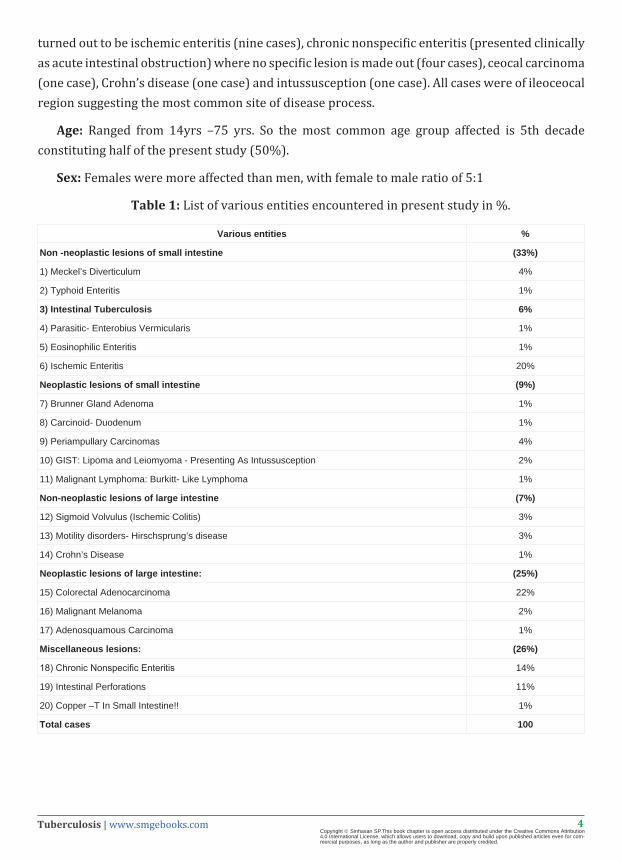
OBSERVATIONSWe studied 100 cases of resected intestinal specimens during the study period of two
years (Table 1). We totally encountered 22 request forms with clinical suspicion of ileoceocal tuberculosis, out of which only 6 cases were proved histologically as cases of TB; the rest were
4Tuberculosis | www.smgebooks.comCopyright Sinhasan SP.This book chapter is open access distributed under the Creative Commons Attribution 4.0 International License, which allows users to download, copy and build upon published articles even for com-mercial purposes, as long as the author and publisher are properly credited.
turned out to be ischemic enteritis (nine cases), chronic nonspecific enteritis (presented clinically as acute intestinal obstruction) where no specific lesion is made out (four cases), ceocal carcinoma (one case), Crohn’s disease (one case) and intussusception (one case). All cases were of ileoceocal region suggesting the most common site of disease process.
Age: Ranged from 14yrs –75 yrs. So the most common age group affected is 5th decade constituting half of the present study (50%).
Sex: Females were more affected than men, with female to male ratio of 5:1
Table 1: List of various entities encountered in present study in %.
Various entities %
Non -neoplastic lesions of small intestine (33%)
1) Meckel’s Diverticulum 4%
2) Typhoid Enteritis 1%
3) Intestinal Tuberculosis 6%
4) Parasitic- Enterobius Vermicularis 1%
5) Eosinophilic Enteritis 1%
6) Ischemic Enteritis 20%
Neoplastic lesions of small intestine (9%)
7) Brunner Gland Adenoma 1%
8) Carcinoid- Duodenum 1%
9) Periampullary Carcinomas 4%
10) GIST: Lipoma and Leiomyoma - Presenting As Intussusception 2%
11) Malignant Lymphoma: Burkitt- Like Lymphoma 1%
Non-neoplastic lesions of large intestine (7%)
12) Sigmoid Volvulus (Ischemic Colitis) 3%
13) Motility disorders- Hirschsprung’s disease 3%
14) Crohn’s Disease 1%
Neoplastic lesions of large intestine: (25%)
15) Colorectal Adenocarcinoma 22%
16) Malignant Melanoma 2%
17) Adenosquamous Carcinoma 1%
Miscellaneous lesions: (26%)
18) Chronic Nonspecific Enteritis 14%
19) Intestinal Perforations 11%
20) Copper –T In Small Intestine!! 1%
Total cases 100
5Tuberculosis | www.smgebooks.comCopyright Sinhasan SP.This book chapter is open access distributed under the Creative Commons Attribution 4.0 International License, which allows users to download, copy and build upon published articles even for com-mercial purposes, as long as the author and publisher are properly credited.
Symptoms: The most common symptoms were pain and mass per abdomen present in all cases, followed by fever and signs and symptoms of acute abdomen (Table No 2). The duration of symptoms varied from 1month to 4years. The pain in abdomen was usually intermittent, often colicky in nature, vague and localized to right ileac fossa or the periumblical region. General symptoms like anorexia and loss of weight were present in all cases. Two cases presented with low-grade fever and none had diarrhea. Two cases presented with acute intestinal obstruction.
Table 2: Showing symptoms of intestinal tuberculosis.
Symptom No. Of Cases Percentage
Pain 6 100%
Mass 6 100%
Fever 2 33.33%
Acute Intestinal obstruction 2 33.33%
Clinical examination findings: Abdominal tenderness was noted in all the cases along with mass in ileoceocal region in all, suggesting that these two are the most common signs (100%) (Table No 3). None of our cases had pulmonary signs in the past or during admission, and X-ray of chest did not reveal evidence of tuberculosis suggesting a diagnosis of ‘primary intestinal tuberculosis’ in all. In the present study routine plain X-ray of abdomen was done preoperatively. Out of six patients, two patients presented with obstructive symptoms (33.33%), had multiple air /fluid levels. Only one patient with perforation had pneumoperitoneum (16.66%).
Table 3: Case details of intestinal tuberculosis.
Sl. No. Age/Sex Pain Mass Fever Acute Intestinal obstruction Clinical Diagnosis HP Diagnosis
1 14yrs /F + + - - Ileoceocal TB Tuberculosis
2 40yrs/M + + - - Ileoceocal TB Tuberculosis
3 40yrs/F + + + + Perforation ? TB. Tuberculosis withPerforation
4 45yrs/F + + - - Ileoceocal TB Tuberculosis
5 21yrs/F + + + + Acute Intestinal obstruction Tuberculosis
6 75yrs/F + + - - Ileoceocal TB Tuberculosis
Operative procedures: All six cases in the present study were treated surgically with limited ileoceocal resection medically with antitubercular regimen. One of the two cases presented with acute intestinal obstruction had a perforation with tubercular peritonitis and associated ascitis. Ileoceocal region was involved in all cases (100%). The single most common finding was of stricture seen in all the cases. Multiple strictures were noted in two cases. Enlarged lymph nodes were noted in two cases. Ileoceocal mass occurred in all cases with adherent mesentery. Lysis of adhesions with ileoceocal resection was carried out.
Grossly: Ileum in all cases showed one or more strictures with dilated adjacent loops. Ileoceocal junction showed stenosis in one case. Ceocum and ascending colon showed marked thickening
6Tuberculosis | www.smgebooks.comCopyright Sinhasan SP.This book chapter is open access distributed under the Creative Commons Attribution 4.0 International License, which allows users to download, copy and build upon published articles even for com-mercial purposes, as long as the author and publisher are properly credited.
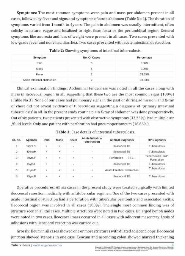
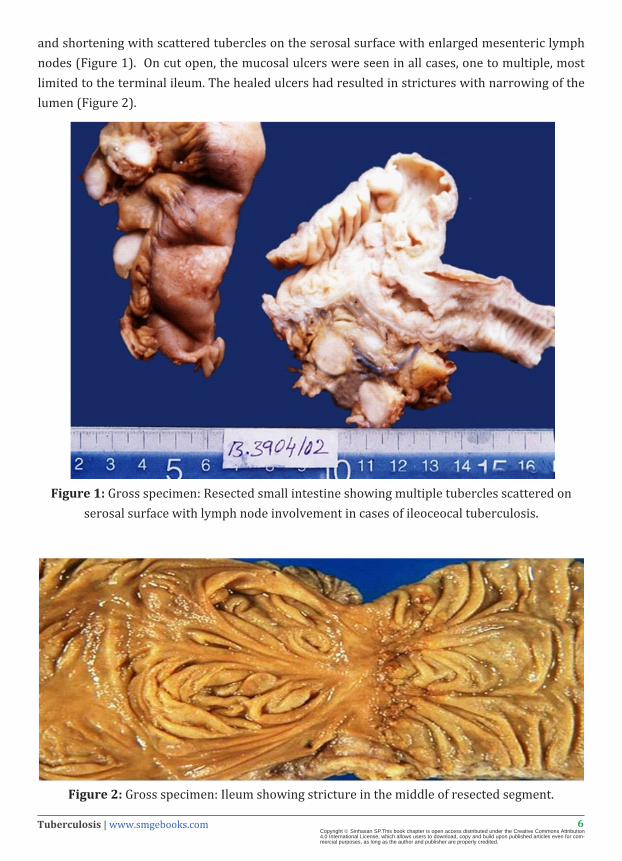
and shortening with scattered tubercles on the serosal surface with enlarged mesenteric lymph nodes (Figure 1). On cut open, the mucosal ulcers were seen in all cases, one to multiple, most limited to the terminal ileum. The healed ulcers had resulted in strictures with narrowing of the lumen (Figure 2).
Figure 1: Gross specimen: Resected small intestine showing multiple tubercles scattered on serosal surface with lymph node involvement in cases of ileoceocal tuberculosis.
Figure 2: Gross specimen: Ileum showing stricture in the middle of resected segment.
7Tuberculosis | www.smgebooks.comCopyright Sinhasan SP.This book chapter is open access distributed under the Creative Commons Attribution 4.0 International License, which allows users to download, copy and build upon published articles even for com-mercial purposes, as long as the author and publisher are properly credited.
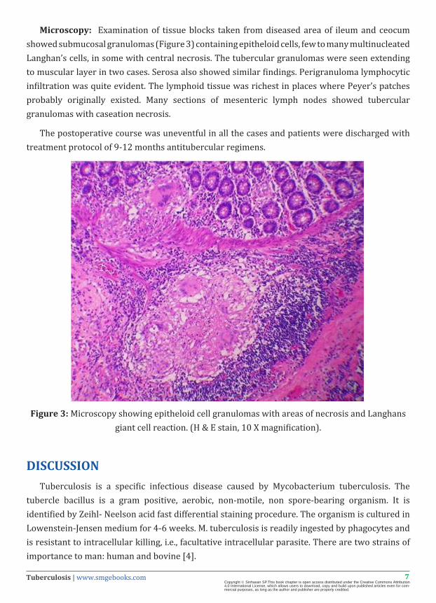
Microscopy: Examination of tissue blocks taken from diseased area of ileum and ceocum showed submucosal granulomas (Figure 3) containing epitheloid cells, few to many multinucleated Langhan’s cells, in some with central necrosis. The tubercular granulomas were seen extending to muscular layer in two cases. Serosa also showed similar findings. Perigranuloma lymphocytic infiltration was quite evident. The lymphoid tissue was richest in places where Peyer’s patches probably originally existed. Many sections of mesenteric lymph nodes showed tubercular granulomas with caseation necrosis.
The postoperative course was uneventful in all the cases and patients were discharged with treatment protocol of 9-12 months antitubercular regimens.
Figure 3: Microscopy showing epitheloid cell granulomas with areas of necrosis and Langhans giant cell reaction. (H & E stain, 10 X magnification).
DISCUSSIONTuberculosis is a specific infectious disease caused by Mycobacterium tuberculosis. The
tubercle bacillus is a gram positive, aerobic, non-motile, non spore-bearing organism. It is identified by Zeihl- Neelson acid fast differential staining procedure. The organism is cultured in Lowenstein-Jensen medium for 4-6 weeks. M. tuberculosis is readily ingested by phagocytes and is resistant to intracellular killing, i.e., facultative intracellular parasite. There are two strains of importance to man: human and bovine [4].
8Tuberculosis | www.smgebooks.comCopyright Sinhasan SP.This book chapter is open access distributed under the Creative Commons Attribution 4.0 International License, which allows users to download, copy and build upon published articles even for com-mercial purposes, as long as the author and publisher are properly credited.
1) Human Source: The most common source of infection is open case of pulmonary tuberculosis whose sputum is positive for tubercle bacilli, who has received no treatment or not treated completely. Such persons can discharge bacilli in their sputum for years. The tubercle bacilli in human case are usually a mixed group. Some multiply very rapidly and some slowly. The rapidly multiplying bacilli strains are more susceptible to bactericidal action of chemotherapeutic agents. The slow multipliers are a source of persistent or dormant bacilli. They can remain alive for years without causing harm to the host, but when the conditions are favorable they may start multiplying again and cause active disease, i.e., they are ‘seeds’ of future relapse [4].
2) Bovine Source: The bovine source of infection is usually infected milk. There is no definitive evidence that bovine tuberculosis is a major problem, because of the practice of boiling milk before consumption.4 But in India, it is common practice in villages, especially in shepherd community to drink goat or cow milk even without heating. This kind of practice may expose them to develop bovine tuberculosis affecting the small intestine.
HOST FACTORSa) Age: Tuberculosis affects all ages. Developing countries show sharp rise in infection rates
from infancy to adolescence. The infection rate climbs up from the age of 15years. The 2/3rd of abdominal tuberculosis patients were in the 3rd and 4th decade of life, while it can affect any age group [6].
b) Sex: Pulmonary tuberculosis is more prevalent in males. The prevalence of abdominal tuberculosis in both sexes varies from study to study.5In our study, we found female predominance.
c) Nutrition: Malnutrition is widely believed to predispose to tuberculosis, but the available evidence on this point is only indirect [6].
d) Immunity: Man has no inherited immunity against tuberculosis. It is acquired as a result of natural infection or BCG vaccination. It is known that both delayed hypersensitivity and acquired resistance to tuberculosis are cell-mediated responses. In most cases cellular immunity proves adequate to limit further multiplication and spread of bacilli [6].
In pre-chemotherapy era, the treatment of tuberculosis was mainly directed at strengthening host defenses by altering general host factors (e.g., rest, fresh air, nutrition) with the advent of chemotherapy, host factors are considered to be less importance in the epidemiology of tuberculosis [6].
With the advent of effective and anti-mycobacterial medication, wide spread use of skin testing, chest X-ray studies, and the pasteurization of milk, the incidence of intestinal tuberculosis fell rapidly in industrialized nations. Very recently, the epidemiology of tuberculosis again changed, predominantly due to the effects of the HIV epidemic and the increased prevalence of multidrug resistant organisms. We have now entered “the age of immunosuppression” or the ‘new’ tuberculosis. 5,10 Nearly 5% to 8% of patients with early disease had enteric involvement; for those with moderately advanced disease the rate rose to 14% to 18% and 70% to 80% of
9Tuberculosis | www.smgebooks.comCopyright Sinhasan SP.This book chapter is open access distributed under the Creative Commons Attribution 4.0 International License, which allows users to download, copy and build upon published articles even for com-mercial purposes, as long as the author and publisher are properly credited.
patients with advanced pulmonary disease demonstrated roengenologic abnormalities consistent with intestinal tuberculosis [5].
PATHOGENEISIntestinal tuberculosis can result from primary infection of the gastrointestinal tract or from
secondary infection from a focus elsewhere in the body, usually the lungs. By definition, primary gastrointestinal tuberculosis occurs in the absence of clinically pulmonary infection [4].
The likely mechanisms for infection of the bowel include direct ingestion of tubercle bacilli from either infected sputum or dairy products, hemotogenous spread from foci elsewhere in the body, bite containing bacilli or direct extension from contiguous organs [4].
The frequent association of tuberculous enteritis and tuberculous laryngitis suggests the role of infected sputum in the pathogenesis of enteric disease. The lipid laden mycobacterial cell wall is relatively resistant to acid digestion and this allows ingested organisms to reach and invade the bowel wall. This hypothesis also clarifies why primary intestinal tuberculosis due to mycobacterium bovis, which was caused by direct ingestion of contaminated dairy products, has been virtually eradicated by the wide spread pasteurization of milk [7].
Hematogenous Infection of the intestine is also presumed cause for some cases of tuberculous enteritis. The importance of the seeding gastrointestinal tract by silent bacillimia in primary pulmonary tuberculosis is not clear. The direct extension of the disease from contiguous infected organs may also play a role in some patients. Primary or secondary infection of the female urogenital tract is known to occur and may cause intestinal disease. However invasion of the bowel from tuberculous involvement of the peritoneum is not believed to occur. The inverse however is true; tuberculous peritonitis can occur after the ulceration of the abdominal lymph node, after free perforation of a tuberculous intestinal ulcer, or as a consequence of transmural infection of the bowel without perforation [7].
ANATOMIC DISTRIBUTIONTuberculosis can affect any region of the gastrointestinal tract from the mouth to the anus.
The ileoceocal region is the most commonly affected site as determined by both autopsy series and clinical reports. The striking predilection for this region is thought to be a consequence of the abundance of the lymphoid tissue (Peyer’s patches) therein, with the increased physiologic stasis, increased rate of fluid and electrolytic absorption and minimal digestive activity permitting grater contact time between the organism and the mucosal surface [6].
PATHOLOGIC APPEARANCE: TUBERCULOSISAfter transport of the organism to a site of stasis, the bacillus becomes localized in the depths
of the glands of the mucosa and initiates an inflammatory reaction. The bacillus is carried through the epithelial layer by phagocytes to the submucosa where the initial lesions form in the lymph follicles or Peyer’s patches. The overlying mucosa, deprived of its blood supply through end arteritis, may slough, forming ulcers [7].
10Tuberculosis | www.smgebooks.comCopyright Sinhasan SP.This book chapter is open access distributed under the Creative Commons Attribution 4.0 International License, which allows users to download, copy and build upon published articles even for com-mercial purposes, as long as the author and publisher are properly credited.
The most active inflammation takes place in the submucosa. It becomes thickened as a result of edema, cell infiltrates, lymphatic hyperplasia, formation of tubercles and fibrosis. Lateral spread and penetration through the wall to the serosa, where tubercles may be visualized on gross examination, occur via lymph channels or by direct contiguity [7].
The mesenteric nodes are invaded through the transportation of tuberculous material along lymph channels. These lymph nodes show complete ranges from hyperplasia to caseation and calcification. In the end stage, lymphatic obstruction results and eventually the mesentery as well as the involved bowel becomes a thick, fixed tuberculous mass [5].
The earliest gross changes occur most frequently in the terminal ileum or ceocum where scattered nodules project into the intestinal lumen. Careful examination frequently reveals that these nodules show evidence of early ulceration and that they generally found overlying lymphoid follicles or Peyer’s patches. As the inflammatory process progress, the characteristic morphological changes of ulcerative, ulcerohypertrophic, or hypertrophic disease occur and infection may extend to the mesenteric lymph nodes [8].
The gross pathologic changes are not correlated with the presence or absence of pulmonary disease, as has been thought in the past, but more likely represent the result of a complex interaction between individual host response and bacterial virulence [8].
1. In ULCERATIVE intestinal tuberculosis: There is in duration and edema of diseased segment, with increase in serosal fat, serosal studdings with nodules and the presence of large ulcers, characteristically transverse and circumferential, which are presumed to be coalescence of multiple smaller ones. The ulcers have thickened, infiltrated, everted, indurated edges, and skip areas of normal appearing mucosa may occur. An end arteritis, with subsequent deprivation of blood flow to the mucosal surface is believed to be an important factor in the pathogenesis of ulcer formation. The ulcers are believed to form relatively slowly and generally do not penetrate the muscularis propria [3].
When perforations do occur, they are ordinarily confined by the inflammatory tissue and fibrosis that accompany tissue perforation. As ulcers heal, the extensive fibrosis may result in strictures (napkin ring) and cause obstructive symptoms [3].
2. The HYPERTROPHIC form: Seen in 10% patients, is characterized by extensive inflammation and marked fibrosis in the submucosa and subserosa. This often results in the adherence of bowel, mesentery and lymph nodes into a mass. This mass may occasionally appear to be an exophytic neoplasm from the mucosal surface. As with the ulcerative form, strictures may occur and result in bowel obstruction. The hyperplastic lesion may develop as a result of reduced virulence of the organism and increased host resistance [3].
3. The ULCEROHYPERTROPHIC form: (Approximately 30%). Displays characteristics of both forms of the disease. Another rare form of intestinal tuberculosis is diffuse colitis; which is endoscopically very similar to ulcerative colitis and cannot be easily distinguished on the basis of the mucosal appearance alone [3].

11Tuberculosis | www.smgebooks.comCopyright Sinhasan SP.This book chapter is open access distributed under the Creative Commons Attribution 4.0 International License, which allows users to download, copy and build upon published articles even for com-mercial purposes, as long as the author and publisher are properly credited.
COMPLICATIONS The most common local complication is intestinal obstruction, which develops in three ways
[8]:
1. Encroachment of the thickened bowel upon the lumen because of hypertrophy and circular contraction of scar tissue.
2. Extensive intraperitoneal adhesion, which form as a result of exudation during the earlier acute phases, eventually contract, resulting in kinking or constriction of the intestine.
3. As a result of retraction of mesentery and shortening of the right colon in the healing phases, the medial wall of the ceocum is drawn cephalad and medially, causing a change in the angle of entrance of the terminal ileum into the ceocum from 90 to nearly 180 degrees. A kink at the ileoceocal junction results, with obstruction [8].
Other rare complications include free perforation, followed by tuberculous peritonitis, intussusception and hemorrhage. Four types of tuberculous peritonitis are recognized-ascitic, encysted, fibrous and purulent. Mesenteric lymph nodes may caseate to form cold abscess in mesentery, conglomerate to produce mass or obstruct intestine, rupture to produce acute tuberculous peritonitis [9].
Tuberculosis can mimic various disease entities, clinically; sometimes morphologically. Vice versa is also true. In our study, operative diagnosis of tubercular enteritis in many instances were finally diagnosed as ischemic enteritis in 12 cases, adenocarcinoma cecum in one case, carcinoid duodenum having stricture in one case and correctly as tuberculosis in only 6 cases, suggesting that tuberculosis can mimic other disease entities, clinically as well as morphologically, as also quoted by Paustian [8]. Reverse is also true that, as seen in present study, one case diagnosed grossly as Crohn’s disease due to skip areas, multiple strictures and classical cobblestone appearance of the mucosa (Figure 4) was diagnosed microscopically as tuberculosis-hyperplastic type. If caseating granulomas are not found, it may be difficult to distinguish between Crohn’s disease and tuberculous enteritis. Large, variable appearing confluent granulomas strongly suggests tuberculosis, as does the presence of multinucleated giant cells. The granulomas of tuberculosis are always surrounded by inflammatory cells, in contrast to those of Crohn’s disease [6]. Submucosal widening and transmural follicular hyperplasia, which are common in Crohn’s disease, are generally absent in tuberculous colitis, while epithelial regeneration and glandular metaplasia are more frequent in tuberculosis [6].
Though the presence of caseation is taken as proof-positive for the diagnosis of tuberculosis, its absence does not negate the possibility of the lesion being tubercular in nature.

12Tuberculosis | www.smgebooks.comCopyright Sinhasan SP.This book chapter is open access distributed under the Creative Commons Attribution 4.0 International License, which allows users to download, copy and build upon published articles even for com-mercial purposes, as long as the author and publisher are properly credited.
Figure 4: Intestine showing multiple strictures and cobblestone appearance of the mucosa masquerading Crohn’s disease.
Two of our cases presented with acute intestinal obstruction, where USG was used to localize the ileoceocal mass. Operative diagnoses of tubercular enteritis in many instances were finally diagnosed as ischemic enteritis in nine cases; presence of strictures in ischemic enteritis cases made us to wrongly diagnose these cases grossly as tuberculosis. We also learnt that, presence of mucosal fibrinopurulent exudates (Figure 5) that are commonly seen in ischemic enteritis, should not be confused for tubercles of tuberculosis. One should remember that the tubercles of tuberculosis should be seen on serosal surface and not on the mucosa.
Figure 5: Case of ischemic enteritis with presence of mucosal fibrinopurulent exudates. These should not be mistaken for tubercles.
13Tuberculosis | www.smgebooks.comCopyright Sinhasan SP.This book chapter is open access distributed under the Creative Commons Attribution 4.0 International License, which allows users to download, copy and build upon published articles even for com-mercial purposes, as long as the author and publisher are properly credited.
Similar presentations were studied by Singhai et al, who quotes that; high index of suspicion is needed to diagnosis a case of tuberculosis intestine [10]. Thomas et al, quotes that, even in centers of excellence, early diagnosis and appropriate treatment of intestinal tuberculosis is frequently delayed because of nonspecific and deceptive clinical presentation of the disease [11].
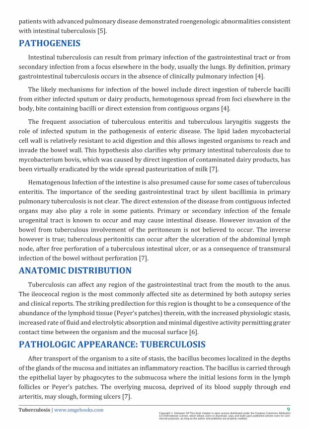
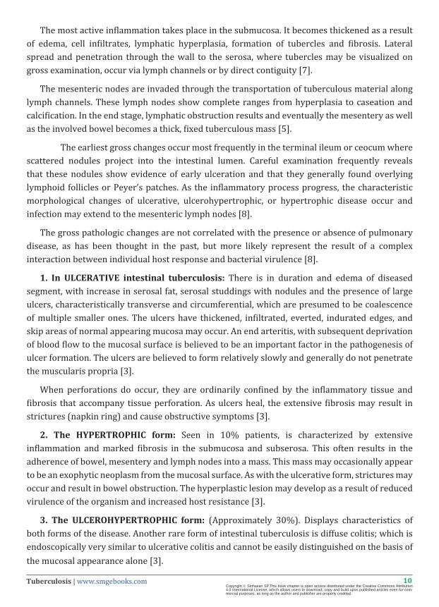
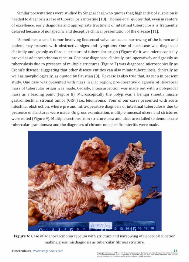
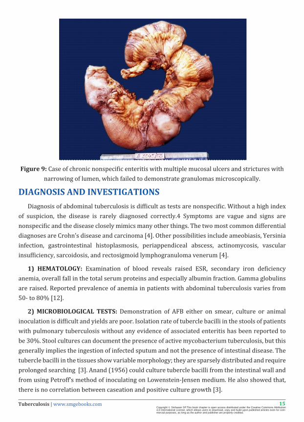
Sometimes, a small tumor involving ileoceocal valve can cause narrowing of the lumen and patient may present with obstructive signs and symptoms. One of such case was diagnosed clinically and grossly as fibrous stricture of tubercular origin (Figure 6); it was microscopically proved as adenocarcinoma ceocum. One case diagnosed clinically, pre-operatively and grossly as tuberculosis due to presence of multiple strictures (Figure 7) was diagnosed microscopically as Crohn’s disease; suggesting that other disease entities can also mimic tuberculosis, clinically as well as morphologically, as quoted by Paustian [8]. Reverse is also true that, as seen in present study. One case was presented with mass in iliac region; pre-operative diagnosis of ileoceocal mass of tubercular origin was made. Grossly, intussusception was made out with a polypoidal mass as a leading point (Figure 8). Microscopically the polyp was a benign smooth muscle gastrointestinal stromal tumor (GIST) i.e., leiomyoma. Four of our cases presented with acute intestinal obstruction, where pre and intra-operative diagnosis of intestinal tuberculosis due to presence of strictures were made. On gross examination, multiple mucosal ulcers and strictures were noted (Figure 9). Multiple sections from stricture area and ulcer area failed to demonstrate tubercular granulomas; and the diagnoses of chronic nonspecific enteritis were made.
Figure 6: Case of adenocarcinoma ceocum with stricture and narrowing of ileoceocal junction making gross misdiagnosis as tubercular fibrous stricture.
14Tuberculosis | www.smgebooks.comCopyright Sinhasan SP.This book chapter is open access distributed under the Creative Commons Attribution 4.0 International License, which allows users to download, copy and build upon published articles even for com-mercial purposes, as long as the author and publisher are properly credited.
Figure 7: Case of Crohn’s disease with multiple skip lesions, ulcers and strictures.
Figure 8: Case of GIST (leiomyoma) presenting as polypoidal mass causing intussusception involving ileocecal junction, clinically suspected as tubercular lesion.
15Tuberculosis | www.smgebooks.comCopyright Sinhasan SP.This book chapter is open access distributed under the Creative Commons Attribution 4.0 International License, which allows users to download, copy and build upon published articles even for com-mercial purposes, as long as the author and publisher are properly credited.
Figure 9: Case of chronic nonspecific enteritis with multiple mucosal ulcers and strictures with narrowing of lumen, which failed to demonstrate granulomas microscopically.
DIAGNOSIS AND INVESTIGATIONSDiagnosis of abdominal tuberculosis is difficult as tests are nonspecific. Without a high index
of suspicion, the disease is rarely diagnosed correctly.4 Symptoms are vague and signs are nonspecific and the disease closely mimics many other things. The two most common differential diagnoses are Crohn’s disease and carcinoma [4]. Other possibilities include ameobiasis, Yersinia infection, gastrointestinal histoplasmosis, periappendiceal abscess, actinomycosis, vascular insufficiency, sarcoidosis, and rectosigmoid lymphogranuloma venerum [4].
1) HEMATOLOGY: Examination of blood reveals raised ESR, secondary iron deficiency anemia, overall fall in the total serum proteins and especially albumin fraction. Gamma globulins are raised. Reported prevalence of anemia in patients with abdominal tuberculosis varies from 50- to 80% [12].
2) MICROBIOLOGICAL TESTS: Demonstration of AFB either on smear, culture or animal inoculation is difficult and yields are poor. Isolation rate of tubercle bacilli in the stools of patients with pulmonary tuberculosis without any evidence of associated enteritis has been reported to be 30%. Stool cultures can document the presence of active mycobacterium tuberculosis, but this generally implies the ingestion of infected sputum and not the presence of intestinal disease. The tubercle bacilli in the tissues show variable morphology; they are sparsely distributed and require prolonged searching [3]. Anand (1956) could culture tubercle bacilli from the intestinal wall and from using Petroff’s method of inoculating on Lowenstein-Jensen medium. He also showed that, there is no correlation between caseation and positive culture growth [3].
16Tuberculosis | www.smgebooks.comCopyright Sinhasan SP.This book chapter is open access distributed under the Creative Commons Attribution 4.0 International License, which allows users to download, copy and build upon published articles even for com-mercial purposes, as long as the author and publisher are properly credited.
3) BIOCHEMICAL TESTS: A peritoneal tap in the patients with tuberculous ascitis reveals a straw colored exudates with a specific gravity >1.016, proteins >3gm%, leukocyte count >250 cell/ mm3 of which 75% are lymphocytes. Ascitic fluid glucose and chlorides are diminished [12].
4) RADIOLOGICAL TESTS: Radiology is the single most important investigative modality for abdominal tuberculosis. A plain film of chest and abdomen should be obtained in all patients. The frequency of associated pulmonary tuberculosis varies from study to study. However, a normal chest X-ray does not exclude the disease [4]. Bhargava reported that, in only 35% of intestinal tuberculosis shows evidence of old or active pulmonary mycobacterial infection. The classic pulmonary changes with cavitations have been observed in only 6% of cases [4]. Abdominal X-rays may show dilated bowel loops, air/fluid levels, ascitis, calcified lymph nodes and enteroliths are supportive of a positive diagnosis but are nonspecific [4].
Barium studies: are mainstays of radiological diagnosis. They are more useful, supporting a diagnosis of intestinal tuberculosis in 66% of the cases [4].
• STIERLIN SIGN: Stierlin in 1911, first noted the value of X-ray in motility studies of the bowel. This is characterized by lack of retention of barium in the diseased segment of ileum and ceocum, a column of barium remaining proximal and distal to the diseased area.1 Brown and Sampson have explained the mechanism of this defect as due to retardation of barium proximal to the disease because of partial obstruction. The area occupied by the lesion fails to visualize because of its hyperirritable state; the barium passes rapidly through it to a region of normal tonus and size [1].
• STRING SIGN: indicates a hyperplastic lesion resulting in persistent narrowing and irregularity of distal ileum.10
• FLEISCHNER’S SIGN: Distortion of ceocum with gaping of ileoceocal valve due to fibrosis, causes a triangular shaped deformity of terminal ileum with its base towards the ceocum.10 The ceocum and ascending colon are contracted and irregular and straightening of the ileoceocal junction occurs in ileoceocal tuberculosis. Ceocum may not be seen in right ileac fossa, instead in right lumbar or hypochondrium, i.e., pulled up ceocum [12].
• Single filling defect in the ceocum are frequently encountered in hyperplastic tuberculosis, but cannot be differentiated from other granulomatous processes or malignancies [1].
e) USG and COMPUTED TOMOGRAPHY: USG and CT abdomen are good in evaluating peritoneal and lymph nodal tuberculosis, but are not effective in evaluating gastrointestinal lesion [6]. They show mesenteric/omental or retroperitoneal adenopathy, thickening of bowel wall, peritoneal nodules, areas of calcification and cold abscess. The presence of enlarged lymph nodes with low-density centers (central necrosis) strongly suggests M.tuberculosis infection [6].
f) ENDOSCOPY: Endoscopy and biopsy are of great value in the diagnosis of colonic and ileoceocal tuberculosis. Colonoscopy shows deformed and contracted ceocum. The ileoceocal
17Tuberculosis | www.smgebooks.comCopyright Sinhasan SP.This book chapter is open access distributed under the Creative Commons Attribution 4.0 International License, which allows users to download, copy and build upon published articles even for com-mercial purposes, as long as the author and publisher are properly credited.
valve is often deformed and may be incompetent. Occasionally nodules of 2 to 4mm in diameter or superficial ulcers can be found on the valve folds. Pseudopolypoid fold and strictures have also been observed. The yield of endoscopic biopsy ranges from 30-80% and relates to the number and site of biopsy specimen [7].
g) LAPEROTOMY: A surgical approach may still be necessary when there is significant uncertainty about correct diagnosis. Exploratory laperotomy will however still be necessary in patients presenting with obstruction or free perforation [7].
h) PCR: Is a promising new method of diagnosing abdominal tuberculosis. It can be performed on any body fluid, specimen including sputum, CSF, urine, ascitic fluid, or even in fixed tissue specimens. In addition to high sensitivity and specificity of PCR, results can be obtained in 48 hours, whereas culture results often take more than 4 weeks, which delay the diagnosis [4].
TREATMENTIleoceocal resection has several advantages over the standard hemicolectomy as quoted by
Bhansali [5].
• The extent of dissection and resection is less and consequently the blood loss is minimal and the procedure is less shocking; no transfusion is required.
• Only a small raw area is left retroperitoneally which can easily be retroperitonealised; it takes less time.
• Presence of functioning ascending colon, hepatic flexure and right 1/3rd to ½ of transverse colon; the incidence of post-operative frequent loose motion is considerably diminished [5].
The treatment for abdominal tuberculosis is no different from that for extra pulmonary tuberculosis at other sites. The mainstay of treatment is medical, using standard regimen for 9 to 12 month course. Surgery has generally been reserved for patients with obstruction, free perforation, fistulas and strictures [9]. Surgery is also required when diagnosis cannot be confirmed with reasonable accuracy or when malignancy cannot be ruled out [12].
CONCLUSION• An increased awareness of intestinal tuberculosis coupled with varied clinical presentations,
nonspecific signs and symptoms, knowledge of path physiology, difficulties in diagnostic methods and need of early and specific treatment should improve the outcome for patients with this disease.
• Tuberculosis can no longer be considered as a rare disease, due in part to the AIDS epidemic. Intestinal tuberculosis needs to be considered in the differential diagnosis when patients with intestinal pathology are encountered. High index of suspicion must be maintained to ensure timely diagnosis and treatment.

18Tuberculosis | www.smgebooks.comCopyright Sinhasan SP.This book chapter is open access distributed under the Creative Commons Attribution 4.0 International License, which allows users to download, copy and build upon published articles even for com-mercial purposes, as long as the author and publisher are properly credited.
• The present study draws attention to the occasional tragic outcome in patients with this curable disease. Even in centers of excellence, early diagnosis and appropriate treatment of intestinal tuberculosis is frequently delayed because of nonspecific and deceptive clinical presentation of the disease.
Acknowledgments: Author expresses his sincere thanks to Dr. Rekha Puranik, Dr. M H Kulkarni (both from Karnataka Institute of Medical Sciences, Hubli, Karnataka) for guiding the case selection. Author also expresses his gratitude to Dr. R V Bhat and Dr. Govindaraj V (both from Indira Gandhi Medical College & Research Institute, Pondicherry) for their moral support in bringing up this article.
References1. Lewis S, Field S. Intestinal and peritoneal tuberculosis. In: Rom WN, Garay SM, editors. Tuberculosis 1st Edition, Boston: Little and
Brown. 1996; 585-597.
2. Singhai SL, Tondan PL, Hafiz MA, Singh R. Abdominal tuberculosis. The Indian J. of Surgery. 1963; 25: 440-50.
3. Das P, Shukla HS. Abdominal tuberculosis demonstration of bacilli in tissues and experimental production of hyperplastic enteric lesions. Br. J. Surg. 1975; 20: 610-17.
4. Schwartz SI, Shires TG, Spencer FC, Daly JM, Fischer JE, et al. Tuberculosis of intestine. Principle of Surgery; 7th Edition, New York; McGraw-Hill co; 1999.
5. Bhansali SK. Abdominal tuberculosis: experiences with 300 cases. Am J Gastroenterol. 1977; 67: 324-327.
6. Fenoglio-Presier CM, Lantz PE, Lantz PE, Listrom MB, Davis M, et al. New York: Raven Press, Gastrointestinal Pathology-An atlas and text; 2nd Edition; 1998.
7. Marshall JB. Tuberculosis for the gastrointestinal tract and peritoneum. Am J Gastroenterol. 1993; 88: 989-999.
8. Paustian FF, Bockus HL. So-called primary ulcerohypertrophic ileocecal tuberculosis. Am J Med. 1959; 27: 509-518.
9. Underwood MJ, Thompson MM, Sayers RD, Hall AW. Presentation of abdominal tuberculosis to general surgeons. Br J Surg. 1992; 79: 1077-1079.
10. Sinhasan SP, Puranik RB, Kulkarni MH. Abdominal tuberculosis May Masquerade Many Diseases. Saudi J Gastroenterol. 2011; 17: 110-113.
11. Horvath KD, Whelan RL. Intestinal tuberculosis: return an old disease. Am J Gastroenterol. 1998; 93: 692-696.
12. Lingenfelser T, Zak J, Marks IN, Steyn E, Halkett J, et al. Abdominal tuberculosis: still a potentially lethal disease. Am J Gastroenterol. 1993; 88: 744-750.