intraarticular injection of heparin-binding insulin-like growth factor 1 sustains delivery of...
TRANSCRIPT
ARTHRITIS & RHEUMATISMVol. 62, No. 12, December 2010, pp 3686–3694DOI 10.1002/art.27709© 2010, American College of Rheumatology
Intraarticular Injection ofHeparin-Binding Insulin-like Growth Factor 1
Sustains Delivery of Insulin-like Growth Factor 1 toCartilage Through Binding to Chondroitin Sulfate
Rachel E. Miller,1 Alan J. Grodzinsky,1 Kiersten Cummings,2 Anna H. K. Plaas,3 Ada A. Cole,3
Richard T. Lee,2 and Parth Patwari2
Objective. Insulin-like growth factor 1 (IGF-1)stimulates cartilage repair but is not a practical therapydue to its short half-life. We have previously modifiedIGF-1 by adding a heparin-binding domain and haveshown that this fusion protein (HB-IGF-1) stimulatessustained proteoglycan synthesis in cartilage. Thisstudy was undertaken to examine the mechanism bywhich HB-IGF-1 is retained in cartilage and to testwhether HB-IGF-1 provides sustained growth factordelivery to cartilage in vivo and to human cartilageexplants.
Methods. Retention of HB-IGF-1 and IGF-1 wasanalyzed by Western blotting. The necessity of heparansulfate (HS) or chondroitin sulfate (CS) glycosamino-glycans (GAGs) for binding was tested using enzymaticremoval and cells with genetic deficiency of HS. Bindingaffinities of HB-IGF-1 and IGF-1 proteins for isolated
GAGs were examined by surface plasmon resonance andenzyme-linked immunosorbent assay.
Results. In cartilage explants, chondroitinasetreatment decreased binding of HB-IGF-1, whereasheparitinase had no effect. Furthermore, HS was notnecessary for HB-IGF-1 retention on cell monolayers.Binding assays showed that HB-IGF-1 bound both CSand HS, whereas IGF-1 did not bind either. Afterintraarticular injection in rat knees, HB-IGF-1 wasretained in articular and meniscal cartilage, but not intendon, consistent with enhanced delivery to CS-richcartilage. Finally, HB-IGF-1 was retained in humancartilage explants but IGF-1 was not.
Conclusion. Our findings indicate that afterintraarticular injection in rats, HB-IGF-1 is specificallyretained in cartilage through its high abundance of CS.Modification of growth factors with heparin-bindingdomains may be a new strategy for sustained andspecific local delivery to cartilage.
Insulin-like growth factor 1 (IGF-1) is known tobe an important anabolic factor in cartilage homeostasis(1). It not only promotes synthesis of aggrecan, linkprotein, and hyaluronan (2–4), but also inhibits proteo-glycan degradation (5–7). IGF-1 is primarily producedby the liver and reaches cartilage through the synovialfluid (8–10), acting on chondrocytes through both auto-crine and paracrine mechanisms (11,12). In multipleanimal models of cartilage injury, chondrocytes trans-fected to overexpress IGF-1 have been successfully usedto enhance cartilage repair (13,14).
While IGF-1 may therefore be a potential thera-peutic agent for cartilage repair, a clinically usefultechnique for acellular IGF-1 delivery to cartilage hasyet to be developed. A successful IGF-1 delivery strategy
Supported in part by the NIH (National Institute of Biomed-ical Imaging and Bioengineering grant EB-003805 to Drs. Grodzinskyand Lee and National Institute of Arthritis and Musculoskeletal andSkin Diseases grant AR-045779 to Dr. Grodzinsky). Dr. Miller’s workwas supported by National Defense Science and Engineering Gradu-ate and National Science Foundation Graduate Research fellowships.Ms Cummings and Drs. Lee and Patwari’s work was supported in partby a collaborative research grant from the Massachusetts Life SciencesCenter with funding from Biomeasure, Inc.
1Rachel E. Miller, PhD, Alan J. Grodzinsky, ScD: Massachu-setts Institute of Technology, Cambridge; 2Kiersten Cummings, BS,Richard T. Lee, MD, Parth Patwari, MD, ScD: Brigham and Women’sHospital and Harvard Medical School, Cambridge, Massachusetts;3Anna H. K. Plaas, PhD, Ada A. Cole, PhD: Rush University MedicalCenter, Chicago, Illinois.
Dr. Lee is a named inventor on a patent related to HB-IGF.Address correspondence and reprint requests to Parth
Patwari, MD, ScD, Partners Research Building, 65 LandsdowneStreet, Room 283, Cambridge, MA 02139. E-mail:[email protected].
Submitted for publication April 10, 2010; accepted in revisedform August 10, 2010.
3686
must overcome 2 major obstacles. First, IGF-1 has ashort half-life of 8–16 hours in vivo when deliveredsystemically (15). Second, systemic delivery of IGF-1must be minimized since long-term excess circulatingIGF-1 has been linked to an increased risk of cancer (16)and high-dose systemic IGF-1 administration causessignificant adverse events (17). The results of studies inwhich IGF-1 was delivered directly to the joint throughfibrin constructs (18–20) have been promising, but rapidclearance of IGF-1 from the joint has prevented intra-articular injections of IGF-1 without a carrier from beingeffective (9) and has been a limiting factor in deliverymethods proposed to date.
We have focused on the family of heparin-binding growth factors as a model for sequestration andsustained local delivery of growth factors to cartilage.Basic fibroblast growth factor (bFGF) or FGF-2, vascu-lar endothelial growth factor, heparin-binding epidermalgrowth factor–like growth factor (HB-EGF), pleiotro-phin, midkine, and platelet-derived growth factor are allmembers of the heparin-binding growth factor familyand have been extensively studied for their ability to beretained in the extracellular matrix (ECM) of varioustissues through their highly positively charged heparin-binding domains (21,22). Heparin-binding domains maybe particularly relevant for localizing growth factors incartilage. In particular, FGF-2 has been shown to bind toisolated highly negatively charged small leucine-richproteoglycan fibromodulin (23) and to the heparansulfate (HS) proteoglycan perlecan (24,25) in cartilage.Binding to ECM maintains a reservoir of FGF-2 that isreleased from the tissue upon cartilage injury or degra-dation (23,26,27), and binding to perlecan has beenshown to protect FGF-2 against proteolytic degradation(28,29).
Motivated by these considerations, we have de-signed a new strategy for local delivery of IGF-1 invarious tissues. We added the heparin-binding domainof HB-EGF to the amino-terminus of IGF-1 to create anew heparin-binding IGF-1 fusion protein, HB-IGF-1(30). We have previously shown that HB-IGF-1 pro-duces long-term delivery of bioavailable IGF-1 to bovinecartilage explants, and a single dose stimulates a sus-tained increase in proteoglycan synthesis compared withIGF-1 (30). However, the mechanism by which HB-IGF-1 is retained in tissues is not yet clear. Heparin-binding domains are all highly positively charged, but therigidity of their secondary structure varies, leading todifferent specificities for binding to HS as opposed toother negatively charged sulfated glycosaminoglycans(GAGs) (31,32). Cartilage ECM contains primarily
chondroitin sulfate (CS), while the pericellular matrix isrich in HS (21,25).
We hypothesized that HB-IGF-1 is retained incartilage by binding HS proteoglycans in the matrix andat the cell surface. In the present study, we tested thishypothesis by measuring the release of bound HB-IGF-1following chondroitinase or heparitinase treatment ofcartilage explants, binding of HB-IGF-1 to cells unableto produce HS, and the binding affinities of HB-IGF-1for isolated HS and CS. Surprisingly, we found thatHB-IGF-1 was retained primarily by binding to CS,whereas HS was not required. This result led us to testwhether intraarticular injection of HB-IGF-1 allowssustained in vivo delivery preferentially to CS-rich ratknee cartilage and whether HB-IGF-1 can bind adulthuman cartilage.
MATERIALS AND METHODS
Protein production. HB-IGF-1 and IGF-1 were ex-pressed in Escherichia coli as Xpress- and hexahistidine-taggedproteins and were purified by nickel–nitrilotriacetic acid affin-ity followed by reverse-phase chromatography, as previouslydescribed in detail (30).
Binding to cartilage after enzymatic removal of GAGs.Cartilage discs (3 mm diameter, 0.5 mm thick, surface re-moved, first and second slices) from calf femoropatellargrooves were cultured in serum-free low-glucose Dulbecco’smodified Eagle’s medium (DMEM) with 500 nM HB-IGF-1 orIGF-1 for 2 days. On day 2, discs were washed with phosphatebuffered saline (PBS) and treated for an additional 48 hourswith either no enzyme, chondroitinase ABC (0.4 units/ml)(E.C. 4.2.2.4; Associates of Cape Cod), or heparitinase(4:1 mixture of heparitinase I and II) (E.C. 4.2.2.8; Associatesof Cape Cod) (0.036 units/ml). These enzymes have beenshown to be specific for CS (33) and HS (34), respectively.Buffers were chosen based on the recommendations of themanufacturer. Chondroitinase buffer consisted of 0.15M NaCl,0.05M Tris (pH 8.0), 1 mM phenylmethylsulfonyl fluoride(PMSF), 2 mM EDTA, 5 mM benzamidine HCl, and 10 mMN-ethylmaleimide (NEM). Heparitinase buffer consisted of0.1M sodium acetate (pH 7.0), 10 mM calcium acetate, 1 mMPMSF, 2 mM EDTA, 5 mM benzamidine HCl, and 10 mMNEM. Discs that were not exposed to enzymes were culturedin PBS with 1 mM PMSF, 2 mM EDTA, 5 mM benzamidineHCl, and 10 mM NEM.
To rule out the possibility that the high abundance ofCS in the tissue could sterically block penetration of hepariti-nase into cartilage and therefore block heparitinase action, onday 4 half of the explants treated with chondroitinase wereincubated with heparitinase (0.036 units/ml); all other discswere incubated in enzyme-free solution (n � 4) (days 4–6). Onday 6, discs were flash-frozen and protein was extracted bypulverization and by incubation with 100 mM NaCl, 50 mMTris, 0.5% Triton X-100 (pH 7.0) with protease inhibitorcocktail (Roche) rotating at 4°C overnight. Protein was quan-tified by bicinchoninic acid (BCA) assay (Thermo Fisher
HB-IGF-1 FOR LOCAL DELIVERY OF IGF-1 TO CARTILAGE 3687
Scientific), and equal amounts of protein were loaded on a4–12% sodium dodecyl sulfate–polyacrylamide gel electrophor-esis gel and analyzed by Western blotting with anti–IGF-1(1:500; Abcam), which recognizes both HB-IGF-1 and IGF-1(30). Five nanograms of recombinant HB-IGF-1 was loaded asa protein standard, and equal protein of an explant incubatedwith 500 nM IGF-1 for 2 days without enzyme treatment orwashing was loaded as a positive control. HB-IGF-1 releasedinto the buffer solution was analyzed by enzyme-linked immu-nosorbent assay (ELISA), as previously described (30). Hep-aritinase activity was confirmed by assaying conditioned buffersolution using anti–HS-stub antibody 3G10 (1:500; Associatesof Cape Cod) by Western blotting. This antibody has previ-ously been shown to detect only neoepitopes generated byheparitinase cleavage and not by chondroitinase (35). Chon-droitinase activity was confirmed by assaying for GAG loss intreated explants using the dimethylmethylene blue dye bindingassay, which showed that �75% of sulfated GAG was removedafter 48 hours.
Binding to Chinese hamster ovary (CHO) cell sur-faces. Mutant CHO cells unable to produce HS (strain pgsD-677) (36) and wild-type CHO (K1) cells were cultured in F-12medium supplemented with 10% fetal bovine serum. At con-fluence, cells were washed with PBS and incubated in serum-free medium with 100 nM HB-IGF-1 or IGF-1. After 3 hours,cells were washed 3 times for 10 minutes each time with PBSand lysed with 50 mM Tris, 150 mM NaCl, 1 mM EDTA, 1%Triton X-100, 1% Igepal CA-630 (Sigma-Aldrich), 2 mMsodium orthovanadate, 1 mM PMSF, and protease inhibitorcocktail. Protein was quantified by BCA assay, and equalamounts of protein were analyzed by Western blotting usinganti–IGF-1 as described above. Five nanograms of recombi-nant IGF-1 was loaded as a control.
Biotinylation of GAGs. HS from bovine kidney (0.88sulfates/disaccharide [37]) (no. H7640; Sigma) and CS-C fromshark cartilage (0.99 sulfates/disaccharide [37]) (no. C4384;Sigma) were biotinylated mid-chain with EZ-link biotin hydra-zide (Thermo Fisher Scientific) as previously described (38)and purified according to the recommendations of the manu-facturer. Biotinylation was confirmed by dot blot using anti-biotin (1:500; Cell Signaling Technology).
Binding analysis via surface plasmon resonance. Allbinding experiments were performed at room temperature at aflow rate of 20 �l/minute on a BIAcore 2000 system (GEHealthcare). Biotinylated HS and CS were immobilized onseparate flow cells of a streptavidin-coated BIAcore chip (GEHealthcare) and coated with �600 response units (RU).Another flow cell was left untreated as a control. HB-IGF-1 orIGF-1 was injected in running buffer consisting of 0.01MHEPES, 0.15M NaCl, 3 mM EDTA, and 0.005% Tween 20(pH 7.4). KinInject was used to inject each IGF-1 over the chipwith association and dissociation times of 5 minutes. Thesurface was regenerated by flowing 1M NaCl over the chipbetween experiments. Three to four concentrations of eachIGF-1, with 3 repeats at each concentration, were performedfor kinetic analyses. Control flow cell curves were subtractedfrom all binding curves in order to account for nonspecificbinding and refractive index change. Association and dissoci-ation rate constants (Ka and Kd, respectively) were determinedby fitting the measured binding curves globally with a 1:1binding model using BIAevaluation software, version 4.1, and
floating Rmax as a local parameter (39). The equilibriumdissociation constant (KD) was calculated as Kd/Ka.
ELISA analysis of binding to biotinylated GAGs. Coat-ing, blocking, and washing buffers and secondary antibody,substrate, and stop solutions were purchased from KPL.Streptavidin-coated microplates (R&D Systems) were coatedwith biotinylated HS and CS at 20 �g/ml overnight at 4°C.Plates were blocked for 15 minutes at room temperaturebefore incubation with 0–500 nM HB-IGF-1 or IGF-1 for 1hour. Plates were washed 3 times and incubated with rabbitanti–IGF-1 (10 �g/ml; Abcam) for 1 hour at room temperatureto detect protein bound to the biotinylated GAGs. Afteradditional washes, horseradish peroxidase–conjugated anti-rabbit IgG (1:500) was applied for 1 hour at room temperature.Following final washes, color was developed by addition ofavidin–biotinylated enzyme complex peroxidase substrate so-lution. Absorbance was measured at 405 nm after quenchingthe wells with stop solution.
Intraarticular injection in rats. Ten micrograms ofHB-IGF-1 in 50 �l of saline (14.3 �M), 10 �g IGF-1 in 50 �lof saline (17.4 �M), or 50 �l of saline alone was injected intothe right knee joint of 2-month-old male Sprague-Dawley rats.After 24 hours, articular cartilage, medial meniscus, patella,and patellar tendon were harvested from the injected joint aswell as a sample of the quadriceps muscle. Tissues wereweighed, pulverized while frozen in liquid nitrogen, and ex-tracted with 10 �l of lysis buffer (100 mM NaCl, 50 mM Tris,0.5% Triton X-100, 5 mM EDTA, 1 mM PMSF, and proteaseinhibitor cocktail [Roche]) per milligram of tissue. Portions ofextracts with equal total protein mass were analyzed by West-ern blotting. Five nanograms of recombinant HB-IGF-1 orIGF-1 were loaded as standards. All animal procedures wereapproved by the Harvard Medical Area Standing Committeeon Animals.
Human cartilage binding assay. Joints from 4 humansubjects were obtained postmortem from the Gift of HopeOrgan and Tissue Donor Network (Elmhurst, IL). Cartilagediscs (3 mm diameter, 0.8 mm thick, with and without intactsuperficial zone) were harvested from femoropatellar groovesof the knee joints of a 26-year-old female (Collins grade 0)(40,41), a 49-year-old female (Collins grade 0), and a 42-year-old female (Collins grade 2) and from the knee joint of a28-year-old male (Collins grade 0) and cultured in 1% insulin–transferrin–selenium serum-free high-glucose DMEM (42)supplemented with 500 nM HB-IGF-1 or IGF-1. After 48hours (day 0), discs were washed with PBS and incubated inIGF-1–free medium. Discs were collected on days 0, 1, 2, and4. Protein was extracted from pulverized discs with lysis buffercontaining nonionic detergent, as described above for bovinecartilage. Portions of extracts containing equal amounts oftotal protein were analyzed for IGF-1 by Western blotting.Five nanograms of recombinant HB-IGF-1 or IGF-1 wasloaded as a standard. Procedures were approved by the Officeof Research Affairs at Rush–Presbyterian–St. Luke’s MedicalCenter and the Committee on Use of Humans as ExperimentalSubjects at Massachusetts Institute of Technology.
Statistical analysis. All data are presented as themean � SEM. Surface plasmon resonance binding constantswere log-transformed and evaluated by Student’s t-test withoutassuming equal variances. Densitometry data for HB-IGF-1binding to human cartilage were rank-transformed and evalu-
3688 MILLER ET AL
ated by paired t-test. Densitometry data for HB-IGF-1 bindingto CHO cells were rank-transformed and evaluated by Stu-dent’s t-test without assuming equal variances. HB-IGF-1 lossto medium ELISA data were analyzed by one-way analysis ofvariance (ANOVA). Immobilized GAG ELISA data wereanalyzed by two-way ANOVA followed by Tukey’s post hoctest. All tests were performed with acceptance level � � 0.05.
RESULTS
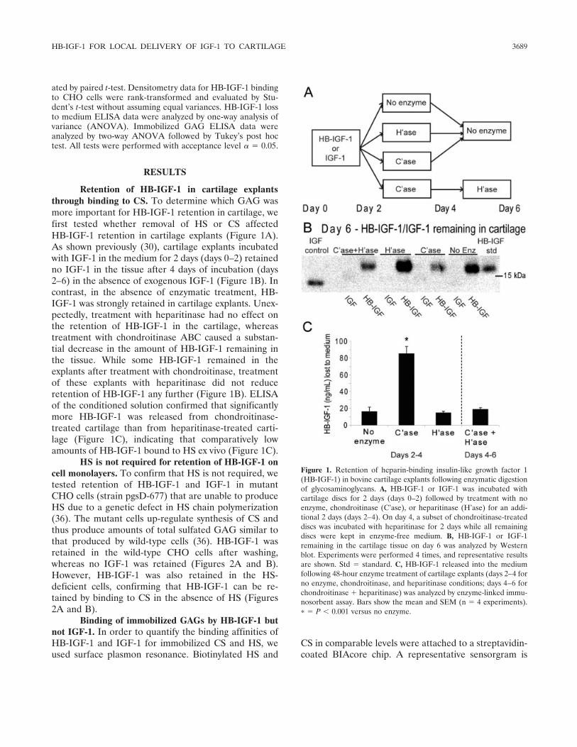
Retention of HB-IGF-1 in cartilage explantsthrough binding to CS. To determine which GAG wasmore important for HB-IGF-1 retention in cartilage, wefirst tested whether removal of HS or CS affectedHB-IGF-1 retention in cartilage explants (Figure 1A).As shown previously (30), cartilage explants incubatedwith IGF-1 in the medium for 2 days (days 0–2) retainedno IGF-1 in the tissue after 4 days of incubation (days2–6) in the absence of exogenous IGF-1 (Figure 1B). Incontrast, in the absence of enzymatic treatment, HB-IGF-1 was strongly retained in cartilage explants. Unex-pectedly, treatment with heparitinase had no effect onthe retention of HB-IGF-1 in the cartilage, whereastreatment with chondroitinase ABC caused a substan-tial decrease in the amount of HB-IGF-1 remaining inthe tissue. While some HB-IGF-1 remained in theexplants after treatment with chondroitinase, treatmentof these explants with heparitinase did not reduceretention of HB-IGF-1 any further (Figure 1B). ELISAof the conditioned solution confirmed that significantlymore HB-IGF-1 was released from chondroitinase-treated cartilage than from heparitinase-treated carti-lage (Figure 1C), indicating that comparatively lowamounts of HB-IGF-1 bound to HS ex vivo (Figure 1C).
HS is not required for retention of HB-IGF-1 oncell monolayers. To confirm that HS is not required, wetested retention of HB-IGF-1 and IGF-1 in mutantCHO cells (strain pgsD-677) that are unable to produceHS due to a genetic defect in HS chain polymerization(36). The mutant cells up-regulate synthesis of CS andthus produce amounts of total sulfated GAG similar tothat produced by wild-type cells (36). HB-IGF-1 wasretained in the wild-type CHO cells after washing,whereas no IGF-1 was retained (Figures 2A and B).However, HB-IGF-1 was also retained in the HS-deficient cells, confirming that HB-IGF-1 can be re-tained by binding to CS in the absence of HS (Figures2A and B).
Binding of immobilized GAGs by HB-IGF-1 butnot IGF-1. In order to quantify the binding affinities ofHB-IGF-1 and IGF-1 for immobilized CS and HS, weused surface plasmon resonance. Biotinylated HS and
CS in comparable levels were attached to a streptavidin-coated BIAcore chip. A representative sensorgram is
Figure 1. Retention of heparin-binding insulin-like growth factor 1(HB-IGF-1) in bovine cartilage explants following enzymatic digestionof glycosaminoglycans. A, HB-IGF-1 or IGF-1 was incubated withcartilage discs for 2 days (days 0–2) followed by treatment with noenzyme, chondroitinase (C’ase), or heparitinase (H’ase) for an addi-tional 2 days (days 2–4). On day 4, a subset of chondroitinase-treateddiscs was incubated with heparitinase for 2 days while all remainingdiscs were kept in enzyme-free medium. B, HB-IGF-1 or IGF-1remaining in the cartilage tissue on day 6 was analyzed by Westernblot. Experiments were performed 4 times, and representative resultsare shown. Std � standard. C, HB-IGF-1 released into the mediumfollowing 48-hour enzyme treatment of cartilage explants (days 2–4 forno enzyme, chondroitinase, and heparitinase conditions; days 4–6 forchondroitinase � heparitinase) was analyzed by enzyme-linked immu-nosorbent assay. Bars show the mean and SEM (n � 4 experiments).� � P � 0.001 versus no enzyme.
HB-IGF-1 FOR LOCAL DELIVERY OF IGF-1 TO CARTILAGE 3689
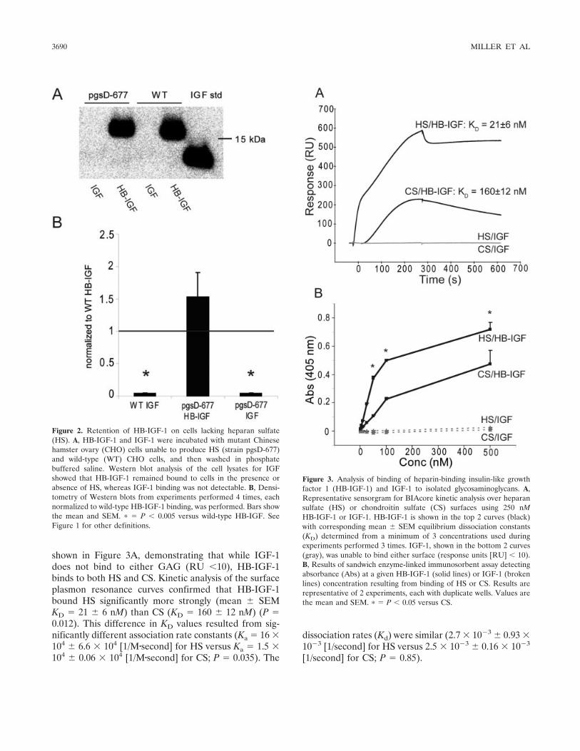
shown in Figure 3A, demonstrating that while IGF-1does not bind to either GAG (RU �10), HB-IGF-1binds to both HS and CS. Kinetic analysis of the surfaceplasmon resonance curves confirmed that HB-IGF-1bound HS significantly more strongly (mean � SEMKD � 21 � 6 nM) than CS (KD � 160 � 12 nM) (P �0.012). This difference in KD values resulted from sig-nificantly different association rate constants (Ka � 16 �104 � 6.6 � 104 [1/M�second] for HS versus Ka � 1.5 �104 � 0.06 � 104 [1/M�second] for CS; P � 0.035). The
dissociation rates (Kd) were similar (2.7 � 10�3 � 0.93 �10�3 [1/second] for HS versus 2.5 � 10�3 � 0.16 � 10�3
[1/second] for CS; P � 0.85).
Figure 2. Retention of HB-IGF-1 on cells lacking heparan sulfate(HS). A, HB-IGF-1 and IGF-1 were incubated with mutant Chinesehamster ovary (CHO) cells unable to produce HS (strain pgsD-677)and wild-type (WT) CHO cells, and then washed in phosphatebuffered saline. Western blot analysis of the cell lysates for IGFshowed that HB-IGF-1 remained bound to cells in the presence orabsence of HS, whereas IGF-1 binding was not detectable. B, Densi-tometry of Western blots from experiments performed 4 times, eachnormalized to wild-type HB-IGF-1 binding, was performed. Bars showthe mean and SEM. � � P � 0.005 versus wild-type HB-IGF. SeeFigure 1 for other definitions.
Figure 3. Analysis of binding of heparin-binding insulin-like growthfactor 1 (HB-IGF-1) and IGF-1 to isolated glycosaminoglycans. A,Representative sensorgram for BIAcore kinetic analysis over heparansulfate (HS) or chondroitin sulfate (CS) surfaces using 250 nMHB-IGF-1 or IGF-1. HB-IGF-1 is shown in the top 2 curves (black)with corresponding mean � SEM equilibrium dissociation constants(KD) determined from a minimum of 3 concentrations used duringexperiments performed 3 times. IGF-1, shown in the bottom 2 curves(gray), was unable to bind either surface (response units [RU] � 10).B, Results of sandwich enzyme-linked immunosorbent assay detectingabsorbance (Abs) at a given HB-IGF-1 (solid lines) or IGF-1 (brokenlines) concentration resulting from binding of HS or CS. Results arerepresentative of 2 experiments, each with duplicate wells. Values arethe mean and SEM. � � P � 0.05 versus CS.
3690 MILLER ET AL
The relative differences in binding affinities ofHB-IGF-1 and IGF-1 to HS and CS were confirmed bysandwich ELISA. Binding of HB-IGF-1 to immobilizedHS and CS increased with concentration, with morebinding to HS at the same given concentration of HS orCS contained in each well (Figure 3B). Therefore,although HB-IGF-1 binds HS with higher affinity, thedata suggest that binding to CS would dominate in
cartilage tissue, where CS is �500–1,000 times moreabundant than HS (25,43).
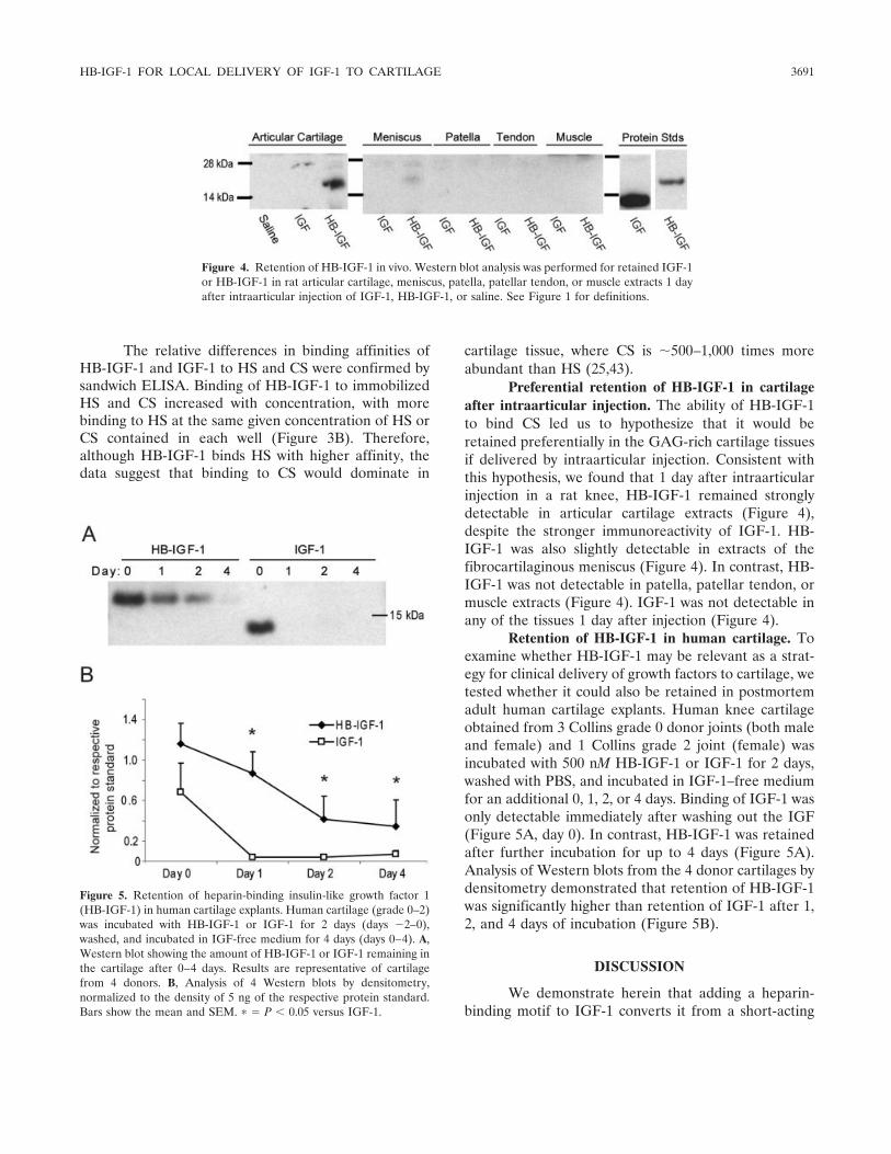
Preferential retention of HB-IGF-1 in cartilageafter intraarticular injection. The ability of HB-IGF-1to bind CS led us to hypothesize that it would beretained preferentially in the GAG-rich cartilage tissuesif delivered by intraarticular injection. Consistent withthis hypothesis, we found that 1 day after intraarticularinjection in a rat knee, HB-IGF-1 remained stronglydetectable in articular cartilage extracts (Figure 4),despite the stronger immunoreactivity of IGF-1. HB-IGF-1 was also slightly detectable in extracts of thefibrocartilaginous meniscus (Figure 4). In contrast, HB-IGF-1 was not detectable in patella, patellar tendon, ormuscle extracts (Figure 4). IGF-1 was not detectable inany of the tissues 1 day after injection (Figure 4).
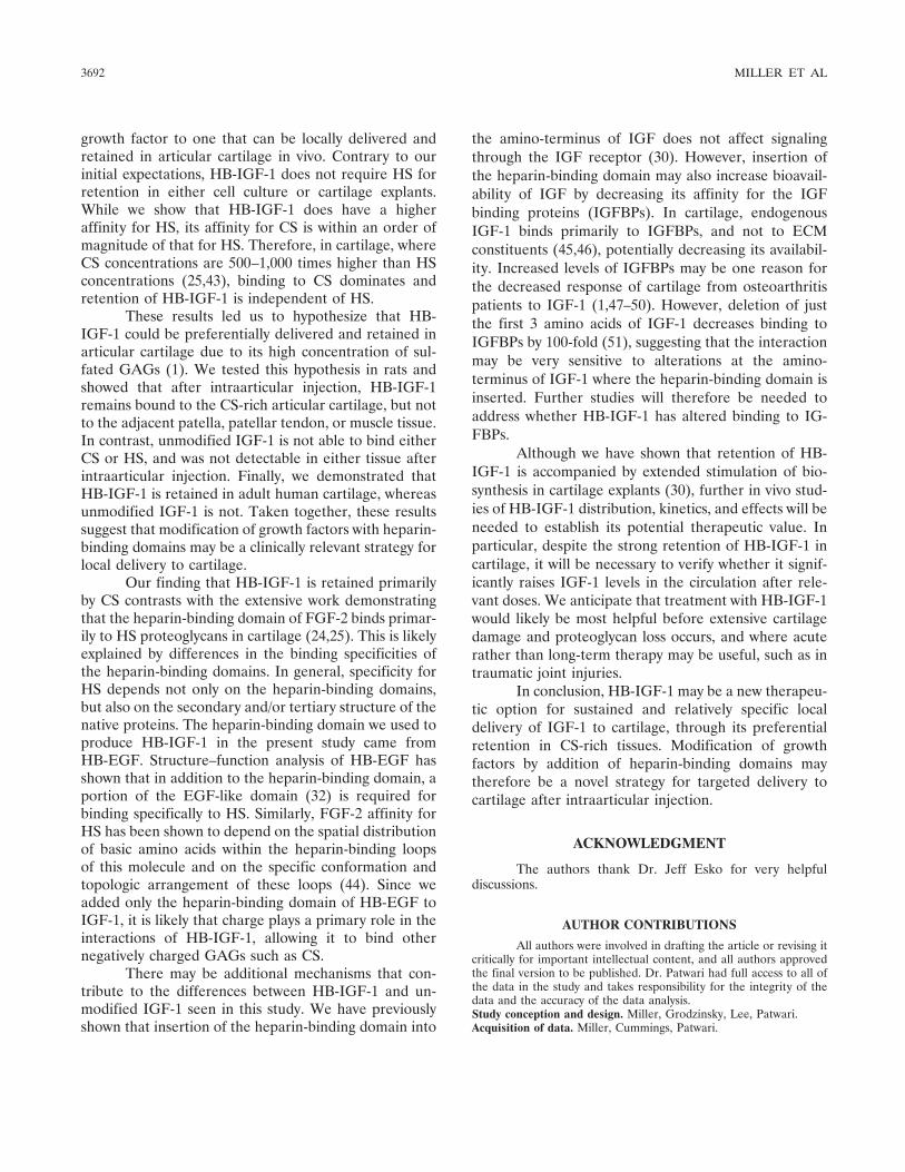
Retention of HB-IGF-1 in human cartilage. Toexamine whether HB-IGF-1 may be relevant as a strat-egy for clinical delivery of growth factors to cartilage, wetested whether it could also be retained in postmortemadult human cartilage explants. Human knee cartilageobtained from 3 Collins grade 0 donor joints (both maleand female) and 1 Collins grade 2 joint (female) wasincubated with 500 nM HB-IGF-1 or IGF-1 for 2 days,washed with PBS, and incubated in IGF-1–free mediumfor an additional 0, 1, 2, or 4 days. Binding of IGF-1 wasonly detectable immediately after washing out the IGF(Figure 5A, day 0). In contrast, HB-IGF-1 was retainedafter further incubation for up to 4 days (Figure 5A).Analysis of Western blots from the 4 donor cartilages bydensitometry demonstrated that retention of HB-IGF-1was significantly higher than retention of IGF-1 after 1,2, and 4 days of incubation (Figure 5B).
DISCUSSION
We demonstrate herein that adding a heparin-binding motif to IGF-1 converts it from a short-acting
Figure 5. Retention of heparin-binding insulin-like growth factor 1(HB-IGF-1) in human cartilage explants. Human cartilage (grade 0–2)was incubated with HB-IGF-1 or IGF-1 for 2 days (days �2–0),washed, and incubated in IGF-free medium for 4 days (days 0–4). A,Western blot showing the amount of HB-IGF-1 or IGF-1 remaining inthe cartilage after 0–4 days. Results are representative of cartilagefrom 4 donors. B, Analysis of 4 Western blots by densitometry,normalized to the density of 5 ng of the respective protein standard.Bars show the mean and SEM. � � P � 0.05 versus IGF-1.
Figure 4. Retention of HB-IGF-1 in vivo. Western blot analysis was performed for retained IGF-1or HB-IGF-1 in rat articular cartilage, meniscus, patella, patellar tendon, or muscle extracts 1 dayafter intraarticular injection of IGF-1, HB-IGF-1, or saline. See Figure 1 for definitions.
HB-IGF-1 FOR LOCAL DELIVERY OF IGF-1 TO CARTILAGE 3691
growth factor to one that can be locally delivered andretained in articular cartilage in vivo. Contrary to ourinitial expectations, HB-IGF-1 does not require HS forretention in either cell culture or cartilage explants.While we show that HB-IGF-1 does have a higheraffinity for HS, its affinity for CS is within an order ofmagnitude of that for HS. Therefore, in cartilage, whereCS concentrations are 500–1,000 times higher than HSconcentrations (25,43), binding to CS dominates andretention of HB-IGF-1 is independent of HS.
These results led us to hypothesize that HB-IGF-1 could be preferentially delivered and retained inarticular cartilage due to its high concentration of sul-fated GAGs (1). We tested this hypothesis in rats andshowed that after intraarticular injection, HB-IGF-1remains bound to the CS-rich articular cartilage, but notto the adjacent patella, patellar tendon, or muscle tissue.In contrast, unmodified IGF-1 is not able to bind eitherCS or HS, and was not detectable in either tissue afterintraarticular injection. Finally, we demonstrated thatHB-IGF-1 is retained in adult human cartilage, whereasunmodified IGF-1 is not. Taken together, these resultssuggest that modification of growth factors with heparin-binding domains may be a clinically relevant strategy forlocal delivery to cartilage.
Our finding that HB-IGF-1 is retained primarilyby CS contrasts with the extensive work demonstratingthat the heparin-binding domain of FGF-2 binds primar-ily to HS proteoglycans in cartilage (24,25). This is likelyexplained by differences in the binding specificities ofthe heparin-binding domains. In general, specificity forHS depends not only on the heparin-binding domains,but also on the secondary and/or tertiary structure of thenative proteins. The heparin-binding domain we used toproduce HB-IGF-1 in the present study came fromHB-EGF. Structure–function analysis of HB-EGF hasshown that in addition to the heparin-binding domain, aportion of the EGF-like domain (32) is required forbinding specifically to HS. Similarly, FGF-2 affinity forHS has been shown to depend on the spatial distributionof basic amino acids within the heparin-binding loopsof this molecule and on the specific conformation andtopologic arrangement of these loops (44). Since weadded only the heparin-binding domain of HB-EGF toIGF-1, it is likely that charge plays a primary role in theinteractions of HB-IGF-1, allowing it to bind othernegatively charged GAGs such as CS.
There may be additional mechanisms that con-tribute to the differences between HB-IGF-1 and un-modified IGF-1 seen in this study. We have previouslyshown that insertion of the heparin-binding domain into
the amino-terminus of IGF does not affect signalingthrough the IGF receptor (30). However, insertion ofthe heparin-binding domain may also increase bioavail-ability of IGF by decreasing its affinity for the IGFbinding proteins (IGFBPs). In cartilage, endogenousIGF-1 binds primarily to IGFBPs, and not to ECMconstituents (45,46), potentially decreasing its availabil-ity. Increased levels of IGFBPs may be one reason forthe decreased response of cartilage from osteoarthritispatients to IGF-1 (1,47–50). However, deletion of justthe first 3 amino acids of IGF-1 decreases binding toIGFBPs by 100-fold (51), suggesting that the interactionmay be very sensitive to alterations at the amino-terminus of IGF-1 where the heparin-binding domain isinserted. Further studies will therefore be needed toaddress whether HB-IGF-1 has altered binding to IG-FBPs.
Although we have shown that retention of HB-IGF-1 is accompanied by extended stimulation of bio-synthesis in cartilage explants (30), further in vivo stud-ies of HB-IGF-1 distribution, kinetics, and effects will beneeded to establish its potential therapeutic value. Inparticular, despite the strong retention of HB-IGF-1 incartilage, it will be necessary to verify whether it signif-icantly raises IGF-1 levels in the circulation after rele-vant doses. We anticipate that treatment with HB-IGF-1would likely be most helpful before extensive cartilagedamage and proteoglycan loss occurs, and where acuterather than long-term therapy may be useful, such as intraumatic joint injuries.
In conclusion, HB-IGF-1 may be a new therapeu-tic option for sustained and relatively specific localdelivery of IGF-1 to cartilage, through its preferentialretention in CS-rich tissues. Modification of growthfactors by addition of heparin-binding domains maytherefore be a novel strategy for targeted delivery tocartilage after intraarticular injection.
ACKNOWLEDGMENT
The authors thank Dr. Jeff Esko for very helpfuldiscussions.
AUTHOR CONTRIBUTIONS
All authors were involved in drafting the article or revising itcritically for important intellectual content, and all authors approvedthe final version to be published. Dr. Patwari had full access to all ofthe data in the study and takes responsibility for the integrity of thedata and the accuracy of the data analysis.Study conception and design. Miller, Grodzinsky, Lee, Patwari.Acquisition of data. Miller, Cummings, Patwari.
3692 MILLER ET AL
Analysis and interpretation of data. Miller, Grodzinsky, Plaas, Cole,Lee, Patwari.
ROLE OF THE STUDY SPONSOR
Biomeasure, Inc. had no role in designing or conducting thestudy or the decision to submit the manuscript for publication.Publication of this article was not contingent upon approval byBiomeasure, Inc.
REFERENCES
1. Goldring MB. Update on the biology of the chondrocyte and newapproaches to treating cartilage diseases. Best Pract Res ClinRheumatol 2006;20:1003–25.
2. Bonassar LJ, Grodzinsky AJ, Frank EH, Davila SG, Bhaktav NR,Trippel SB. The effect of dynamic compression on the response ofarticular cartilage to insulin-like growth factor-I. J Orthop Res2001;19:11–7.
3. McQuillan DJ, Handley CJ, Campbell MA, Bolis S, Milway VE,Herinton AC. Stimulation of proteoglycan biosynthesis by serumand insulin-like growth factor-I in cultured bovine articular carti-lage. Biochem J 1986;240:423–30.
4. Curtis AJ, Devenish RJ, Handley CJ. Modulation of aggrecan andlink-protein synthesis in articular cartilage. Biochem J 1992;288:721–6.
5. Hui W, Rowan AD, Cawston T. Modulation of the expression ofmatrix metalloproteinase and tissue inhibitors of metalloprotein-ases by TGF-�1 and IGF-1 in primary human articular and bovinenasal chondrocytes stimulated with TNF-�. Cytokine 2001;16:31–5.
6. Luyten FP, Hascall VC, Nissley SP, Morales TI, Reddi AH.Insulin-like growth factors maintain steady-state metabolism ofproteoglycans in bovine articular cartilage explants. Arch BiochemBiophys 1988;267:416–25.
7. Tyler JA. Insulin-like growth factor I can decrease degradationand promote synthesis of proteoglycan in cartilage exposed tocytokines. Biochem J 1989;260:543–8.
8. Schneiderman R, Rosenberg N, Hiss J, Lee P, Liu F, Hintz RL, etal. Concentration and size distribution of insulin-like growthfactor-1 in human normal and osteoarthritic synovial fluid andcartilage. Arch Biochem Biophys 1995;324:173–88.
9. Schmidt MB, Chen EH, Lynch SE. A review of the effects ofinsulin-like growth factor and platelet derived growth factor on invivo cartilage healing and repair. Osteoarthritis Cartilage 2006;14:403–12.
10. Matsumoto T, Gargosky SE, Iwasaki K, Rosenfeld RG. Identifi-cation and characterization of insulin-like growth factors (IGFs),IGF-binding proteins (IGFBPs), and IGFBP proteases in humansynovial fluid. J Clin Endocrinol Metab 1996;81:150–5.
11. Loeser RF, Shanker G. Autocrine stimulation by insulin-likegrowth factor 1 and insulin-like growth factor 2 mediates chon-drocyte survival in vitro. Arthritis Rheum 2000;43:1552–9.
12. Nixon A, Saxer R, Brower-Toland BD. Exogenous insulin-likegrowth factor-I stimulates an autoinductive IGF-I autocrine/para-crine response in chondrocytes. J Orthop Res 2001;19:26–32.
13. Madry H, Kaul G, Cucchiarini M, Stein U, Zurakowski D,Remberger K, et al. Enhanced repair of articular cartilage defectsin vivo by transplanted chondrocytes overexpressing insulin-likegrowth factor I (IGF-I). Gene Ther 2005;12:1171–9.
14. Goodrich LR, Hidaka C, Robbins PD, Evans CH, Nixon AJ. Ge-netic modification of chondrocytes with insulin-like growth fac-tor-1 enhances cartilage healing in an equine model. J Bone JointSurg Br 2007;89:672–85.
15. Laron Z. Somatomedin-1 (recombinant insulin-like growth factor-
1): clinical pharmacology and potential treatment of endocrineand metabolic disorders. BioDrugs 1999;11:55–70.
16. Chan JM, Stampfer MJ, Giovannucci E, Gann PH, Ma J, Wilkin-son P, et al. Plasma insulin-like growth factor-I and prostate cancerrisk: a prospective study. Science 1998;279:563–6.
17. Jabri N, Schalch D, Schwartz S, Fischer J, Kipnes M, Radnik B, etal. Adverse effects of recombinant human insulin-like growthfactor I in obese insulin-resistant type II diabetic patients. Diabe-tes 1994;43:369–74.
18. Fortier LA, Mohammed HO, Lust G, Nixon AJ. Insulin-likegrowth factor-I enhances cell-based repair of articular cartilage.J Bone Joint Surg Br 2002;84:276–88.
19. Nixon AJ, Fortier LA, Williams J, Mohammed H. Enhancedrepair of extensive articular defects by insulin-like growth factor-I-laden fibrin composites. J Orthop Res 1999;17:475–87.
20. Hunziker EB, Rosenberg LC. Repair of partial-thickness defectsin articular cartilage: cell recruitment from the synovial mem-brane. J Bone Joint Surg Am 1996;78:721–33.
21. Farach-Carson MC, Hecht JT, Carson DD. Heparan sulfateproteoglycans: key players in cartilage biology. Crit Rev EukaryotGene Expr 2005;15:29–48.
22. Kirn-Safran CB, Gomes RR, Brown AJ, Carson DD. Heparansulfate proteoglycans: coordinators of multiple signaling pathwaysduring chondrogenesis. Birth Defects Res C Embryo Today 2004;72:69–88.
23. Heinegard D. Proteoglycans and more—from molecules to biol-ogy. Int J Exp Pathol 2009;90:575–86.
24. Govindraj P, West L, Smith S, Hassell JR. Modulation of FGF-2binding to chondrocytes from the developing growth plate byperlecan. Matrix Biol 2006;25:232–9.
25. Vincent TL, McLean CJ, Full LE, Peston D, Saklatvala J. FGF-2is bound to perlecan in the pericellular matrix of articular carti-lage, where it acts as a chondrocyte mechanotransducer. Osteoar-thritis Cartilage 2007;15:752–63.
26. Vincent T, Hermansson M, Bolton M, Wait R, Saklatvala J. BasicFGF mediates an immediate response of articular cartilage tomechanical injury. Proc Natl Acad Sci U S A 2002;99:8259–64.
27. Vincent TL, Hermansson MA, Hansen UN, Amis AA, SaklatvalaJ. Basic fibroblast growth factor mediates transduction of mechan-ical signals when articular cartilage is loaded. Arthritis Rheum2004;50:526–33.
28. Saksela O, Moscatelli D, Sommer A, Rifkin DB. Endothelialcell-derived heparan sulfate binds basic fibroblast growth factorand protects it from proteolytic degradation. J Cell Biol 1988;107:743–51.
29. Flaumenhaft R, Moscatelli D, Saksela O, Rifkin DB. Role ofextracellular matrix in the action of basic fibroblast growth factor:matrix as a source of growth factor for long-term stimulation ofplasminogen activator production and DNA synthesis. J CellPhysiol 1989;140:75–81.
30. Tokunou T, Miller R, Patwari P, Davis ME, Segers VF, Grodzin-sky AJ, et al. Engineering insulin-like growth factor-1 for localdelivery. FASEB J 2008;22:1886–93.
31. Cardin AD, Weintraub HJ. Molecular modeling of protein-glyco-saminoglycan interactions. Arterioscler Thromb Vasc Biol 1989;9:21–32.
32. Thompson SA, Higashiyama S, Wood K, Pollitt NS, Damm D,McEnroe G, et al. Characterization of sequences within heparin-binding EGF-like growth factor that mediates interaction withheparin. J Biol Chem 1994;269:2541–9.
33. Oike Y, Kimata K, Shinomura T, Nakazawa K, Suzuki S. Struc-tural analysis of chick-embryo cartilage proteoglycan by selectivedegradation with chondroitin lyases (chondroitinases) and endo-�-D-galactosidase (keratanase). Biochem J 1980;191:193–207.
34. Silverberg I, Havsmark B, Fransson LA. The substrate specificityof heparan sulphate lyase and heparin lyase from Flavobacteriumheparinum. Carbohydr Res 1985;137:227–38.
HB-IGF-1 FOR LOCAL DELIVERY OF IGF-1 TO CARTILAGE 3693
35. David G, Bai XM, van der Schueren B, Cassiman JJ, van denBerghe H. Developmental changes in heparan sulfate expression:in situ detection with mAbs. J Cell Biol 1992;119:961–75.
36. Lidholt K, Weinke JL, Kiser CS, Lugemwa FN, Bame KJ, CheifetzS, et al. A single mutation affects both N-acetylglucosaminyltrans-ferase and glucuronosyltransferase activities in a Chinese hamsterovary cell mutant defective in heparan sulfate biosynthesis. ProcNatl Acad Sci U S A 1992;89:2267–71.
37. Ley K, Cerrito M, Arfors KE. Sulfated polysaccharides inhibitleukocyte rolling in rabbit mesentery venules. Am J Physiol1991;260:H1667–73.
38. Friedrich U, Blom AM, Dahlback B, Villoutreix BO. Structuraland energetic characteristics of the heparin-binding site in anti-thrombotic protein C. J Biol Chem 2001;276:24122–8.
39. Ricard-Blum S, Feraud O, Lortat-Jacob H, Rencurosi A, Fukai N,Dkhissi F, et al. Characterization of endostatin binding to heparinand heparan sulfate by surface plasmon resonance and molecularmodeling: role of divalent cations. J Biol Chem 2004;279:2927–36.
40. Muehleman C, Bareither D, Huch K, Cole AA, Kuettner KE.Prevalence of degenerative morphological changes in the joints ofthe lower extremity. Osteoarthritis Cartilage 1997;5:23–37.
41. Collins DH. The pathology of articular and spinal diseases.London: Edward Arnold; 1949. p. 76–9.
42. Sui Y, Lee JH, DiMicco MA, Vanderploeg EJ, Blake SM, HungHH, et al. Mechanical injury potentiates proteoglycan catabolisminduced by interleukin-6 with soluble interleukin-6 receptor andtumor necrosis factor � in immature bovine and adult humanarticular cartilage. Arthritis Rheum 2009;60:2985–96.
43. Govindraj P, West L, Koob TJ, Neame P, Doege K, Hassell JR.Isolation and identification of the major heparan sulfate proteo-glycans in the developing bovine rib growth plate. J Biol Chem2002;277:19461–9.
44. Raman R, Venkataraman G, Ernst S, Sasisekharan V, Sasisekha-ran R. Structural specificity of heparin binding in the fibroblastgrowth factor family of proteins. Proc Natl Acad Sci U S A2003;100:2357–62.
45. Garcia AM, Szasz N, Trippel SB, Morales TI, Grodzinsky AJ,Frank EH. Transport and binding of insulin-like growth factor Ithrough articular cartilage. Arch Biochem Biophys 2003;415:69–79.
46. Bhakta NR, Garcia AM, Frank EH, Grodzinsky AJ, Morales TI.The insulin-like growth factors (IGFs) I and II bind to articularcartilage via the IGF-binding proteins. J Biol Chem 2000;275:5860–6.
47. Martel-Pelletier J, Di Battista JA, Lajeunesse D, Pelletier JP.IGF/IGFBP axis in cartilage and bone in osteoarthritis pathogen-esis. Inflamm Res 1998;47:90–100.
48. Martin JA, Ellerbroek SM, Buckwalter JA. Age-related decline inchondrocyte response to insulin-like growth factor-I: the role ofgrowth factor binding proteins. J Orthop Res 1997;15:491–8.
49. Loeser RF, Shanker G, Carlson CS, Gardin JF, Shelton BJ,Sonntag WE. Reduction in the chondrocyte response to insulin-like growth factor 1 in aging and osteoarthritis: studies in anon-human primate model of naturally occurring disease. ArthritisRheum 2000;43:2110–20.
50. Dore S, Pelletier JP, DiBattista JA, Tardif G, Brazeau P, Martel-Pelletier J. Human osteoarthritic chondrocytes possess an in-creased number of insulin-like growth factor 1 binding sites but areunresponsive to its stimulation: possible role of IGF-1–bindingproteins. Arthritis Rheum 1994;37:253–63.
51. Bagley CJ, May BL, Szabo L, McNamara PJ, Ross M, Francis GL,et al. A key functional role for the insulin-like growth factor 1N-terminal pentapeptide. Biochem J 1989;259:665–71.
3694 MILLER ET AL