intracranial arachnoid cysts in dogs free ce - david · pdf file ·...
TRANSCRIPT

Intracranial Arachnoid Cysts in Dogs FREE
CE
CE Article 1
Intracranial Arachnoid Cysts in Dogs
Intracranial arachnoid cyst (IAC), also called intracranial intraarachnoid cystand quadrigeminal cyst, is a develop-
mental brain disorder in which cerebro-spinal fl uid (CSF) is thought to accumulate within a split of the arachnoid membrane.1
Although IACs have been reported to occur in several locations in humans, all reported canine cases have been in the
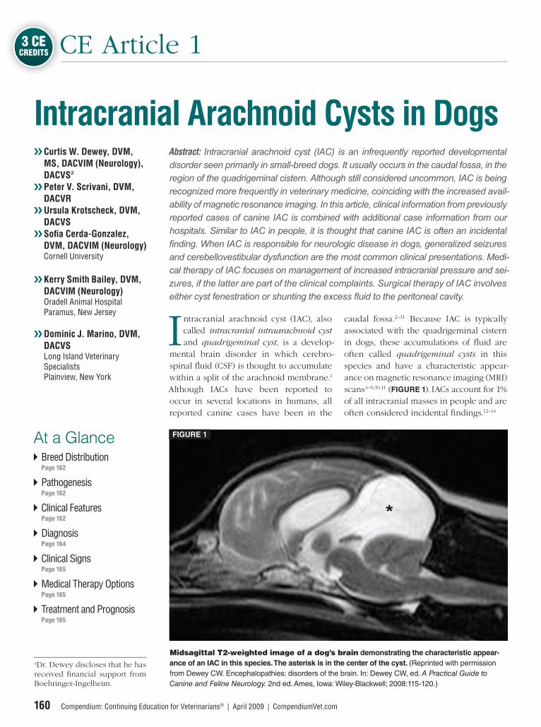
caudal fossa.2–11 Because IAC is typically associated with the quadrigeminal cistern in dogs, these accumulations of fl uid are often called quadrigeminal cysts in this species and have a characteristic appear-ance on magnetic resonance imaging (MRI) scans4–6,10,11 (FIGURE 1). IACs account for 1% of all intracranial masses in people and are often considered incidental fi ndings.12–14
❯❯ Curtis W. Dewey, DVM, MS, DACVIM (Neurology), DACVSa
❯❯ Peter V. Scrivani, DVM, DACVR
❯❯ Ursula Krotscheck, DVM, DACVS
❯❯ Sofi a Cerda-Gonzalez, DVM, DACVIM (Neurology)Cornell University
❯❯ Kerry Smith Bailey, DVM, DACVIM (Neurology)Oradell Animal HospitalParamus, New Jersey
❯❯ Dominic J. Marino, DVM, DACVSLong Island Veterinary SpecialistsPlainview, New York
3 CECREDITS
Breed Distribution Page 162
Pathogenesis Page 162
Clinical Features Page 162
Diagnosis Page 164
Clinical Signs Page 165
Medical Therapy Options Page 165
Treatment and Prognosis Page 165
At a Glance
160 Compendium: Continuing Education for Veterinarians® | April 2009 | CompendiumVet.com
Abstract: Intracranial arachnoid cyst (IAC) is an infrequently reported developmental disorder seen primarily in small-breed dogs. It usually occurs in the caudal fossa, in the region of the quadrigeminal cistern. Although still considered uncommon, IAC is being recognized more frequently in veterinary medicine, coinciding with the increased avail-ability of magnetic resonance imaging. In this article, clinical information from previously reported cases of canine IAC is combined with additional case information from our hospitals. Similar to IAC in people, it is thought that canine IAC is often an incidental fi nding. When IAC is responsible for neurologic disease in dogs, generalized seizures and cerebellovestibular dysfunction are the most common clinical presentations. Medi-cal therapy of IAC focuses on management of increased intracranial pressure and sei-zures, if the latter are part of the clinical complaints. Surgical therapy of IAC involves either cyst fenestration or shunting the excess fl uid to the peritoneal cavity.
Midsagittal T2-weighted image of a dog’s brain demonstrating the characteristic appear-
ance of an IAC in this species. The asterisk is in the center of the cyst. (Reprinted with permission
from Dewey CW. Encephalopathies: disorders of the brain. In: Dewey CW, ed. A Practical Guide to Canine and Feline Neurology. 2nd ed. Ames, Iowa: Wiley-Blackwell; 2008:115-120.)
FIGURE 1
aDr. Dewey discloses that he has received fi nancial support from Boehringer-Ingelheim.

Intracranial Arachnoid Cysts in Dogs
CompendiumVet.com | April 2009 | Compendium: Continuing Education for Veterinarians® 161
FREE
CE
CompendiumVet.com | April 2009 | Compendium: Continuing Education for Veterinarians® 161
Dr. Dewey is an associate
professor of neurology and
neurosurgery and chief of
the section of neurology at
Cornell University.

Intracranial Arachnoid Cysts in Dogs
162 Compendium: Continuing Education for Veterinarians® | April 2009 | CompendiumVet.com
FREE
CE
There are 10 clinical reports of IAC in dogs in the veterinary literature.2–11 This review combines these reported cases with three additional cases from our hospitals to present information regarding 56 dogs with IAC. Most reported cases of IAC in dogs are in small breeds, with a predominance of brachycephalic animals.2–11 Shih tzus may be overrepresented.11,15 Male sex also appears to be a predisposing factor. Clinical signs attributable to IAC in dogs are most often related to cerebral or cerebellar com-pression by the cyst; generalized seizures and central vestibular dysfunction are most commonly noted.15 Similar to human IACs, a large proportion of reported IACs in dogs were suspected to be incidental.4–6,11 Medical and surgical options are available to treat IAC in dogs.
PathogenesisIACs are believed to represent a developmen-tal abnormality caused by an aberrant split in the arachnoid membrane during embryo-genesis.1,12 The developing neural tube is sur-rounded by a loose layer of mesenchymal
tissue called the perimedullary mesh; this tissue eventually becomes the pia and arach-noid layers of the meninges. In normal devel-opment, pulsatile CSF fl ow from the choroid plexuses is thought to divide the perimedul-lary mesh into the pia and arachnoid layers, effectively creating the subarachnoid space. It is postulated that some aberration of CSF fl ow from the choroid plexuses during this stage of development forces a separation within the forming arachnoid layer, eventu-ally leading to the creation of an IAC.1,12 The intraarachnoid location of IACs has been demonstrated via light and electron micros-copy in people.1 Depending on whether these cysts communicate with the subarach-noid space or the ventricular system, they are sometimes referred to as communicating or noncommunicating.1
The mechanisms by which an IAC con-tinues to expand with fluid are unknown, but several theories have been proposed.1,12 There is evidence that arachnoid cells lining the IAC may have secretory capacity.1,12,16 Fluid may also move into the cyst via an osmotic pressure gradient. However, consid-ering that the fluid within the IAC is nearly identical to CSF, this theory is unlikely.1,12 In addition, there have been documented cases in people in which small slits exist between the IAC and the subarachnoid space; these slits act as one-way valves, diverting CSF into the cyst during systole and preventing its return to the subarachnoid space during diastole.1,12,17
Clinical FeaturesMost reported dogs with IAC have been small breeds, and many had brachycephalic confor-mation. The following information was ob tained by combining the IAC cases reported in the literature with three additional cases from our hospitals. The breed distribution of these 56 dogs is listed in BOX 1. Approximately 66% of the dogs (37 of 56) were male.2–11
There is a wide age range at clinical pre-sentation for dogs with IAC (2 months to 12 years), with an approximate average age of 4 years. The most common clinical signs (BOX 2) seen with IAC are refl ective of forebrain or central vestibular (cerebellovestibular) dysfunc-tion. Other reported clinical signs include neck pain, paraparesis, and tetraparesis.2–11
Breed Distribution of Reported Intracranial Arachnoid Cysts in Dogs
Shih tzu . . . . . . . . . . . . . . . . . . . . . . . . . 15 dogs Maltese . . . . . . . . . . . . . . . . . . . . . . . . . . . . . . . .4 Pug . . . . . . . . . . . . . . . . . . . . . . . . . . . . . . . . . . . .4 Lhasa apso . . . . . . . . . . . . . . . . . . . . . . . . . . . . .4 Cavalier King Charles spaniel . . . . . . . . . . . .4 Yorkshire terrier . . . . . . . . . . . . . . . . . . . . . . . .4 Chihuahua. . . . . . . . . . . . . . . . . . . . . . . . . . . . . .3 Staffordshire bull terrier . . . . . . . . . . . . . . . . .3 Bulldog. . . . . . . . . . . . . . . . . . . . . . . . . . . . . . . . .3 Pekingese . . . . . . . . . . . . . . . . . . . . . . . . . . . . . .2 West Highland white terrier . . . . . . . . . . . . . .2 Bichon frise . . . . . . . . . . . . . . . . . . . . . . . . . . . .1 Pomeranian . . . . . . . . . . . . . . . . . . . . . . . . . . . .1 Cairn terrier. . . . . . . . . . . . . . . . . . . . . . . . . . . . .1 Jack Russell (Parson Russell) terrier . . . . . .1 Terrier mix. . . . . . . . . . . . . . . . . . . . . . . . . . . . . .1 Beagle . . . . . . . . . . . . . . . . . . . . . . . . . . . . . . . . .1 Miniature schnauzer. . . . . . . . . . . . . . . . . . . . .1 German shorthaired pointer . . . . . . . . . . . . .1
BOX 1
When there is evi-dence of a large IAC and another disease (e.g., granulomatous meningoencepha-lomyelitis, hydro-cephalus) in the same patient, the optimal response to treatment may require treating both conditions.
QuickNotes

No matter how long you have been in this profession, it’s always humbling to know that you are such a trusted partner in the human–animal bond.
Knowing that my clients can access their Pet Portal® to fi nd good medical advice, review their pet’s medical records, and receive medication reminders allows me to consistently meet their needs — and exceed their expectations — even when I have no extra time.
Vetstreet ensures client satisfaction and compliance without additional work for me or my staff.
Easy to set up and easy to use, Vetstreet® is a powerful practice communication and management tool that keeps you in touch with your clients via Pet Portals. To discover how
Vetstreet can help you increase client satisfaction, build compliance, and enhance your bottom line, visit Vetstreet.com, call toll-free 888-799-8387, or email [email protected].
Exceeding client expectations
Gary Edlin, DVMOwner, East Louisville Animal HospitalLouisville, KY
”
“
Vetstreet and Pet Portal are registered trademarks of VetInsite.com, Inc.

Intracranial Arachnoid Cysts in Dogs
164 Compendium: Continuing Education for Veterinarians® | April 2009 | CompendiumVet.com
FREE
CE
Transaxial Computed Tomography ImagesFIGURE 2
Preoperative (A) and postoperative (B) transaxial computed tomography images from a dog with an IAC treated with cystoperitoneal shunting. In A, the arrowheads are point-
ing to the ventral aspect of the cyst and the outlined arrowheads are pointing to the dorsal aspect
of the cyst. In B, the arrow is pointing to the rostral aspect of the shunt. (Reprinted with permission
from Dewey CW, Krotscheck U, Bailey KS, Marino DJ. Craniotomy with cystoperitoneal shunting for treat-
ment of intracranial arachnoid cysts in dogs. Vet Surg 2007;36:416-422.)
A B
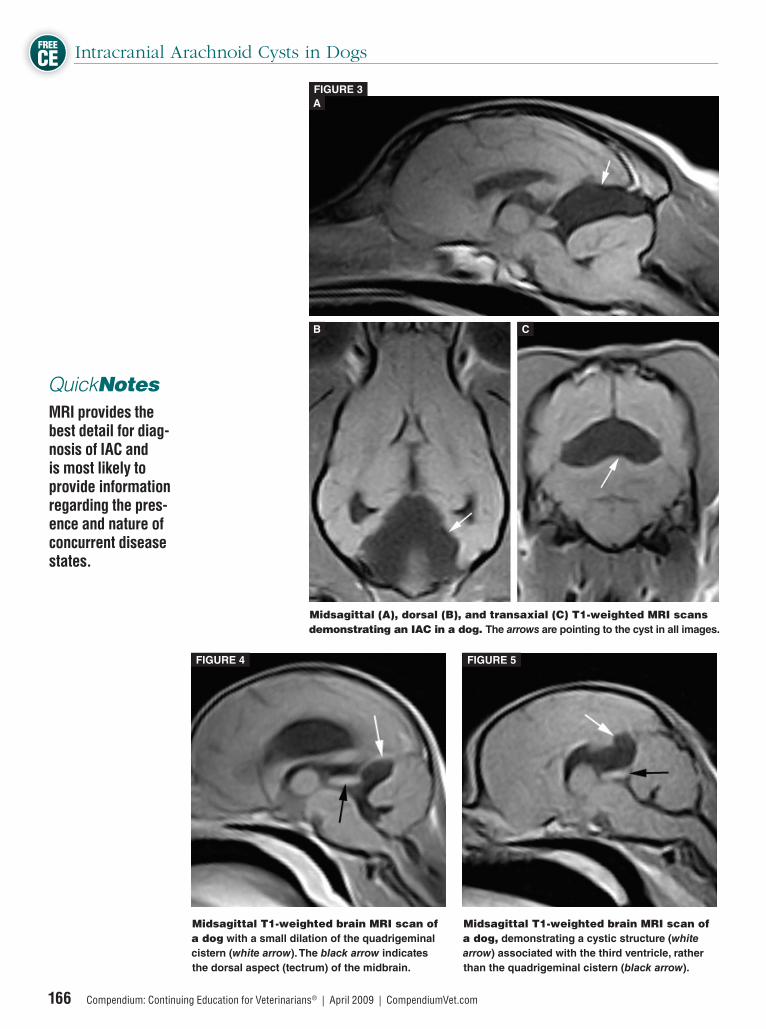
DiagnosisDiagnosis of IAC in dogs is typically made via computed tomography (FIGURE 2) or, prefer-ably, MRI.2–5,7–10 IACs may also be visualized using ultrasound (via the foramen magnum, temporal window, or persistent bregmatic fontanelle), especially in younger dogs.6 The characteristic ultrasonographic appearance of IAC is a large, fl uid-fi lled structure, isoechoic with the CSF spaces (e.g., lateral ventricle) and located between the occipital lobe of the cere-brum and rostral lobe of the cerebellum.6 MRI provides the best detail for diagnosis of IAC and is most likely to provide information regarding the presence and nature of concurrent disease states. The typical appearance of IAC on MRI is a well-demarcated, cystic-appearing structure that is hypointense on T1-weighted images, hyperintense on T2-weighted images, and non-contrast enhancing with intrave-nous gadolinium administration and that sup-presses on FLAIR (fl uid attenuation inversion recovery) images2–5,10,11 (FIGURE 3). Because IAC may be an incidental fi nding, it is important to rule out concurrent infl ammatory disease with a CSF examination. In the absence of an additional brain disorder, the CSF is typically normal in dogs with IAC. In our opinion, it is often diffi cult or impos-sible to discern whether a large IAC in the
presence of another brain disorder is purely an incidental fi nding. We have seen a number of patients with relatively small dilations of the quadrigeminal cistern (FIGURE 4), which may represent a variant of normal structure or may be evidence of nascent IACs that may be of no clinical signifi cance. Conversely, very large IACs have been described both as sole disease entities and as suspected incidental fi ndings in patients with other intracranial disease processes. Anecdotally, we have also observed similar cystic structures in the brain that do not appear to be associated with the quadrigeminal cistern (FIGURE 5); consider-ing that IACs occur in multiple locations in the human brain, this fi nding may simply imply that the term quadrigeminal cyst is too restrictive in describing IAC in dogs.12 Since the presence of a large, fl uid-fi lled struc-ture within the cranial vault likely decreases intracranial compliance, some IACs may be contributory rather than incidental fi ndings. In other words, when there is evidence of a large IAC and another disease (e.g., granu-lomatous meningoencephalomyelitis, hydro-cephalus) in the same patient, the optimal response to treatment may require treating both conditions. In addition, the combined presence of hydrocephalus and IAC in a patient does
Intracranial Arachnoid Cysts in Dogs
CompendiumVet.com | April 2009 | Compendium: Continuing Education for Veterinarians® 165
FREE
CE
not necessarily make one of the disorders (IAC) incidental, nor does this combination ensure that surgically treating one disorder will address the other. Hydrocephalus can be secondary to an IAC, developing because of mechanical obstruction of normal CSF fl ow by the expanding cyst (i.e., obstructive hydro-cephalus).10,18,19 It is unlikely that an IAC can be distinguished as communicating or noncom-municating based on standard MRI sequences; such a distinction would likely require either contrast studies or phase-contrast (cine) MRI. In one report of two dogs with IAC, intracys-tic hemorrhage, which was suspected to have contributed to the development of neurologic dysfunction, was verifi ed at surgery.3
In a recent study, the degree of brain com-pression by an IAC was calculated from MRI scans of dogs with the disorder, and these measurements were correlated with the pres-ence or absence of clinical dysfunction. It was found that dogs with >14% compression of the occipital lobe always displayed clini-cal signs and that dogs with compression of both the cerebellum and the occipital lobe were signifi cantly more likely to display clini-cal signs than dogs with compression of only one region or dogs with no apparent brain compression.11
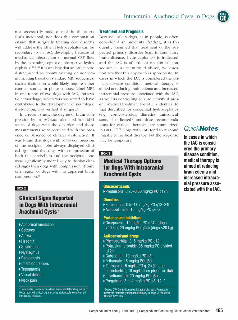
Treatment and PrognosisBecause IAC in dogs, as in people, is often considered an incidental finding, it is fre-quently assumed that treatment of the sus-pected primary disorder (e.g., infl ammatory brain disease, hydrocephalus) is indicated and the IAC is of little or no clinical con-sequence. As mentioned above, we ques-tion whether this approach is appropriate. In cases in which the IAC is considered the pri-mary disease condition, medical therapy is aimed at reducing brain edema and increased intracranial pressure associated with the IAC, as well as controlling seizure activity if pres-ent. Medical treatment for IAC is identical to that described for congenital hydrocephalus (e.g., cortico steroids, diuretics, anticonvul-sants if indicated), and dose recommenda-tions for various therapies are summarized in BOX 3.20,21 Dogs with IAC tend to respond initially to medical therapy, but the response may be temporary.
Medical Therapy Options for Dogs With Intracranial Arachnoid Cysts
Glucocorticoids Prednisone: 0.25–0.50 mg/kg PO q12h
Diuretics Furosemide: 0.5–4.0 mg/kg PO q12–24h Acetazolamide: 10 mg/kg PO q6–8h
Proton pump inhibitors Omeprazole: 10 mg/kg PO q24h (dogs <20 kg); 20 mg/kg PO q24h (dogs >20 kg)
Anticonvulsant drugs Phenobarbital: 3–5 mg/kg PO q12h Potassium bromide: 35 mg/kg PO divided q12h Gabapentin: 10 mg/kg PO q8h Felbamate: 15 mg/kg PO q8h Zonisamide: 5 mg/kg PO q12h (if not on phenobarbital; 10 mg/kg if on phenobarbital) Levetiracetam: 20 mg/kg PO q8h Pregabalin: 3 to 4 mg/kg PO q8–12h*
*Dewey CW, Cerda-Gonzalez S, Levine JM, et al. Pregabalin therapy for refractory idiopathic epilepsy in dogs. J Vet Intern Med 2008;22:765.
BOX 3
In cases in which the IAC is consid-ered the primary disease condition, medical therapy is aimed at reducing brain edema and increased intracra-nial pressure asso-ciated with the IAC.
QuickNotes
Clinical Signs Reported in Dogs With Intracranial Arachnoid Cysts*
Abnormal mentation Seizures Ataxia Head tilt Strabismus Nystagmus Paraparesis Intention tremors Tetraparesis Visual defi cits Neck pain
*Because IAC is often considered an incidental fi nding, some of these reported clinical signs may be attributable to concurrent intracranial diseases.
BOX 2
Intracranial Arachnoid Cysts in Dogs
166 Compendium: Continuing Education for Veterinarians® | April 2009 | CompendiumVet.com
FREE
CE
Midsagittal (A), dorsal (B), and transaxial (C) T1-weighted MRI scans demonstrating an IAC in a dog. The arrows are pointing to the cyst in all images.
FIGURE 3
A
B C
Midsagittal T1-weighted brain MRI scan of a dog with a small dilation of the quadrigeminal
cistern (white arrow). The black arrow indicates
the dorsal aspect (tectrum) of the midbrain.
Midsagittal T1-weighted brain MRI scan of a dog, demonstrating a cystic structure (white
arrow) associated with the third ventricle, rather
than the quadrigeminal cistern (black arrow).
FIGURE 4 FIGURE 5
MRI provides the best detail for diag-nosis of IAC and is most likely to provide information regarding the pres-ence and nature of concurrent disease states.
QuickNotes
Intracranial Arachnoid Cysts in Dogs
CompendiumVet.com | April 2009 | Compendium: Continuing Education for Veterinarians® 167
FREE
CE
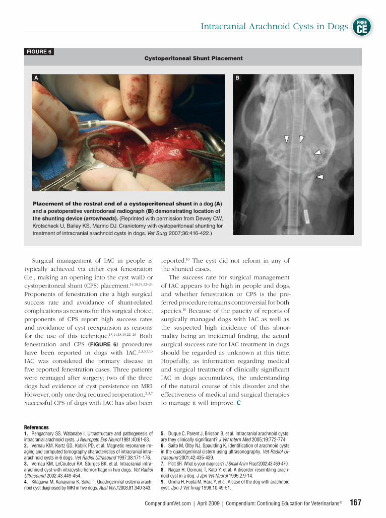
Surgical management of IAC in people is typically achieved via either cyst fenestration (i.e., making an opening into the cyst wall) or cystoperitoneal shunt (CPS) placement.14,18,19,22–24
Proponents of fenestration cite a high surgical success rate and avoidance of shunt-related complications as reasons for this surgical choice; proponents of CPS report high success rates and avoidance of cyst reexpansion as reasons for the use of this technique.13,14,18,19,22–26 Both fenestration and CPS (FIGURE 6) procedures have been reported in dogs with IAC.2,3,5,7,10
IAC was considered the primary disease in fi ve reported fenestration cases. Three patients were reimaged after surgery; two of the three dogs had evidence of cyst persistence on MRI. However, only one dog required reoperation.2,3,7 Successful CPS of dogs with IAC has also been
reported.10 The cyst did not reform in any of the shunted cases. The success rate for surgical management of IAC appears to be high in people and dogs, and whether fenestration or CPS is the pre-ferred procedure remains controversial for both species.10 Because of the paucity of reports of surgically managed dogs with IAC as well as the suspected high incidence of this abnor-mality being an incidental fi nding, the actual surgical success rate for IAC treatment in dogs should be regarded as unknown at this time. Hopefully, as information regarding medical and surgical treatment of clinically signifi cant IAC in dogs accumulates, the understanding of the natural course of this disorder and the effectiveness of medical and surgical therapies to manage it will improve.
Cystoperitoneal Shunt PlacementFIGURE 6
Placement of the rostral end of a cystoperitoneal shunt in a dog (A)
and a postoperative ventrodorsal radiograph (B) demonstrating location of
the shunting device (arrowheads). (Reprinted with permission from Dewey CW,
Krotscheck U, Bailey KS, Marino DJ. Craniotomy with cystoperitoneal shunting for
treatment of intracranial arachnoid cysts in dogs. Vet Surg 2007;36:416-422.)
A B
References1. Rengachary SS, Watanabe I. Ultrastructure and pathogenesis of intracranial arachnoid cysts. J Neuropath Exp Neurol 1981;40:61-83.2. Vernau KM, Kortz GD, Koblik PD, et al. Magnetic resonance im-aging and computed tomography characteristics of intracranial intra-arachnoid cysts in 6 dogs. Vet Radiol Ultrasound 1997;38:171-176.3. Vernau KM, LeCouteur RA, Sturges BK, et al. Intracranial intra-arachnoid cyst with intracystic hemorrhage in two dogs. Vet Radiol Ultrasound 2002;43:449-454. 4. Kitagawa M, Kanayama K, Sakai T. Quadrigeminal cisterna arach-noid cyst diagnosed by MRI in fi ve dogs. Aust Vet J 2003;81:340-343.
5. Duque C, Parent J, Brisson B, et al. Intracranial arachnoid cysts: are they clinically signifi cant? J Vet Intern Med 2005;19:772-774.6. Saito M, Olby NJ, Spaulding K. Identifi cation of arachnoid cysts in the quadrigeminal cistern using ultrasonography. Vet Radiol Ul-trasound 2001;42:435-439.7. Platt SR. What is your diagnosis? J Small Anim Pract 2002;43:469-470.8. Nagae H, Oomura T, Kato Y, et al. A disorder resembling arach-noid cyst in a dog. J Jpn Vet Neurol 1995;2:9-14.9. Orima H, Fujita M, Hara Y, et al. A case of the dog with arachnoid cyst. Jpn J Vet Imag 1998;10:49-51.
Intracranial Arachnoid Cysts in Dogs
168 Compendium: Continuing Education for Veterinarians® | April 2009 | CompendiumVet.com
FREE
CE
1. In dogs, all reported cases of IAC have been in the _________ fossa.
a. rostral b. middle c. caudal d. none of the above
2. Formation of IACs in dogs is believed to be due to
a. a split in the arachnoid meningeal layer during embryogenesis.
b. failure of the neuroectoderm and nonneural ectoderm to separate during embryogenesis.
c. compensatory fl uid accumulation following an in utero brain infarction (stroke).
d. failure of the cerebellar vermis to form correctly during embryogenesis.
3. Proposed theories to explain progressive expansion of IACs in dogs include
a. active secretion by the arachnoid cells lining the cyst cavity.
b. movement of fl uid into the cyst cavity along an osmotic pressure gradient.
c. movement of fl uid into the cyst from the neighboring subarachnoid space via slit-like openings (one-way valves) into the cyst lumen.
d. all of the above
4. Which is most characteristic of the typical signalment for dog with an IAC?
a. 10-year-old female spayed German shepherd
b. 4-year-old male castrated shih tzu c. 2-year-old female greyhound d. There is no typical signalment.
5. Clinical signs associated with IAC in dogs include
a. abnormal mentation. b. generalized seizures. c. cerebellar dysfunction. d. all of the above
6. The preferred imaging modality for diagnosis of IAC in dogs is
a. ultrasound. b. scintigraphy. c. magnetic resonance imaging. d. computed tomography.
7. The characteristic MRI appearance of an IAC in a dog is a large, well-demarcated, cyst-like structure that is
a. hypointense on T1-weighted images. b. hyperintense on T2-weighted images. c. contrast-enhancing and hyperintense
on FLAIR images. d. a and b
8. In a study of IAC cases in which brain
compression by the cyst was measured, dogs were found to be most likely to exhibit clinical dysfunction if
a. the cyst compressed more than 14% of the occipital lobe of the cerebrum.
b. both the occipital lobe of the cerebrum and the cerebellum were compressed by the cyst.
c. a and b d. none of the above
9. Medical therapy for IAC in dogs is directed at
a. decreasing brain edema associated with the cyst.
b. controlling seizure activity if present. c. minimizing increases in intracranial
pressure. d. all of the above
10. Which statement regarding surgical management of IAC is false?
a. Both cyst fenestration and cystoperitoneal shunting procedures have been described in dogs.
b. Cystoperitoneal shunt (CPS) placement has been shown to be superior to fenes-tration in dogs and humans with IAC.
c. The success rate for surgical management of IAC appears to be high in people and dogs.
d. a and b
3 CECREDITS CE TEST 1 This article qualifi es for 3 contact hours of continuing education credit from the Auburn University College of Veterinary
Medicine. Subscribers may take individual CE tests online and get real-time scores at CompendiumVet.com. Those who wish to apply this credit to fulfi ll state relicensure requirements should consult their respective state authorities regarding the applicability of this program.
10. Dewey CW, Krotscheck U, Bailey KS, Marino DJ. Craniotomy with cystoperitoneal shunting for treatment of intracranial arachnoid cysts in dogs. Vet Surg 2007;36:416-422.11. Matiasek LA, Platt SR, Shaw S, Dennis R. Clinical and magnetic resonance imaging characteristics of quadrigeminal cysts in dogs. J Vet Intern Med 2007;21:1021-1026.12. Cincu R, Agrawal A, Eiras J. Intracranial arachnoid cysts: cur-rent concepts and treatment alternatives. Clin Neurol Neurosurg2007;109:837-843.13. Erdincler P, Kaynar MY, Bozkus H, et al. Posterior fossa arachnoid cysts. Br J Neurosurg 1999;13:10-17.14. Kandenwein JA, Richter HP, Borm W. Surgical therapy of symp-tomatic arachnoid cysts—an outcome analysis. Acta Neurochir (Wien)2004;146:1317-1322.15. Dewey CW. Encephalopathies: disorders of the brain. In: Dewey CW, ed. A Practical Guide to Canine and Feline Neurology. 2nd ed. Ames, Iowa: Wiley-Blackwell; 2008:115-120.16. Go KG, Houthoff HJ, Blaauw EH, et al. Arachnoid cyst of the sylvian fi ssure: evidence of fl uid secretion. J Neurosurg 1984;60:803-813.17. Santamarta D, Aguas J, Ferrer E. The natural history of arach-noid cysts: endoscopic and cine-mode MRI evidence of a slit-valve mechanism. Minim Invasive Neurosurg 1995;38:133-137.
18. Raffel C, McComb JG. To shunt or fenestrate: which is the best sur-gical treatment for arachnoid cysts in pediatric patients? Neurosurgery1988;23:338-342.19. Locatelli D, Bonfanti N, Sfogliarini R, et al. Arachnoid cysts: di-agnosis and treatments. Childs Nerv Syst 1987;3:121-124.20. Coates JR, Axlund TW, Dewey CW, Smith J. Hydrocephalus in dogs and cats. Compend Contin Educ Pract Vet 2006;28:136-146.21. Dewey CW. Anticonvulsant therapy in dogs and cats. Vet Clin North Am Small Anim Pract 2006;36:1107-1127.22. Ciricillo SF, Cogen PH, Harsh GT, et al. Intracranial arachnoid cysts in children: a comparison of the effects of fenestration and shunting. J Neurosurg 1991;74:230-235.23. Kaplan BK, Mickle JP, Parkhurst R. Cystoperitoneal shunting for congenital arachnoid cysts. Childs Brain 1984;11:304-311.24. Stein SC. Intracranial developmental cysts in children: treatment by cystoperitoneal shunting. Neurosurgery 1981;8:647-650.25. Kim SK, Cho BK, Chung YN, et al. Shunt dependency in shunt-ed arachnoid cyst: a reason to avoid shunting. Pediatr Neurosurg2002;37:178-185.26. Gangemi M, Maiuri F, Colella G, et al. Endoscopic treatment of quadrigeminal cistern arachnoid cysts. Minim Invasive Neurosurg2005;48:289-292.