intracranial pressure 2015
TRANSCRIPT

Intracranial pressure 2015
SAMIR EL ANSARY

Global Critical Carehttps://www.facebook.com/groups/1451610115129555/#!/groups/145161011512
9555/ Wellcome in our new group ..... Dr.SAMIR EL ANSARY
Normal intracranial pressure in adults is 8 to 18mm Hg
And in babies the pressure is 10-20 mm.
Less when measured through a lumbar puncture.
ICP is not a static state, but one that is influenced by several factors.
The recording of ICP shows 2 forms of pressure fluctuations.
There is a rise with cardiac systole (due to distention of intracranial arteriolar tree
which follows )
And a slower change in pressure with respiration, falling with each inspiration
and rising with expiration.

Straining, compression of neck veins can also cause sudden, considerable rise in
pressure.
The conception of the cranium acting as a near rigid container of virtually
incompressible substances in the form of brain, blood & CSF in known as the
Monro Kellie doctrine.
Monro Kellie doctrine.
CSF can be displaced through the foramen magnum into spinal theca.
The spinal dural sheath can accept a quantity of CSF as it does not fit the
canal closely, being surrounded by a layer of loose areolar tissue & plexus of
epidural veins.
In addition, in states of increased ICP there is increase in passage of blood through venous emissaries.
Intracranial pressure is a result of at least 2 factors, the volume of the brain (about 1400ml in an adult)
being constant.
(a)CSF which is constantly secreted & after circulating absorbed at an equal rate. CSF circulation is slow (500 to 700
ml/day). At a given time the cranium contains
75 ml of CSF.
(b) Intracranial circulation of blood which is about 1000 litres per day delivered at a pressure of 100 mmHg and at a given
time, the cranium contains 75 ml
Any obstruction to venous outflow will entail an increase in the volume of intracranial
blood and ICP. As the ICP increases, the cerebral venous
pressure increases in parallel so as to remain 2 to 5 mm higher or else the venous system
would collapse.
Because of this relationship CPP (mean art pressure - venous
pressure or mean ICP)Can be satisfactorily estimated from
Mean art pressure – ICP.
Lundberg has described 3 wave patterns ICP waves (A, B, and C waves).
A waves are pathological. There is a rapid rise in ICP up to 50-100 mm Hg
Followed by a variable period during which the ICP remains elevated followed by a rapid fall to the
baseline and when they persist for longer periods, they are called 'plateau' waves which are
pathological.

'Truncated' or atypical ones, that do not exceed an elevation of 50 mm
Hg, are early indicators of neurological deterioration.
B & C waves are related to respiration and 'Traube-Hering-
Mayer' waves respectively and are of little clinical significance.
Cerebral blood flow (CBF)

The brain accounts for only 2% of total body weight, yet its blood flow represents 15% of resting cardiac output and uses 20% total
amount of oxygen consumed.
Each 24 hours brain requires 1000 liters in order to obtain
71 lit of oxygen and 100 gm of glucose.

The CBF remains constant over a wide range of arterial pressures (between 60 to 150 mm hg)
when the mean arterial pressure is increased beyond 150 mm hg there is increased blood flow.
CBF ceases when art. mean pressure drops to 20mm Hg.
In chronically hypertensive this auto regulation limits appear to be reset.
The exact nature of this auto regulation is not known.
(a) Myogenic theory suggests direct reaction of the cerebral arterial smooth muscles to the
stretch.
(b) The humoral theory involves regulations by the direct effect of by- products of metabolism
(c) Neurogenic theory rests on perivascularnerves.
The auto regulation is influenced by various factors.

With normal cerebrovascular system and BP, even moderate alterations of pCO2 are
capable of markedly altering CBF.
Within the range of 30 to 60 mm Hg there is a 2.5% change in CBF as the
pCO2 changes by 1 mmHg.
With less than 20 and more than 80 mmHg there is no further change.
In old age and arteriosclerosis, there is marked decrease in pCO2 influence.

The effects of pO2 are not as marked as CO2 Changes.
Moderate variation of O2 above and below the normal level do not affect CBF.
pO2 causes constriction of a non ischemic brain along with reduction in CBF.
In ischemic hemisphere, increasing the pO2 has no effect.
Cerebral vaso dilatation begins with pO2 of 50 mm Hg & CBF increases.
When pO2 falls to 30 mmHg, CBF may have tripled.
The ICP influences the CBF through the cerebral perfusion pressure (CPP) which is
the difference between mean arterial pressure (MAP) and ICP.
Raise in ICP would lead to a fall in CPP and every effort should be taken to maintain
the CPP to 50 mm Hg or more during treatment of raised ICP.

Pathophysiology of increased intracranial pressure
Increased ICP is defined as a sustained elevation in pressure above 20mm of
Hg/cm of H20.
The craniospinal cavity may be considered as a balloon.
During slow increase in volume in a continuous mode, the ICP raises to a
plateau level at which the increase level of CSF absorption keeps pace with the
increase in volume.
Intermittent expansion causes onlyA transient rise in ICP at first.
When sufficient CSF has been absorbed to accommodate the
volume the ICP returns to normal.

Expansion to a critical volumecause persistent raise in ICP which
thereafter increases logarithmically with increasing volume
(Volume - pressure relationship).
The ICP finally raises to the level of arterial pressure which it self begins to
increase, accompanied bybradycardia or other disturbances of
heart rhythm (Cushing response).

(Cushing response) is accompanied by dilatation of small pial arteries and some slowing of venous flow which is followed
by pulsatile venous flow.
The rise in ICP to the level of systemic arterial pressure extinguishes cerebral
circulation which will restart only if arterial pressure raises sufficiently beyond the ICP
to restore CBF.
If it fails, brain death occurs.

The cause of raise in ICP and the rate at which it occurs are also
important
Cause axial distortion to impair brainstem perfusion.

Many patients with benign ICT or obstructive hydrocephalus show little or no ill effect, the
reason being the brain it self is normal and auto regulation is probably intact.
In patients with parenchymal lesion (tumor, hematoma and contusion), because of the shift of brain and disturbed auto regulation, CBF may by compromised with relatively low levels of ICP.
In acute hydrocephalus, there is rapid deterioration as there is no time for
compensation.

The raise in ICP disturbs brain function by
(1) Reduction in CBF
(2) Transtentorial or foramen magnum herniation resulting in selective
compression and ischaemia in the brain stem.
Transtentorial herniation with brainstem compression can lead to clinical
deterioration even with adequate CBF.
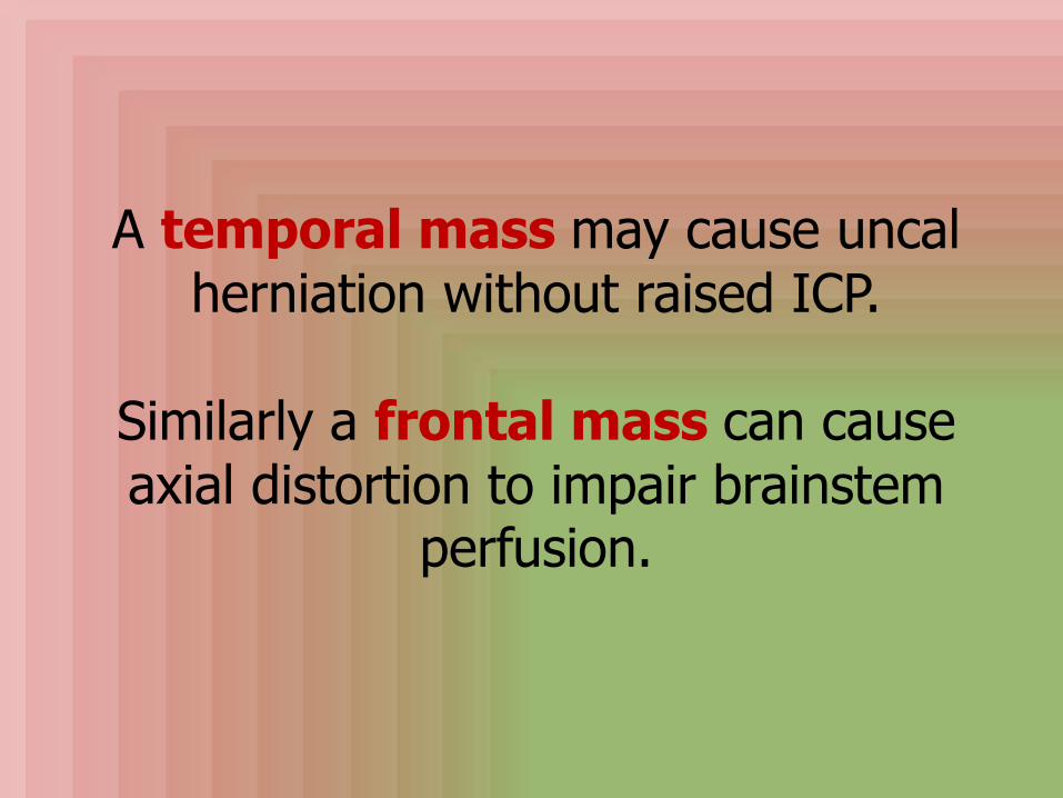
A temporal mass may cause uncalherniation without raised ICP.
Similarly a frontal mass can cause axial distortion to impair brainstem
perfusion.

Clinicalfeatures if raised ICP

Raised ICP causes arterial hypertension,
bradycardia(Cushing's response)
And respiratory changes.
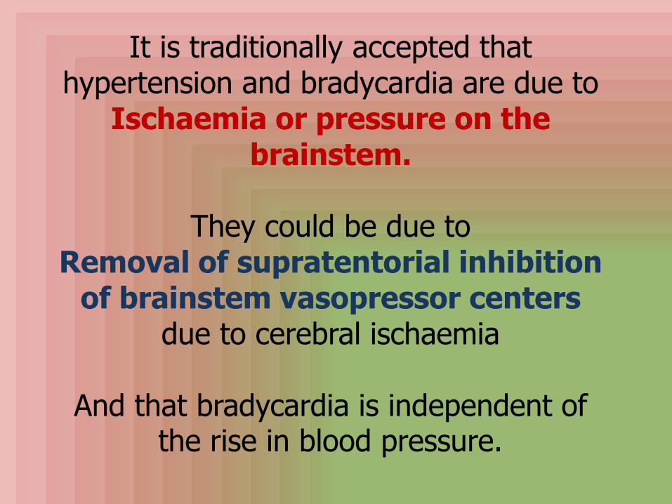
It is traditionally accepted that hypertension and bradycardia are due to
Ischaemia or pressure on the brainstem.
They could be due toRemoval of supratentorial inhibition
of brainstem vasopressor centersdue to cerebral ischaemia
And that bradycardia is independent of the rise in blood pressure.

The respiratory changes depend on the level of
brainstem involved.
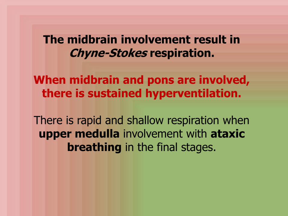
The midbrain involvement result in Chyne-Stokes respiration.
When midbrain and pons are involved, there is sustained hyperventilation.
There is rapid and shallow respiration when upper medulla involvement with ataxic
breathing in the final stages.
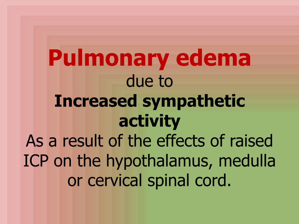
Pulmonary edemadue to
Increased sympathetic activity
As a result of the effects of raised ICP on the hypothalamus, medulla
or cervical spinal cord.

ICPMonitoring
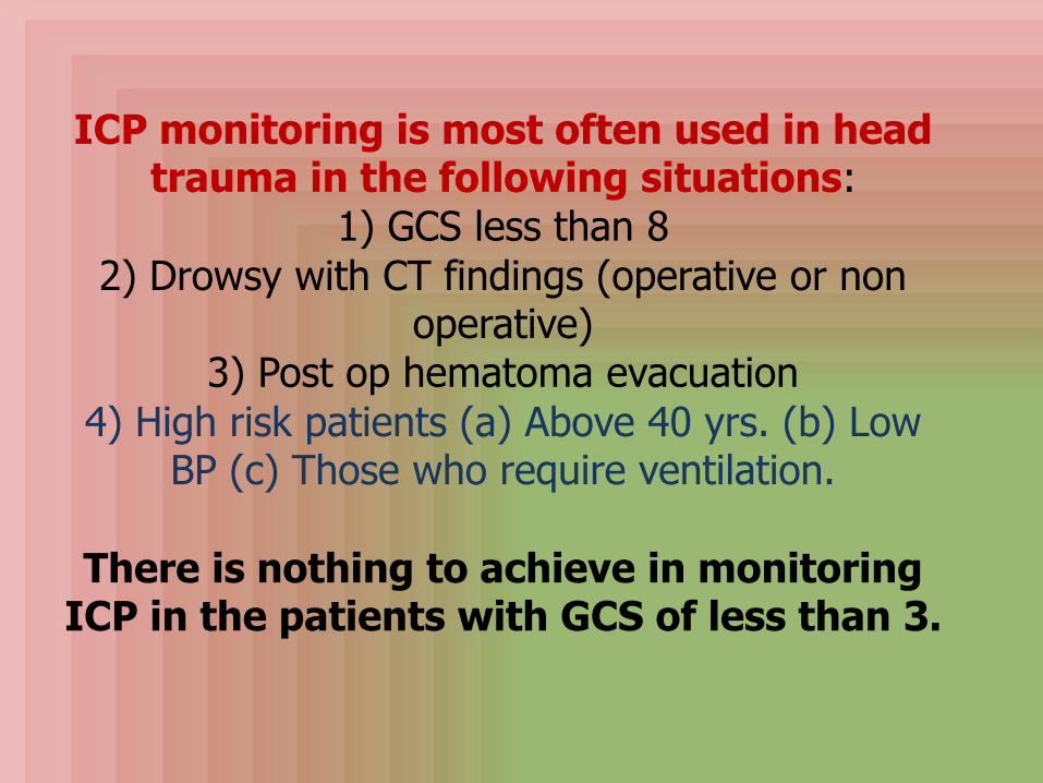
ICP monitoring is most often used in head trauma in the following situations:
1) GCS less than 8
2) Drowsy with CT findings (operative or non operative)
3) Post op hematoma evacuation
4) High risk patients (a) Above 40 yrs. (b) Low BP (c) Those who require ventilation.
There is nothing to achieve in monitoring ICP in the patients with GCS of less than 3.

Non invasive methods
Invasive methods

Non invasive methods:
(1)Clinical deterioration in neurological status is widely considered as sign of increased ICP.
Bradycardia, increased pulse pressure, pupillary dilation are normally accepted
as signs of increased ICP.
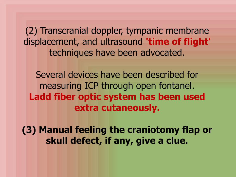
(2) Transcranial doppler, tympanic membrane displacement, and ultrasound 'time of flight'
techniques have been advocated.
Several devices have been described for measuring ICP through open fontanel.
Ladd fiber optic system has been used extra cutaneously.
(3) Manual feeling the craniotomy flap or skull defect, if any, give a clue.
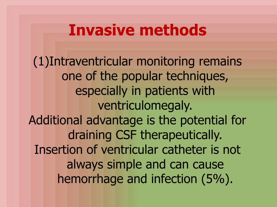
Invasive methods
(1)Intraventricular monitoring remains one of the popular techniques,
especially in patients with ventriculomegaly.
Additional advantage is the potential for draining CSF therapeutically.
Insertion of ventricular catheter is not always simple and can cause
hemorrhage and infection (5%).

(2) Other most commonly used
devices are the hollow screw and bolt devices, and the sub dural
catheter.
Richmond screw and Becker bolt are used extra durally.
A fluid filled catheter in the subdural space, connected to arterial pressure
monitoring system is cost effective and serves the purpose adequately.

(3) Ladd deviceis currently in wide use.
It employs a fibre optic system to detect the distortion of a tiny
mirror within with balloon system.
It can be used in the subdural , extradural and even extra
cutaneously.

(4) A mechanically coupled surface monitoring device is the
'cardio search pneumatic sensor'
used subdurally or extradurally. These systems are not widely used.
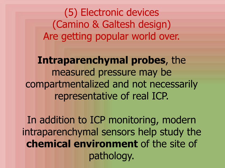
(5) Electronic devices(Camino & Galtesh design)
Are getting popular world over.
Intraparenchymal probes, the measured pressure may be
compartmentalized and not necessarily representative of real ICP.
In addition to ICP monitoring, modern intraparenchymal sensors help study the chemical environment of the site of
pathology.
(6) Fully implantable devices are valuable in a small group who requires
long term ICP monitoring for brain tumors, hydrocephalus or other chronic
brain diseases. Cosmon intrcranial pressure telesensor
can be implanted as a part of shunt system.
Ommaya reservoir is an alternative which can be punctured & CSF pressure
readings are obtained.
(7) Lumbar puncture and measurement of CSF pressure
for obvious reasons is not recommended.
Benefits ofICP monitoring
Monitoring is the only means by which therapy can be selectively employed and the effectiveness
of therapy can be accurately studied.

1) Where ever clinical monitoring is not possible, such as during
Hyper ventilation therapy and high dose barbiturate therapy
ICP monitoring helps.
2) Pre op monitoringHelps in assessment of NPH before a
shunting procedure.
3) Cerebral perfusion pressure (CPP), regulation of cerebral blood flow, and volume, CSF absorption capacity, brain compensatory reserve, and content of
vasogenic events can be studied with ICP monitoring.
Some of these parameters help in prediction of prognosis of survival
following head injury and optimization of' 'CPP guided therapy'.

4) It can provide additional assessment of brain death.
Brain perfusion effectively ceases in nearly all, once ICP exceeds diastolic
blood pressure.
The problems of ICP monitoring are cost, infection, and hemorrhage.
The effective maintenance requires a dedicated team effort.

Treatmentof
increased ICP
There is no doubt the best treatment for increased ICP is the removal of the
causative lesion such as tumors, hydrocephalus, and hematomas.
Post operative increased ICP should be uncommon
these days with increased use of microscope and special techniques to
avoid brain retraction.
As we so often see, a basal meningiomaonce completely removed, has a smooth post op period, whereas a convexity or even falx meningioma may be easily
removed but post operative period may be stormy, mainly due to impairment of
venous drainage
Either due tointraoperative injury to veins and
post operative diuretic therapyas practiced in some centers.

There is still a debate whether increased ICP is the cause or result of the brain damage.
Many feel both compliment each other.
Not all the midline shift seen in CTs indicate increased ICP.
It just means ICP was high during the shift.
The shift takes longer to reverse even after ICP returns to normal .

It is widely acceptedthe increased ICP is a
temporary phenomenonlasting for a short time
unless there is a fresh secondary injury due to
A clot, hypoxia or electrolyte disturbance.
Treatment is aimed at preventing the secondary events.
Clinical and ICP monitoring will help.

1) I line of management:
General measures form the I line of treatment essentially making the patient comfortable and
ABC of trauma management are effectively instituted.
Careful attention to nutrition and electrolytes, bladder and bowel functions and appropriate
treatment of infections are instituted promptly.
Adequate analgesia is often forgotten; it is a must even in unconscious patients.

2) II line of management
Induced cerebral vasoconstriction -Hyperventilation, hyper baric O2,
hypothermia
Osmotherapy - Mannitol, glycerol ,urea
Anesthetic agents - Barbiturates, gamma hydroxybutyrate,
Etomidate,
Surgical decompression -Many do not recommend decompressive
surgery.
This aims at combating increased ICP which is assumed when there is
neurological deterioration or if ICP monitoring is available and the ICP
goes above 25 cm of H2O.
There is a small group of surgeons who start the II line in conditions where ICP is
expected to raise without waiting for a rise.
Many feel that institution of measures to reduce ICP invariably compromises
CBF and wait for the raise in ICP before starting the II line of
management.

Debate continues in the II line of management as well.
Some prefer osmotherapy alone as the II line.
Some prefer measures to induce cerebral vasoconstriction, thereby reducing CBF
and reduce ICP. Some go for both.
Hyperventilation
Aims at keeping the pCO2 down to 30-25 mm Hg so that CBF falls and cerebral blood volume is
reduced and thereby reducing the ICP. Prolonged hyperventilation should be
avoided and becomes ineffective after about 24 hrs.
In addition it causes hypotension due to decreased venous return .
It is claimed a pCO2 under 20 results in ischemia, although there is no experimental proof for the
same.

The present trend is to maintain normal ventilation with pCO2 in the range of 30
- 35 mmHg and pO2 of 120 - 140 mmHg.
When there is clinical deterioration such as pupillary dilatation or
widened pulse pressure, hyperventilation may be instituted (preferably with an Ambu bag) until the
ICP comes down.

Hyper baric O2, hypothermia are still in experimental stage,
especially in Japan .
They basically induce cerebral vasoconstriction and reduce the cerebral blood volume and the ICP.

Osmotherapy
Is useful in the cytotoxic edema stage, when capillary permeability is intact, by increasing the
serum osmolality.
Mannitol is still the magic drug to reduce to ICP, but only if used properly:
it is the most common osmotic diuretic used. It may also act as a free radical scavenger.

Mannitol is not inert and harmless. Glycerol and urea are hardly used these days.
Several theories have been advanced concerning the mechanism by which it reduces ICP.
1) It increases the erythrocyte flexibility, which decreases blood viscosity and causes a
reflex vasoconstriction that reduces cerebral blood volume and decreases
ICP and may reduce CSF production by the choroids plexus.

MannitolIn small doses protects the
brain from ischemic insults due to increased erythrocyte
flexibility.
2) The diuretic effect is mainly around the lesion, where blood brain barrier integrity
is impairedAnd there is no significant effect on
normal brain.
As one would have observed, intraaxiallesions respond better than extra
axial lesions.
3) Another theory ismannitol withdraws water
across the ependyma of the ventricles
in a manner analogous to that produced by ventricular
drainage.
The traditional dose is 1 gm/kg/24 hr of 20% to 25% i.v. either as a bolus or
more commonly intermittently.
There is no role for dehydration.
Mannitol effect on ICP is maximal 1/2 hr after infusion and lasts for 3 or 4 hrs as a
rule.
The correct dose is the smallest dose which will have sufficient effect on ICP.

When repeated doses are required, the base line serum osmolality gradually increases and when this exceeds 330 mosm/1 mannitol
therapy should cease.
Further use is ineffective and likely to induce renal failure.
Diuretics such as frusemide, either alone or in conjunction with mannitol help to hasten its
excretion and reduce the baseline serum osmolality prior to next dose.

Some claim, that frusemide compliments mannitol and increases the output.
Some give frusemide before mannitol, so that it reduces circulatory overload.
The so called rebound phenomenon is due to reversal of osmotic gradient as a result of
progressive leak of the osmotic agent across defective blood brain barrier, or is due to
recurrence of increased ICP.

Barbiturates
Can lower the ICP when other measures fail; but have no prophylactic value.
They inhibit free radical mediated lipid peroxidation
And suppress cerebral metabolism cerebral metabolic requirements and
thereby cerebral blood volume are reduced resulting in the reduction of ICP.
Phenobarbital is most widely used.
A loading dose of 10mg/kg over 30 minutes and 1-3mg/kg every hour is widely employed.
Facilities for close monitoring of ICP and hemodynamic instability should
accompany any barbiturate therapy.
High dose steroidTherapy was popular some years ago and still
used by some.
It restores cell wall integrity and helps in recovery and reduce edema.
Barbiturates and other anesthetic agents reduce CBF and arterial pressure thereby reducing ICP.
In addition it reduces brain metabolism and energy demand which facilitate better healing.
Surgical decompression
Decompressive craniotomies such as sub temporal decompression are recommended only in highly
selected patients these days.
Herniation of brain thro' defect, cause further injury, further edema and further increased ICP.
But in occasional cases, when every other measure has failed, such decompression
craniotomy may be justified.

Global Critical Carehttps://www.facebook.com/groups/1451610115129555/#!/groups/145161011512
9555/ Wellcome in our new group ..... Dr.SAMIR EL ANSARY

GOOD LUCK
SAMIR EL ANSARY
ICU PROFESSOR
AIN SHAMS
CAIRO