intrastromal corneal ring segments for astigmatism...
TRANSCRIPT

Research ArticleIntrastromal Corneal Ring Segments for AstigmatismCorrection after Deep Anterior Lamellar Keratoplasty
Júlio C. D. Arantes,1 Sandro Coscarelli,2 Paulo Ferrara,3 Luana P. N. Araújo,4
Marcos Ávila,1 and Leonardo Torquetti5
1Center of Reference in Ophthalmology, Federal University of Goiás, Goiânia, Brazil2Ennio Coscarelli Eye Clinic, Belo Horizonte, Brazil3Paulo Ferrara Eye Clinic, Belo Horizonte, Brazil4Altino Ventura Foundation, Recife, Brazil5Center of Excellence in Ophthalmology, Pará de Minas, Brazil
Correspondence should be addressed to Júlio C. D. Arantes; [email protected]
Received 13 March 2017; Accepted 17 July 2017; Published 29 August 2017
Academic Editor: Daniel Gore
Copyright © 2017 Júlio C. D. Arantes et al. This is an open access article distributed under the Creative Commons AttributionLicense, which permits unrestricted use, distribution, and reproduction in any medium, provided the original work isproperly cited.
Background. To evaluate the change in corneal astigmatism after intrastromal corneal ring segment (ICRS) implantation inkeratoconus patients with previous deep anterior lamellar keratoplasty (DALK). Design was a longitudinal, retrospective,interventional study. The study included 25 eyes of 24 patients with keratoconus who had DALK performed at least two yearsprior to ICRS implantation. All patients had a clear corneal graft with up to 8.00D of corneal astigmatism and intolerance tocontact lenses. The studied parameters were age, sex, corrected distance visual acuity (CDVA), maximum keratometry (K1),minimum keratometry (K2), spherical equivalent, and astigmatism. There was a statistically significant decrease in thepostintervention analysis as follows: 3.5D reduction in K1 (p < 0 001); 1.53D in K2 (p = 0 005); and 2.52D (p < 0 001) in theaverage K. The spherical equivalent reduced from −3.67D (±2.74) to −0.71D (±2.35) (p < 0 001). The topographic astigmatismreduced from 3.87D preoperatively to 1.90D postoperatively (p < 0 001). The CDVA improved from 0.33 (±0.10) to 0.20(±0.09, p < 0 001). ICRS implantation is a useful option for the correction of astigmatism after DALK as it yields significantvisual, topographic, and refractive results.
1. Introduction
Keratoconus is a noninflammatory, progressive ectaticdisease characterized by thinning and protrusion of the cor-nea. It causes progressive myopia and irregular astigmatism,decreasing the quality of vision [1].
Treatment of keratoconus is indicated according todisease severity. Initially, correction is achieved by wearingglasses, followed by the fitting of rigid gas permeable contactlenses (RGPCL). When these methods fail, surgery is indi-cated to improve the corneal surface. [2]. When the corneastill appears transparent, the implantation of intrastromalcorneal ring segments (ICRS) may be a successful option,and in many cases, it represents an alternative to cornealtransplantation [3–6].
Penetrating keratoplasty (PKP) used to be the methodof choice for treating ectatic corneal disease and has beenthe standard procedure for the keratoconus patient. A dis-advantage of this method is its greater potential for graftrejection, which can lead to a significant reduction in visualacuity. [7–10]. The alternative approach has been deepanterior lamellar keratoplasty (DALK), in which a split-thickness graft is sutured to the receptor, sparing the host’sendothelium and Descemet’s membrane and avoiding therisk of endothelial rejection, with similar visual results [9].DALK can be used in virtually all cases of corneal opacitynot involving the endothelium, and it proved to be a valu-able alternative to PKP for treating keratoconus [10–13].
The surgical duration and risk of ocular perforation havedecreased, and the technique [11] has also provided an
HindawiJournal of OphthalmologyVolume 2017, Article ID 8689017, 7 pageshttps://doi.org/10.1155/2017/8689017
optical surface of excellent quality. In cases of a high residualpost-DALK astigmatism, one alternative is intrastromalcorneal ring segment (ICRS) implantation, which may beindicated for transparent corneas. [14, 15] The main goal isto reshape the cornea without removing tissue or weakeningits central or paracentral region [16].
This study evaluated the use of intrastromal corneal ringsegments implanted using a femtosecond laser as a surgicaloption for treating patients undergoing DALK due to kerato-conus. No other studies were found that have evaluated theresults of employing this implant in the same manner asproposed here.
2. Methodology
This was a retrospective, longitudinal study with secondarydata collected from the medical records of patients diagnosedwith keratoconus who underwent intrastromal corneal ringimplantation for residual astigmatism correction after DALK.
An informed consent was given to all eligible patientsprior to data collection, requesting permission for theresearch and use of data from their medical records relatingto the pre- and postoperative periods. All bioethical princi-ples were considered in accordance with the Declaration ofHelsinki and Brazilian regulations.
The following were considered inclusion criteria: a clearand transparent corneal graft, a minimum of 2.50 diopters(D) and a maximum of 8.00D of astigmatism, intoleranceto contact lenses, and at least two years of follow-up afterDALK before implanting the intrastromal corneal ring.Exclusion criteria were age under 18 years, having undergoneDALK to treat a disorder other than keratoconus, any ocularsurgery other than those proposed in the present study,history of corneal graft rejection, and any other associatedocular disease.
To evaluate the preoperative parameters, the data usedwere taken from the information recorded on the last visitprior to when surgery was indicated. All patients had com-pleted at least one year of follow-up after ICRS implantation,and the second evaluation was based on information takenafter this period had elapsed. Clinical variables werecorrected distance visual acuity (CDVA), measured withthe patient at a distance of 6 meters from the Snellen table,without cycloplegy. To analyze CDVA, the Snellen fractionswere converted to LogMAR. Maximum keratometry (K1)and minimum keratometry (K2) were measured in diopters,using EyeSys topography (EyeSys Vision, Houston, UnitedStates). The spherical equivalent, in cylindrical diopters(DC), was obtained during refractometry performed withthe patient at a distance of 6 meters from the Snellen table,without cycloplegy. Topographic astigmatism was measuredin diopters, using EyeSys topography (EyeSys Vision,Houston, United States).
All ICRS implantations were performed by the samesurgeon (SC). The procedures were performed from June2012 to September 2013. These interventions were madeusing a standard technique, as previously describe [17].
DALK surgery was performed under peribulbar anesthe-sia. The cornea was partially trephined at a diameter of
7.5mm. A radial incision was made with a diamond bladeat the 90° meridian, which reached 90% of the corneal depth,from which stromal tunnels were created using a spatula. Airwas injected into the stromal tunnels to create a deep cleav-age between Descemet’s membrane and the posterior stroma.Atraumatic Vannas scissors were used to remove the anteriorstromal tissue along the edge of the existing partial trepana-tion. The donor cornea was trephined with a diameter0.25mm greater than that used in the recipient. The endothe-lium of the graft was completely removed. The donor buttonwas fixed with 16 separate sutures using mononylon 10.0.Postoperatively, 0.3% moxifloxacin eye drops combined with0.1% dexamethasone, four times a day for a period of sixweeks, were prescribed. Lubricant eye drops were usedseveral times a day, according to each patient’s needs. Thesutures removal began three months after surgery.
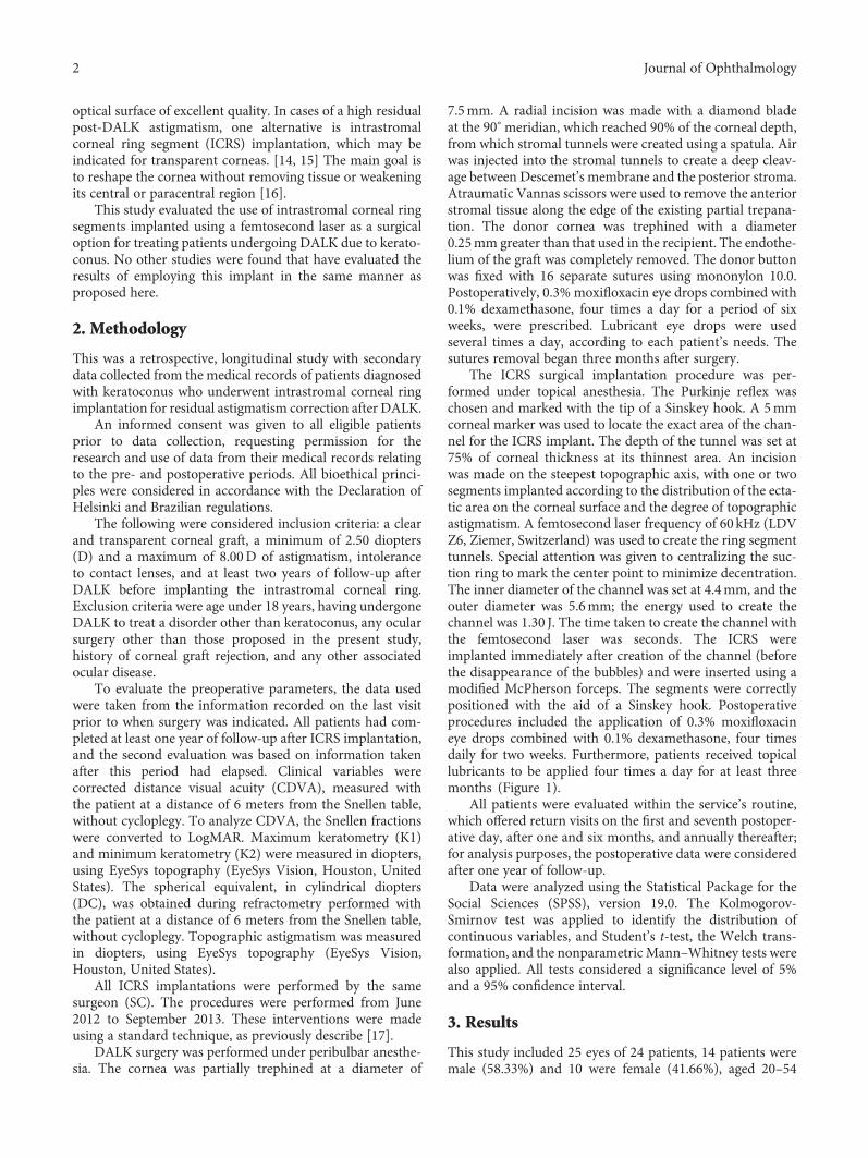
The ICRS surgical implantation procedure was per-formed under topical anesthesia. The Purkinje reflex waschosen and marked with the tip of a Sinskey hook. A 5mmcorneal marker was used to locate the exact area of the chan-nel for the ICRS implant. The depth of the tunnel was set at75% of corneal thickness at its thinnest area. An incisionwas made on the steepest topographic axis, with one or twosegments implanted according to the distribution of the ecta-tic area on the corneal surface and the degree of topographicastigmatism. A femtosecond laser frequency of 60 kHz (LDVZ6, Ziemer, Switzerland) was used to create the ring segmenttunnels. Special attention was given to centralizing the suc-tion ring to mark the center point to minimize decentration.The inner diameter of the channel was set at 4.4mm, and theouter diameter was 5.6mm; the energy used to create thechannel was 1.30 J. The time taken to create the channel withthe femtosecond laser was seconds. The ICRS wereimplanted immediately after creation of the channel (beforethe disappearance of the bubbles) and were inserted using amodified McPherson forceps. The segments were correctlypositioned with the aid of a Sinskey hook. Postoperativeprocedures included the application of 0.3% moxifloxacineye drops combined with 0.1% dexamethasone, four timesdaily for two weeks. Furthermore, patients received topicallubricants to be applied four times a day for at least threemonths (Figure 1).
All patients were evaluated within the service’s routine,which offered return visits on the first and seventh postoper-ative day, after one and six months, and annually thereafter;for analysis purposes, the postoperative data were consideredafter one year of follow-up.
Data were analyzed using the Statistical Package for theSocial Sciences (SPSS), version 19.0. The Kolmogorov-Smirnov test was applied to identify the distribution ofcontinuous variables, and Student’s t-test, the Welch trans-formation, and the nonparametric Mann–Whitney tests werealso applied. All tests considered a significance level of 5%and a 95% confidence interval.
3. Results
This study included 25 eyes of 24 patients, 14 patients weremale (58.33%) and 10 were female (41.66%), aged 20–54
2 Journal of Ophthalmology

years (mean: 35.96; standard deviation: ±8.69 years). Theindication for ring implantation was to correct residual astig-matism after DALK. Twenty-three patients had one eyetreated, and only one patient had both eyes treated.
All clinical variables showed a statistically significantdecrease from preoperative to postoperative. There wasa reduction of 3.50D in K1 (p < 0 001) and 1.53D inK2 (p = 0 005), and the reduction in the mean K was 2. D(p < 0 001) (Figure 2).
The spherical equivalent (SE) reduced from −3.67D(±2.74) before the procedure to −0.71D (±2.35) after theprocedure (p < 0 001). Topographic astigmatism reducedfrom 3.87D preoperatively to 1.90DC postoperatively(p < 0 001) (Table 1). The corrected distance visual acuity(CDVA), in LogMAR, increased from 0.33 (±0.10) to 0.20(±0.09; p < 0 001).
Vector analysis (double-angle plot) revealed a significantdecrease in the centroid between the preoperative andpostoperative data in both topographic astigmatism andrefractive astigmatism (Figure 3).
The preoperative refractive astigmatism centroid was1.87D× 4.66° ± 3.76 (p = 1 55), and the postoperative cen-troid was 1.01D× 21.31° ± 1.84 (p = 0 83). After implantationof the ICRS, the refractive astigmatism centroid was 0.86D,and the standard deviation in astigmatism was reduced by a
Figure 1: Transparent cornea one week after the ICRS implant.
Figure 2: Preoperative and postoperative corneal topography after ICRS implantation.
Table 1: Preoperative and postoperative data after ICRSimplantation.
Preop Postopp∗
Mean SD Mean SD
Maximum keratometry 47.20 ±2.50 43.70 ±3.45 <0.001Minimum keratometry 43.33 ±2.00 41.80 ±3.53 0.005
Mean keratometry 45.26 ±2.03 42.74 ±3.44 <0.001Spherical equivalent −3.67 ±2.74 −0.71 ±2.35 <0.001Topographic astigmatism 3.87 ±1.95 1.90 ±1.16 <0.001∗Student’s t-test for paired samples, p value. SD: standard deviation.
3Journal of Ophthalmology

factor of 1.92 (3.76D/1.84D). Relocation of the centroidnearer to the origin and contraction of the ellipse in thedouble-angle plots showed improvement (Figure 3).
Figure 4 shows the double-angle plot for preoperativeand postoperative keratometric astigmatism. The preopera-tive centroid was 0.97D× 91.97° ± 1.74, (p = 0 95), and thepostoperative centroid was 0.75D× 98.59° ± 1.50, (p = 0 82).Although there was a reduction in mean keratometricastigmatism, it was considerably smaller than the decreasein refractive astigmatism.
4. Discussion
The most common cause of decreased vision after cornealtransplantation is the astigmatism. It is commonly acceptedthat the average postoperative cylinder after keratoplasty var-ies from three to five diopters; [18] about 10–27% of patientsundergoing corneal transplantation evolve with high astig-matism, and for high astigmatism, it is understood as therefractive cylinder of more than four diopters (D) [19].
In the present study, using ICRS implantation to treathigh astigmatism after the DALK significantly reduced Kvalues, spherical equivalent, and the topographic astigma-tism. Tunnels made with the femtosecond laser have anadvantage over those implanted using the manual techniquebecause they cause lower traction at the junction where therecipient cornea meets the donor button, avoiding possibledehiscence in the surgical wound [3] (Figure 5).
The choice of a Ferrara ICRS with a 5mm optical zonealso has advantages over choosing those that are implanted
in a 6 or 7mm optic zone, as the former theoretically causesgreater corneal flattening because the refractive results areinversely proportional to the implant diameter [16]. Further-more, a smaller optical zone provides greater distancebetween the ICRS and graft/host junction, reducing thechance of stromal neovascularization/dehiscence of thejunction. In turn, a smaller optical zone increases the risk ofpostoperative halos. Excimer laser photorefractive keratec-tomy (PRK) and laser in situ keratomileusis (LASIK) can alsobe used to treat posttransplant astigmatism [20].
The fact that the ICRS implant did not affect the cen-tral region of the cornea, avoiding the risk of opacity onthe visual axis and the reversibility of this method, meansthat this can be considered theoretically more beneficialthan the correction of astigmatism using LASIK, whichhas limited efficacy due to the stromal thickness of thegraft and the high remaining ametropia [20].It also doesnot provide results as good as those seen in corneas thathave never been treated, with less predictability andgreater chance of complications such as epithelializationdefects, flap displacement, dry eye, and corneal graft failure[18, 21, 22]. PRK can cause a significant haze in corneal graftsand induce progressive astigmatism [20].
Another surgical option for the treatment of post-DALKastigmatism is the implantation of a phakic intraocular lens(PIOL). Barraquer and Rodriguez-Barraquer [23] evaluatedthe Artisan PIOL implant in the treatment of high myopiaafter PKP. In that study, there was an improvement in CDVAwithout significant loss of endothelial cells in the first sixpostoperative months [23].
Preoperative centroid: 1.87 D×4.66º± 3.76, p= 1.55Postoperative centroid: 1.01 D×21.31º± 1.84, p= 0.83
Each ring = 1.0 DOuter ring = 6.0 D
75º
90º
105º
120º135º
150º
165º
0º
15º
30º45º
60º
Figure 3: Double-angle plot of preoperative and postoperative refractive astigmatism. The standard deviation is represented by the areaaround the centroid (p=shape factor). Each ring = 1.0D/outer ring = 6.0D.
4 Journal of Ophthalmology

Another study demonstrated that a PIOL implant inpost-PKP eyes reduces refractive error and improves theCDVA, in addition to being a predictable and stable method.However, even if the refractive error is significantly reduced,corneal abnormalities will still be present. Alternatively, anICRS implant decreases these abnormalities, improving thequality of vision. In addition, as it is a less invasive cornealprocedure, the risks related to intraocular surgery are avoided[24]. Alfonso et al. [25] evaluated the efficacy and safety ofthe implantable collamer posterior chamber intraocularlenses to correct refractive errors that occurred after PKP in15 eyes of 15 patients. In this study, no eye lost more thanone line of vision, two eyes gained one line, five eyes gainedmore than one line, and eight eyes remained with unchangedvision [25, 26].
In the current study, when evaluating the CDVA 12months after ICRS implantation, stability was observed in28% (seven eyes); 36% (nine eyes) gained one line of vision;20% (five eyes) gained two lines of vision; 8% (two eyes)gained three lines of vision; and 8% (two eyes) gained fourlines of vision. No eyes lost lines of vision.
Other scientific studies using a post-PKP manual intras-tromal corneal ring implant technique showed K reductionvalues higher than those found in our study. A study con-ducted in Spain, with the participation of nine patients (nineeyes), found a reduction in K1 values greater than 5D and areduction in K2 greater than 2D, while the mean K decreasedby more than 4D [3]. In another publication using a similartechnique conducted in Brazil evaluating patients (59 eyes),K values were also reduced by more than the amount foundin our study, with the maximum K decreasing by 3.92Dand the minimum K by 2.44D [17].
In another study with 30 patients (32 eyes) using afemtosecond laser for post-PKP intrastromal corneal ringimplantation, lower reductions in K2 (0.53D) and K1(3.65D) were found, a result similar to that of our study [14].
Arriola-Villalobos [27] found that the spherical equiva-lent decreased by 3.05D, and Coscarelli et al. [16] reporteda decrease of 3.68D. These values are similar to those foundin our study, in which we observed a decrease of 2.96D. Inthe study of Lisa et al. [13], a smaller reduction in thespherical equivalent of 1.67D was observed.
Figure 5: Optical coherence tomography (OCT) of the cornea one year after the procedure, showing the position of the ICRS.
Preoperative centroid: 0.97 D×91.97º± 1.74, p= 0.95Postoperative centroid: 0.75 D×98.59º± 1.50, p= 0.82
Each ring = 1.0 DOuter ring = 6.0 D
75º
90º
105º
120º135º
150º
165º
0º
15º
30º45º
60º
Figure 4: Relationship between preoperative and postoperative keratometric astigmatism. The standard deviation is represented by the areaaround the centroid (p=shape factor). Each ring = 1.0D/outer ring = 6.0D.
5Journal of Ophthalmology
Topographic astigmatism decreased 1.97 in our study,whereas in another similar study, a decrease of 2.59 wasobtained [9].
We hypothesize that the results, in terms of reduction ofkeratometry and astigmatism, are less significant in cases ofICRS implantation after DALK, in comparison of ICRS afterPKP due to biomechanics properties of the cornea Thepreservation of Descemet’s membrane and endothelium inDALK provides an additional strength to the graft, compar-ing to PKP, which in turns, leads to less response to cornealflattening/remodeling after ICRS implantation in thesecases. Moreover, the irregularity at host-donor interfacemay play a role in the corneal changes after ICRS implantationafter PKP.
In our investigation, there were no complications duringthe surgery or postoperative follow-up. The use of the femto-second laser in the construction of stromal tunnels is saferthan the manual technique, providing a significant reductionin complications such as ICRS extrusion because of theaccuracy in implant depth [27–29].
One limitation of the study was the lack of standardiza-tion in the medical records, making the data difficult tocollect and increasing the time required to populate thedatabase, although all the information necessary for theproposed evaluations were obtained in the documents.
One of the strengths of this study is the fact that no otherstudies were found that evaluates the results of ICRS implan-tation using a femtosecond laser in patients previouslyundergoing DALK due to keratoconus. The number ofpatients selected was lower than ideal due to the rigorousselection of the studied eyes. The strict inclusion andexclusion criteria limited the sample to 25 eyes, but statisticalsignificance was achieved.
The results of this study indicate that ICRS implantedusing a femtosecond laser is a surgical treatment option forpatients having undergone DALK due to keratoconus thatoffers significant visual, topographic, and refractive results.Prospective studies with longer follow-up periods are neededto verify the long-term safety and stability of the procedure.
4.1. What Was Known before This Study. The use of a femto-second laser for intrastromal corneal ring implantation is asafe option for treating astigmatism after PKP.
4.2. What This Study Has Added. This is the first publishedstudy of ICRS implantation with a femtosecond laser to cor-rect high astigmatism in eyes that were previously subjectedto DALK due to keratoconus.
In a series of 25 eyes, the ICRS implant produced a signif-icant reduction in topographic astigmatism and improvedthe CDVA in patients previously undergoing deep anteriorlamellar keratoplasty.
5. Synopsis
ICRS implantation can successfully improve the visualacuity and reduce the astigmatism after deep anteriorlamellar keratoplasty.
Disclosure
Ferrara has financial interest in Ferrara intrastromal cornearing segments. No other author has a financial or proprietaryinterest in any material or method mentioned.
Conflicts of Interest
The authors declare that they have no conflicts of interest.
References
[1] L. S. Lim, R. Beuerman, L. Lim, and D. T. Tan, “Late-onsetdeep stromal scarring after riboflavin-UV-A corneal collagencross-linking for mild keratoconus,” Archives of Ophthalmol-ogy, vol. 129, pp. 360–362, 2011.
[2] Y. S. Rabinowitz, “Keratoconus,” Survey of Ophthalmology,vol. 42, pp. 297–319, 1998.
[3] N. Avni-Zauberman and D. S. Rootman, “Cross-linking andintracorneal ring segments—review of the literature,” Eye &Contact Lens, vol. 40, pp. 365–370, 2015.
[4] G. Ferrara, L. Torquetti, P. Ferrara, and J. Merayo-Lloves,“Intrastromal corneal ring segments: visual outcomes from alarge case series,” Clinical & Experimental Ophthalmoogy,vol. 40, pp. 433–439, 2012.
[5] L. Torquetti, G. Ferrara, F. Almeida et al., “Intrastromal cor-neal ring segments implantation in patients with keratoconus:10-year follow-up,” Journal of Refractive Surgery, vol. 30,pp. 22–26, 2014.
[6] J. S. Parker, K. DijkVan, and G. R. Melles, “Treatment optionsfor advanced keratoconus: a review,” Survey Ophthalmology,vol. 60, pp. 459–480, 2015.
[7] A. Cano-Ortiz and A. Villarrubia, “Corneal transplantation inkeratoconus: penetrating keratoplasty versus deep anteriorlamellar keratoplasty with Melles technique,” Archivos de laSociedad Espanola de Oftalmologia, vol. 90, pp. 4–8, 2015.
[8] R. Donoso, C. Díaz, and P. Villavicencio, “Comparative studyof keratoconus between Anwar’s deep anterior lamellar kera-toplasty versus converted penetrating keratoplasty,” Archivosde la Sociedad Espanola de Oftalmologia, vol. 90, pp. 257–263, 2015.
[9] M. Keane, D. Coster, M. Ziaei, and K.Williams, “Deep anteriorlamellar keratoplasty versus penetrating keratoplasty fortreating keratoconus,” Cochrane Database Systematic Review,vol. 7, article CD009700, 2014.
[10] S. L. Watson, S. J. Tuft, and J. K. Dart, “Patterns of rejectionafter deep lamellar keratoplasty,” Ophthalmology, vol. 113,pp. 556–560, 2006.
[11] M. Anwar and K. D. Teichmann, “Big bubble technique tobare Descemet’s membrane in anterior lamellar keratoplasty,”Journal of Cataract & Refractive Surgery, vol. 28, no. 3,pp. 398–403, 2002.
[12] S. Feizi, S. Daryabari, D. Najdi, M. A. Javadi, and F. Karimian,“Big-bubble deep anterior lamellar keratoplasty using centralvs peripheral air injection: a clinical trial,” European JournalOphthalmology, vol. 26, no. 4, pp. 297–302, 2016.
[13] C. Lisa, M. García-Fernández, D. Madrid-Costa, L. Torquetti,J. Merayo-Lloves, and J. F. Alfonso, “Femtosecond laser-assisted intrastromal corneal ring segment implantation forhigh astigmatism correction after penetrating keratoplasty,”
6 Journal of Ophthalmology

Journal of Cataract & Refractive Surgery, vol. 39, no. 11,pp. 1660–1667, 2013.
[14] J. Colin, C. Buestel, and D. Touboul, “Unusual secondarydisplacement of Intacs segments—superimposition of distalends,” Journal of Refractive Surgery, vol. 26, no. 12, pp. 924-925, 2010.
[15] S. Coscarelli, G. Ferrara, J. F. Alfonso et al., “Intrastromalcorneal ring segment implantation to correct astigmatism afterpenetrating keratoplasty,” Journal of Refractive Surgery,vol. 38, no. 6, pp. 1006–1013, 2012.
[16] S. A. Coscarelli, R. C. Neves, and J. E. Boteon, “Deep lamellarkeratectomy using the big-bubble technique in patients withkeratoconus,” Arquivos Brasileiros de Oftalmologia, vol. 75,pp. 20–23, 2012.
[17] S. Coscarelli and L. Torquetti, “Easy bubble: Pachymetry-assisted big bubble deep anterior lamellar keratoplasty,”Journal of Emmetropia, vol. 3, pp. 151–154, 2012.
[18] A. D. Jensen and A. E. Maumenee, “Refractive errors followingkeratoplasty,” Transactions of the American Ophtalmologic,vol. 72, pp. 123–131, 1974.
[19] G. I. Genvert, E. J. Cohen, J. J. Arentsen, and P. R. Laibson,“Fitting gas-permeable contact lenses after penetrating kerato-plasty,” American Journal of Ophtalmology, vol. 5, no. 5,pp. 511–514, 1985.
[20] J. I. Barraquer, “Modification of refraction by means of intra-corneal inclusions,” International Ophthalmology Clinics,vol. 6, pp. 53–78, 1966.
[21] S. Kwitko, D. R. Marinho, S. Rymer, and F. S. Ramos,“Laser in situ keratomileusis after penetrating keratoplasty,”Journal of Cataract & Refractive Surgery, vol. 27, no. 3,pp. 374–379, 2001.
[22] E. D. Donnenfeld, H. S. Kornstein, A. Amin et al., “Laser in situkeratomileusis for correction of myopia and astigmatism afterpenetrating keratoplasty,” Ophthalmology, vol. 106, no. 10,pp. 1966–1974, 1999.
[23] C. Barraquer and T. Rodriguez-Barraquer, “Five-year results oflaser in-situ keratomileusis (LASIK) after penetrating kerato-plasty,” Cornea, vol. 23, no. 3, pp. 243–248, 2004.
[24] M. Moshirfar, C. A. Barsam, and J. W. Parker, “Implantationof an artisan phakic intraocular lens for the correction of highmyopia after penetrating keratoplasty,” Journal of Cataract &Refractive Surgery, vol. 30, no. 7, pp. 1578–1581, 2004.
[25] J. F. Alfonso, C. Puchades, L. Fernández-Vega, R. Montés-Micó, B. Valcárcel, and T. Ferrer-Blasco, “Visual acuity com-parison of 2 models of bifocal aspheric intraocular lenses,”Journal of Cataract & Refractive Surgery, vol. 35, no. 4,pp. 672–676, 2009.
[26] J. F. Alfonso, L. Fernández-Vega, B. Baamonde, D. Madrid-Costa, and R. Montés-Micó, “Refractive lens exchange withspherical diffractive intraocular lens implantation afterhyperopic laser in situ keratomileusis,” Journal of Cataract &Refractive Surgery, vol. 35, no. 10, pp. 1744–1750, 2009.
[27] P. Arriola-Villalobos, D. Díaz-Valle, J. L. Güell et al.,“Intrastromal corneal ring segment implantation for highastigmatism after penetrating keratoplasty,” Journal of Cata-ract & Refractive Surgery, vol. 35, no. 11, pp. 1878–1884, 2009.
[28] D. Ortiz, D. Piñero, M. H. Shabayek, F. Arnalich-Montiel, andJ. L. Alió, “Corneal biomechanical properties in normal,post-laser in situ keratomileusis, and keratoconic eyes,”Journal of Cataract & Refractive Surgery, vol. 33, no. 8,pp. 1371–1375, 2007.
[29] A. Ertan and G. Kamburoğlu, “Intacs implantation using afemtosecond laser for management of keratoconus: compari-son of 306 cases in different stages,” Journal of Cataract &Refractive Surgery, vol. 34, no. 7, pp. 1521–1526, 2008.
7Journal of Ophthalmology

Submit your manuscripts athttps://www.hindawi.com
Stem CellsInternational
Hindawi Publishing Corporationhttp://www.hindawi.com Volume 2014
Hindawi Publishing Corporationhttp://www.hindawi.com Volume 2014
MEDIATORSINFLAMMATION
of
Hindawi Publishing Corporationhttp://www.hindawi.com Volume 2014
Behavioural Neurology
EndocrinologyInternational Journal of
Hindawi Publishing Corporationhttp://www.hindawi.com Volume 2014
Hindawi Publishing Corporationhttp://www.hindawi.com Volume 2014
Disease Markers
Hindawi Publishing Corporationhttp://www.hindawi.com Volume 2014
BioMed Research International
OncologyJournal of
Hindawi Publishing Corporationhttp://www.hindawi.com Volume 2014
Hindawi Publishing Corporationhttp://www.hindawi.com Volume 2014
Oxidative Medicine and Cellular Longevity
Hindawi Publishing Corporationhttp://www.hindawi.com Volume 2014
PPAR Research
The Scientific World JournalHindawi Publishing Corporation http://www.hindawi.com Volume 2014
Immunology ResearchHindawi Publishing Corporationhttp://www.hindawi.com Volume 2014
Journal of
ObesityJournal of
Hindawi Publishing Corporationhttp://www.hindawi.com Volume 2014
Hindawi Publishing Corporationhttp://www.hindawi.com Volume 2014
Computational and Mathematical Methods in Medicine
OphthalmologyJournal of
Hindawi Publishing Corporationhttp://www.hindawi.com Volume 2014
Diabetes ResearchJournal of
Hindawi Publishing Corporationhttp://www.hindawi.com Volume 2014
Hindawi Publishing Corporationhttp://www.hindawi.com Volume 2014
Research and TreatmentAIDS
Hindawi Publishing Corporationhttp://www.hindawi.com Volume 2014
Gastroenterology Research and Practice
Hindawi Publishing Corporationhttp://www.hindawi.com Volume 2014
Parkinson’s Disease
Evidence-Based Complementary and Alternative Medicine
Volume 2014Hindawi Publishing Corporationhttp://www.hindawi.com