introducing bioorthogonal functionalities into proteins in living cells
DESCRIPTION
ZIYANG HAO, SENLIAN HONG, XING CHEN, AND PENG R. CHEN, 2011TRANSCRIPT
742 ’ ACCOUNTS OF CHEMICAL RESEARCH ’ 742–751 ’ 2011 ’ Vol. 44, No. 9 Published on the Web 06/02/2011 www.pubs.acs.org/accounts10.1021/ar200067r & 2011 American Chemical Society
Introducing Bioorthogonal Functionalities intoProteins in Living Cells
ZIYANG HAO,† SENLIAN HONG,†, ‡ XING CHEN,*, † ANDPENG R. CHEN*, †, §
†Beijing National Laboratory for Molecular Sciences and Department ofChemical Biology, College of Chemistry and Molecular Engineering,
‡School of Life Sciences, Peking University, Beijing 100871, China, and §Laboratoryof Chemical Genomics, School of Chemical Biology and Biotechnology, Shenzhen
Graduate School of Peking University, Shenzhen 518055, China
RECEIVED ON MARCH 1, 2011
CONS P EC TU S
P roteins are the workhorses of the cell, playingcrucial roles in virtually every biological pro-
cess. The revolutionary ability to visualize andmonitor proteins in living systems, which is largelythe result of the development of green fluorescenceprotein (GFP) and its derivatives, has dramaticallyexpanded our understanding of protein dynamicsand function. Still, GFPs are ill suited in manycircumstances; one major drawback is their rela-tively large size, which can significantly perturb thefunctions of the native proteins to which they are fused.
To bridge this gap, scientists working at the chemistry�biology interface have developed methods to install bioorthogonalfunctional groups into proteins in living cells. The bioorthogonal group is, by definition, a non-native and nonperturbing chemicalgroup. But more importantly, the installed bioorthogonal handle is able to react with a probe bearing a complementaryfunctionality in a highly selective fashion and with the cell operating in its physiological state. Although extensive efforts have beendirected toward the development of bioorthogonal chemical reactions, introducing chemical functionalities into proteins in livingsystems remains an ongoing challenge. In this Account, we survey recent progress in this area, focusing on a genetic codeexpansion approach.
In nature, a cell uses posttranslational modifications to append the necessary functional groups into proteins that are beyondthose contained in the canonical 20 amino acids. Taking lessons from nature, scientists have chosen or engineered certain enzymesto modify target proteins with chemical handles. Alternatively, one can use the cell's translational machinery to genetically encodebioorthogonal functionalities, typically in the form of unnatural amino acids (UAAs), into proteins; this can be done in a residue-specific or a site-specific manner. For studying protein dynamics and function in living cells, site-specific modification by means ofgenetic code expansion is usually favored.
A variety of UAAs bearing bioorthogonal groups as well as other functionalities have been genetically encoded into proteins ofinterest. Although this approach is well established in bacteria, tagging proteins in mammalian cells is challenging. A facilepyrrolysine-based system, which might potentially become the “one-stop shop” for protein modification in both prokaryotic andeukaryotic cells, has recently emerged. This technology can effectively introduce a series of bioorthogonal handles into proteins inmammalian cells for subsequent chemical conjugation with small-molecule probes. Moreover, the method may provide moreprecise protein labeling than GFP tagging. These advancements build the foundation for studyingmore complex cellular processes,such as the dynamics of important receptors on living mammalian cell surfaces.
IntroductionProteins are the most abundant biomolecule within cells
and participate in essentially all processes. Dissecting these
processes in many cases requires the ability to label
proteins in the context of living cells.1 The genetically
encoded green fluorescent protein (GFP) and its variants
are undeniably the most powerful tool for protein labeling
andvisualizationandhavebeenwidely utilized for studying

Vol. 44, No. 9 ’ 2011 ’ 742–751 ’ ACCOUNTS OF CHEMICAL RESEARCH ’ 743
Introducing Bioorthogonal Functionalities into Proteins Hao et al.
protein expression, trafficking, and localization in living
systems.2 However, the GFP fusion technique also has some
limitations. The relatively large size of GFP tags can cause
significant structural perturbation and thus influence the
expression, localization, or function of the proteins of inter-
est. Second, the fusion of GFP is largely confined to the N- or
C-terminus of the target proteins. Furthermore, GFP tags can
only be visualized by fluorescence methods and are not
directly applicable for other imaging techniques. These
limitations have therefore promoted the recent expansion
of the arsenal of bioorthogonal chemical reactions used to
label proteins in their native settings.3�13
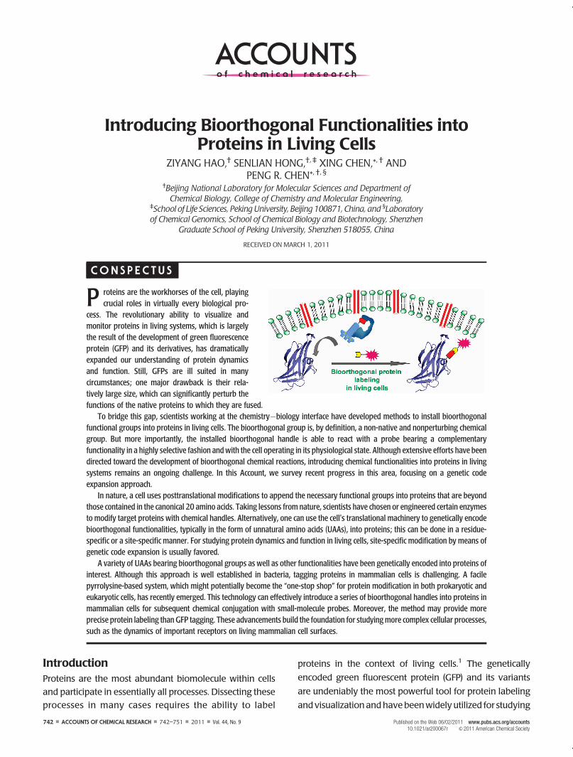
Bioorthogonal protein labeling involves the incorpora-
tion of a unique chemical group into the protein of interest,
followed by a bioorthogonal reaction to covalently attach a
biophysical probe bearing a complementary functional
group (Figure 1).11,12 A bioorthogonal reaction typically
requires the two participating components (bioorthogonal
reaction pair) to be mutually reactive while remaining inert
to the surrounding molecules under the physiological envi-
ronment. So far, only a limited number of chemical reactions
have been developed to meet the requisite requirements,
exemplified by the azide�phosphine Staudinger�Bertozzi
ligation and the different versions of azide�alkyne cyclo-
addition “click” chemistry that will be discussed in details
below.14�19 These reactions have been widely utilized for
labeling proteins, as well as nucleic acids, glycans, and lipids
in the context of living cells and whole organisms.1
One of the key challenges in using this chemical strategy
for protein labeling is to selectively incorporate the bio-
orthogonal chemical groups into the target proteins, which
allows the subsequent conjugation with probes bearing the
complementary functionalities. These bioorthogonal groups
have to be non-native and nonperturbing in order to satisfy
the aforementioned stringent “bioorthogonal” requirements.
A variety of strategies have therefore been developed in
recent years toward installation of these chemical handles
into proteins of interest, typically in the form of the sub-
strates of protein modification enzymes or unnatural ami-
no acids (UAAs), which are the main focus of this
Account.3�5,7,10,13,20�23
Enzymatic and Residue-Specific IncorporationEnzymatic Conjugation or Conversion. In nature, addi-
tional chemistries beyond the functional groups contained
in the canonical 20 amino acids are often required to carry
out a protein's physiological functions. A cell uses enzyme
cofactors and posttranslational modification (PTM) machi-
neries to fulfill this requirement at the posttranslational
stage.5,22�24 Mimicking PTM, enzymatic tools have been
developed to conjugate bioorthogonal groups onto proteins
of interest.5,23 The target protein is fused or inserted with a
peptide tag to which an enzyme can ligate its substrate with
high specificity. For protein labeling, a substrate analogue
containing a bioorthogonal group is usually used, and the
enzyme is chosen or engineered so that it can tolerate the
substrate modification. For example, Ting and co-workers
inserted a 22-amino-acid sequence that can be efficiently
recognized by Escherichia. coli lipoic acid ligase, LplA, into
target proteins. The ligase can introduce an azidoalkanoic
acid in place of lipoic acid, and the ligated azide was then
selectively derivatized with fluorescent probes by a bio-
orthogonal reaction.25 Many other enzymes and their sub-
strate mimics have been developed in a similar fashion,
which was well reviewed previously.5,23 It is worthwhile to
mention that this strategy is versatile in that some enzymes
can be engineered to directly conjugate the whole bulky
labeling probes.5,23,26
Alternatively, a natural amino acid side chain might be
directly converted by an enzymatic reaction to carry a
bioorthogonal chemical group. Bertozzi and co-workers
recently utilized formylglycine-generating enzyme (FGE) to
convert the cysteine residue in a 13- or 6-residue consensus
sequence to aldehyde containing formylglycine in bacteria
and mammalian cells.7,13 This approach eliminates the
need for enzyme substrate engineering so that it might be
advantageous for certain applications. Looking for addi-
tional enzymes capable of directly converting a natural
amino acid side chain to an unnatural moiety will be an
exciting avenue to explore in the future.
Residue-Specific Incorporation. Instead of installing the
chemical groups on the residues in a protein by enzymatic
modifications (conjugation or conversion), an alternative
strategy involves using the cell's translational machinery
to genetically encode bioorthogonal functionalities in
the form of UAAs into proteins. Cell-free translation systems
were first used to chemically charge UAAs to the correspond-
ing tRNAs,whichwere subsequently delivered into living cells
bymicroinjection or transfection to facilitate the in vivo study
of protein structure and functions.27,28 However, this method
is limited by the fragile nature and the stoichiometric usage of
the aminocyl-tRNAs synthesized, as well as the disruptive
delivery methods in some cases. Another approach takes
advantage of bacterial strains that are auxotrophic for one of
the common 20 amino acids to globally replace that amino
744 ’ ACCOUNTS OF CHEMICAL RESEARCH ’ 742–751 ’ 2011 ’ Vol. 44, No. 9
Introducing Bioorthogonal Functionalities into Proteins Hao et al.
acid with its UAA analogue.29 By this means, all sites of that
residue in newly synthesized proteins can be installed with
bioorthogonal groups including azides, alkynes, and ketones.
Subsequently, the installed functional groups are reactedwith
imaging probes or enrichment tags linked to the comple-
mentary bioorthogonal groups4 (also see Tirrell's review in
this issue). Although this residue-specific incorporation has
been very useful for protein labeling, such a “global tagging”
method may interfere with the protein's structure and func-
tion. Under many circumstances, a more precise strategy,
such as the site-specific introduction of bioorthogonal func-
tionalities into proteins, is desired.
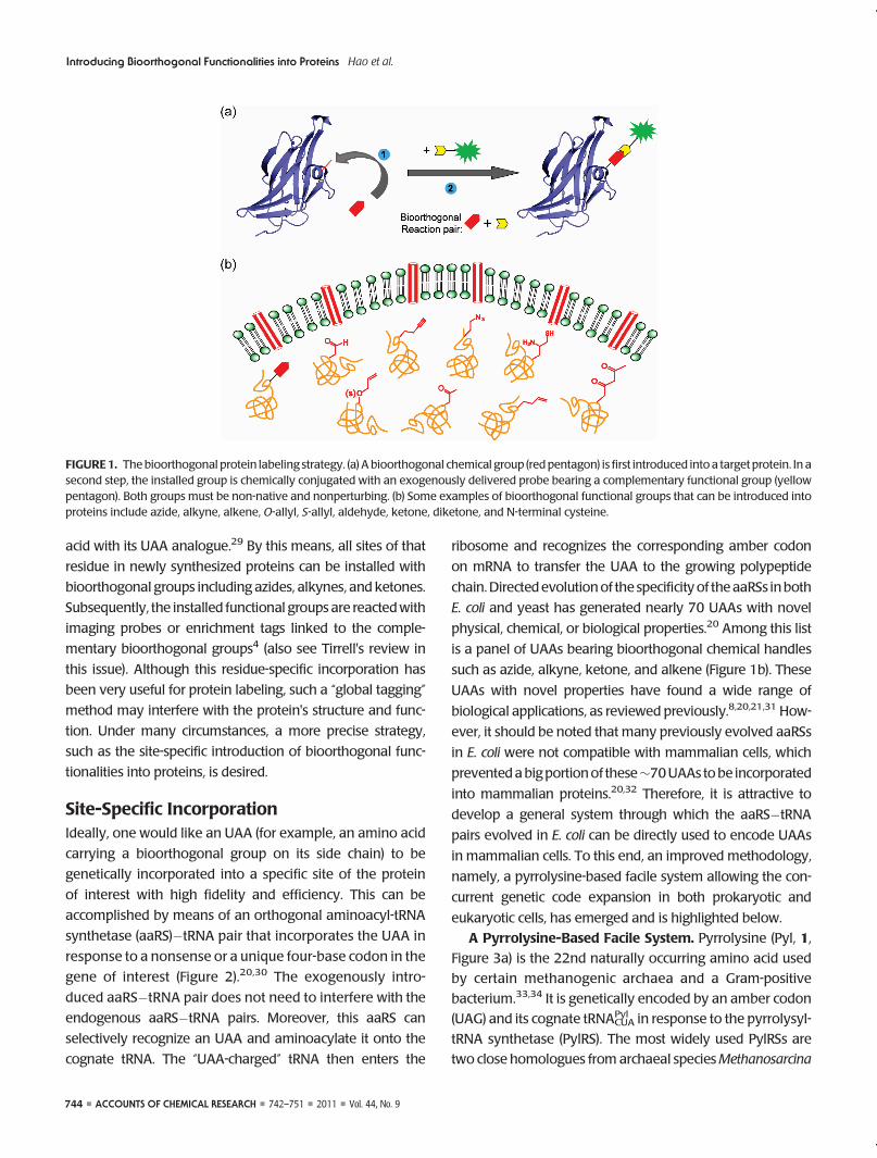
Site-Specific IncorporationIdeally, one would like an UAA (for example, an amino acid
carrying a bioorthogonal group on its side chain) to be
genetically incorporated into a specific site of the protein
of interest with high fidelity and efficiency. This can be
accomplished by means of an orthogonal aminoacyl-tRNA
synthetase (aaRS)�tRNA pair that incorporates the UAA in
response to a nonsense or a unique four-base codon in the
gene of interest (Figure 2).20,30 The exogenously intro-
duced aaRS�tRNA pair does not need to interfere with the
endogenous aaRS�tRNA pairs. Moreover, this aaRS can
selectively recognize an UAA and aminoacylate it onto the
cognate tRNA. The “UAA-charged” tRNA then enters the
ribosome and recognizes the corresponding amber codon
on mRNA to transfer the UAA to the growing polypeptide
chain.Directedevolutionof the specificity of theaaRSs inboth
E. coli and yeast has generated nearly 70 UAAs with novel
physical, chemical, or biological properties.20 Among this list
is a panel of UAAs bearing bioorthogonal chemical handles
such as azide, alkyne, ketone, and alkene (Figure 1b). These
UAAs with novel properties have found a wide range of
biological applications, as reviewed previously.8,20,21,31 How-
ever, it should be noted that many previously evolved aaRSs
in E. coli were not compatible with mammalian cells, which
preventedabigportionof these∼70UAAs tobe incorporated
into mammalian proteins.20,32 Therefore, it is attractive to
develop a general system through which the aaRS�tRNA
pairs evolved in E. coli can be directly used to encode UAAs
in mammalian cells. To this end, an improved methodology,
namely, a pyrrolysine-based facile system allowing the con-
current genetic code expansion in both prokaryotic and
eukaryotic cells, has emerged and is highlighted below.
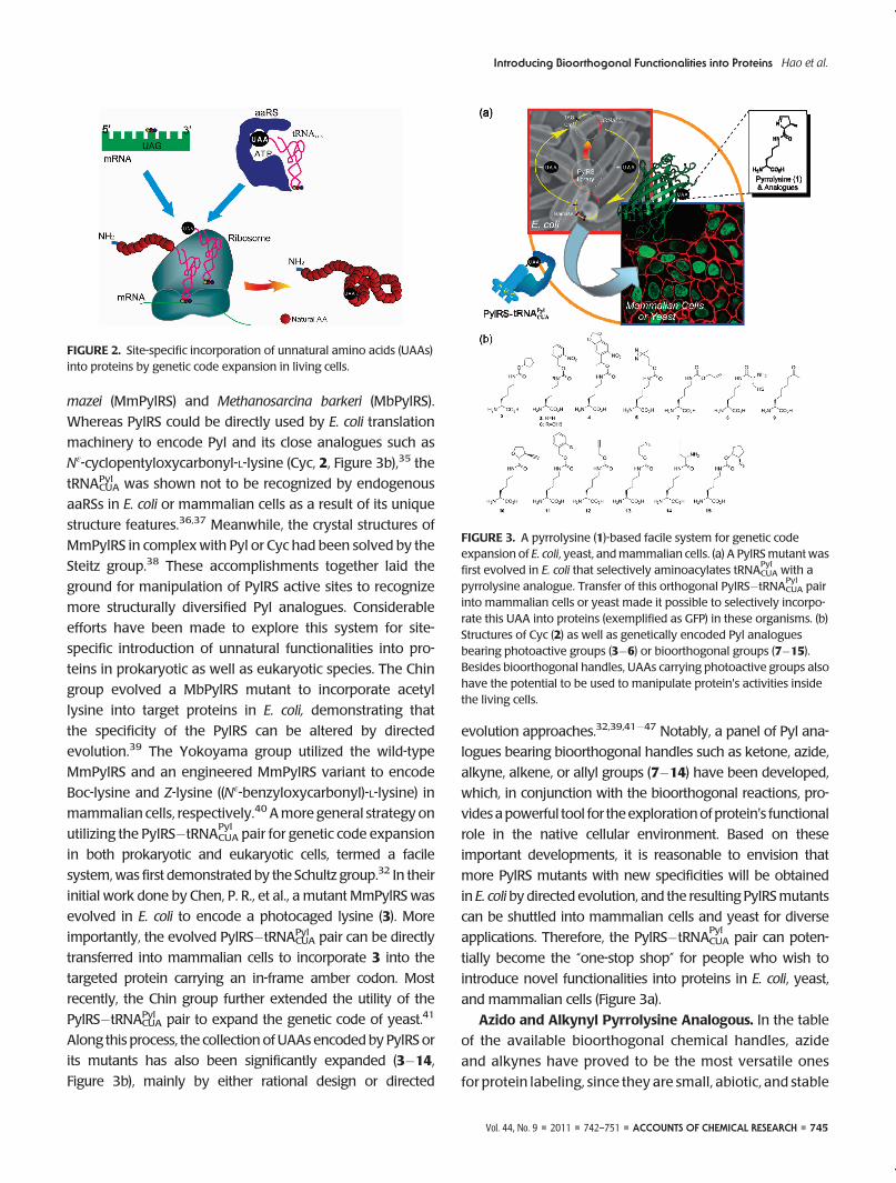
A Pyrrolysine-Based Facile System. Pyrrolysine (Pyl, 1,
Figure 3a) is the 22nd naturally occurring amino acid used
by certain methanogenic archaea and a Gram-positive
bacterium.33,34 It is genetically encoded by an amber codon
(UAG) and its cognate tRNACUAPyl in response to the pyrrolysyl-
tRNA synthetase (PylRS). The most widely used PylRSs are
two close homologues from archaeal speciesMethanosarcina
FIGURE1. Thebioorthogonal protein labeling strategy. (a) A bioorthogonal chemical group (red pentagon) is first introduced into a target protein. In asecond step, the installed group is chemically conjugated with an exogenously delivered probe bearing a complementary functional group (yellowpentagon). Both groups must be non-native and nonperturbing. (b) Some examples of bioorthogonal functional groups that can be introduced intoproteins include azide, alkyne, alkene, O-allyl, S-allyl, aldehyde, ketone, diketone, and N-terminal cysteine.
Vol. 44, No. 9 ’ 2011 ’ 742–751 ’ ACCOUNTS OF CHEMICAL RESEARCH ’ 745
Introducing Bioorthogonal Functionalities into Proteins Hao et al.
mazei (MmPylRS) and Methanosarcina barkeri (MbPylRS).
Whereas PylRS could be directly used by E. coli translation
machinery to encode Pyl and its close analogues such as
Nε-cyclopentyloxycarbonyl-L-lysine (Cyc, 2, Figure 3b),35 the
tRNACUAPyl was shown not to be recognized by endogenous
aaRSs in E. coli or mammalian cells as a result of its unique
structure features.36,37 Meanwhile, the crystal structures of
MmPylRS in complex with Pyl or Cyc had been solved by the
Steitz group.38 These accomplishments together laid the
ground for manipulation of PylRS active sites to recognize
more structurally diversified Pyl analogues. Considerable
efforts have been made to explore this system for site-
specific introduction of unnatural functionalities into pro-
teins in prokaryotic as well as eukaryotic species. The Chin
group evolved a MbPylRS mutant to incorporate acetyl
lysine into target proteins in E. coli, demonstrating that
the specificity of the PylRS can be altered by directed
evolution.39 The Yokoyama group utilized the wild-type
MmPylRS and an engineered MmPylRS variant to encode
Boc-lysine and Z-lysine ((Nε-benzyloxycarbonyl)-L-lysine) in
mammalian cells, respectively.40 Amore general strategy on
utilizing the PylRS�tRNACUAPyl
pair for genetic code expansion
in both prokaryotic and eukaryotic cells, termed a facile
system,was first demonstrated by the Schultz group.32 In their
initial work done by Chen, P. R., et al., a mutant MmPylRS was
evolved in E. coli to encode a photocaged lysine (3). More
importantly, the evolved PylRS�tRNACUAPyl pair can be directly
transferred into mammalian cells to incorporate 3 into the
targeted protein carrying an in-frame amber codon. Most
recently, the Chin group further extended the utility of the
PylRS�tRNACUAPyl pair to expand the genetic code of yeast.41
Along this process, the collection of UAAs encoded by PylRS or
its mutants has also been significantly expanded (3�14,
Figure 3b), mainly by either rational design or directed
evolution approaches.32,39,41�47 Notably, a panel of Pyl ana-
logues bearing bioorthogonal handles such as ketone, azide,
alkyne, alkene, or allyl groups (7�14) have been developed,
which, in conjunction with the bioorthogonal reactions, pro-
videsapowerful tool for theexplorationofprotein's functional
role in the native cellular environment. Based on these
important developments, it is reasonable to envision that
more PylRS mutants with new specificities will be obtained
in E. coli by directed evolution, and the resulting PylRSmutants
can be shuttled into mammalian cells and yeast for diverse
applications. Therefore, the PylRS�tRNACUAPyl
pair can poten-
tially become the “one-stop shop” for people who wish to
introduce novel functionalities into proteins in E. coli, yeast,
and mammalian cells (Figure 3a).
Azido and Alkynyl Pyrrolysine Analogous. In the table
of the available bioorthogonal chemical handles, azide
and alkynes have proved to be the most versatile ones
for protein labeling, since they are small, abiotic, and stable
FIGURE 3. A pyrrolysine (1)-based facile system for genetic codeexpansion of E. coli, yeast, andmammalian cells. (a) A PylRSmutant wasfirst evolved in E. coli that selectively aminoacylates tRNACUA
Pylwith a
pyrrolysine analogue. Transfer of this orthogonal PylRS�tRNACUAPyl
pairinto mammalian cells or yeast made it possible to selectively incorpo-rate this UAA into proteins (exemplified as GFP) in these organisms. (b)Structures of Cyc (2) as well as genetically encoded Pyl analoguesbearing photoactive groups (3�6) or bioorthogonal groups (7�15).Besides bioorthogonal handles, UAAs carrying photoactive groups alsohave the potential to be used to manipulate protein's activities insidethe living cells.
FIGURE 2. Site-specific incorporation of unnatural amino acids (UAAs)into proteins by genetic code expansion in living cells.
746 ’ ACCOUNTS OF CHEMICAL RESEARCH ’ 742–751 ’ 2011 ’ Vol. 44, No. 9
Introducing Bioorthogonal Functionalities into Proteins Hao et al.
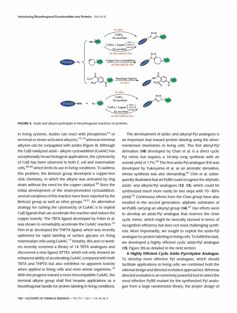
in living systems. Azides can react with phosphines14 or
terminal or strain-activated alkynes,15,19 whereas terminal
alkynes can be conjugated with azides (Figure 4). Although
the Cu(I)-catalyzed azide�alkyne cycloaddition (CuAAC) has
exceptionally broad biological applications, the cytotoxicity
of Cu(I) has been observed in both E. coli and mammalian
cells,48,49 which limits its use in living conditions. To address
this problem, the Bertozzi group developed a copper-free
click chemistry, in which the alkyne was activated by ring
strain without the need for the copper catalyst.50 Since the
initial development of the strain-promoted cycloaddition,
several variations of this reaction have been reported by the
Bertozzi group as well as other groups.19,51 An alternative
strategy for curbing the cytotoxicity of CuAAC is to exploit
Cu(I) ligands that can accelerate the reaction and reduce the
copper toxicity. The TBTA ligand developed by Fokin et al.
was shown to remarkably accelerate the CuAAC reaction.16
Finn et al. developed the THPTA ligand, which was recently
optimized for rapid labeling of surface glycans on living
mammalian cells using CuAAC.17 Notably, Wu and co-work-
ers recently screened a library of 14 TBTA analogues and
discovered a new ligand, BTTES, which not only showed an
enhanced ability of accelerating CuAAC comparedwith both
TBTA and THPTA but also exhibited no apparent toxicity
when applied to living cells and even whole organisms.18
With the progress toward amore biocompatible CuAAC, the
terminal alkyne group shall find broader applications as a
bioorthogonal handle for protein labeling in living conditions.
The development of azido- and alkynyl-Pyl analogues is
an important step toward protein labeling using the afore-
mentioned chemistries in living cells. The first alknyl-Pyl
derivative (10) developed by Chan et al. is a direct cyclic
Pyl mimic but requires a 16-step long synthesis with an
overall yield of 17%.43 The first azido-Pyl analogue (11) was
developed by Yukoyama et al. as an aromatic derivative,
whose synthesis was also demanding.42 Chin et al. subse-
quently illustrated that wt-PylRS could recognize the aliphatic
azido- and alkyne-Pyl analogues (12, 13), which could be
synthesized much more easily (in two steps with 70�80%
yield).52 Continuous efforts from the Chan group have also
resulted in the second generation, aliphatic substrates of
wt-PylRS carrying an alkynyl group (14).47 Our efforts were
to develop an azido-Pyl analogue that reserves the close
cyclic mimic, which might be sterically favored in terms of
recognition efficiency but does not need challenging synth-
esis. More importantly, we sought to exploit the azido-Pyl
analogue for protein labeling in living cells. To fulfill this task,
we developed a highly efficient cyclic azido-Pyl analogue
(15, Figure 3b) as detailed in the next section.
A Highly Efficient Cyclic Azido Pyrrolysine Analogue.
To develop more effective Pyl analogues, which should
facilitate applications in living cells, we combined both the
rational design anddirected evolution approaches.Whereas
directed evolution is an extremely powerful tool to select the
most effective PylRS mutant for the synthesized Pyl analo-
gue from a large randomized library, the proper design of
FIGURE 4. Azide and alkyne participate in bioorthogonal reactions on proteins.
Vol. 44, No. 9 ’ 2011 ’ 742–751 ’ ACCOUNTS OF CHEMICAL RESEARCH ’ 747
Introducing Bioorthogonal Functionalities into Proteins Hao et al.
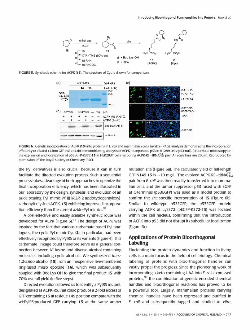
the Pyl derivatives is also crucial, because it can in turn
facilitate the directed evolution process. Such a sequential
process takes advantage of both approaches to optimize the
final incorporation efficiency, which has been illustrated in
our laboratory by the design, synthesis, and evolution of an
azide-bearing Pyl mimic Nε-(((1R,2R)-2-azidocyclopentyloxy)-
carbonyl)-L-lysine (ACPK, 15) exhibiting improved incorpora-
tion efficiency than the current azido-Pyl mimics.53
A cost-effective and easily scalable synthetic route was
developed for ACPK (Figure 5).53 The design of ACPK was
inspired by the fact that various carbamate-based Pyl ana-
logues, the cyclic Pyl mimic Cyc (2), in particular, had been
effectively recognized by PylRS or its variants (Figure 4). This
carbamate linkage could therefore serve as a general con-
nection between Nε-lysine and diverse alcohol-containing
molecules including cyclic alcohols. We synthesized trans-
1,2-azido alcohol (18) from an inexpensive five-membered
ring-fused meso epoxide (16), which was subsequently
coupled with Boc-Lys-OH to give the final product 15 with
70% overall yield (in five steps).
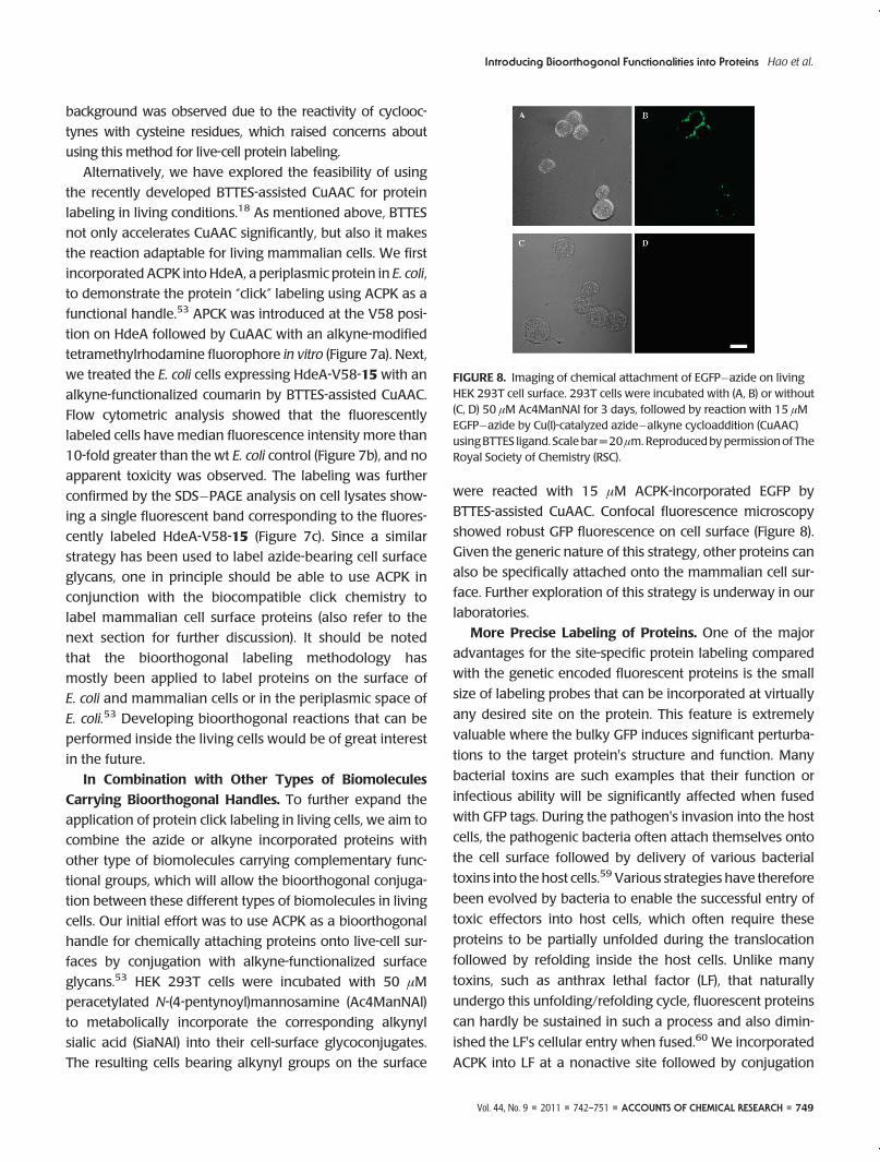
Directed evolution allowed us to identify a PylRS mutant,
designated asACPK-RS, that could produce a 2-fold excess of
GFP containing 15 at residue 149 position compare with the
wt-PylRS-produced GFP carrying 15 at the same amber
mutation site (Figure 6a). The calculated yield of full-length
GFP-N149-15 is ∼10 mg/L. The evolved ACPK-RS�tRNACUAPyl
pair from E. coli was then readily transferred into mamma-
lian cells, and the tumor suppressor p53 fused with EGFP
at C-terminus (p53EGFP) was used as a model protein to
confirm the site-specific incorporation of 15 (Figure 6b).
Similar to wild-type p53EGFP, the p53EGFP protein
carrying ACPK at Lys372 (pEGFP-K372-15) was located
within the cell nucleus, confirming that the introduction
of ACPK into p53 did not disrupt its subcellular localization
(Figure 6c).
Applications of Protein BioorthogonalLabelingElucidating the protein dynamics and function in living
cells is a main focus in the field of cell biology. Chemical
labeling of proteins with bioorthogonal handles can
vastly propel the progress. Since the pioneering work of
incorporating a keto-containing UAA into E. coli-expressed
proteins,54 the combination of genetic encoded chemical
handles and bioorthogonal reactions has proved to be
a powerful tool. Largely, mammalian proteins carrying
chemical handles have been expressed and purified in
E. coli and subsequently tagged and studied in vitro.
FIGURE 5. Synthesis scheme for ACPK (15). The structure of Cyc is shown for comparison.
FIGURE 6. Genetic incorporation of ACPK (15) into proteins in E. coli and mammalian cells. (a) SDS�PAGE analysis demonstrating the incorporationefficiency of 13 and 15 into GFP in E. coli. (b) Immunoblotting analysis of ACPK incorporated p53 in H1299 cells (p53-null). (c) Confocal microscopy onthe expression and localization of p53EGFP-K372-15 in HEK293T cells harboring ACPK-RS�tRNACUA
Pyl pair. All scale bars are 20 μm. Reproduced bypermission of The Royal Society of Chemistry (RSC).

748 ’ ACCOUNTS OF CHEMICAL RESEARCH ’ 742–751 ’ 2011 ’ Vol. 44, No. 9
Introducing Bioorthogonal Functionalities into Proteins Hao et al.
The availability of the recently developed pyrrolysine-
based facile system has set the stage for such studies in
living mammalian cells.
Labeling of Purified Proteins. Using advanced optical
techniques, proteins tagged with fluorophores, particularly
in a site-specific manner, have been studied in solution and
shed light on the structural and dynamic information. For
example, Chanand co-workers incorporated10 at the Thr34
position of calmodulin (CaM), a calcium binding protein.
After purification, CaM functionalized with alkyne was con-
jugated with azidocoumarin by CuAAC.43 An acceptor fluor-
ophore Fluor Alexa 488was then attached to an engineered
cysteine at position 134, which enabled the F€orster reso-
nance energy transfer (FRET) between these two fluorescent
dyes. Upon addition of Ca(II) and the regulatory peptide
M13, changes in FRET efficiencywere observed, indicating
a conformational change on CaM. We site-specifically
introduced 15 into the heme recognition domain (NEAT)
of a bacterial heme-transfer protein IsdA, which allowed
us to conjugate DBCO-Fluor 488 (a variation of cyclooctyne-
conjugated dye) near the heme binding pocket by the
“copper free” click chemistry. A reversible fluorescence
quenching effect was observed, caused by the direct energy
transfer between heme and Fluor 488.53 Since heme is
utilized by mammalian hosts to tightly control the essential
iron element as an effective defense mechanism, this site-
specific “click” labeling tool can be used to study heme
transfer among heme-acquisition proteins in Gram-positive
pathogens.
Simultaneously incorporating two distinct UAAs into a
single protein was first demonstrated by Schultz and co-
workers by incorporating L-homoglutamine andO-methyl-L-
tyrosine in response to a quadruplet codon and an amber
codon, respectively.55 Two bioorthogonal handles can also
be installed on a single protein in this way. Recently, Chin
and co-workers evolved an orthogonal ribosome that can
decode a series of quadruplet codons.56 Together with two
mutually orthogonal aaRS�tRNA pairs (MjTyrRS-tRNACUA
and MbPylRS-tRNACUA), they demonstrated the encoding
of an azide-bearing UAA and an alkyne-bearing UAA in
response to two new codons created on the same orthogo-
nal mRNA.56 By mutating the PylRS�tRNA pair to suppress
the ochre UAA codon, Liu et al. also showed that the
combination of this engineered PylRS�tRNAUAA pair with
MjTyrRS�tRNACUA pair was able to genetically incorporate
two distinct UAAs into a single protein in E. coli.57 However,
the efficiency for simultaneous incorporation of two UAAs
into the same protein still waits for further improvements
before it can be applied for versatile bioorthogonal protein
labeling with diverse probes.
Protein Labeling in Living Cells. To understand protein
function and dynamics in a physiologically relevant context
requires a means to label proteins in living cells. Whereas
the in vitro protein labeling strategy has been widely ex-
plored, expanding this methodology for protein labeling in
living cells is still at its infancy. Using genetic code expansion
and bioorthogonal groups for labeling of proteins in living
conditions was first demonstrated on the E. coli surface. The
acetylphenylalanine was site-specifically incorporated into
an outer-membrane protein LamB followed by conjugation
with fluorescein hydrazide derivatives for imaging LamB on
the living E. coli cell surface.58 “Click” labeling was also
applied to proteins metabolically incorporated with azide
or alkyne containing methionine analogues, which allowed
the imaging of the newly synthesized proteins.4 Since CuACC
is not compatible with living mammalian cells, Tirrell and
Bertozzi and co-workers employed the strain-promoted cyclo-
addition to conjugate cellular proteins carrying metaboli-
cally introduced azide handles with membrane-permeable
coumarin-cyclooctynes inside the living cells.9 Elevated
FIGURE7. Click labelingof HdeA-V58-15 in vitro and in livingbacteria: (a) SDS�PAGEof fluorescent labeledHdeA-V58-15byCuAACandalk-TMR; (b)flowcytometric analysis onbiocompatible CuAAC-mediated fluorescent labelingof living E. coli cells expressingHdeA-V58-15orwtHdeAas a control;(c) SDS�PAGE analysis on lysate of E. coli cells expressing wt HdeA or HdeA-V58-15 or without HdeA after being reacted with alk-coumarin bybiocompatible CuAAC in living bacteria. Reproduced by permission of The Royal Society of Chemistry (RSC).
Vol. 44, No. 9 ’ 2011 ’ 742–751 ’ ACCOUNTS OF CHEMICAL RESEARCH ’ 749
Introducing Bioorthogonal Functionalities into Proteins Hao et al.
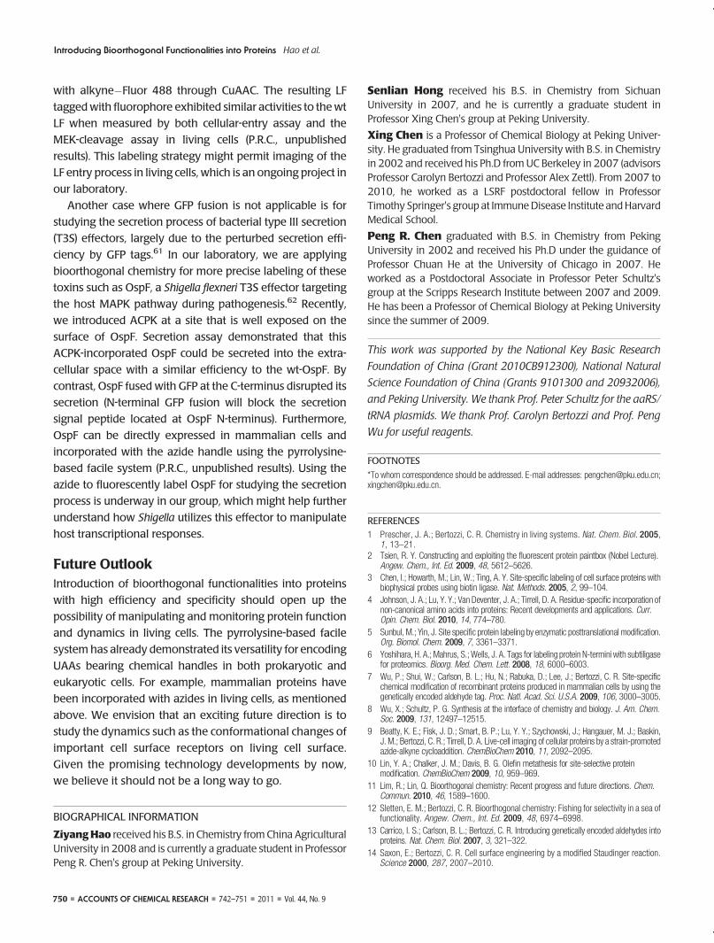
background was observed due to the reactivity of cyclooc-
tynes with cysteine residues, which raised concerns about
using this method for live-cell protein labeling.
Alternatively, we have explored the feasibility of using
the recently developed BTTES-assisted CuAAC for protein
labeling in living conditions.18 As mentioned above, BTTES
not only accelerates CuAAC significantly, but also it makes
the reaction adaptable for living mammalian cells. We first
incorporated ACPK into HdeA, a periplasmic protein in E. coli,
to demonstrate the protein “click” labeling using ACPK as a
functional handle.53 APCK was introduced at the V58 posi-
tion on HdeA followed by CuAAC with an alkyne-modified
tetramethylrhodamine fluorophore in vitro (Figure 7a). Next,
we treated the E. coli cells expressing HdeA-V58-15 with an
alkyne-functionalized coumarin by BTTES-assisted CuAAC.
Flow cytometric analysis showed that the fluorescently
labeled cells have median fluorescence intensity more than
10-fold greater than the wt E. coli control (Figure 7b), and no
apparent toxicity was observed. The labeling was further
confirmed by the SDS�PAGE analysis on cell lysates show-
ing a single fluorescent band corresponding to the fluores-
cently labeled HdeA-V58-15 (Figure 7c). Since a similar
strategy has been used to label azide-bearing cell surface
glycans, one in principle should be able to use ACPK in
conjunction with the biocompatible click chemistry to
label mammalian cell surface proteins (also refer to the
next section for further discussion). It should be noted
that the bioorthogonal labeling methodology has
mostly been applied to label proteins on the surface of
E. coli and mammalian cells or in the periplasmic space of
E. coli.53 Developing bioorthogonal reactions that can be
performed inside the living cells would be of great interest
in the future.
In Combination with Other Types of Biomolecules
Carrying Bioorthogonal Handles. To further expand the
application of protein click labeling in living cells, we aim to
combine the azide or alkyne incorporated proteins with
other type of biomolecules carrying complementary func-
tional groups, which will allow the bioorthogonal conjuga-
tion between these different types of biomolecules in living
cells. Our initial effort was to use ACPK as a bioorthogonal
handle for chemically attaching proteins onto live-cell sur-
faces by conjugation with alkyne-functionalized surface
glycans.53 HEK 293T cells were incubated with 50 μM
peracetylated N-(4-pentynoyl)mannosamine (Ac4ManNAl)
to metabolically incorporate the corresponding alkynyl
sialic acid (SiaNAl) into their cell-surface glycoconjugates.
The resulting cells bearing alkynyl groups on the surface
were reacted with 15 μM ACPK-incorporated EGFP by
BTTES-assisted CuAAC. Confocal fluorescence microscopy
showed robust GFP fluorescence on cell surface (Figure 8).
Given the generic nature of this strategy, other proteins can
also be specifically attached onto the mammalian cell sur-
face. Further exploration of this strategy is underway in our
laboratories.
More Precise Labeling of Proteins. One of the major
advantages for the site-specific protein labeling compared
with the genetic encoded fluorescent proteins is the small
size of labeling probes that can be incorporated at virtually
any desired site on the protein. This feature is extremely
valuable where the bulky GFP induces significant perturba-
tions to the target protein's structure and function. Many
bacterial toxins are such examples that their function or
infectious ability will be significantly affected when fused
with GFP tags. During the pathogen's invasion into the host
cells, the pathogenic bacteria often attach themselves onto
the cell surface followed by delivery of various bacterial
toxins into the host cells.59 Various strategies have therefore
been evolved by bacteria to enable the successful entry of
toxic effectors into host cells, which often require these
proteins to be partially unfolded during the translocation
followed by refolding inside the host cells. Unlike many
toxins, such as anthrax lethal factor (LF), that naturally
undergo this unfolding/refolding cycle, fluorescent proteins
can hardly be sustained in such a process and also dimin-
ished the LF's cellular entry when fused.60 We incorporated
ACPK into LF at a nonactive site followed by conjugation
FIGURE 8. Imaging of chemical attachment of EGFP�azide on livingHEK 293T cell surface. 293T cells were incubated with (A, B) or without(C, D) 50 μM Ac4ManNAl for 3 days, followed by reaction with 15 μMEGFP�azide by Cu(I)-catalyzed azide�alkyne cycloaddition (CuAAC)usingBTTES ligand. Scalebar=20μm.Reproducedbypermissionof TheRoyal Society of Chemistry (RSC).
750 ’ ACCOUNTS OF CHEMICAL RESEARCH ’ 742–751 ’ 2011 ’ Vol. 44, No. 9
Introducing Bioorthogonal Functionalities into Proteins Hao et al.
with alkyne�Fluor 488 through CuAAC. The resulting LF
taggedwith fluorophore exhibited similar activities to thewt
LF when measured by both cellular-entry assay and the
MEK-cleavage assay in living cells (P.R.C., unpublished
results). This labeling strategy might permit imaging of the
LF entry process in living cells, which is an ongoing project in
our laboratory.
Another case where GFP fusion is not applicable is for
studying the secretion process of bacterial type III secretion
(T3S) effectors, largely due to the perturbed secretion effi-
ciency by GFP tags.61 In our laboratory, we are applying
bioorthogonal chemistry for more precise labeling of these
toxins such as OspF, a Shigella flexneri T3S effector targeting
the host MAPK pathway during pathogenesis.62 Recently,
we introduced ACPK at a site that is well exposed on the
surface of OspF. Secretion assay demonstrated that this
ACPK-incorporated OspF could be secreted into the extra-
cellular space with a similar efficiency to the wt-OspF. By
contrast, OspF fused with GFP at the C-terminus disrupted its
secretion (N-terminal GFP fusion will block the secretion
signal peptide located at OspF N-terminus). Furthermore,
OspF can be directly expressed in mammalian cells and
incorporated with the azide handle using the pyrrolysine-
based facile system (P.R.C., unpublished results). Using the
azide to fluorescently label OspF for studying the secretion
process is underway in our group, which might help further
understand how Shigella utilizes this effector to manipulate
host transcriptional responses.
Future OutlookIntroduction of bioorthogonal functionalities into proteins
with high efficiency and specificity should open up the
possibility of manipulating and monitoring protein function
and dynamics in living cells. The pyrrolysine-based facile
systemhas already demonstrated its versatility for encoding
UAAs bearing chemical handles in both prokaryotic and
eukaryotic cells. For example, mammalian proteins have
been incorporated with azides in living cells, as mentioned
above. We envision that an exciting future direction is to
study the dynamics such as the conformational changes of
important cell surface receptors on living cell surface.
Given the promising technology developments by now,
we believe it should not be a long way to go.
BIOGRAPHICAL INFORMATION
ZiyangHao received his B.S. in Chemistry from China AgriculturalUniversity in 2008 and is currently a graduate student in ProfessorPeng R. Chen's group at Peking University.
Senlian Hong received his B.S. in Chemistry from SichuanUniversity in 2007, and he is currently a graduate student inProfessor Xing Chen's group at Peking University.
Xing Chen is a Professor of Chemical Biology at Peking Univer-sity. He graduated from Tsinghua University with B.S. in Chemistryin 2002 and received his Ph.D from UC Berkeley in 2007 (advisorsProfessor Carolyn Bertozzi and Professor Alex Zettl). From 2007 to2010, he worked as a LSRF postdoctoral fellow in ProfessorTimothy Springer's group at ImmuneDisease Institute andHarvardMedical School.
Peng R. Chen graduated with B.S. in Chemistry from PekingUniversity in 2002 and received his Ph.D under the guidance ofProfessor Chuan He at the University of Chicago in 2007. Heworked as a Postdoctoral Associate in Professor Peter Schultz'sgroup at the Scripps Research Institute between 2007 and 2009.He has been a Professor of Chemical Biology at Peking Universitysince the summer of 2009.
This work was supported by the National Key Basic ResearchFoundation of China (Grant 2010CB912300), National NaturalScience Foundation of China (Grants 9101300 and 20932006),and Peking University. We thank Prof. Peter Schultz for the aaRS/tRNA plasmids. We thank Prof. Carolyn Bertozzi and Prof. PengWu for useful reagents.
FOOTNOTES
*To whom correspondence should be addressed. E-mail addresses: [email protected];[email protected].
REFERENCES1 Prescher, J. A.; Bertozzi, C. R. Chemistry in living systems. Nat. Chem. Biol. 2005,
1, 13–21.2 Tsien, R. Y. Constructing and exploiting the fluorescent protein paintbox (Nobel Lecture).
Angew. Chem., Int. Ed. 2009, 48, 5612–5626.3 Chen, I.; Howarth, M.; Lin, W.; Ting, A. Y. Site-specific labeling of cell surface proteins with
biophysical probes using biotin ligase. Nat. Methods. 2005, 2, 99–104.4 Johnson, J. A.; Lu, Y. Y.; Van Deventer, J. A.; Tirrell, D. A. Residue-specific incorporation of
non-canonical amino acids into proteins: Recent developments and applications. Curr.Opin. Chem. Biol. 2010, 14, 774–780.
5 Sunbul, M.; Yin, J. Site specific protein labeling by enzymatic posttranslational modification.Org. Biomol. Chem. 2009, 7, 3361–3371.
6 Yoshihara, H. A.; Mahrus, S.;Wells, J. A. Tags for labeling protein N-termini with subtiligasefor proteomics. Bioorg. Med. Chem. Lett. 2008, 18, 6000–6003.
7 Wu, P.; Shui, W.; Carlson, B. L.; Hu, N.; Rabuka, D.; Lee, J.; Bertozzi, C. R. Site-specificchemical modification of recombinant proteins produced in mammalian cells by using thegenetically encoded aldehyde tag. Proc. Natl. Acad. Sci. U.S.A. 2009, 106, 3000–3005.
8 Wu, X.; Schultz, P. G. Synthesis at the interface of chemistry and biology. J. Am. Chem.Soc. 2009, 131, 12497–12515.
9 Beatty, K. E.; Fisk, J. D.; Smart, B. P.; Lu, Y. Y.; Szychowski, J.; Hangauer, M. J.; Baskin,J. M.; Bertozzi, C. R.; Tirrell, D. A. Live-cell imaging of cellular proteins by a strain-promotedazide-alkyne cycloaddition. ChemBioChem 2010, 11, 2092–2095.
10 Lin, Y. A.; Chalker, J. M.; Davis, B. G. Olefin metathesis for site-selective proteinmodification. ChemBioChem 2009, 10, 959–969.
11 Lim, R.; Lin, Q. Bioorthogonal chemistry: Recent progress and future directions. Chem.Commun. 2010, 46, 1589–1600.
12 Sletten, E. M.; Bertozzi, C. R. Bioorthogonal chemistry: Fishing for selectivity in a sea offunctionality. Angew. Chem., Int. Ed. 2009, 48, 6974–6998.
13 Carrico, I. S.; Carlson, B. L.; Bertozzi, C. R. Introducing genetically encoded aldehydes intoproteins. Nat. Chem. Biol. 2007, 3, 321–322.
14 Saxon, E.; Bertozzi, C. R. Cell surface engineering by a modified Staudinger reaction.Science 2000, 287, 2007–2010.
Vol. 44, No. 9 ’ 2011 ’ 742–751 ’ ACCOUNTS OF CHEMICAL RESEARCH ’ 751
Introducing Bioorthogonal Functionalities into Proteins Hao et al.
15 Rostovtsev, V. V.; Green, L. G.; Fokin, V. V.; Sharpless, K. B. A stepwise Huisgencycloaddition process: Copper(I)-catalyzed regioselective “ligation” of azides andterminal alkynes. Angew. Chem., Int. Ed. 2002, 41, 2596–2599.
16 Chan, T. R.; Hilgraf, R.; Sharpless, K. B.; Fokin, V. V. Polytriazoles as copper(I)-stabilizingligands in catalysis. Org. Lett. 2004, 6, 2853–2855.
17 Hong, V.; Steinmetz, N. F.; Manchester, M.; Finn, M. G. Labeling live cells by copper-catalyzed alkyne�azide click chemistry. Bioconjugate Chem. 2010, 21, 1912–1916.
18 Soriano Del Amo, D.; Wang, W.; Jiang, H.; Besanceney, C.; Yan, A. C.; Levy, M.; Liu, Y.;Marlow, F. L.; Wu, P. Biocompatible copper(I) catalysts for in vivo imaging of glycans. J. Am.Chem. Soc. 2010, 132, 16893–16899.
19 Jewett, J. C.; Bertozzi, C. R. Cu-free click cycloaddition reactions in chemical biology.Chem. Soc. Rev. 2010, 39, 1272–1279.
20 Liu, C. C.; Schultz, P. G. Adding new chemistries to the genetic code. Annu. Rev. Biochem.2010, 79, 413–444.
21 Wang, Q.; Parrish, A. R.; Wang, L. Expanding the genetic code for biological studies. Chem.Biol. 2009, 16, 323–336.
22 Walsh, C. T.; Garneau-Tsodikova, S.; Gatto, G. J., Jr. Protein posttranslationalmodifications: The chemistry of proteome diversifications. Angew. Chem., Int. Ed.2005, 44, 7342–7372.
23 Foley, T. L.; Burkart, M. D. Site-specific protein modification: Advances and applications.Curr. Opin. Chem. Biol. 2007, 11, 12–19.
24 Lin, M. Z.; Wang, L. Selective labeling of proteins with chemical probes in living cells.Physiology (Bethesda) 2008, 23, 131–141.
25 Fernandez-Suarez, M.; Baruah, H.; Martinez-Hernandez, L.; Xie, K. T.; Baskin, J. M.;Bertozzi, C. R.; Ting, A. Y. Redirecting lipoic acid ligase for cell surface protein labelingwith small-molecule probes. Nat. Biotechnol. 2007, 25, 1483–1487.
26 Uttamapinant, C.; White, K. A.; Baruah, H.; Thompson, S.; Fernandez-Suarez, M.;Puthenveetil, S.; Ting, A. Y. A fluorophore ligase for site-specific protein labelinginside living cells. Proc. Natl. Acad. Sci. U.S.A. 2010, 107, 10914–10919.
27 Rodriguez, E. A.; Lester, H. A.; Dougherty, D. A. In vivo incorporation of multiple unnaturalamino acids through nonsense and frameshift suppression. Proc. Natl. Acad. Sci. U.S.A.2006, 103, 8650–8655.
28 Kohrer, C.; Yoo, J. H.; Bennett, M.; Schaack, J.; RajBhandary, U. L. A possible approach totwo different unnatural site-specific insertion of amino acids into proteins in mammaliancells via nonsense suppression. Chem. Biol. 2003, 10, 1095–1102.
29 Link, A. J.; Mock, M. L.; Tirrell, D. A. Non-canonical amino acids in protein engineering.Curr. Opin. Biotechnol. 2003, 14, 603–609.
30 Wang, L.; Schultz, P. G. Expanding the genetic code. Angew.Chem., Int. Ed.2004, 44, 34–66.31 Chin, J.W.Modular approaches to expanding the functions of livingmatter. Nat. Chem. Biol.
2006, 2, 304–311.32 Chen, P. R.; Groff, D.; Guo, J.; Ou, W.; Cellitti, S.; Geierstanger, B. H.; Schultz, P. G. A facile
system for encoding unnatural amino acids in mammalian cells. Angew. Chem., Int. Ed.2009, 48, 4052–4055.
33 Srinivasan, G.; James, C. M.; Krzycki, J. A. Pyrrolysine encoded by UAG in Archaea:Charging of a UAG-decoding specialized tRNA. Science 2002, 296, 1459–1462.
34 Hao, B.; Gong, W.; Ferguson, T. K.; James, C. M.; Krzycki, J. A.; Chan, M. K. A new UAG-encoded residue in the structure of a methanogen methyltransferase. Science 2002, 296,1462–1466.
35 Polycarpo, C. R.; Herring, S.; Berube, A.; Wood, J. L.; Soll, D.; Ambrogelly, A. Pyrrolysineanalogues as substrates for pyrrolysyl-tRNA synthetase. FEBS Lett. 2006, 580, 6695–6700.
36 Nozawa, K.; O'Donoghue, P.; Gundllapalli, S.; Araiso, Y.; Ishitani, R.; Umehara, T.; Soll, D.;Nureki, O. Pyrrolysyl-tRNA synthetase-tRNA(Pyl) structure reveals the molecular basis oforthogonality. Nature 2009, 457, 1163–1167.
37 Polycarpo, C.; Ambrogelly, A.; Berube, A.; Winbush, S. M.; McCloskey, J. A.; Crain, P. F.;Wood, J. L.; Soll, D. An aminoacyl-tRNA synthetase that specifically activates pyrrolysine.Proc. Natl. Acad. Sci. U.S.A. 2004, 101, 12450–12454.
38 Kavran, J. M.; Gundliapalli, S.; O'Donoghue, P.; Englert, M.; Soell, D.; Steitz, T. A. Structureof pyrrolysyl-tRNA synthetase, an archaeal enzyme for genetic code innovation. Proc. Natl.Acad. Sci. U.S.A. 2007, 104, 11268–11273.
39 Neumann, H.; Peak-Chew, S. Y.; Chin, J. W. Genetically encoding N-epsilon-acetyllysine inrecombinant proteins. Nat. Chem. Biol. 2008, 4, 232–234.
40 Mukai, T.; Kobayashi, T.; Hino, N.; Yanagisawa, T.; Sakamoto, K.; Yokoyama, S. AddingL-lysine derivatives to the genetic code of mammalian cells with engineered pyrrolysyl-tRNAsynthetases. Biochem. Biophys. Res. Commun. 2008, 371, 818–822.
41 Hancock, S. M.; Uprety, R.; Deiters, A.; Chin, J. W. Expanding the genetic code of yeast forincorporation of diverse unnatural amino acids via a pyrrolysyl-tRNA synthetase/tRNA pair.J. Am. Chem. Soc. 2010, 132, 14819–14824.
42 Yanagisawa, T.; Ishii, R.; Fukunaga, R.; Kobayashi, T.; Sakamoto, K.; Yokoyama, S.Multistep engineering of pyrrolysyl-tRNA synthetase to genetically encodeN-epsilon-(o-Azidobenzyloxycarbonyl) lysine for site-specific protein modification.Chem. Biol. 2008, 15, 1187–1197.
43 Fekner, T.; Li, X.; Lee, M.M.; Chan,M. K. A pyrrolysine analogue for protein click chemistry.Angew. Chem., Int. Ed. 2009, 48, 1633–1635.
44 Li, X.; Fekner, T.; Ottesen, J. J.; Chan, M. K. A pyrrolysine analogue for site-specific proteinubiquitination. Angew. Chem., Int. Ed. 2009, 48, 9184–9187.
45 Huang, Y.; Wan,W.; Russell, W. K.; Pai, P. J.; Wang, Z. Y.; Russell, D. H.; Liu,W. S. Geneticincorporation of an aliphatic keto-containing amino acid into proteins for their site-specificmodifications. Bioorg. Med. Chem. 2010, 20, 878–880.
46 Fekner, T.; Li, X.; Chan, M. K. Pyrrolysine analogs for translational incorporation intoproteins. Eur. J. Org. Chem. 2010, 4171–4179.
47 Li, X.; Fekner, T.; Chan, M. K. N6-(2-(R)-propargylglycyl)lysine as a clickable pyrrolysinemimic. Chem.;Asian J. 2010, 5, 1765–1769.
48 Link, A. J.; Tirrell, D. A. Cell surface labeling of Escherichia coli via copper(I)-catalyzed [3þ2] cycloaddition. J. Am. Chem. Soc. 2003, 125, 11164–11165.
49 Link, A. J.; Vink, M. K.; Tirrell, D. A. Presentation and detection of azide functionality inbacterial cell surface proteins. J. Am. Chem. Soc. 2004, 126, 10598–10602.
50 Agard, N. J.; Prescher, J. A.; Bertozzi, C. R. A strain-promoted [3 þ 2] azide-alkynecycloaddition for covalent modification of biomolecules in living systems. J. Am. Chem.Soc. 2004, 126, 15046–15047.
51 Ning, X.; Guo, J.; Wolfert, M. A.; Boons, G. J. Visualizing metabolically labeledglycoconjugates of living cells by copper-free and fast huisgen cycloadditions.Angew. Chem., Int. Ed. 2008, 47, 2253–2255.
52 Nguyen, D. P.; Lusic, H.; Neumann, H.; Kapadnis, P. B.; Deiters, A.; Chin, J. W. Geneticencoding and labeling of aliphatic azides and alkynes in recombinant proteins via apyrrolysyl-tRNA synthetase/tRNA(CUA) pair and click chemistry. J. Am. Chem. Soc. 2009,131, 8720–8721.
53 Hao, Z.; Song, Y.; Lin, S.; Yang, M.; Liang, Y.; Wang, J.; Chen, P. R. A readily synthesizedcyclic pyrrolysine analogue for site-specific protein “click” labeling. Chem. Commun. 2011,47 (15), 4502–4504.
54 Wang, L.; Zhang, Z.; Brock, A.; Schultz, P. G. Addition of the keto functional group to thegenetic code of Escherichia coli. Proc. Natl. Acad. Sci. U.S.A. 2003, 100, 56–61.
55 Anderson, J. C.; Wu, N.; Santoro, S. W.; Lakshman, V.; King, D. S.; Schultz, P. G. Anexpanded genetic code with a functional quadruplet codon. Proc. Natl. Acad. Sci. U.S.A.2004, 101, 7566–7571.
56 Neumann, H.; Wang, K.; Davis, L.; Garcia-Alai, M.; Chin, J. W. Encoding multiple unnaturalamino acids via evolution of a quadruplet-decoding ribosome. Nature 2010, 464, 441–444.
57 Wan, W.; Huang, Y.; Wang, Z.; Russell, W. K.; Pai, P. J.; Russell, D. H.; Liu, W. R. A facilesystem for genetic incorporation of two different noncanonical amino acids into one proteinin Escherichia coli. Angew. Chem., Int. Ed. 2010, 49, 3211–3214.
58 Zhang, Z.; Smith, B. A.; Wang, L.; Brock, A.; Cho, C.; Schultz, P. G. A new strategyfor the site-specific modification of proteins in vivo. Biochemistry 2003, 42, 6735–6746.
59 Enninga, J.; Sansonetti, P.; Tournebize, R. Roundtrip explorations of bacterial infection:From single cells to the entire host and back. Trends Microbiol. 2007, 15, 483–490.
60 Zornetta, I.; Brandi, L.; Janowiak, B.; Dal Molin, F.; Tonello, F.; Collier, R. J.; Montecucco, C.Imaging the cell entry of the anthrax oedema and lethal toxins with fluorescent proteinchimeras. Cell. Microbiol. 2010, 12, 1435–1445.
61 Enninga, J.;Mounier, J.; Sansonetti, P.; Tran VanNhieu, G. Secretion of type III effectors intohost cells in real time. Nat. Methods 2005, 2, 959–965.
62 Li, H.; Xu, H.; Zhou, Y.; Zhang, J.; Long, C.; Li, S.; Chen, S.; Zhou, J. M.; Shao, F. Thephosphothreonine lyase activity of a bacterial type III effector family. Science 2007, 315,1000–1003.