introduction - boston university
TRANSCRIPT

1
INTRODUCTION
This is a brief introduction to the molecular biology and biochemistry necessaryto understand what follows in the book. Those readers who are already familiarwith the basics of these subjects should feel free to skip this chapter. For thoseof you, who need some introductory material, the next fifteen pages is an e↵ortto get you started. Obviously, this material cannot substitute serious coursesin biochemistry and molecular biology. Hopefully, as you get familiar with thematerial of the book, you will be find the questions we try address in the bookinteresting enough. This interest, in turn, would encourage you to study thesesubjects further. In fact, this chapter contains a bit more than the absoluteminimum of information necessary to understand the rest of the book. That isby choice. The idea is to enable you to engage in discussions in biology andperhaps find a research problem of your own. Biology o↵ers an inexhaustiblenumber of such problems for those interested in quantitative modeling, as wewill see.
1.1 Life and evolution
Life on Earth started about 4 billion years ago within a billion year of the So-lar system forming and the planets coming into existence. Life has been aroundfor about a third of the period the Universe has been around. The world earlylife coped with was very di↵erent from the world we see now. As physical envi-ronment on Earth changed, life changed accordingly. All of us, bacteria, fungi,plants, worms, insects, all the way to vertebrate animals, come from commonancestors. These early organisms possibly resembled eubacteria and archea. Itis not surprising that these many of these bacteria are highly evolved and areextremely adaptive: ready for many di↵erent contingencies. The early single cellorganisms have a relatively simple structure. The cell does not have separateenvelope holding the genetic material, DNA (more on it shortly). More or less,everything happens in the bag that is inside the cell wall. These organisms arecalled prokaryotes. Some prokaryotes, found a way of using sunlight for energy,and created oxygen as a byproduct two and a half billion years ago. It changedthe whole dynamics of evolution.
About two billion years ago, the eukaryotes, the ones with well defined nu-cleus, came into being. This is the branch we, humans, come from. Eukaryoticcells are far more complex and compartmentalized. We will discuss this in greaterdetail in a later section. Life was still unicellular. However, one could make a casethat the molecular machinery needed for more complex multicellular life was al-ready being developed, albeit, for a di↵erent purpose.
1
2 INTRODUCTION
Early Prokaryot es
Eukaryot es
Archea
Eubact eria
Prot ozoa
Mult icellular Organisms
Mammals
Humans MouseFungi
Plant sFishAmphibiansRept iles
3by
4by
2by
1by
500my
100my
CambrianExplosion
LandPlant s
FloweringPlant s
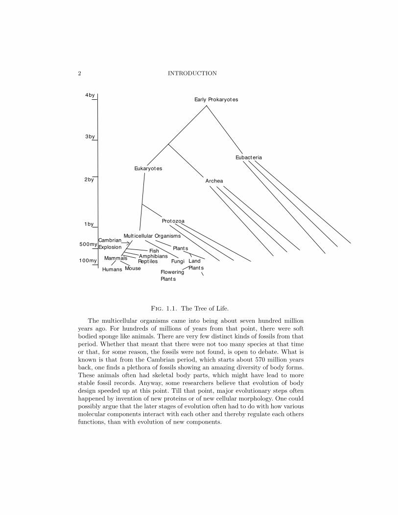
Fig. 1.1. The Tree of Life.
The multicellular organisms came into being about seven hundred millionyears ago. For hundreds of millions of years from that point, there were softbodied sponge like animals. There are very few distinct kinds of fossils from thatperiod. Whether that meant that there were not too many species at that timeor that, for some reason, the fossils were not found, is open to debate. What isknown is that from the Cambrian period, which starts about 570 million yearsback, one finds a plethora of fossils showing an amazing diversity of body forms.These animals often had skeletal body parts, which might have lead to morestable fossil records. Anyway, some researchers believe that evolution of bodydesign speeded up at this point. Till that point, major evolutionary steps oftenhappened by invention of new proteins or of new cellular morphology. One couldpossibly argue that the later stages of evolution often had to do with how variousmolecular components interact with each other and thereby regulate each othersfunctions, than with evolution of new components.

STRUCTURE OF A CELL 3
Bacillusmegat erium
Candidaalbicans
MouseL-cell
Fig. 1.2. Prokaryotic vs Eukaryotic Cell (courtesy Dr. Kaiser and Dr. Car-bonne).
At the end of the Cambrian explosion, as that enormous diversification iscalled, almost all the di↵erent kinds of body plans that we see today have alreadybeen invented. Some have called this process biology’s big bang. In the next fourhundred million years saw the arrival of fish, reptiles, mammals and birds. Italso saw the evolution of land plants which later gave rise to flowering plants.We would be able to relate to the world a hundred million years ago. Hominids(human like apes) are recent phenomenon: 20-25 million years old. We humanshave been around for only one and half million years. If life on Earth was anhour long movie, human existence would occupy a blink of an eye.
Much of the story described so far is from fossil record combined with geo-logical inferences. However we could also get a record of the history from the lifeforms present today, by looking at their relations at a microscopic and ultimatelymolecular level. For that we need to understand what is in our cells.
4 INTRODUCTION
Type Prokaryote EukaryoteSize 0.1� 10µm 10� 100µm
Genome Single circular DNA Multiple chromosomesOrganelles None Mitochondria and others
Metabolism Many strategies Mostly oxidativeInternal membranes None Complex folded ER
Motility Flagella Complex undulipodiumTable 1.1 Some di↵erences between prokaryotic cells and eukaryotic cells, otherthan existence of a nuclear envelope.
1.2 Structure of a Cell
The basic unit of all living beings are cells 1. Cells are enclosed within cellwalls. The figure shows basic components of prokaryotic and eukaryotic cells andcontrast their structures. In prokaryotes, there is no compartmentalization of thebasic processes making proteins from DNA. Eukaryotes distinguish themselvesby having a nuclear membrane which encloses the genetic material, DNA. RNA, amolecule that copies information about genes, from DNA, carries the instructionsout of nucleus into a rather convoluted surface called the endoplasmic reticulum(ER), where proteins are made. Eukaryotes also have many additional organelles,some of which (like mitochondria and chloroplasts, for plants) come with theirown genetic material. It is believed that such components may have come fromfree living prokaryotes at one point of time.
1.3 Biopolymers: DNA, RNA and Proteins
The nucleic acids, DNA and RNA, and the polypeptide chains making proteinsare biopolymers of fundamental importance. Without some basic familiarity withthem, and understanding of the relationships between these polymers, it is im-possible to appreciate why modern molecular biology is so powerful.
1.3.1 The Nucleic Acids
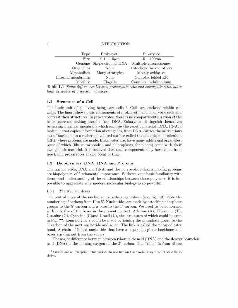
The central piece of the nucleic acids is the sugar ribose (see Fig. 1.3). Note thenumbering of carbons from 1’ to 5’. Nucleotides are made by attaching phosphategroups to the 5’ carbon and a base to the 1’ carbon. We need to be concernedwith only five of the bases in the present context: Adenine (A), Thyamine (T),Guanine (G), Cytosine (C)and Uracil (U), the structures of which could be seenin Fig. ??. Long polymers could be made by joining the phosphate group to the3’ carbon of the next nucleotide and so on. The link is called the phospodiesterbond. A chain of linked nucleotide thus have a sugar phosphate backbone andbases sticking out from the sugars.
The major di↵erence between between ribonucleic acid (RNA) and the deoxyribonucleicacid (DNA) is the missing oxygen at the 3’ carbon. The “ribo-” is from ribose
1Viruses are an exception. But viruses do not live on their own. They need other cells tothrive.
BIOPOLYMERS: DNA, RNA AND PROTEINS 5
Fig. 1.3. Building blocks of nucleic acids.
and “nucleic” has got to do with being from the nucleus of cells. These are weakacids, the phosphate group being negatively charged having lost a proton. DNAutilizes the bases A,T, G and C.
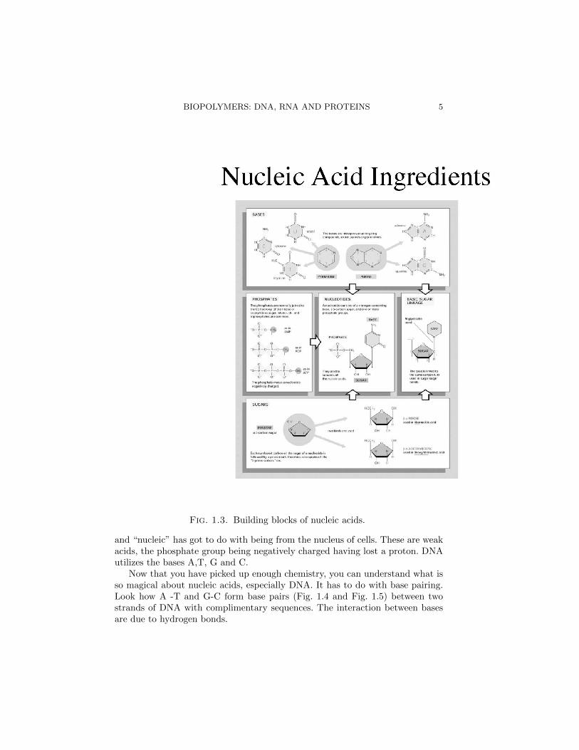
Now that you have picked up enough chemistry, you can understand what isso magical about nucleic acids, especially DNA. It has to do with base pairing.Look how A -T and G-C form base pairs (Fig. 1.4 and Fig. 1.5) between twostrands of DNA with complimentary sequences. The interaction between basesare due to hydrogen bonds.

6 INTRODUCTION
This is a good place for an aside on hydrogen bonds, which play an extremelyimportant role in molecular biology. A hydrogen bond is a kind of attractiveintermolecular force between partial electric charges on a hydrogen atom anda strongly electronegative atom (like oxygen, nitrogen or fluorine). Hydrogenbonds are quite strong but are much weaker than covalent bonds or full strengthionic bonds. They are strong enough to provide stability to a molecular structurebut weak enough to be broken when a biological function demands structuralchanges.
As these base pairs are formed, sugar phosphate backbones twirl around eachother making this pretty staircase. DNA is, universally, the repository of geneticinformation across biological species2. One of the important things about thedouble helix structure is that it immediately lead to a hypothesis about howspecificity of base pairing could be utilized for copying genetic information. Wewill discuss that when we come to replication in the next section.
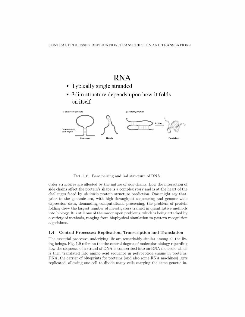
RNA has, another base, uracil (U) instead of thyamine (T), which base pairswith A. RNA is more often found in the single stranded form. As a result, itcould form base pairs with itself, making complex three dimensional structuresas in Fig. 1.6. Such structure is important for RNA molecules, like t-RNA, whichhave special functions.
RNA plays the role of the messenger in the flow of genetic information (seethe coming sections on transcription and translation) as well as form moleculeswith enzymatic activity. Enzymes are biomolecules ( in most cases proteins)that speed up certain biochemical reactions. In other words, they play the role ofcatalysts. Some hypothesize that in the earliest form of life, RNA was the geneticmaterial as well as the constituent of the enzymes encoded in the genes. Whetherthis hypothesis, known as the “RNA” world, is true or not, recently there hasbeen surge of interest in additional role RNA plays in the genetic networks. Theregulatory role played by small RNA pieces is beginning to be explored andhas already been used as a very powerful molecular tool for perturbing geneexpression patterns.
1.3.2 Proteins
Much as we have come to appreciate the di↵erent roles of RNA, past and present,the fact remains that the proteins are the workhorses of present day cells. Pro-teins are made of amino acids, which get linked by peptide bond formation,shown in fig. 1.7. Individual polymers, made this way by stringing of aminoacids, are known as polypeptide chains. A single protein molecule might consistof one or more of these chains.
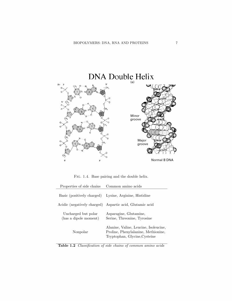
There are 20 amino acids (out of about 80, found in nature) which make upalmost all proteins. these amino acids di↵er from each other in their side chains.The biologically relevant amino acids are often classified based on the propertiesof the side chains into basic, acidic, polar etc. See Table 1.2.
2Some viruses use RNA as genetic material. But then, as we mentioned before viruses arespecial, somewhere between the living and the dead.
BIOPOLYMERS: DNA, RNA AND PROTEINS 7
Fig. 1.4. Base pairing and the double helix.
Properties of side chains Common amino acids
Basic (positively charged) Lysine, Arginine, Histidine
Acidic (negatively charged) Aspartic acid, Glutamic acid
Uncharged but polar Asparagine, Glutamine,(has a dipole moment) Serine, Threonine, Tyrosine
Alanine, Valine, Leucine, Isoleucine,Nonpolar Proline, Phenylalanine, Methionine,
Tryptophan, Glycine,Cysteine
Table 1.2 Classification of side chains of common amino acids

8 INTRODUCTION
Fig. 1.5. 3-d structure of the double helix.
Proper functioning of the protein depends upon its three dimensional struc-ture, and therefore, folding of the polypeptide into the right structure is ex-tremely important. For most proteins, the ultimate structure is determined bythe precise sequence of amino acids of individual polypeptide chains. This se-quence, that is determined genetically, as we will see, is often referred to as theprimary structure.
The next level of structure refers to certain repeating themes found in manyproteins. Secondary structure, like alpha helix or beta sheet, is formed whenthe positively charged hydrogen from the amine group is brought close to thenegatively charged oxygen on the carboxyl group. This is one more example ofhydrogen bonds playing an important role in biomolecular structure. Furtherhigher level organization, namely, tertiary structures has to do with how thesehelices, sheets and unstructured loops interact with each other to form a stablestructure for a polypeptide chain. How multiple polypeptide chains weave to-gether in a protein often goes by the name of quaternary structure. These higher
CENTRAL PROCESSES: REPLICATION, TRANSCRIPTION AND TRANSLATION9
Fig. 1.6. Base pairing and 3-d structure of RNA.
order structures are a↵ected by the nature of side chains. How the interaction ofside chains a↵ect the protein’s shape is a complex story and is at the heart of thechallenges faced by ab initio protein structure prediction. One might say that,prior to the genomic era, with high-throughput sequencing and genome-wideexpression data, demanding computational processing, the problem of proteinfolding drew the largest number of investigators trained in quantitative methodsinto biology. It is still one of the major open problems, which is being attacked bya variety of methods, ranging from biophysical simulation to pattern recognitionalgorithms.
1.4 Central Processes: Replication, Transcription and Translation
The essential processes underlying life are remarkably similar among all the liv-ing beings. Fig. 1.9 refers to the the central dogma of molecular biology regardinghow the sequence of a strand of DNA is transcribed into an RNA molecule whichis then translated into amino acid sequence in polypeptide chains in proteins.DNA, the carrier of blueprints for proteins (and also some RNA machines), getsreplicated, allowing one cell to divide many cells carrying the same genetic in-

10 INTRODUCTION
Fig. 1.7. Building proteins from amino acids.
formation. We have already discussed the biomolecules involved. Now we discussthese basic processes, replication, transcription and translation, and the machin-ery involved in carrying them out.
1.4.1 Replication
One of the the essential features of life is cell division and each cell receiving areliable copy of the genetic information coded in DNA (more about how it iscoded in the subsection to come). The celebrated understatement, “It has notescaped our attention that the specific pairing we have postulated immediatelysuggests a possible copying mechanism for the genetic material.” , made in thepaper that elucidated the molecular structure of DNA ( J.D. Watson and F.H.C.Crick. Molecular structure of nucleic acids: a structure for deoxyribose nucleicacid. Nature, 171:737–738, 1953.), relied on the following observation. The two

CENTRAL PROCESSES: REPLICATION, TRANSCRIPTION AND TRANSLATION11
Structural Motifs in Proteins
a helix
b sheet
Black, Grey: Carbon
Blue: Nitrogen
Red: Oxygen
Fig. 1.8. The alpha helix and the beta sheet.
strands of DNA were held together by hydrogen bonds of the base pairing. Sincehydrogen bonds are much weaker than covalent bonds, the two strands couldbe easily made to come apart, allowing each to be used as a template for fur-ther synthesis of two double stranded DNA molecules. This would be a possiblemechanism of DNA replication, the process by which double stranded DNA gen-erates a copy of itself. This picture turned out to be essentially correct, withsome interesting twists.
The simplest model would be that DNA opens up and replication happenson both strands continouously (Fig. 1.10). Cosidering the model carefully, onenotices that it requires one strand to be synthesized in the 5’ to 3’ directionwhereas the other to be in the 3’ to 5’ direction. This would require two kinds ofpolymerase (polymerases are proteins that catalyzes the formation of polymers,like single stranded nucleic acids, from monomers, like nucleotides) actions. After

12 INTRODUCTION
Fig. 1.9. Central biological processes.
5’3’
3’ 5’ 3’ 5’
5’ 3’
Fig. 1.10. Continuous replication model.
the discovery of the DNA polymerase, it became clear that the enzyme onlysynthesizes DNA in the 5’ to 3’ direction. No one has found a DNA polymerasethat makes DNA in the 3’ to 5’ orientation. As resolution of the puzzle, ReijiOkazaki suggested that the DNA replication might happen in a discontinuousfashion. Today we know that the process is semi-discontinuous (Fig. 1.11). In

CENTRAL PROCESSES: REPLICATION, TRANSCRIPTION AND TRANSLATION13
5’3’
3’ 5’ 3’ 5’
5’ 3’
Fig. 1.11. Semi-discontinuous replication model and Okazaki fragments.
fact, it is quite complicated. Enzymes called primases make short RNA fragmentshybridized to DNA to act as primers (starting points) for the DNA polymerase tocome along and make the so-called Okazaki fragments. Finally the RNA parts areexcised and replaced by DNA and then another enzyme called DNA ligase joins(“ligates” , in technical terms) all these discontinuous DNA fragments. Quiteremarkably, all this happens very fast: at a rate of about 1000 base pairs/second.This speed is good enough to replicate prokaryotic genomes with few millionbases in less than an hour. Prokaryotes therefore usually have only one locationon their DNA from which an open replication bubble spreads. This locationcalled the origin of replication. Eukaryotic genomes are much bigger and henceneed multiple origins of replication. Another di↵erence between prokaryotes andeukaryotes is that the fidelity of replication. Prokaryotes have an error rate of10�6 � 10�7 per base per replication. In eukaryotes, it is about 10�9 per baseper replication, thanks to additional proof-reading mechanisms.
1.4.2 Transcription
Transcription is the process by which RNA is made from DNA. The key playerin this process is another polymerase, one that makes RNA instead of DNA.RNA polymerase copies a the information on a stretch of DNA, usually a fewthousand bases, into a piece of RNA. This process is called transcription. Thetranscribed stretch of DNA, along with regions around it important for control-ling transcription rates, l is the basic unit of hereditary material, often referredto as a gene. To start the process of transcription, the RNA polymerase has to

14 INTRODUCTION
Non-template strand
5’ 3’
3’ 3’ 5’ RNA5’ Template strand
Fig. 1.12. Transcription.
bind upstream (to the 5’ end) of the transcribed sequence (regarded as runningfrom 5’ to 3’ end of the non-template strand, see Fig. 1.12) in DNA. Much ofgenetic regulation happens through controlling this step. Once the polymerasebinds, it forms the so-called open complex, by opening a DNA bubble. It thengoes ahead making RNA, using one of the two strands as a template, as shown inthe same figure (Fig. 1.12). In this process, called elongation, the RNA is madein the 5’ to 3’ direction.
In prokaroytes like E. coli, the initial recognition happens because the coreenzyme (the essential parts of RNA polymerase) come in complex with a sigmafactor, which is a DNA binding protein. It recognizes certain sequences upstreamof the gene. This process can be regulated by additional DNA binding proteinsbound to neighboring sites. Some proteins interact with the polymerase andenhance its binding. Other regulatory proteins, often known as repressors, get inthe way of the initial RNA polymerase binding or a↵ects the process of elongationnegatively.
Transcription is far more complicated for eukaryotes. In eukaryotes, the DNAis packed around nucleosomes, which are complexes of proteins called histones.Nucleosomes are then packed into higher order structures, forming chromatin.DNA, tightly packed this way, does not bind proteins necessary for transcrip-tion. In many cases, there are regulatory DNA sequences, typically upstreamof the coding sequence, known as upstream activator sequences (UAS). Sucha sequence binds an activator, a regulatory protein, which in turn can recruit
CENTRAL PROCESSES: REPLICATION, TRANSCRIPTION AND TRANSLATION15
other proteins that could modify chromatin structure making it more permis-sive for transcription. This modification exposes sequences (like the so-calledTATA box, a sequence similar to TATAAAA) to which Transcription factor IIDor TFIID complex binds, beginning the recruitment of many other componentsfinally leading to transcription.
The speed with which RNA polymerase synthesizes RNA is an order of mag-nitude lower than the rate for DNA synthesis by DNA polymerase. In prokaryotesit is of the order of 30-85 bases/sec.
1.4.3 Translation
The information contained in RNA is converted to a polypeptide by the ribosome,an enzyme made of RNA itself and some proteins. The steps to protein synthesis,subsequent to making of the primary RNA transcript are di↵erent in prokaryptesand eukaryotes. In prokaryotes, the ribosomes come attach themselves to specificribosome binding sequences on the nascent RNA (Fig. 1.13(a)). Prokaryotic RNAoften encodes, in tandem, for multiple proteins, with the ribosome binding sites,also called the Shine Dalgarno sequences, between the regions coding for di↵erentproteins. Eukaryotic primary transcript goes through far more processing (Fig.1.13(b)). The RNA gets capped on the 5’ end by a methylated G. This cap isfinally what the eukaryotic ribosomes recognize. The regions in RNA which doesnot code for proteins, the introns, are spliced out. A poly-A tail is added tothe 3’ end. The resulting messenger RNA (mRNA) is exported out of nucleus.The mRNA finally arrives at the endoplasmic retiuculum (ER) where it meetsribosomes.
The process of making a polypeptide, once the ribosome is bound to themRNA, is very similar across all living beings. The translation apparatus movesfrom the 5’ to the 3’ end. It starts from an AUG, coding for Methionine. RNA isread in non overlapping triplets coding for di↵erent amino acids. It goes on till itrecognizes any of the three triplets that code for a stop (UAA, UAG and UGA).The recognition happens by transfer RNAs (t-RNA) for each amino acid havinga triplet complimentary to the code. The t-RNA comes covalently bonded withthe amino acid, binds to mRNA at the right place and o↵ers the amino acid forjoining to the polypeptide chain. The process is indicated in Fig. 1.14.
The rule book of triplets to amino acids (Fig. 1.15) is nearly universal, witha few exceptions, like for mitochondrial genes. Universality of the genetic code,once more, indicate the common origin of life. Whether there is any deep reasonwhy the code is this way or whether this is just “a frozen accident” (as FrancisCrick puts it) we don’t know.
We have studied how information on DNA is converted into to a proteinmolecule. Scientifically, it was a remarkable achievement to have solved the es-sential aspects of this process within two decades, the 1950s and the 1960s. Itturns out that the whole process is heavily regulated. Nature has used any knobit could find to tweak the rate of protein synthesis adaptively. We will come backto this aspect when we study genetic networks.

16 INTRODUCTION
Fig. 1.13. From transcription to translation.
Fig. 1.14. Translation.

CENTRAL PROCESSES: REPLICATION, TRANSCRIPTION AND TRANSLATION17
Fig. 1.15. The genetic code.