introduction to anatomy contents :. anatomical position. anatomical terms.. anatomical planes.....
Post on 19-Dec-2015
261 views
TRANSCRIPT
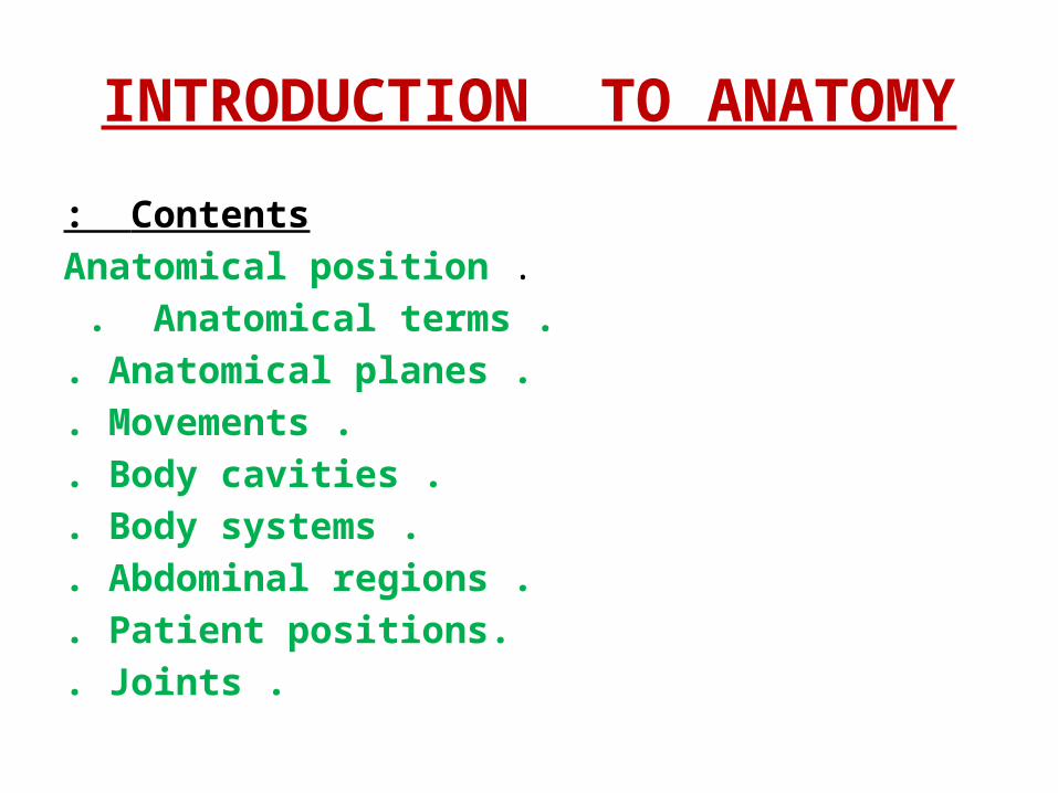
INTRODUCTION TO ANATOMYContents:
.Anatomical position .Anatomical terms . .Anatomical planes.
.Movements. .Body cavities. .Body systems.
.Abdominal regions. .Patient positions.
.Joints.
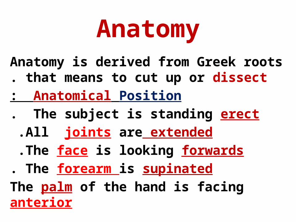
AnatomyAnatomy is derived from Greek roots that means to
cut up or dissect .Anatomical Position: The subject is standing erect.
All joints are extended .The face is looking forwards .The forearm is supinated .
The palm of the hand is facing anterior
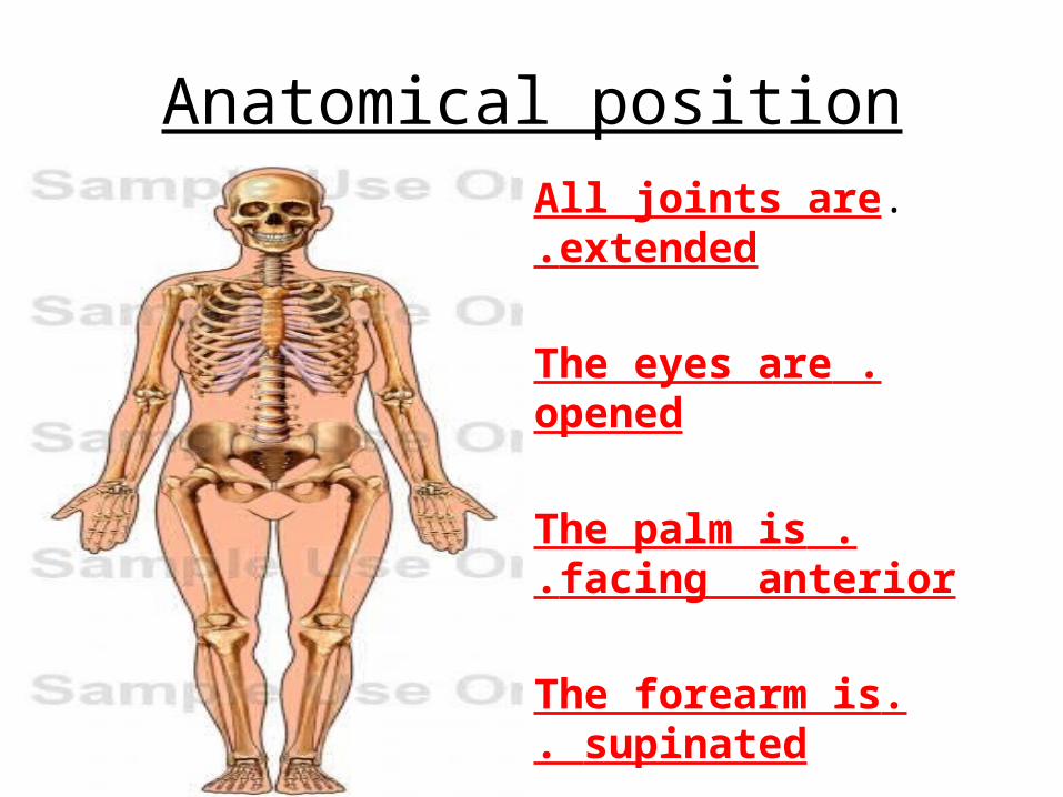
Anatomical position.All joints are extended.
.The eyes are opened
.The palm is facing anterior.
.The forearm is supinated.
.The face is forwards
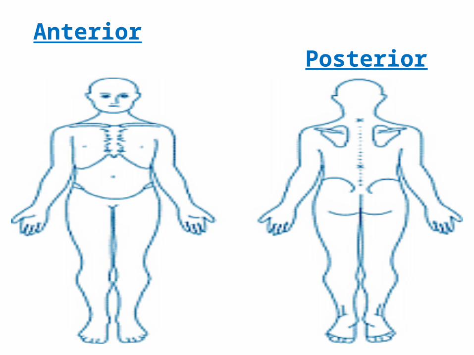
Anterior Posterior
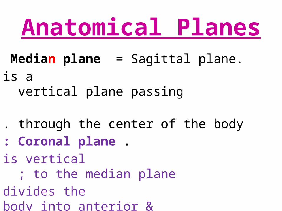
Anatomical Planes.Median plane = Sagittal plane
is a vertical plane passing through the center of the body.
.Coronal plane: is vertical to the median plane ;
divides the body into anterior & posterior .Transverse plane : divides the body into
upper & lower parts .It is at right angle
to sagittal & coronal planes.
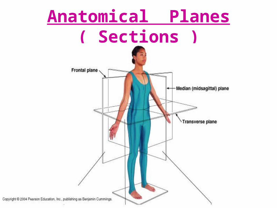
Anatomical Planes ( Sections )
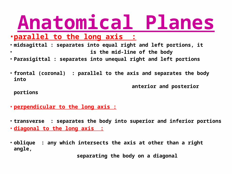
Anatomical Planes• parallel to the long axis :• midsagittal : separates into equal right and left portions, it • is the mid-line of the body• Parasigittal : separates into unequal right and left portions
• frontal (coronal) : parallel to the axis and separates the body into anterior and posterior portions
• perpendicular to the long axis :
• transverse : separates the body into superior and inferior portions• diagonal to the long axis :
• oblique : any which intersects the axis at other than a right angle, separating the body on a diagonal
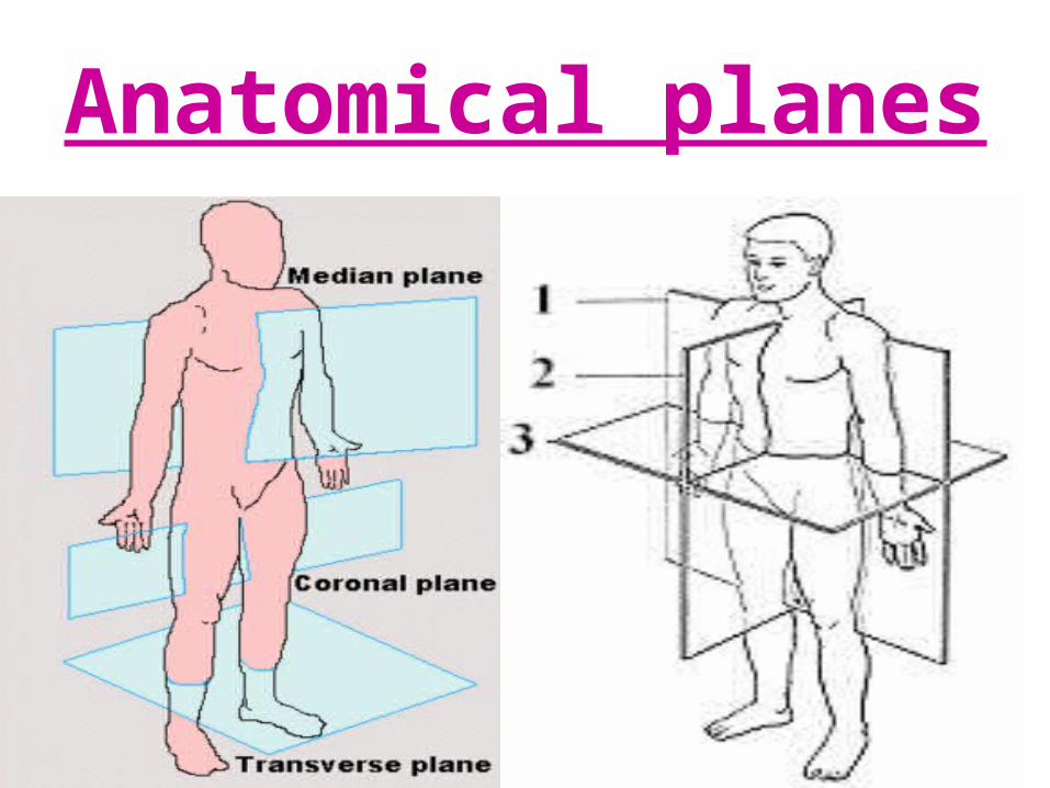
Anatomical planes
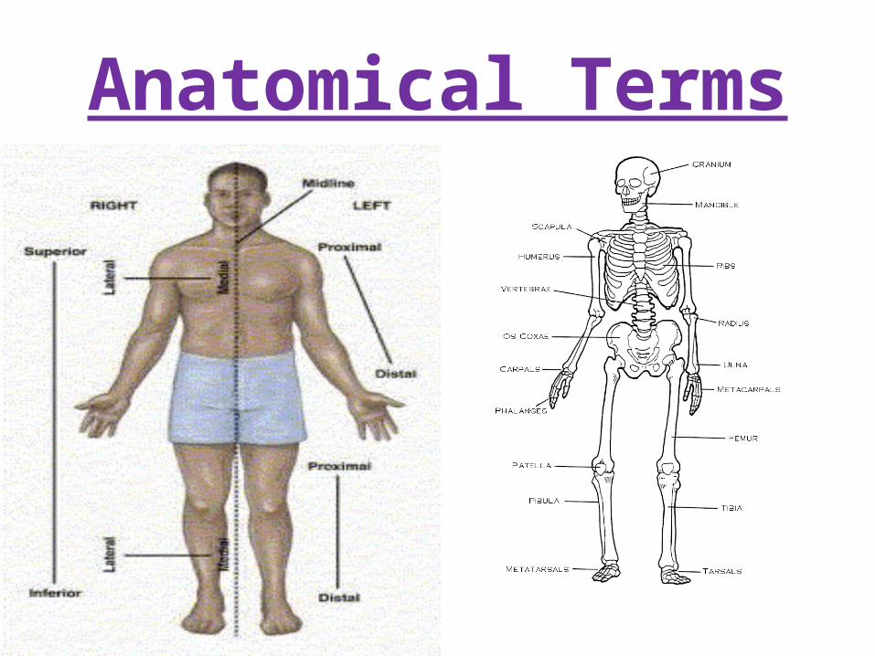
Anatomical Terms
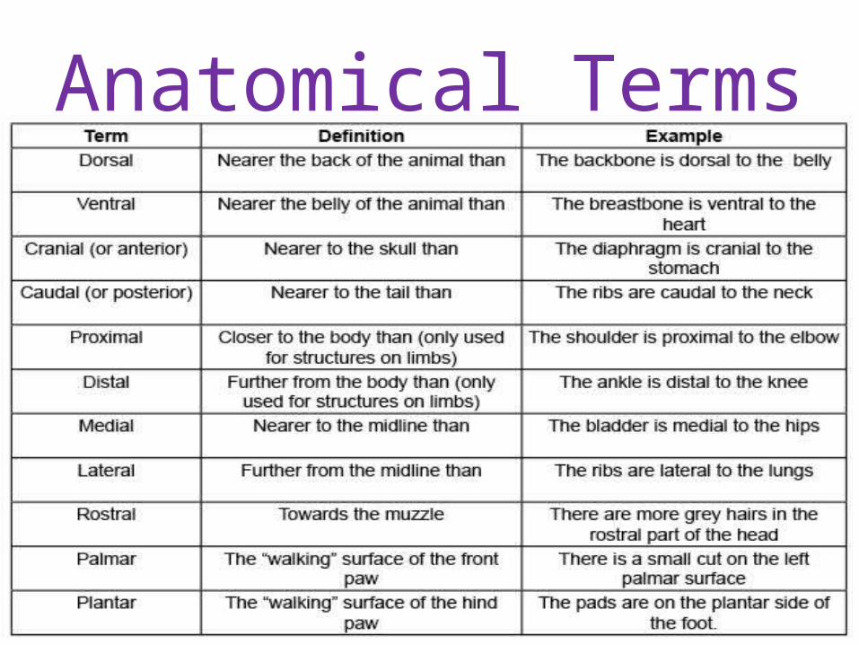
Anatomical Terms
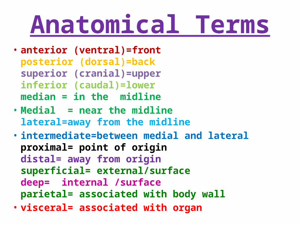
Anatomical Terms• anterior (ventral)=front
posterior (dorsal)=backsuperior (cranial)=upperinferior (caudal)=lowermedian = in the midline
• Medial = near the midlinelateral=away from the midline
• intermediate=between medial and lateralproximal= point of origindistal= away from originsuperficial= external/surfacedeep= internal /surfaceparietal= associated with body wall
• visceral= associated with organ
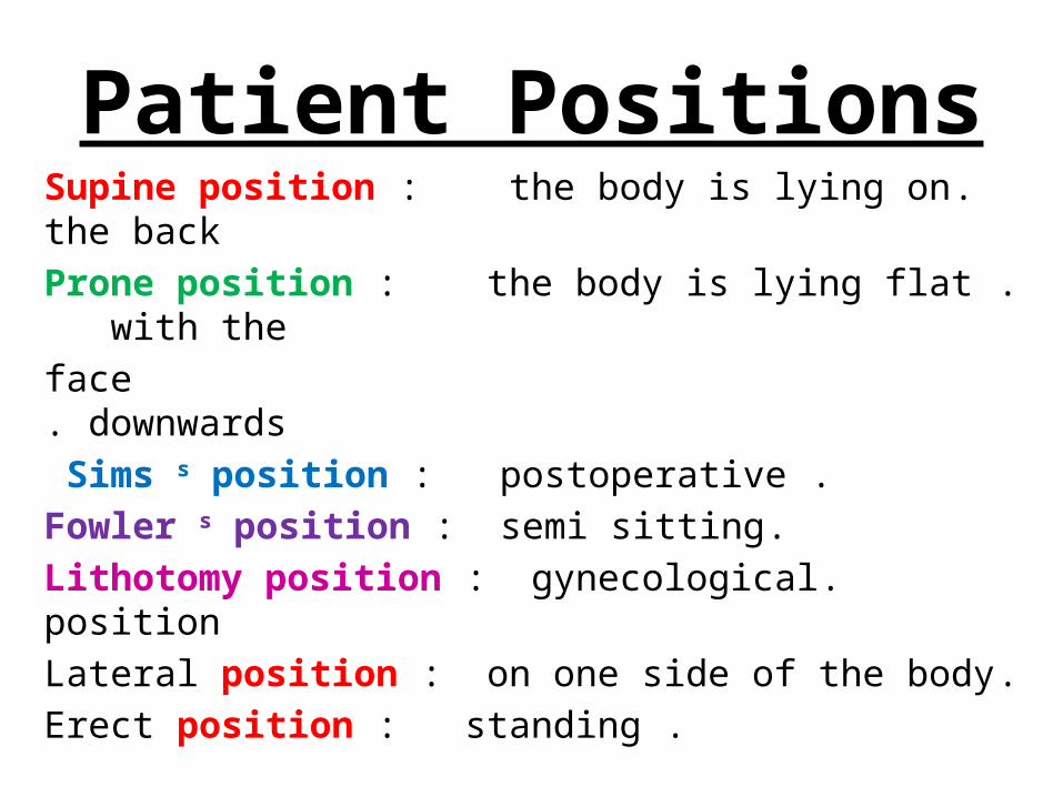
Patient Positions.Supine position : the body is lying on the back
.Prone position : the body is lying flat with the face downwards.
.Sims s position : postoperative .Fowler s position : semi sitting
.Lithotomy position : gynecological position.Lateral position : on one side of the body
.Erect position : standing
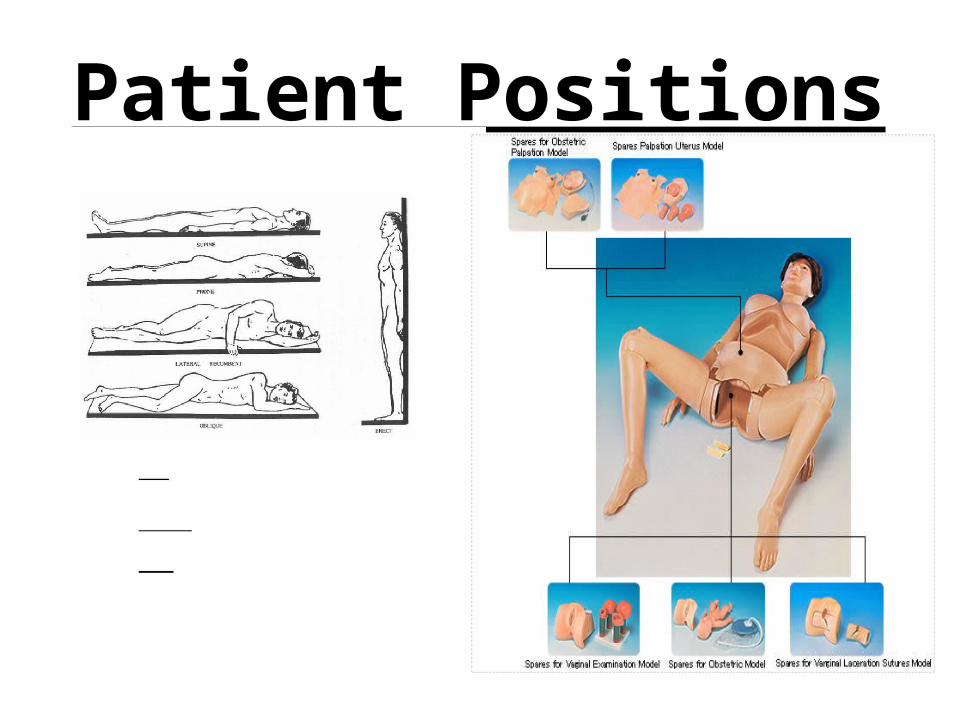
Patient Positions
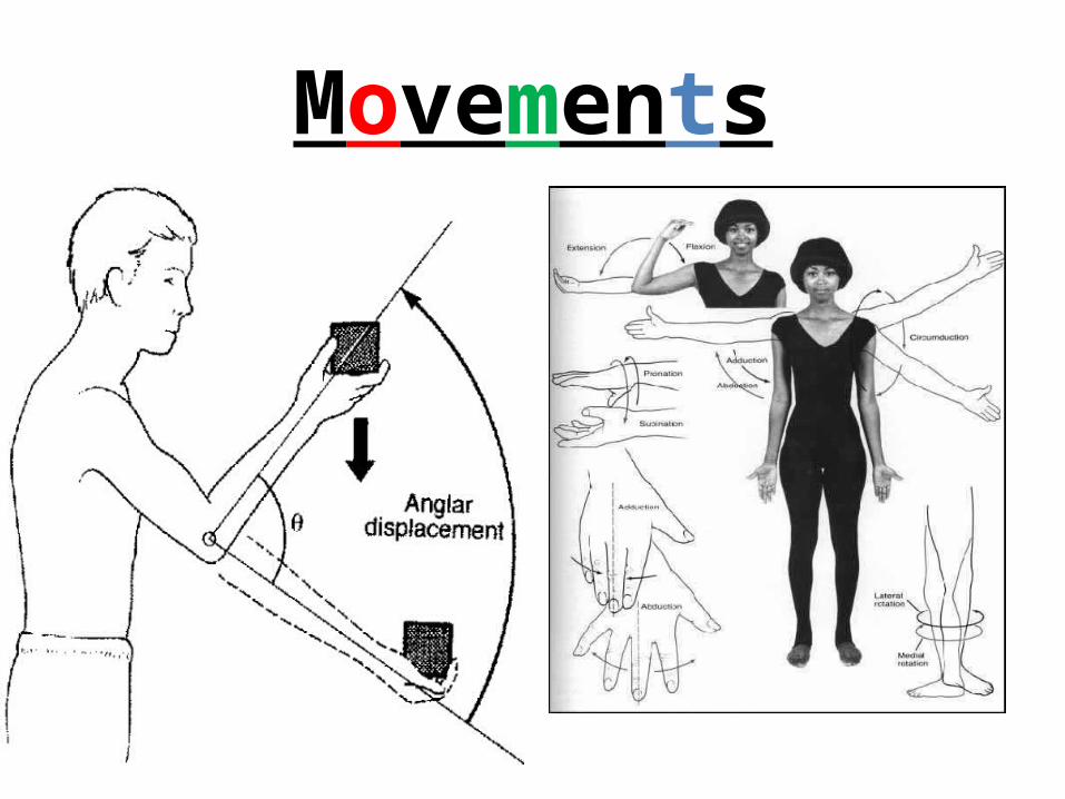
Movements
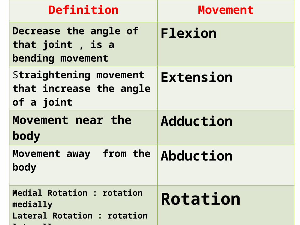
Definition Movement
Decrease the angle of that joint , is a bending movement
Flexion
Straightening movement that increase the angle of a joint
Extension
Movement near the body Adduction
Movement away from the body Abduction
Medial Rotation : rotation mediallyLateral Rotation : rotation laterally RotationSummation of flexion ,adduction ,extension ,abduction Circumduction
The ulna & radius are parallel The radius crosses over the ulna
Supination Pronation
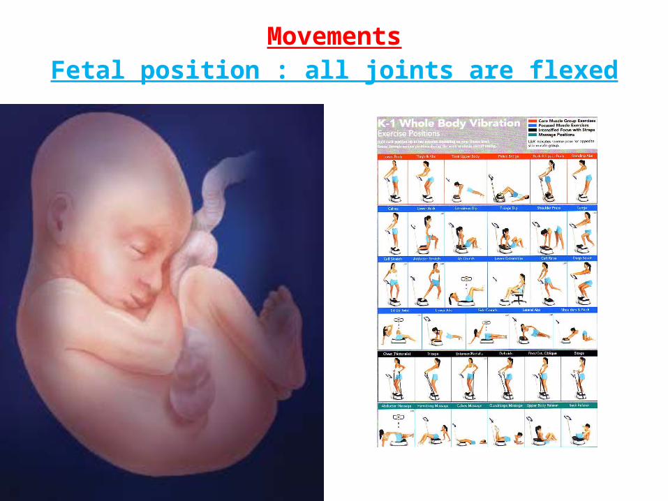
MovementsFetal position : all joints are flexed
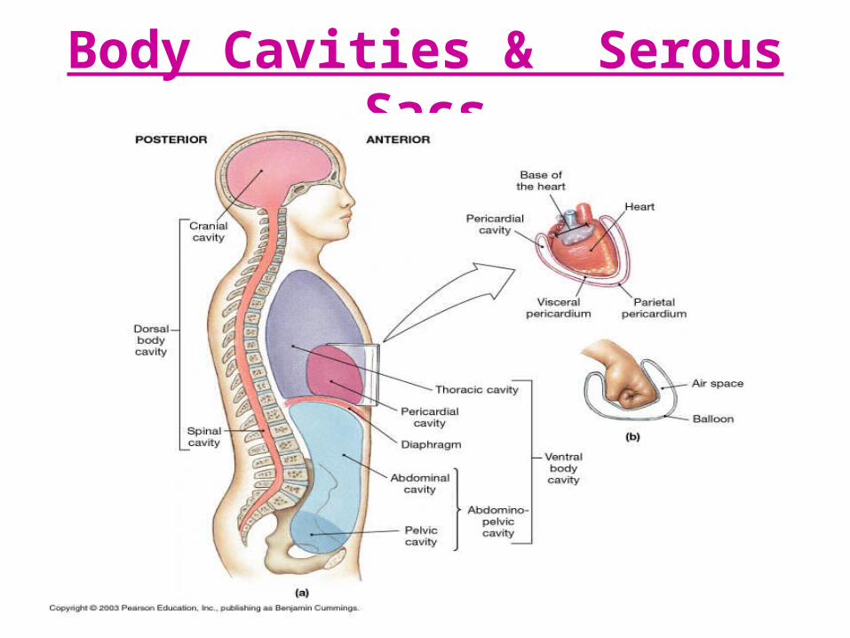
Body Cavities & Serous Sacs
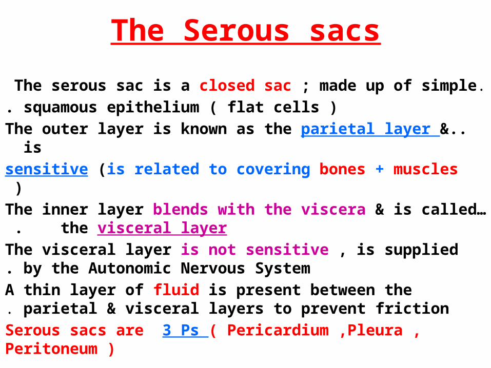
The Serous sacs
.The serous sac is a closed sac ; made up of simple squamous epithelium ( flat cells ).
..The outer layer is known as the parietal layer & is sensitive (is related to covering bones + muscles )
…The inner layer blends with the viscera & is called the visceral layer .
The visceral layer is not sensitive , is supplied by the Autonomic Nervous System.
A thin layer of fluid is present between the parietal & visceral layers to prevent friction.
Serous sacs are 3 Ps ( Pericardium ,Pleura , Peritoneum )
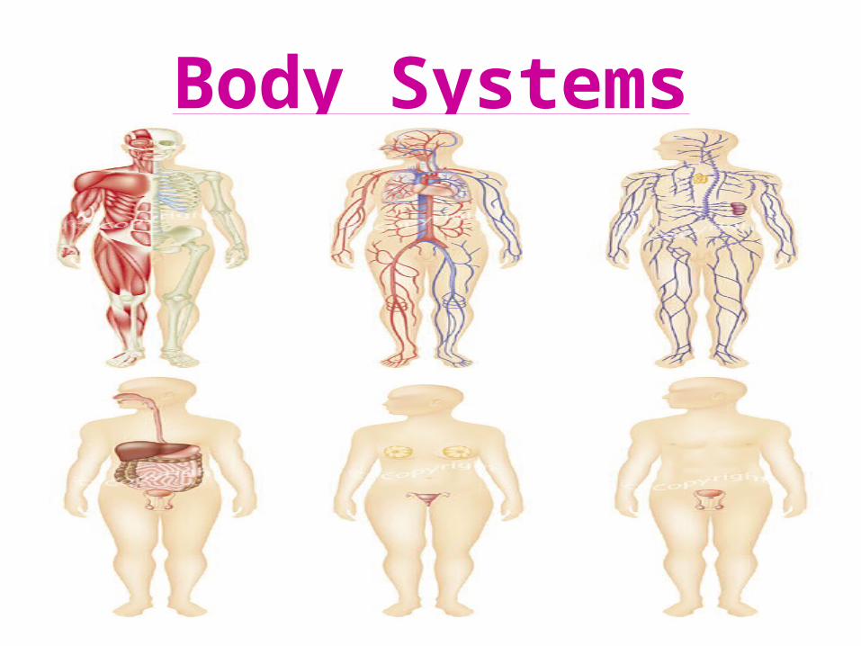
Body Systems
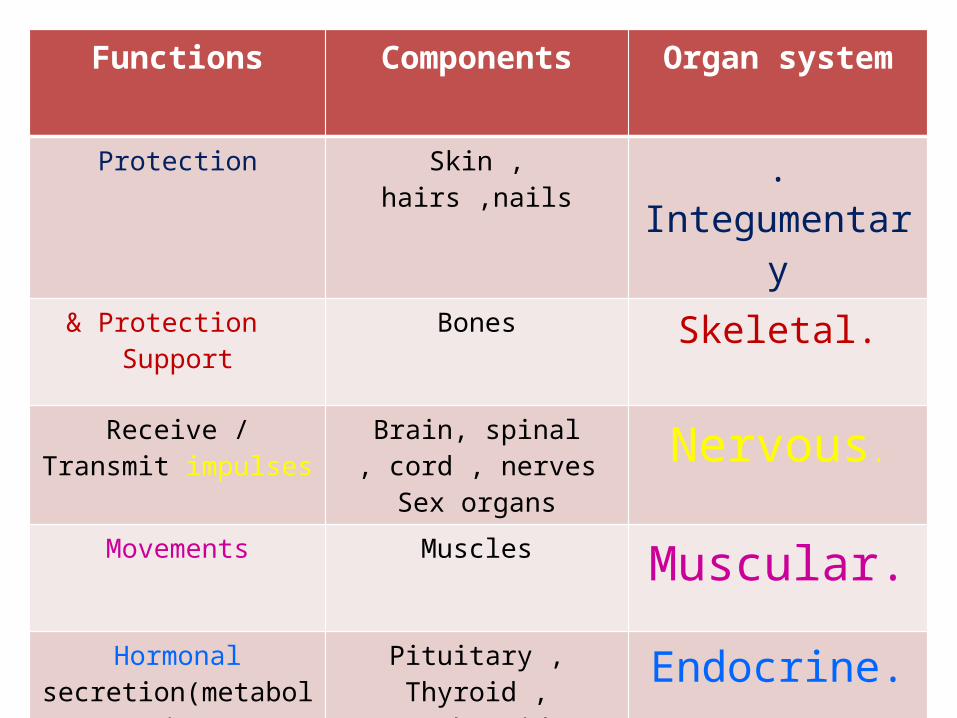
Functions Components Organ system
Protection Skin , hairs ,nails .Integumentary
Protection & Support
Bones .Skeletal
Receive / Transmit impulses
Brain, spinal cord , nerves ,
Sex organs
.Nervous
Movements Muscles .Muscular
Hormonal secretion(metabolism/
Homeostasis)
Pituitary , Thyroid , Parathyroid , Adrenal, Pancreas, ovary , testis
.Endocrine
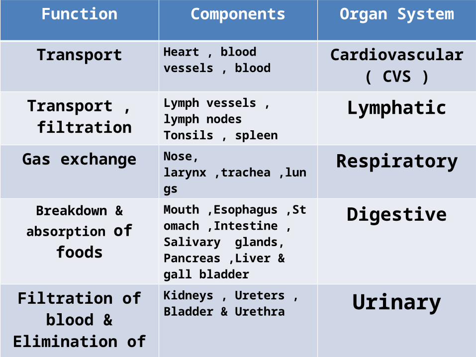
Function Components Organ System
Transport Heart , blood vessels , blood Cardiovascular( CVS )
Transport , filtration Lymph vessels , lymph nodesTonsils , spleen Lymphatic
Gas exchange Nose, larynx ,trachea ,lungs Respiratory
Breakdown & absorption of foods
Mouth ,Esophagus ,Stomach ,Intestine , Salivary glands, Pancreas ,Liver & gall bladder
Digestive
Filtration of blood & Elimination of wastes
Kidneys , Ureters , Bladder & Urethra Urinary
Propagation Gonads Reproduction
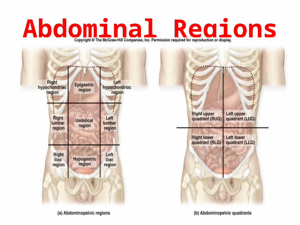
Abdominal Regions
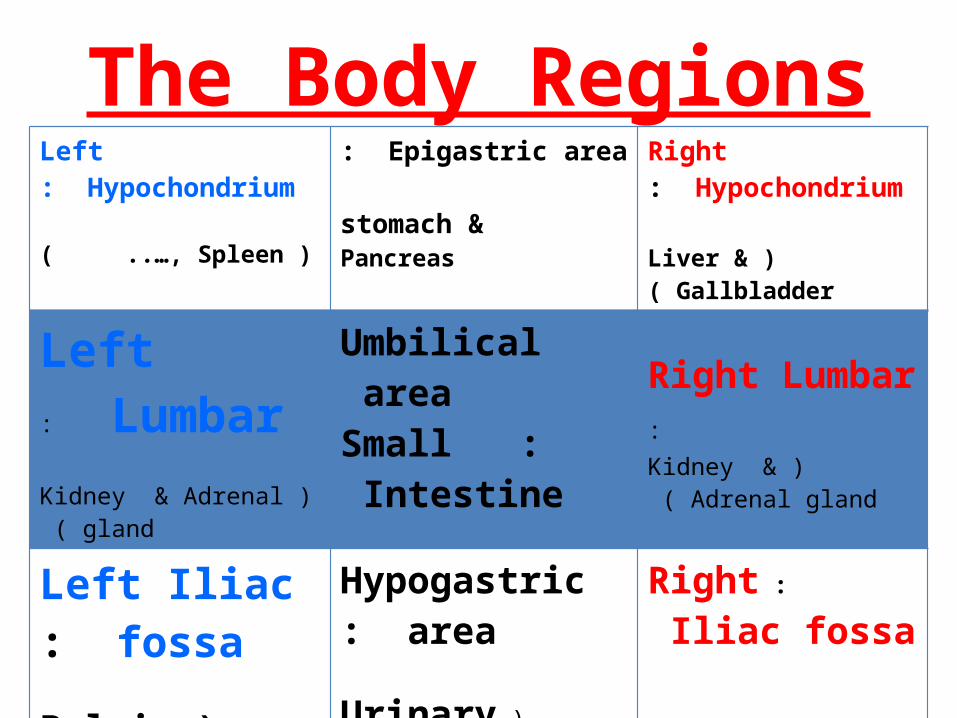
The Body Regions Left Hypochondrium:
(Spleen ) ..…,
Epigastric area:
stomach & Pancreas
Right Hypochondrium :
(Liver & Gallbladder)
Left Lumbar :
(Kidney & Adrenal gland )
Umbilical area :Small Intestine
Right Lumbar : ( Kidney & Adrenal gland )
Left Iliac fossa :
(Pelvic Colon , ………)
Hypogastric area:
(Urinary bladder, ..…………)
: Right Iliac fossa
Appendix, Cecum
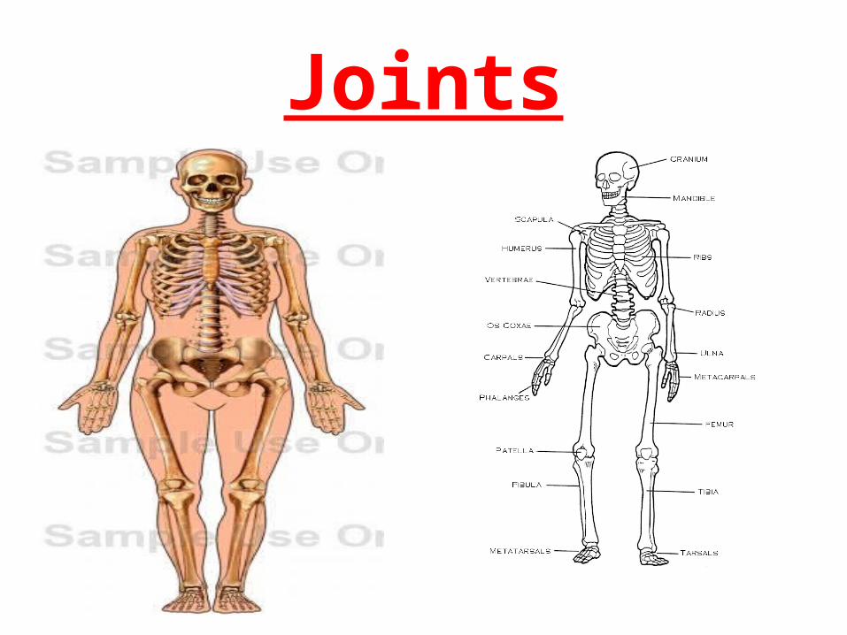
Joints
The JointsThe joint is the meeting of two or more bones.
According to the type & mode of connection 3 types of joints are known:
. Fibrous joint
. Cartilaginous joint
. Synovial joint
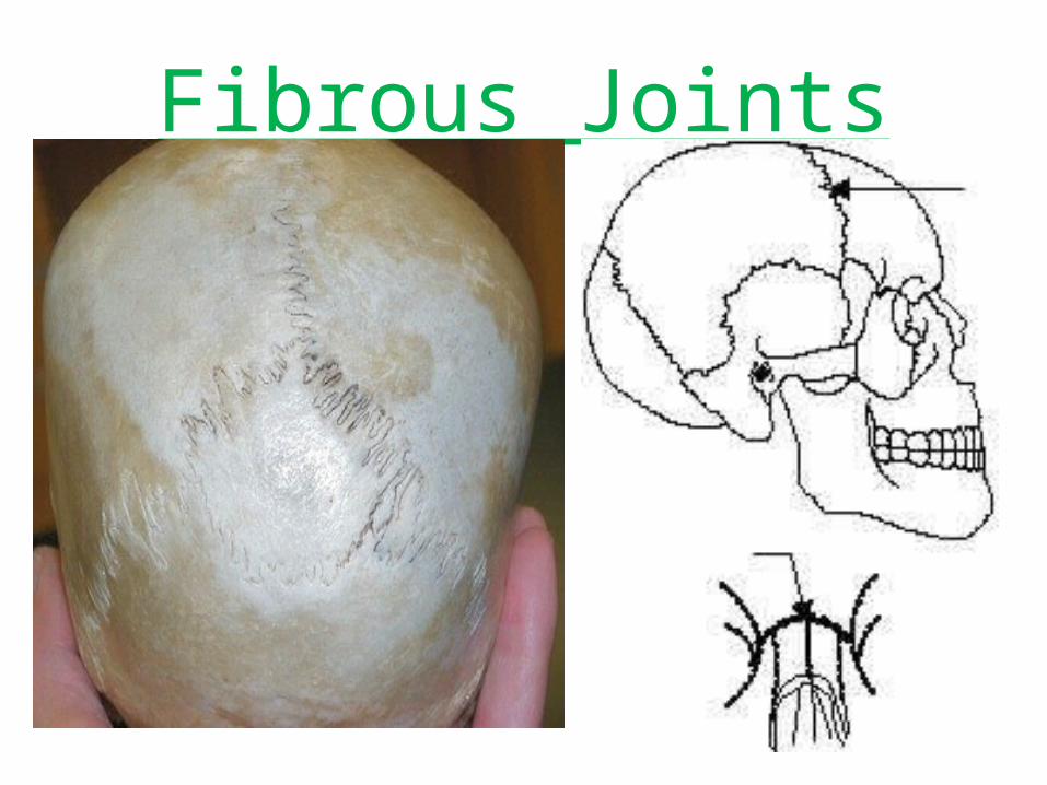
Fibrous Joints
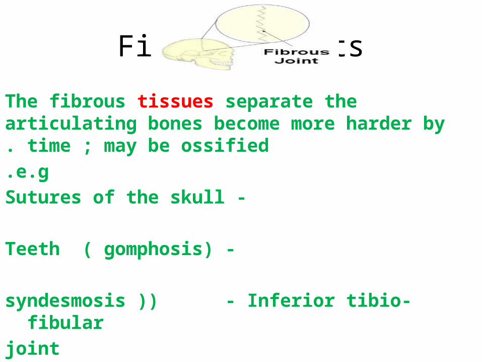
Fibrous Joints
The fibrous tissues separate the articulating bones become more harder by time ; may be ossified.
e.g. - Sutures of the skull
- Teeth ( gomphosis)
syndesmosis )) - Inferior tibio-fibular joint
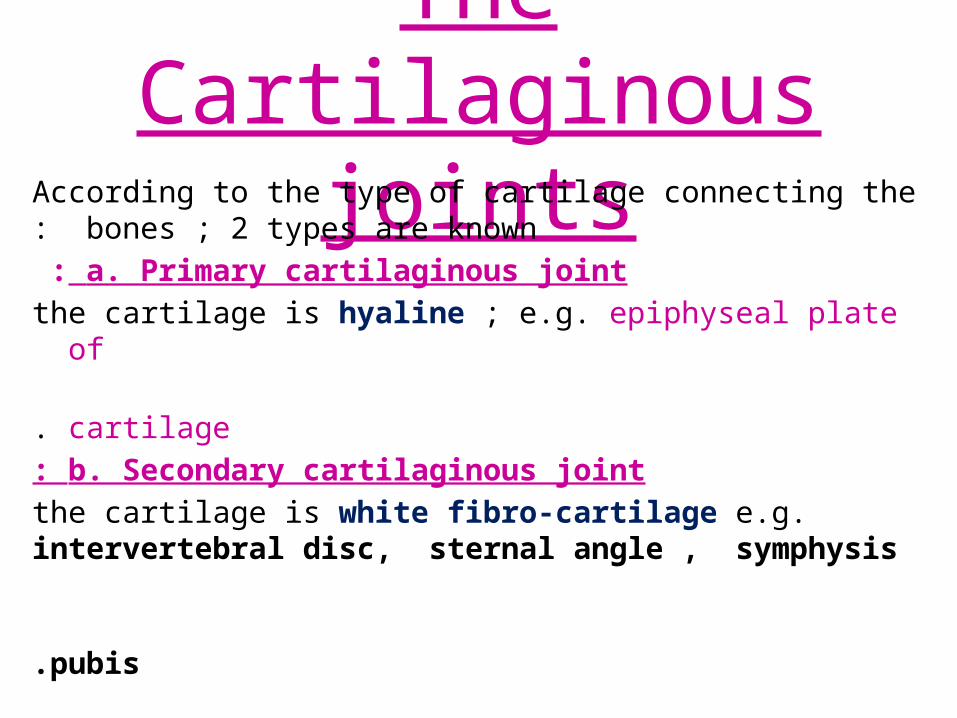
The Cartilaginous jointsAccording to the type of cartilage connecting the
bones ; 2 types are known: a. Primary cartilaginous joint :
the cartilage is hyaline ; e.g. epiphyseal plate of cartilage .
b. Secondary cartilaginous joint: the cartilage is white fibro-cartilage e.g.
intervertebral disc, sternal angle , symphysis pubis.
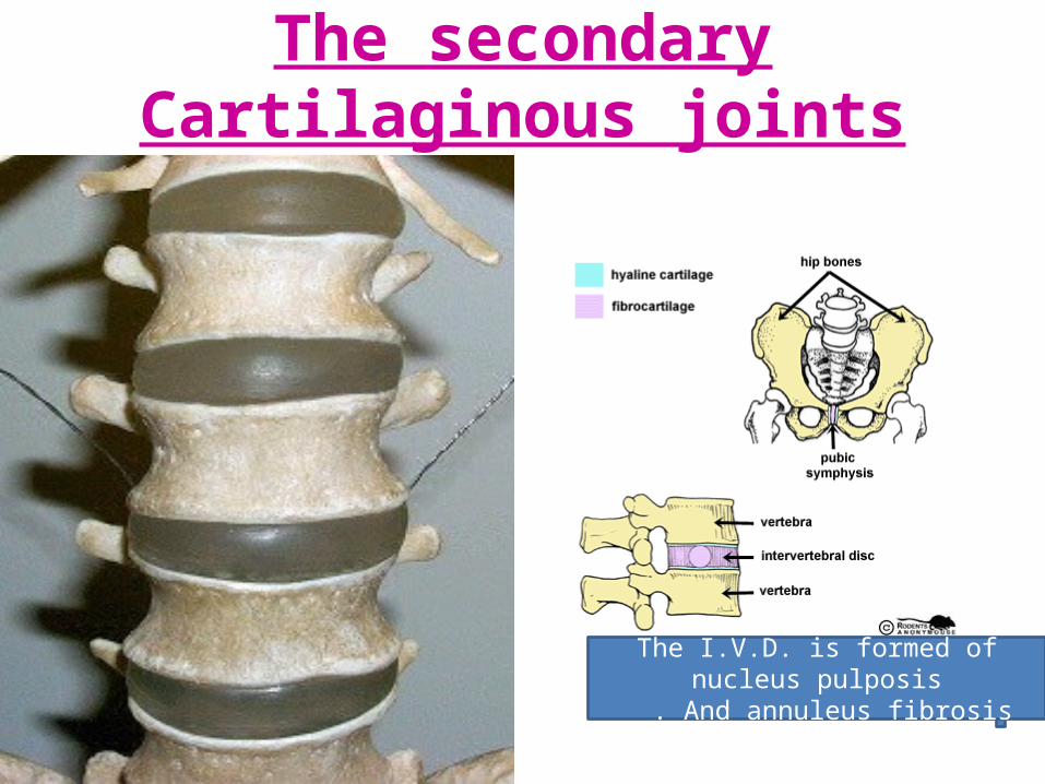
The secondary Cartilaginous joints
The I.V.D. is formed of nucleus pulposisAnd annuleus fibrosis .
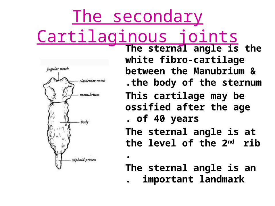
The secondary Cartilaginous jointsThe sternal angle is the white fibro-cartilage between the Manubrium & the body of the sternum.
This cartilage may be ossified after the age of 40 years.
The sternal angle is at the level of the 2nd rib.
The sternal angle is an important landmark.
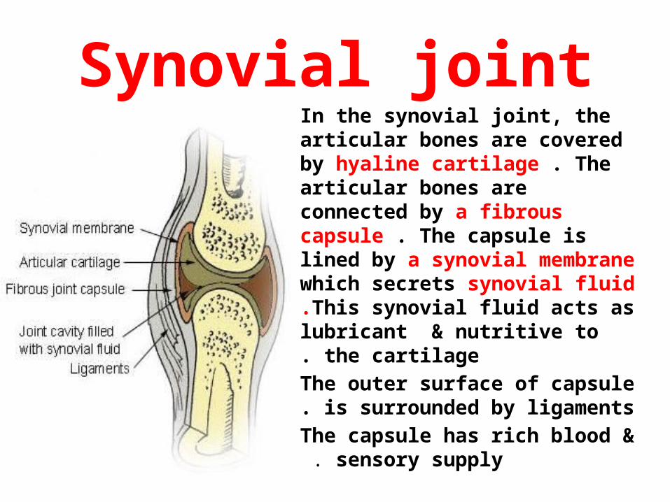
Synovial jointIn the synovial joint, the articular bones are covered by hyaline cartilage . The articular bones are connected by a fibrous capsule . The capsule is lined by a synovial membrane which secrets synovial fluid .This synovial fluid acts as lubricant
& nutritive to the cartilage. The outer surface of capsule is
surrounded by ligaments. The capsule has rich blood &
sensory supply .
Synovial joint
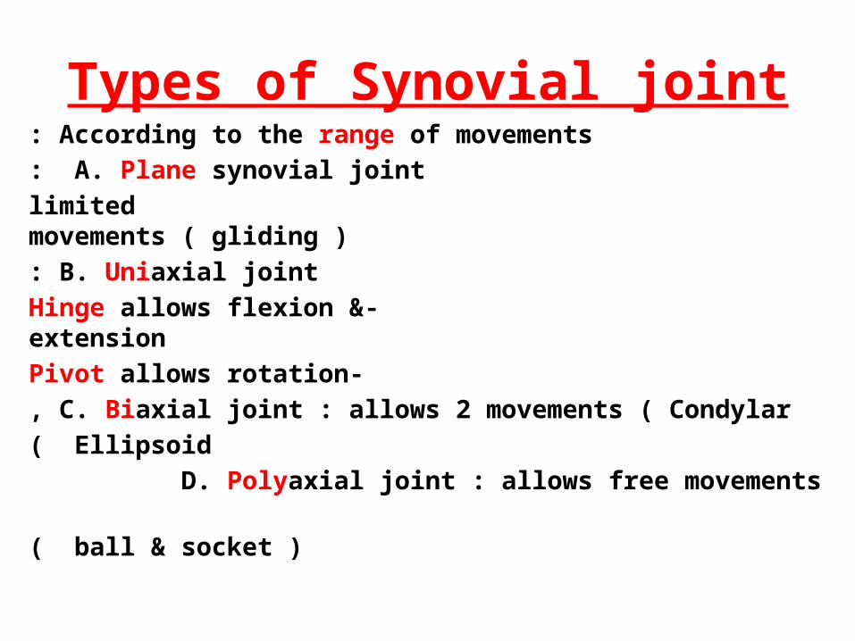
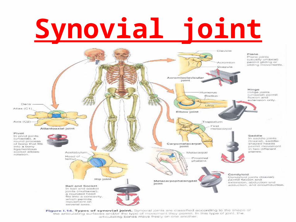
Types of Synovial jointAccording to the range of movements: A. Plane synovial joint:
limited movements ( gliding )B. Uniaxial joint:
- Hinge allows flexion & extension- Pivot allows rotation
C. Biaxial joint : allows 2 movements ( Condylar, Ellipsoid)
D. Polyaxial joint : allows free movements ( ball & socket)