introduction to electron microscopy andres kaech
TRANSCRIPT
24.04.2012
1
Introduction to
Electron Microscopy
Andres Kaech
Instrumentation and Image Formation
Center for Microscopy and Image Analysis
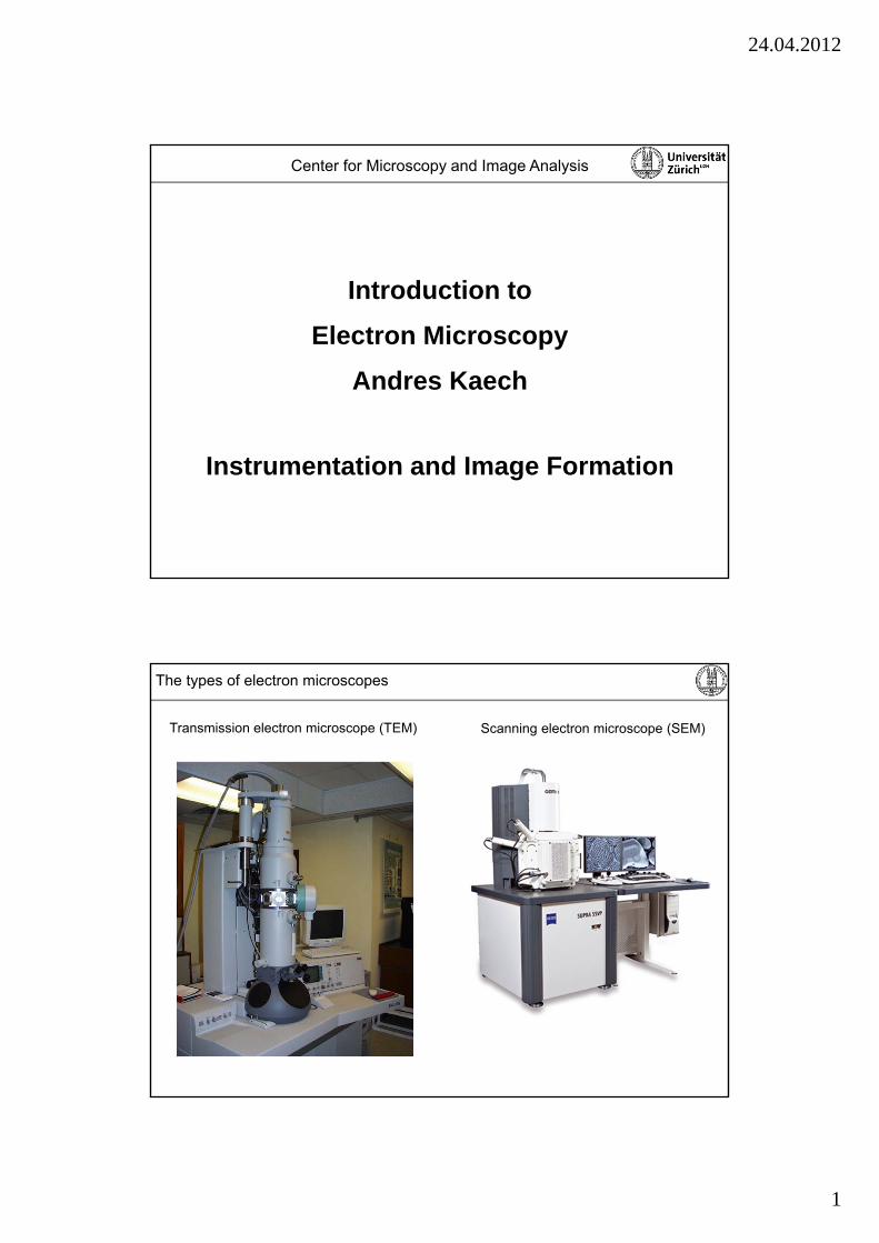
The types of electron microscopes
Scanning electron microscope (SEM)Transmission electron microscope (TEM)
24.04.2012
2
Scanning electron microscope (SEM)Transmission electron microscope (TEM)
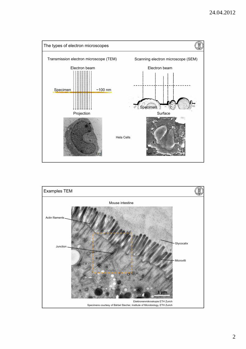
The types of electron microscopes
Electron beam
Specimen ~100 nm
Electron beam
Specimen
Projection Surface
Hela Cells
Elektronenmikroskopie ETH Zurich
1 µm
Specimens courtesy of Bärbel Stecher, Institute of Microbiology, ETH Zurich
Examples TEM
Mouse intestine
Microvilli
GlycocalixJunction
Actin filaments
24.04.2012
3
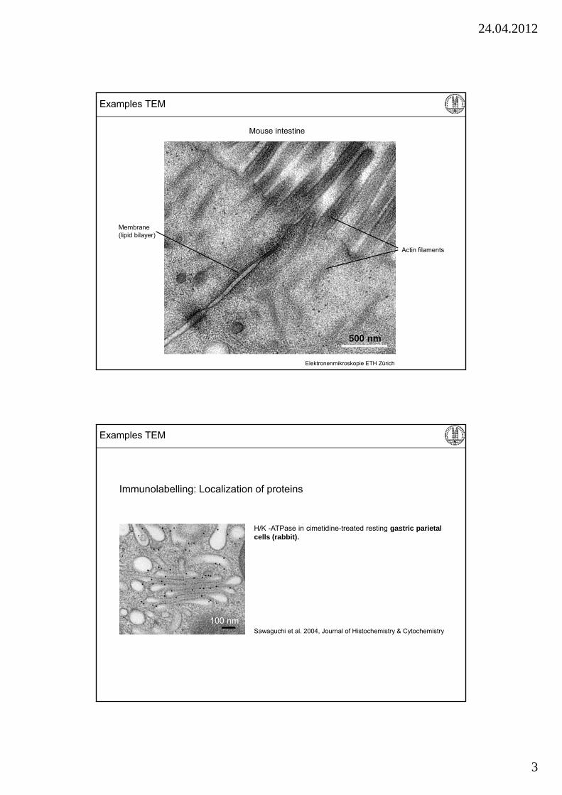
Elektronenmikroskopie ETH Zürich
500 nm
Mouse intestine
Membrane(lipid bilayer)
Actin filaments
Examples TEM
H/K -ATPase in cimetidine-treated resting gastric parietalcells (rabbit).
Immunolabelling: Localization of proteins
Sawaguchi et al. 2004, Journal of Histochemistry & Cytochemistry
100 nm
Examples TEM
24.04.2012
4
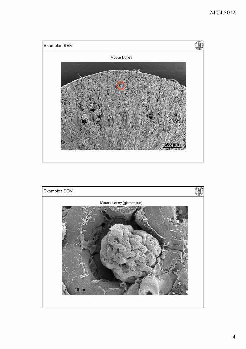
Examples SEM
Mouse kidney
500 µm
Mouse kidney (glomerulus)
10 µm
Examples SEM
24.04.2012
5
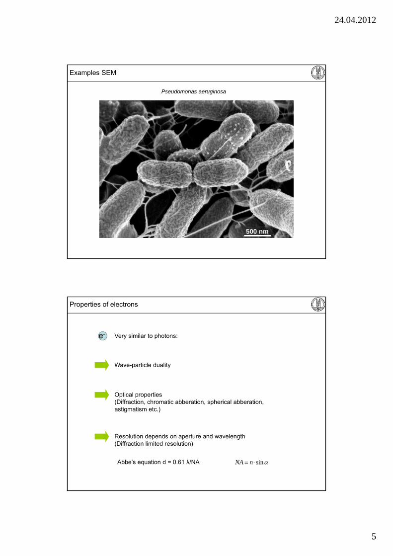
Pseudomonas aeruginosa
500 nm
Examples SEM
Wave-particle duality
Resolution depends on aperture and wavelength (Diffraction limited resolution)
Optical properties(Diffraction, chromatic abberation, spherical abberation, astigmatism etc.)
Abbe’s equation d = 0.61 λ/NA sin nNA
e-
Properties of electrons
Very similar to photons:
24.04.2012
6
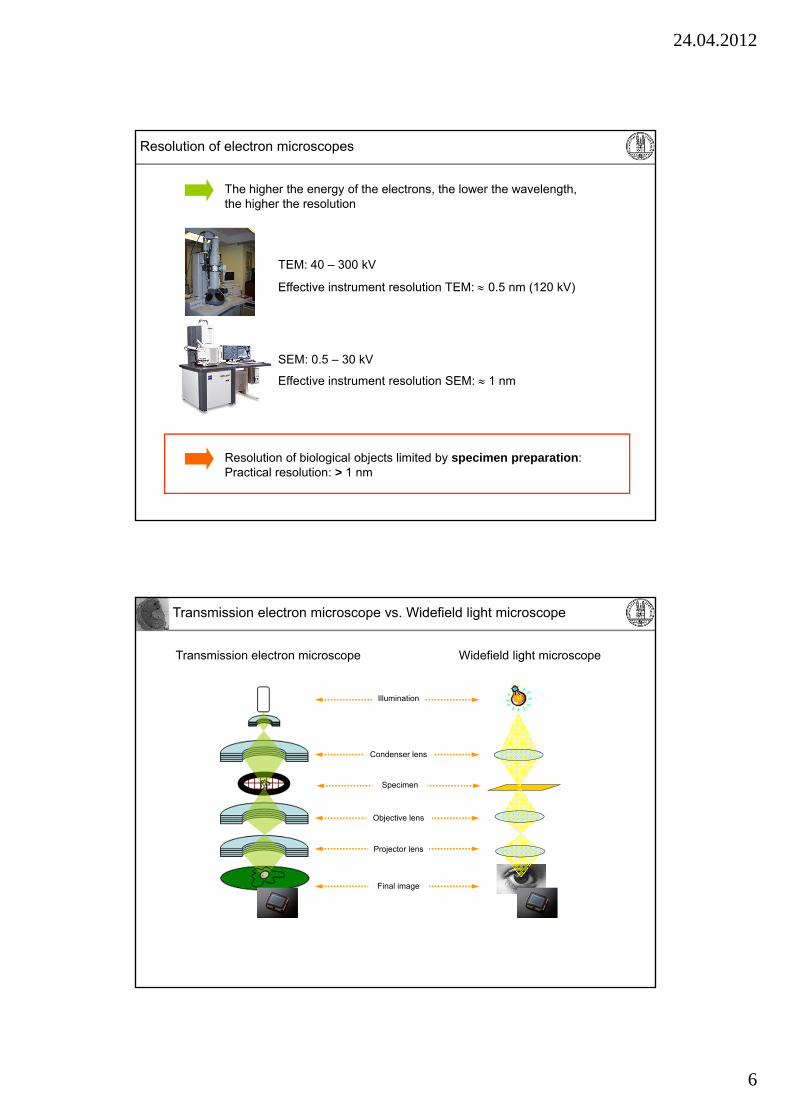
Resolution of biological objects limited by specimen preparation: Practical resolution: > 1 nm
TEM: 40 – 300 kV
Effective instrument resolution TEM: 0.5 nm (120 kV)
Effective instrument resolution SEM: 1 nm
Resolution of electron microscopes
The higher the energy of the electrons, the lower the wavelength, the higher the resolution
SEM: 0.5 – 30 kV
Widefield light microscopeTransmission electron microscope
Condenser lens
Objective lens
Projector lens
Specimen
Illumination
Final image
Transmission electron microscope vs. Widefield light microscope

24.04.2012
7
Confocal laser scanning microscopeScanning electron microscope
Beam scanner
Detector
Lens system
Lens system
Specimen
Illumination
Scanning electron microscope vs. Confocal laser scanning microscope
Example: Transmission electron microscope
Cathode
Specimen holder
Viewing screen
TMP
RP
IGP
IGP
Ion getter pump
Turbo molecular pump
Oil diffusion pump
Rotary pump
Atmosphere: 1000 mbar
10-5 - 10-7 mbar
10-0 - 10-2 mbar
10-7 - 10-10 mbar
Electron microscopes are high vacuum systems
24.04.2012
8
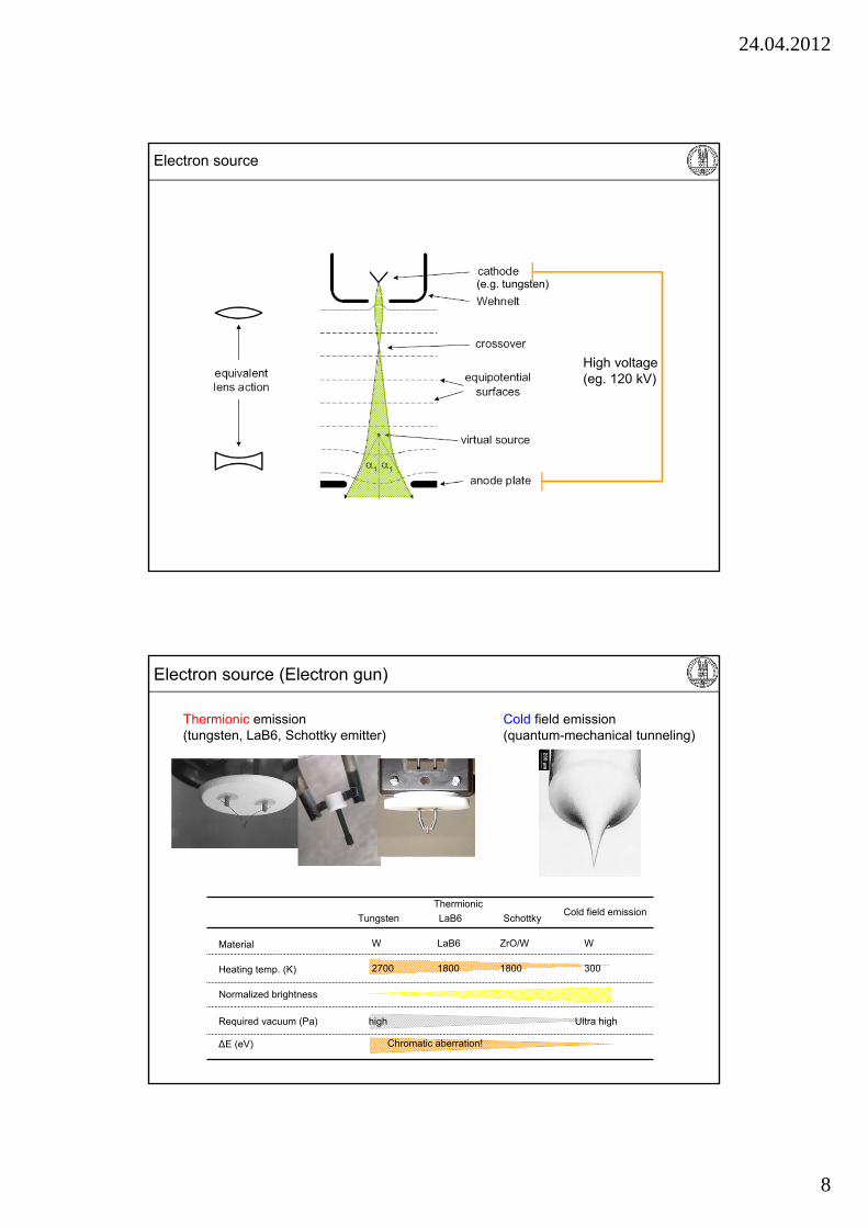
High voltage(eg. 120 kV)
Electron source
(e.g. tungsten)
Thermionic emission(tungsten, LaB6, Schottky emitter)
Electron source (Electron gun)
Cold field emission(quantum-mechanical tunneling)
Thermionic
Tungsten LaB6 SchottkyCold field emission
Material
Heating temp. (K)
Normalized brightness
Required vacuum (Pa)
∆E (eV)
W
2700
LaB6
1800
ZrO/W
1800
W
300
Chromatic aberration!
Ultra highhigh
24.04.2012
9
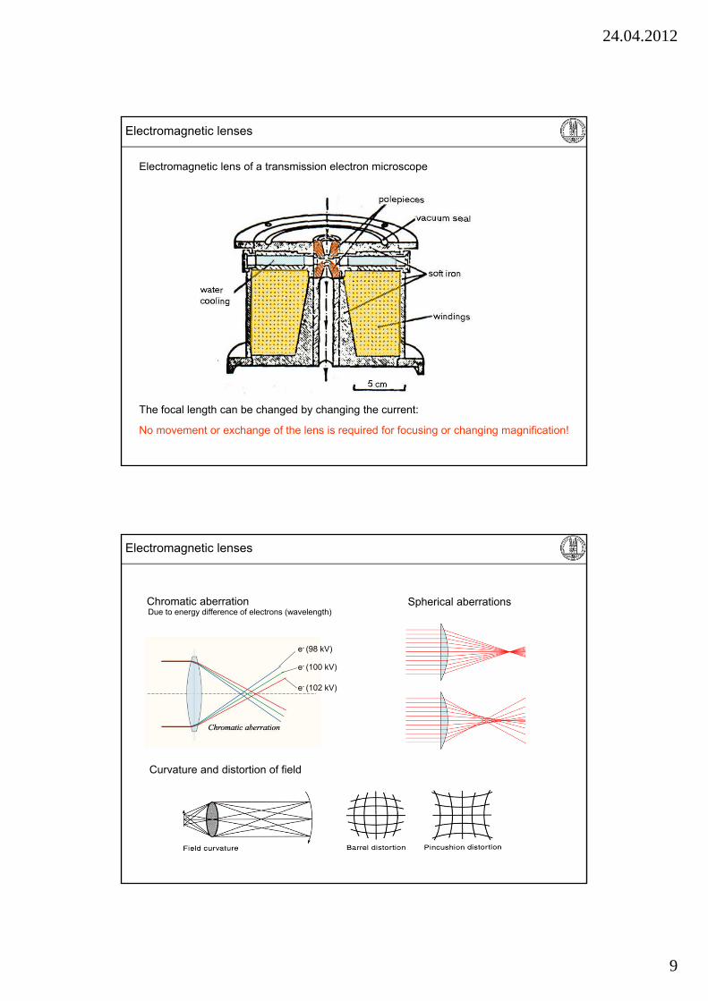
Electromagnetic lenses
Electromagnetic lens of a transmission electron microscope
The focal length can be changed by changing the current:
No movement or exchange of the lens is required for focusing or changing magnification!
Chromatic aberration
Electromagnetic lenses
Spherical aberrationsDue to energy difference of electrons (wavelength)
e- (98 kV)
e- (100 kV)
e- (102 kV)
Curvature and distortion of field
24.04.2012
10
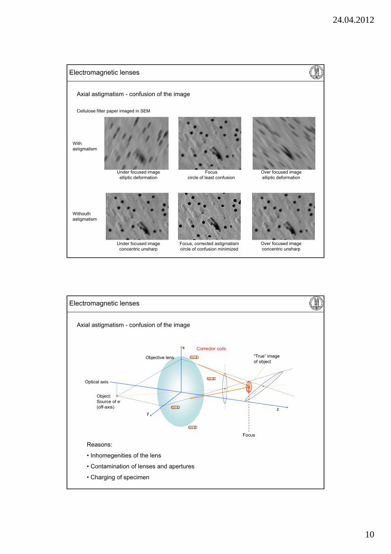
Axial astigmatism - confusion of the image
Electromagnetic lenses
Under focused imageelliptic deformation
Over focused imageelliptic deformation
Focuscircle of least confusion
Cellulose filter paper imaged in SEM
Focus, corrected astigmatismcircle of confusion minimized
With astigmatism
Withouth astigmatism
Under focused imageconcentric unsharp
Over focused imageconcentric unsharp
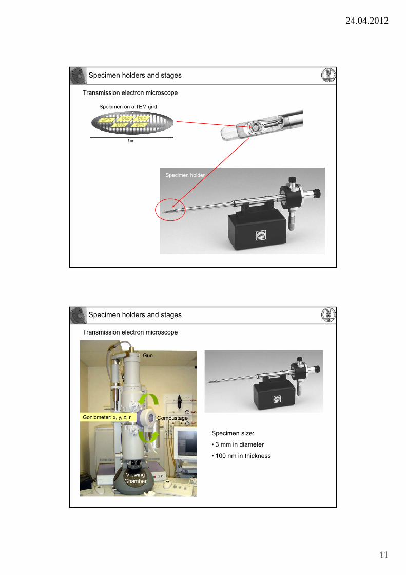
Electromagnetic lenses
Reasons:
• Inhomegenities of the lens
• Contamination of lenses and apertures
• Charging of specimen
Object: Source of e-
(off-axis)
Objective lens
Optical axis
“True” image of object
z
x
y
Focus
Corrector coils
Axial astigmatism - confusion of the image
24.04.2012
11
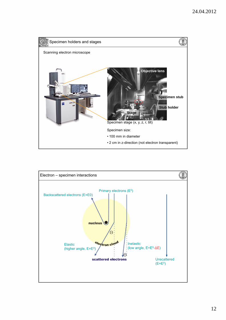
Specimen holder
Specimen on a TEM grid
Specimen holders and stages
3 mm3 mm
Transmission electron microscope
Specimen holders and stages
Transmission electron microscope
Goniometer: x, y, z, r
Specimen size:
• 3 mm in diameter
• 100 nm in thickness
24.04.2012
12
Specimen holders and stages
Scanning electron microscope
Specimen stage (x, y, z, r, tilt)
Objective lens
Stage
Specimen stub
Stub holder
Specimen size:
• 100 mm in diameter
• 2 cm in z-direction (not electron transparent)
Electron – specimen interactions
Inelastic(low angle, E=E0-∆E)
Unscattered(E=E0)
Primary electrons (E0)Backscattered electrons (E=E0)
Elastic(higher angle, E=E0)
24.04.2012
13
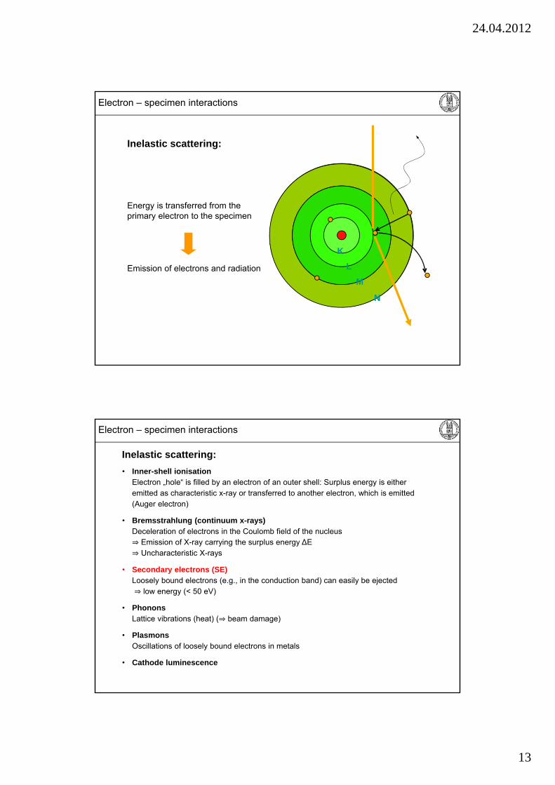
Electron – specimen interactions
Emission of electrons and radiation
Inelastic scattering:
Energy is transferred from the primary electron to the specimen
K
L
M
N
1
2
K
L
M
N
Electron – specimen interactions
• Inner-shell ionisationElectron „hole“ is filled by an electron of an outer shell: Surplus energy is eitheremitted as characteristic x-ray or transferred to another electron, which is emitted(Auger electron)
• Bremsstrahlung (continuum x-rays)Deceleration of electrons in the Coulomb field of the nucleus⇒ Emission of X-ray carrying the surplus energy ∆E⇒ Uncharacteristic X-rays
• Secondary electrons (SE)Loosely bound electrons (e.g., in the conduction band) can easily be ejected⇒ low energy (< 50 eV)
• PhononsLattice vibrations (heat) (⇒ beam damage)
• PlasmonsOscillations of loosely bound electrons in metals
• Cathode luminescence
Inelastic scattering:
24.04.2012
14
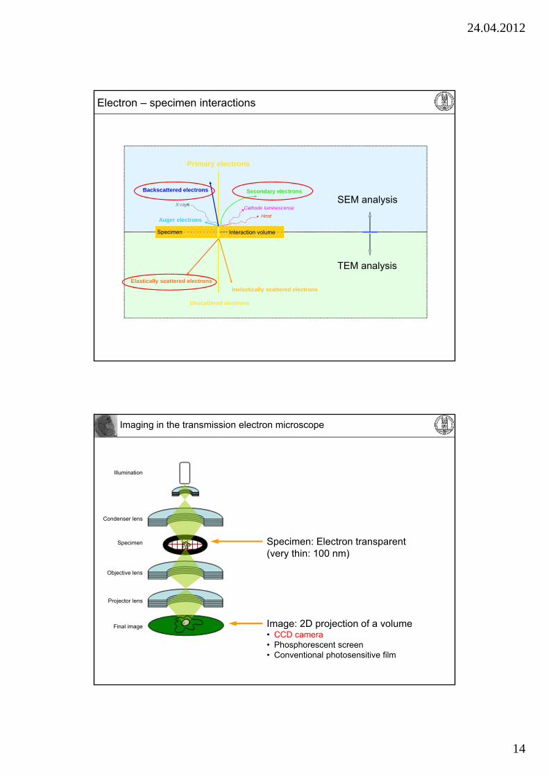
Electron – specimen interactions
Primary electrons
Unscattered electrons
Inelastically scattered electrons
Elastically scattered electrons
Secondary electronsBackscattered electrons
Auger electronsHeat
Cathode luminescenseX-rays
Specimen Interaction volume
SEM analysis
TEM analysis
Imaging in the transmission electron microscope
Specimen: Electron transparent(very thin: 100 nm)
Image: 2D projection of a volume• CCD camera• Phosphorescent screen• Conventional photosensitive film
Condenser lens
Objective lens
Projector lens
Specimen
Illumination
Final image
24.04.2012
15
Contrast formation in TEM
NOTE: Mechanisms occur at the same time (superposition)
Imaging in the transmission electron microscope
Absorption contrast
Scattering/phase contrast
Contrast formation in TEM
Biological specimen consist of light elements:
Absorption contrast weak
Scattering/phase contrast weak
Contrast enhancement required:
Treatment with heavy metals (Ur, Pb, Os)!
“LOW CONTRAST”
Heavy metals attach differently to different components
Imaging in the transmission electron microscope
24.04.2012
16
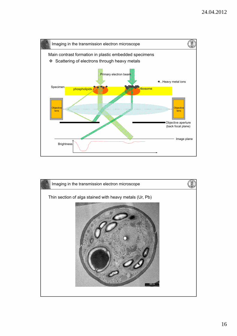
Main contrast formation in plastic embedded specimens
Scattering of electrons through heavy metals
Specimen
Image plane
phospholipids ribosome
…Heavy metal ions
Imaging in the transmission electron microscope
Objective aperture(back focal plane)
Primary electron beam
Brightness
Objective lens
Objective lens
Thin section of alga stained with heavy metals (Ur, Pb)
Imaging in the transmission electron microscope

24.04.2012
17
Thin section of alga without heavy metal staining
1 µm
Imaging in the transmission electron microscope
Underfocus OverfocusFocusMinimum contrast
100 nm
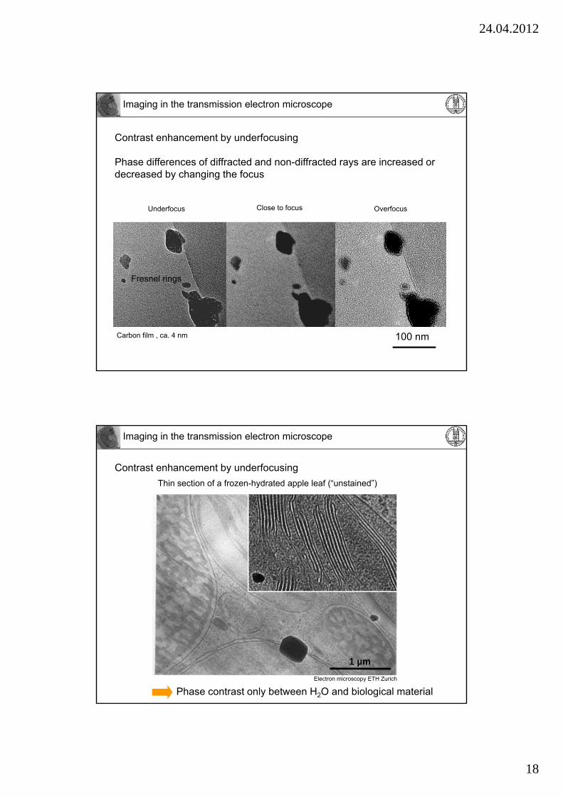
Contrast enhancement by underfocusing
Imaging in the transmission electron microscope
24.04.2012
18
Close to focus
100 nm
Fresnel rings
OverfocusUnderfocus
Carbon film , ca. 4 nm
Contrast enhancement by underfocusing
Phase differences of diffracted and non-diffracted rays are increased or decreased by changing the focus
Imaging in the transmission electron microscope
Contrast enhancement by underfocusing
Thin section of a frozen-hydrated apple leaf (“unstained”)
1 µm
Phase contrast only between H2O and biological material
Electron microscopy ETH Zurich
Imaging in the transmission electron microscope
24.04.2012
19
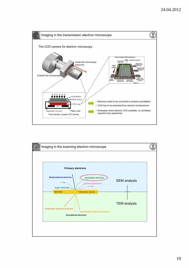
The CCD camera for electron microscopy
Outside the microscope
Inside the microscope (vacuum)
• Electrons need to be converted to photons (scintillator)
• CCD has to be protected from electron bombardment
Imaging in the transmission electron microscope
• Nowadays direct electron CCD available, no scintillator required (very expensive)
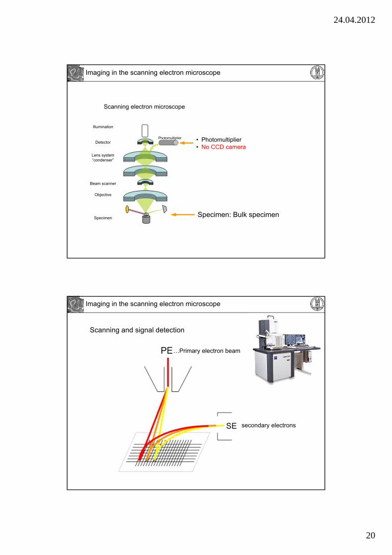
Primary electrons
Unscattered electrons
Inelastically scattered electrons
Elastically scattered electrons
Secondary electronsBackscattered electrons
Auger electronsHeat
Cathode luminescenseX-rays
Specimen Interaction volume
SEM analysis
TEM analysis
Imaging in the scanning electron microscope
24.04.2012
20
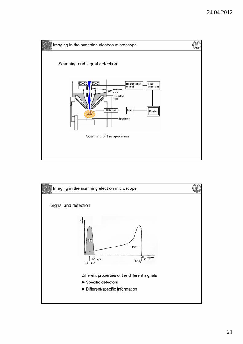
Imaging in the scanning electron microscope
Photomultiplier
Scanning electron microscope
Specimen: Bulk specimen
• Photomultiplier• No CCD camera
Beam scanner
Detector
Lens system“condenser”
Objective
Specimen
Illumination
Scanning and signal detection
…Primary electron beam
secondary electrons
Imaging in the scanning electron microscope
24.04.2012
21
Scanning and signal detection
Scanning of the specimen
Imaging in the scanning electron microscope
Signal and detection
Different properties of the different signals
►Specific detectors
►Different/specific information
Imaging in the scanning electron microscope
24.04.2012
22
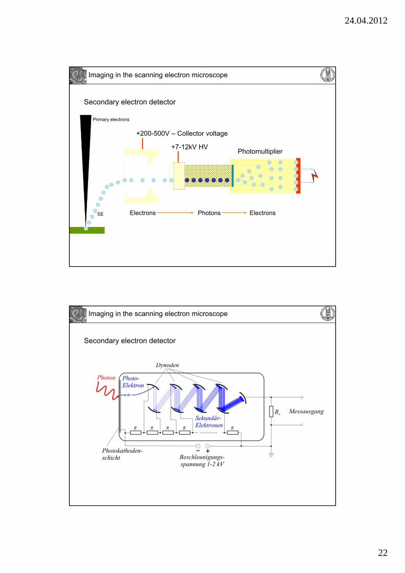
Secondary electron detector
+7-12kV HVPhotomultiplier
+200-500V – Collector voltage
Photons ElectronsElectrons
Primary electrons
SE
Imaging in the scanning electron microscope
Secondary electron detector
Imaging in the scanning electron microscope
24.04.2012
23
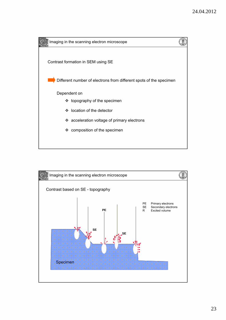
Contrast formation in SEM using SE
Different number of electrons from different spots of the specimen
topography of the specimen
location of the detector
acceleration voltage of primary electrons
composition of the specimen
Dependent on
Imaging in the scanning electron microscope
PE Primary electronsSE Secondary electronsR Excited volume
Contrast based on SE - topography
SE
PE
R
RSE
Specimen
Imaging in the scanning electron microscope
24.04.2012
24
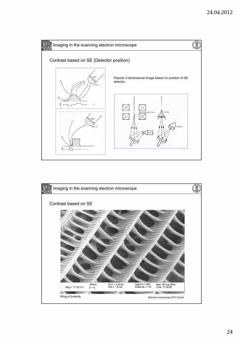
Contrast based on SE (Detector position)
Pseudo 3-dimensional image based on position of SE detector
Imaging in the scanning electron microscope
Wing of butterfly
Contrast based on SE
Electron microscopy ETH Zurich
Imaging in the scanning electron microscope
24.04.2012
25
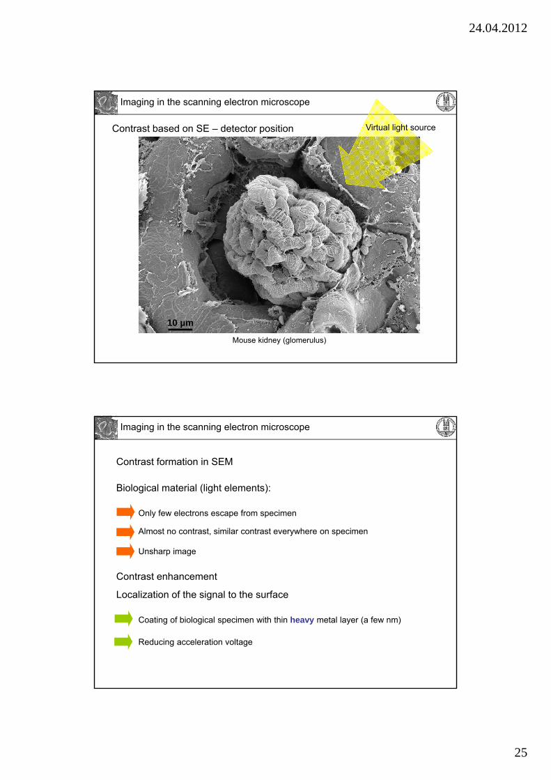
Mouse kidney (glomerulus)
10 µm
Contrast based on SE – detector position Virtual light source
Imaging in the scanning electron microscope
Biological material (light elements):
Only few electrons escape from specimen
Almost no contrast, similar contrast everywhere on specimen
Contrast enhancement
Localization of the signal to the surface
Coating of biological specimen with thin heavy metal layer (a few nm)
Reducing acceleration voltage
Unsharp image
Contrast formation in SEM
Imaging in the scanning electron microscope
24.04.2012
26
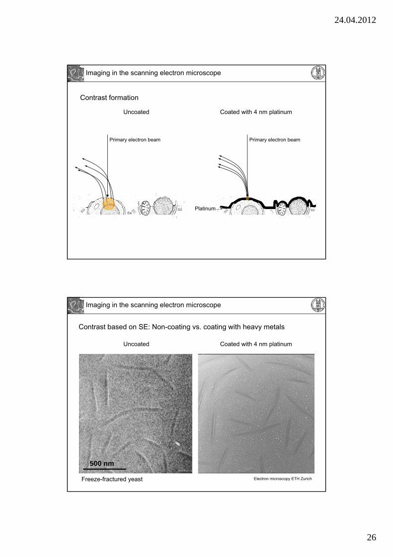
Primary electron beam
Contrast formation
Platinum
Primary electron beam
Uncoated Coated with 4 nm platinum
Imaging in the scanning electron microscope
Freeze-fractured yeast
500 nm
Contrast based on SE: Non-coating vs. coating with heavy metals
Uncoated Coated with 4 nm platinum
Electron microscopy ETH Zurich
Imaging in the scanning electron microscope

24.04.2012
27
Low mag. High mag.
768 px
1024 px
768 px
1024 px
Object
Imaging in the scanning electron microscope
Focusing and magnification in electron microscopy
Focusing and magnification in light microscopy
TEM:
Focusing:
Change current in magnetic lenses for focusing (objective lens)
Move holder in z
Magnification:
Change current in magnetic lenses (projective lenses) and combine several projective lenses.
SEM:
Focusing:
Change current in magnetic lenses for focusing (objective lens)
Move stage in z
Magnification:
Change scanning field (scan a smaller or larger area with the same number of pixels), pixel size changes.
Widefield light microscopy:
Focusing:
Moving objective or
stage in z
Magnification:
Changing the whole objective.
Confocal scanning laser microscopy:
Focusing:
Moving objective or
stage in z
Magnification:
Change scanning field (scan a smaller or larger area with the same number of pixels), pixel size changes.
Changing the whole objective
Imaging in the scanning electron microscope
24.04.2012
28
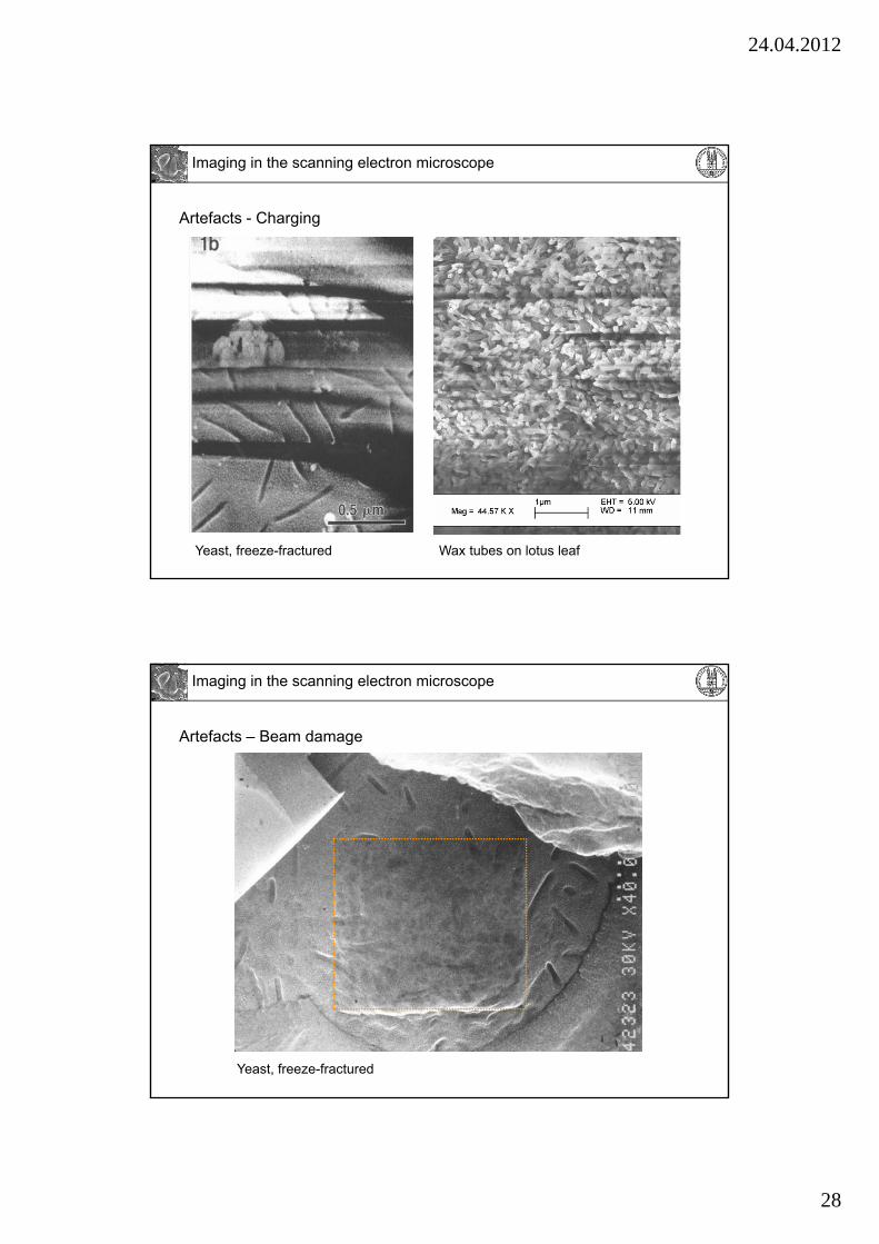
Artefacts - Charging
Yeast, freeze-fractured Wax tubes on lotus leaf
Imaging in the scanning electron microscope
Artefacts – Beam damage
Yeast, freeze-fractured
Imaging in the scanning electron microscope
24.04.2012
29
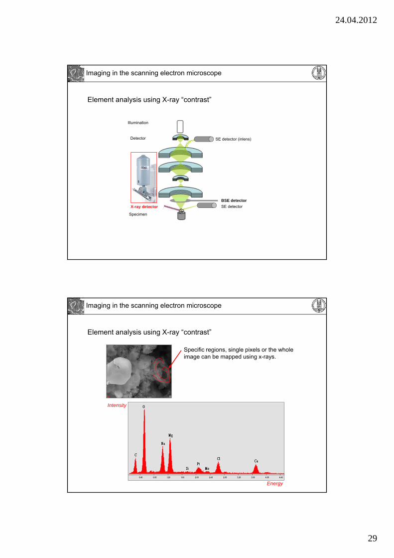
Element analysis using X-ray “contrast”
Detector
Specimen
Illumination
SE detector (inlens)
SE detector
BSE detector
X-ray detector
Imaging in the scanning electron microscope
Element analysis using X-ray “contrast”
Specific regions, single pixels or the whole image can be mapped using x-rays.
Energy
Intensity
Imaging in the scanning electron microscope