introduction to mycology · diagnosis •diagnosis of fungal infections is based on a combination...
TRANSCRIPT

Introduction to Mycology
By: Nader Alaridah
Medical mycology is the study of mycoses of man and their etiologic agents.
Mycoses are the diseases caused by fungi. Of the several thousands of species of fungi that are known, less than 300 are pathogenic to man.
fungal invasion of human tissue was recognized in the early 1800s before the science of bacteriology was developed.

What is a Fungus ?
• Kingdom fungi
• Eukaryotic – a true nucleus , heterotrophic, do not contain chlorophyll
• Yeasts & filamentous structures (hyphae)
• Produce spores (sexual & asexual reproduction)
• Saprophytic ( on dead tissue).Parasitic (on living organism).
• All fungi required organic source of Carbon associated with decaying matter

• Cell wall consist of chitin and B-glucan ,both are polysaccharide which is the site of action of some antifungal drugs.
• Cell membrane consist of ergosterol Ergosterol is the site of action of some antifungal.
• Most fungi are obligatory aerobes .

Fungal Cell

The Importance of Fungi
1-They are common cause of damage to crops and food chain.
2- Few species of fungi can cause disease in human (300/200,000). However, fungal infections are increasing due to AIDS and other immunosuppressant conditions.
3. Production of antibiotics e.g Penicillin.
General mycology
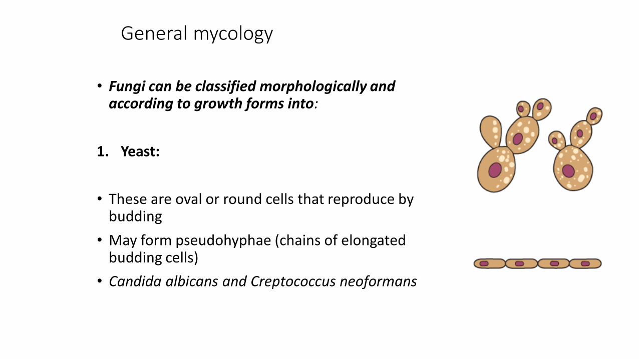
• Fungi can be classified morphologically and according to growth forms into:
1. Yeast:
• These are oval or round cells that reproduce by budding
• May form pseudohyphae (chains of elongated budding cells)
• Candida albicans and Creptococcus neoformans

General mycology / yeasts cont’d
• Common in immunocompromised patients and can cause multisystem infections such as meningitis, arthritis and respiratory infections.
• C. neoformans found in soil and pigeon faeces and it commonly infects lung initially.

General mycology
2. Filamentous fungi ( Molds):
• They have branching tubular filaments (hyphae ) which may be septate or non-septate
• Mycelium: mass of branching, interlinking hyphae
• Also may produce asexual spores at the tip or side of the hyphae
• Asexual spores may be contained in a sac called sporangiospores
• e.g Zygomycetes, Aspergillus and Dermatophytes
General mycology
3. Dimorphic fungi
• These occur in two forms: yeast form in tissues or when grow at 37°C& filamentous form when grow at 22°C
• Examples:
Blastomyces dermatitidis
Coccidioides immitis
Histoplasma capsulatum

Figure 22.2

General mycology• Fungal diseases
• Fungal infections have recently emerged as a growing threat to human health, especially to persons whose immune systems are compromised in some way.
1. Fungal allergies
• Molds grow on any damp organic surface, and spores are constantly in the air.
• Inhaled Spores & volatile fungal toxins may play a role in producing allergic manifestations such as asthmatic reaction ( rapid broncho-constriction mediated by IgE) and eosinophilia.
• Notable in Aspergillus fumigatus

General mycology
2. Fungal toxins ( mycotoxicosis ):
• Aflatoxicosis
• Aflatoxicosis is a poisoning condition & it results from ingestion of aflatoxins in contaminated food
• Aflatoxins are group of structurally related toxic compounds produced by certain strains of fungi (Aspergillus flavus& A. parasiticus)
• Under favorable conditions of temperature & humidity, these fungi grow on certain foods& resulting in production of aflatoxins.

Aflatoxicosis
• The most pronounced contamination has been encountered in treenuts, peanuts& other oilseeds including corn.
• Aflatoxins are metabolized in the liver to epoxide, which is potentcarcinogenic.
• Aflatoxin B1 induce mutation in the p53 human suppressor gene,leading to loss of growth control in hepatocytes.

General mycology
3. Fungal infection ( mycoses ):
• Fungal infections range from superficial infections to overwhelming infections that are rapidly fatal in compromised host
• The infection with fungi is increasing in frequency as a result of increased use of antibiotics, corticosteroids& cytotoxic drugs (immunosuppression).
General mycology
• Human fungal infections are commonly classified as:
Superficial & Cutaneous infections
• Infections involve the skin, mucous membrane, nail or hair with or without tissue destruction & immunological reaction
• e.g pityriasis versicolor , Tinea nigra
• e.g cutaneous candidiaisis & dermatophytes.

General mycology
Subcutaneous
• Infection is confined to sub- cutaneous tissue without dissemination to distant organs
e.g Chromoblastomycosis
Systemic mycoses
• Are primarily pulmonary lesion that may disseminate to any organ.
• e.g COCCIDIOIDOMYCOSIS , HISTOPLASMOSIS
Opportunistic Mycoses
• e.g Candida spp. Cryptococcus

Diagnosis
• Diagnosis of fungal infections is based on a combination of clinical observation and laboratory investigation.
• Clinical investigation
• The first indication that a patient may have a systemic mycosis is often their failure to respond to antibacterial antibiotics.

• Laboratory diagnosis
• Recognition of the pathogen in tissue by microscopy
• Isolation of the causal fungus in culture
• The use of serological tests
• detection of fungal DNA by PCR

Types of specimen
• Skin scales, nail clippings and scrapings of the scalp that include hair stubs and skin scales are the most suitable specimens for the diagnosis of ringworm; these are collected into folded paper squares for transport to the laboratory.

• Swabs should be taken from suspected Candida infections from the mucous membranes and preferably sent to the laboratory in ‘clear’ transport medium.
• For subcutaneous infections the most suitable specimens are scrapings and crusts, aspirated pus and biopsies.
• In suspected systemic infection, specimens should be taken from appropriate sites.

Stains and Direct Microscopic Examination
• Most specimens can be examined satisfactorily in wet mounts after partial digestion of the tissue with 10–20% potassium hydroxide.
• Addition of Calcofluor white and subsequent examination by fluorescence microscopy enhances the detection of most fungi as the fluorescent hydroxide– Calcofluor binds to the fungal cell walls
• special stains (methylene blue, lactophenol blue, periodic acid-Schiff (PAS), ink, etc.)

KOH wet mount

Culture
• Most pathogenic fungi are easy to grow in culture.
Sabouraud dextrose medium:
• commonly used
• may be supplemented with chloramphenicol to minimize bacterial contamination and cycloheximide to reduce contamination with saprophytic fungi.

Antifungal therapy
• The drugs used to treat bacterial diseases have no effect on fungal infection.
• it depends on presence of ergosterol in fungal cell membranes
• Amphotercin B and nystatin are polynes (Fungicidal) & various azoles (fungistatic) are commonly used for treatment of fungal infection.

General mycology
1. Polyene derivatives• Amphotericin B
• Nystatin
2. Azoles• Ketoconazole
• Fluconazole
• Itraconazole
• Voriconazole
• Posaconazole

General mycology
3. Griseofulvin
4. 5-fluorocytosine (5-FC)
5. Allylamines
-Terbinafine (Lamasil)
6. Echinocandins- Caspofungin

The End
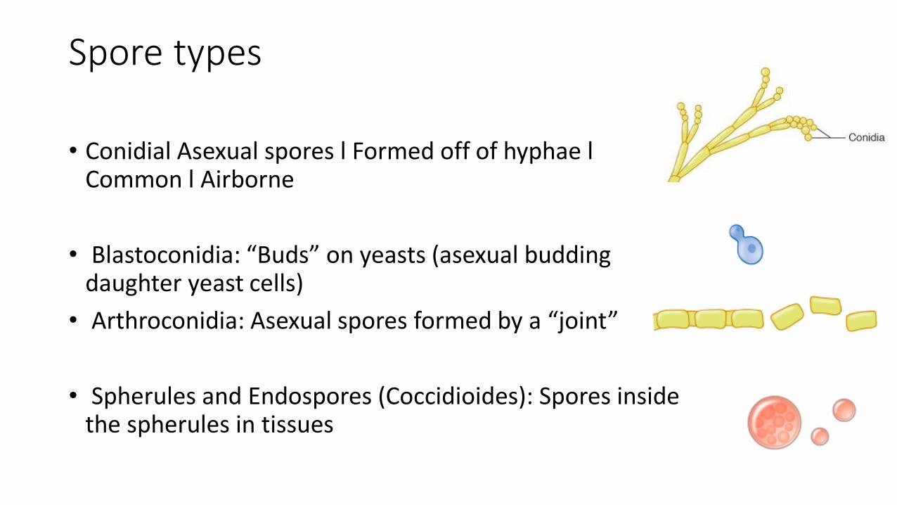
Spore types
• Conidial Asexual spores l Formed off of hyphae l Common l Airborne
• Blastoconidia: “Buds” on yeasts (asexual budding daughter yeast cells)
• Arthroconidia: Asexual spores formed by a “joint”
• Spherules and Endospores (Coccidioides): Spores inside the spherules in tissues