intussusception secondary to a carcinoid tumor in an adult patient
TRANSCRIPT

I
IDS
a
ARRAA
KAIICI
1
cpedSipoa
2
t
Cco
1T
(
2h
CASE REPORT – OPEN ACCESSInternational Journal of Surgery Case Reports 5 (2014) 265–267
Contents lists available at ScienceDirect
International Journal of Surgery Case Reports
journa l h omepage: www.caserepor ts .com
ntussusception secondary to a carcinoid tumor in an adult patient�
sidoro Wiener-Carrillo, Carlos González-Alvarado ∗, Mario Cervantes-Valladolid,enis Echaverry-Navarrete, Gregorio Zubieta-O’Farrill, Andrés Gudino-Chávez
urgery Department, Angeles Pedregal Hospital, Mexico City, Mexico
r t i c l e i n f o
rticle history:eceived 10 August 2013eceived in revised form 1 January 2014ccepted 29 January 2014vailable online 20 March 2014
eywords:dult intussusception
ntussusceptionleocolic carcinoidarcinoid
ntestinal obstruction
a b s t r a c t
INTRODUCTION: Intussusception in adult patients represents 5% of all intussusceptions and 1–5% of bowelobstructions in adults. In contrast to pediatric patients, 90% of the time, in adults, it’s caused by well-established pathologic mechanisms, such as carcinoma, polyps, diverticula, Meckel diverticula, stenosis,or benign neoplasms. Small intestine intussusceptions are more frequent, but colonic intussusceptionsare caused 50% of the time by malignant neoplasms, especially adenocarcinoma.PRESENTATION OF CASE: We present a 70-year-old woman, with no relevant familial history, who pre-sented with a 3-day symptomatology consisting of epigastric, colic, diffuse, abdominal pain of moderateintensity, which progressed till reaching a severe intensity, also referring abdominal distension, nausea,and gastrointestinal-content vomits.DISCUSSION: In adult patients, the exact mechanism of intussusception is unknown in 8–20% of the cases,however, secondary intussusception can occur with any lesion of the intestinal wall or any irritant factor inits lumen that alters normal peristaltic activity and that could serve as a trigger to start an intussusception
of one bowel segment over another the most common site is the small intestine.CONCLUSION: Intussusception represents an unusual problem in adult patients; it requires a high clin-ical suspicion, mainly as a differential diagnosis in patients with intestinal obstruction, and it clinicallypresents as a subacute or chronic illness. CT represents the most useful diagnostic tool. An attempt todures a forblish
perform reduction procecolonic intussusceptions,
© 2014 The Authors. Pu
. Introduction
Intussusception in adult patients represents 5% of all intussus-eptions and 1–5% of bowel obstructions in adults. In contrast toediatric patients, 90% of the time, in adults, it’s caused by well-stablished pathologic mechanisms, such as carcinoma, polyps,iverticula, Meckel diverticula, stenosis, or benign neoplasms.1
mall intestine intussusceptions are more frequent, but colonicntussusceptions are caused 50% of the time by malignant neo-lasms, especially adenocarcinoma. The most common symptomsf presentation in adults are nausea, vomit, intermittent, colicbdominal pain, and constipation.2
. Case report
We present a 70-year-old woman, with no relevant familial his-ory, who presented with a 3-day symptomatology consisting of
� This is an open-access article distributed under the terms of the Creativeommons Attribution-NonCommercial-ShareAlike License, which permits non-ommercial use, distribution, and reproduction in any medium, provided theriginal author and source are credited.∗ Corresponding author at: Angeles Pedregal Hospital, Camino a Santa Teresa055, Col. Heroes de Padierna, Magdalena Contreras, 10700 Mexico City, Mexico.el.: +52 1 55 2727 9579.
E-mail addresses: [email protected], [email protected]. González-Alvarado).
210-2612/$ – see front matter © 2014 The Authors. Published by Elsevier Ltd. on behalf
ttp://dx.doi.org/10.1016/j.ijscr.2014.01.022
in small intestine intussusceptions can be done, however, in ileocolic ormal resection of the segment is recommended.ed by Elsevier Ltd. on behalf of Surgical Associates Ltd. All rights reserved.
epigastric, colic, diffuse, abdominal pain, of moderate intensity,which progressed till reaching a severe intensity, also referringabdominal distension, nausea, and 4 gastrointestinal-content vom-its. Upon physical examination at arrival, the patient was foundconscious, oriented, with skin paleness, poorly hydrated; thoraxwith adequate vesicular murmur, without wheezing or rattles,adequate amplexion and amplexation, cardiac sounds with onlyS1 and S2, without aggregated sounds; soft, distended abdomen,with increased-frequency peristalsis, diffuse pain upon palpation,and rebound tenderness (Blumberg sign); and adequate peripheralpulses.
While in the Emergency Room, laboratorial tests reported val-ues of hemoglobin at 16.7 g/dL, 51.2% hematocrit, 551,000 mm−3
platelets, 23,800 mm−3 leukocytes, 88% neutrophilia, and bloodchemistry within normal parameters. Because of the frank leuko-cytosis and signs of acute abdomen, a computed abdomentomography (CT) was performed, with reported findings suggestingileo-colic intussusception, with a tumoral image of pedicle aspectadjacent to the terminal ileum, with probable secondary intesti-nal oclusion, nodular images of unknown origin on the liver, andBosniak category I cystic images on the right kidney (Figs. 1 and 2).
Considering this CT result, coagulation times and tumor markers
(CEA, AFP, CA 125, and CA 19–9) were submitted, finding normalresults.The patient was submitted to an exploratory laparotomy, withfindings of a 6 cm tumor on the ileo-caecal segment, responsible
of Surgical Associates Ltd. All rights reserved.
CASE REPORT – OPEN ACCESS266 I. Wiener-Carrillo et al. / International Journal of Surgery Case Reports 5 (2014) 265–267
Fig. 1. CT showing the classical “Target” image.
fhrib
rt
parison, our patient’ onset of symptoms was slightly slower, and
Fig. 2. CT showing nodular images in the liver.
or an intussusception of that segment, as well as multiple bilateralepatic metastases (Fig. 3). A radical right hemicolectomy, withesection of the terminal ileum tumor (20 cm), and side-to-sideleotransverse anastomosis was performed, in addition to a hepaticiopsy.
The surgical samples were sent to Pathology department, whicheported a massive, ileo-caecal, transmural, classical carcinoidumor, with extension to pericaecal tissues (confined tumor of
Fig. 3. Laparotomy finding of an ileo-caecal mass.
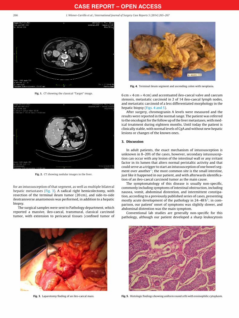
Fig. 4. Terminal ileum segment and ascending colon with neoplasia.
6 cm × 4 cm × 4 cm) and accentuated ileo-caecal valve and caecumstenosis, metastatic carcinoid in 2 of 14 ileo-caecal lymph nodes,and metastatic carcinoid of a less differentiated morphology in thehepatic biopsy (Figs. 4 and 5).
After surgery, chromogranin A levels were measured and theresults were reported in the normal range. The patient was referredto the oncologist for the follow up of the liver metastases, with med-ical treatment during eighteen months. Until today the patient isclinically stable, with normal levels of CgA and without new hepaticlesions or changes of the known ones.
3. Discussion
In adult patients, the exact mechanism of intussusception isunknown in 8–20% of the cases, however, secondary intussuscep-tion can occur with any lesion of the intestinal wall or any irritantfactor in its lumen that alters normal peristaltic activity and thatcould serve as a trigger to start an intussusception of one bowel seg-ment over another1; the most common site is the small intestine,just like it happened in our patient, and with afterwards identifica-tion of an ileo-caecal carcinoid tumor as the main cause.
The symptomatology of this disease is usually non-specific,commonly including symptoms of intestinal obstruction, includingnausea, vomit, abdominal distention, and intermittent constipa-tion, according to a previously published series of cases, presentingmostly acute development of the pathology in 24–48 h3; in com-
abdominal distention was the main symptom.Conventional lab studies are generally non-specific for this
pathology, although our patient developed a sharp leukocytosis
Fig. 5. Histologic findings showing uniform round cells with eosinophilic cytoplasm.

– Ornal o
womaasCiobdtso
stuiorbc
ndd1tt
4
pdpud
1
2
3
4
5
OTpc
CASE REPORTI. Wiener-Carrillo et al. / International Jou
ith neutrophilia; however, the main diagnostic support consistsn imaging studies. On pediatric patients, the diagnosis is mainlyade using ultrasound or barium-enema studies; however, on
dult patients, the CT is the main diagnostic tool, being addition-lly helpful in establishing the underlying cause.4 The distinctiveign in early stages is the “target” image, which was found in theT of our patient (Fig. 1). Enhanced CT has become a valuable tool
n the diagnosis of bowel obstruction, because it allows imagingf structures other than just mucosal detail, which may outlineoth the site as well and as the etiology of the obstruction. It alsoemonstrates changes in the intestinal wall and associated mesen-ery. IV enhanced CT doesn’t have the detrimental effects one canee with oral agents, so its indicated in both partial and completebstruction.5
Typically, the initial diagnosis is ignored because of the acute,ubacute or chronic presence of nonspecific symptoms; however,he therapeutic decision is established intraoperatively, and in sit-ations in which the colon is involved, a formal resection that
ncludes lymph-node dissection is indicated, because up to 50%f the time, a malignant lesion is associated (in our patient, aight hemicolectomy with lymph-node dissection was performedecause of the finding of a 6 cm × 4 cm × 4 cm tumor on the ileo-aecal valve).
Carcinoid tumors represent the most frequent neuroendocrineeoplasms in the gastrointestinal (GI) tract, and they basicallyerive from enterochromaffin (or Kulchitsky) cells.6 There’s a greativersity in its bodily distribution; however, in a study made in1,427 patients with carcinoid-tumor diagnosis, 54.5% occurred inhe GI tract, followed by lungs and bronchi (30.1%). Within the GIract, the small intestine was the most common site: 44.7%.7
. Conclusion
Intussusception represents an unusual problem in adultatients; it requires a high clinical suspicion, mainly as a differential
iagnosis in patients with intestinal obstruction, and it clinicallyresents as a subacute or chronic illness. CT represents the mostseful diagnostic tool. An attempt to perform reduction proce-ures in small intestine intussusceptions can be done, however, in67
pen Accesshis article is published Open Access at sciencedirect.com. It is distribermits unrestricted non commercial use, distribution, and reproductredited.
PEN ACCESSf Surgery Case Reports 5 (2014) 265–267 267
ileocolic or colonic intussusceptions, a formal resection of the seg-ment is recommended.
Conflict of interest
No conflict of interest for any of the authors in this case report.
Funding
None.
Ethical Approval
Written informed consent was obtained from the patient forpublication of this case report and accompanying images. A copyof the written consent is available for review by the Editor-in-Chiefof this journal on request.
Author contributions
Carlos Gonzalez Alvarado and Gregorio Zubieta-O’Farrill per-formed data collection, analysis and writting. Isidoro Wiener, MarioCervantes and Denis Echaverry did data collection while AndresGudino-Chavez did the writing job.
References
. Wang N, Cui XY, Liu Y, Long J, Xu YH, Guo RX, et al. Adult intussusception: aretrospective review of 41 cases. World J Gastroenterol 2009;15(July (26)):3303–8.
. Loukas M, Pellerin M, Kimball Z, de la Garza-Jordan J, Tubbs RS, Jordan R. Intus-susception an anatomical perspective with review of the literature. Clin Anat2011;24(July (5)):552–61.
. Begos D, Sandor A, Modlin I. The diagnosis and management of adult intussus-ception. Am J Surg 1997;173:88–94.
. Byrne A, Goeghegan T, Govender P, Lyburn I, Colhoun E, Torreggiania W. Theimaging of intussusception. Clin Radiol 2005;60:39–46.
. Yakan S, Calıskan C, Makay O, Denecl A, Korkut M. Intussusception in adults:clinical characteristics, diagnosis and operative strategies. World J Gastroenterol2009;15(April (16)):1985–9.
. Rebecca S, Herbert C. Carcinoid tumors. Surg Oncol Clin N Am 2006;15:463–78.
. Maggard M, O’Connell J, Ko C. Updated population-based review of carcinoid
tumors. Ann Surg 2004;240:117–22.uted under the IJSCR Supplemental terms and conditions, whichion in any medium, provided the original authors and source are