influence of sequence and covalent modifications on … · influence of sequence and covalent...
TRANSCRIPT

Influence of Sequence and Covalent Modifications on Yeast tRNADynamicsXiaoju Zhang,† Ross C. Walker,‡,§ Eric M. Phizicky,† and David H. Mathews*,†
†Department of Biochemistry and Biophysics and Center for RNA Biology, University of Rochester Medical Center, Rochester, NewYork 14642, United States‡San Diego Supercomputer Center, University of California San Diego, La Jolla, California 92093, United States§Department of Chemistry and Biochemistry, University of California San Diego, La Jolla, California 92093, United States
*S Supporting Information
ABSTRACT: Modified nucleotides are prevalent in tRNA.Experimental studies reveal that these covalent modificationsplay an important role in tuning tRNA function. In this study,molecular dynamics (MD) simulations were used toinvestigate how modifications alter tRNA dynamics. The X-ray crystal structures of tRNA(Asp), tRNA(Phe), andtRNA(iMet), both with and without modifications, wereused as initial structures for 333 ns explicit solvent MDsimulations with AMBER. For each tRNA molecule, threeindependent trajectory calculations were performed, giving anaggregate of 6 μs of total MD across six molecules. The global root-mean-square deviations (RMSD) of atomic positions showthat modifications only introduce significant rigidity to the global structure of tRNA(Phe). Interestingly, RMSDs of theanticodon stem-loop (ASL) suggest that modified tRNA has a more rigid structure compared to the unmodified tRNA in thisdomain. The anticodon RMSDs of the modified tRNAs, however, are higher than those of corresponding unmodified tRNAs.These findings suggest that the rigidity of the anticodon stem-loop is finely tuned by modifications, where rigidity in theanticodon arm is essential for tRNA translocation in the ribosome, and flexibility of the anticodon is important for codonrecognition. Sugar pucker and water residence time of pseudouridines in modified tRNAs and corresponding uridines inunmodified tRNAs were assessed, and the results reinforce that pseudouridine favors the 3′-endo conformation and has a highertendency to interact with water. Principal component analysis (PCA) was used to examine correlated motions in tRNA.Additionally, covariance overlaps of PCAs were compared for trajectories of the same molecule and between trajectories ofmodified and unmodified tRNAs. The comparison suggests that modifications alter the correlated motions. For the anticodonbases, the extent of stacking was compared between modified and unmodified molecules, and only unmodified tRNA(Asp) hassignificantly higher percentage of stacking time. Overall, the simulations reveal that the effect of covalent modification on tRNAdynamics is not simple, with modifications increasing flexibility in some regions of the structure and increasing rigidity in otherregions.
1. INTRODUCTIONNaturally occurring covalent modifications to the standardRNA chemistry are prevalent in tRNA. In 561 sequencedtRNAs, from a wide range of organisms, modifications aredetected on 11.9% of the residues.1−3 In the yeastSaccharomyces cerevisiae, 16.2% of the residues of the 28 uniquesequenced cytoplasmic tRNA species hold modifications. Therange is from 7 to 17 modifications per tRNA.1 With theirfrequent occurrence in tRNAs, modifications are recognized asan important device for tuning tRNA structure and function.4−7
tRNAs are usually 74−95 nucleotides long, and theirsequences (primary structure) typically allow extensive basepairing (secondary structure) and noncanonical interactions(tertiary structure). tRNA secondary structure is nearlyuniversally arranged in a cloverleaf shape, as first realized byHolley et al.8 A common numbering scheme for tRNAnucleotides is used, which relies on the position of a base
relative to the canonical secondary structure. On the basis ofthe cloverleaf structure, tRNA is composed of four regions: theanticodon stem loop (ASL, nucleotides 27−43), acceptor stem(nucleotides 1−7 and 66−76), D stem loop (nucleotides 10−25), and T stem loop containing the conserved TΨC sequence(nucleotides 49−65). The first tertiary structure wasdetermined by X-ray crystallography studies in 1973.9,10 TheL-shaped tertiary structure is formed by two coaxial stacks ofhelices in the cloverleaf secondary structure. The acceptor stemand T stem coaxially stack and form one of the arms, and theother arm is formed by coaxial stacking of the D stem andanticodon stem.The structure and conformation of several unmodified yeast
tRNAs have been studied with T7 polymerase transcription. It
Received: February 9, 2014
Article
pubs.acs.org/JCTC
© XXXX American Chemical Society A dx.doi.org/10.1021/ct500107y | J. Chem. Theory Comput. XXXX, XXX, XXX−XXX

is shown that fully modified native tRNA and unmodified tRNAhave similar overall structure.11−16 Melting experiments revealthat both modified and unmodified tRNAs are stabilized byaddition of Mg2+. Unmodified tRNA, however, has moreplasticity than modified tRNA at physiological Mg2+ concen-tration and is less stable than modified tRNA in the absence ofMg2+ or at low Mg2+ concentration.15 A nuclease probing studyfound a greater accessibility of the D- and T-loops of thetranscribed tRNA, indicating that the absence of modificationmay disrupt the tertiary structure of the D−T loop corner intRNA.15
The anticodon loop is the domain that directly interacts withmRNA and the ribosome. Therefore, alteration to tRNAstructure at this location by modification directly changes theinteraction between tRNA and other partners of translation.This loop is a prominent location for modifications, especiallyat positions of 34 and 37, where 34, 35, and 36 are the positionsof the anticodon nucleotides. Modifications of uridine atposition 34 and purine at position 37 are ubiquitous, and onlyseven tRNAs in Escherichia coli are read by tRNAs withoutmodifications at N34 or R37.17
There is also another way that modification can alter tRNAperformance, and that is by tuning the dynamics of the tRNA.Most of the modifications that exist in the T loop and D loopare believed to contribute to dynamics.18 The absence of them1A58 modification from initiator tRNA (tRNAiMet) results inits degradation.19,20 This degradation might be due to aweakening of a tertiary interaction between D and T loops thatis unique to tRNAiMet.20,21 In addition, modification can applyits influence via stabilizing effects that restrict conformationalflexibility of tRNA on the angstrom scale.18 This feature is oftenobserved in NMR studies and molecular dynamics simu-lations.22−25 For example, with a methyl group added to the 2′hydroxyl moiety, 2′-O-methylated nucleotides increase basestacking.26 In addition, 2′-O-methylation significantly stabilizesthe 3′-endo sugar pucker conformation in pyrimidines becauseof the steric repulsion between the 2-carbonyl group, the 2′-O-methyl group and the 3′-phosphate group in the C2′-endoform.26
Pseudouridylation also changes dynamics. It is initiated bythe cleavage of the N1−C1′ glycosidic bond, followed by a180° rotation along the N3−C6 axis of the base and rejoiningwith the sugar by the formation of a C5−C1′ bond.27 With thisaltered base-ribose linkage, an additional imino group isavailable as a hydrogen bond donor, while the functionalgroups on the Watson−Crick base pairing face remain intact.Molecular dynamics simulations and experimental data indicatethat the pseudouridines (Ψs), in ASL and TΨC loops, areinvolved in a water mediated base-to-backbone interactions,where the additional (Ψ)N1−H imino group is used toestablish an N1−H···O hydrogen bond with water. With thisinteraction, the conformational sampling of the Ψ nucleotide isreduced.28−33 In the tRNALys, 3, ASL loop, the conserved Ψ39increases the melting temperature of the ASL.34
With the power to investigate structural and dynamicalinformation on macro-molecular structure in atomic detail,there is a rich history of application of molecular dynamics tostudy tRNA molecules. Harvey et al. reported the firstinvestigations of the intramolecular dynamics of a tRNAPhe
molecule by computer simulation.35 In their model, solvationwas implicit and the effect of the counterions was approximatedby reducing the atomic charges on the phosphate groups, andthe main features of the crystal structure were preserved in 12
ps of dynamics. Auffinger et al. reported a series of studies onthe dynamics of a fully solvated tRNAAsp molecule andanticodon hairpin, in explicit SPC/E water.22,33,36,37 For thewhole tRNAAsp, the simulation reached 500 ps.22 The MDsimulation showed that Ψ32 stabilizes a water molecule,bridging the two adjacent C31 and Ψ32 anionic oxygen atomsthrough an N1−H···O hydrogen bond. McCrate et al. reportedan MD simulation study on the human tRNALys anticodonstem-loop to elucidate roles of tRNA modified bases in mRNArecognition. The simulations were performed on the anticodonstem-loop using eight distinct combinations of modified basesincluding an unmodified ASL molecule. Each molecule wassimulated for 4 ns, except the wild type molecule, which wasrun to 40 ns to ensure the simulation was stable. This studyconcluded that the ms2t6 modification at position 37 is requiredfor maintenance of the canonical anticodon stair-steppedconformation and reduces solvent accessibility of U36, whilems2t6A37 generates hydrogen bonds across the loop, whichmay prevent U36 from rotating into solution and hencestabilizes the loop. In addition, Ψ39 was shown to stabilizetRNA structure through a water-mediated hydrogen-bondingnetwork.38
Although experimental studies have been carried out ontRNA modification to get a better understanding of its function,there are few reports about the structural mechanism ofmodification for tuning tRNA activity. By comparing MDsimulations between tRNA molecules with or withoutmodification, this study investigated the structural anddynamical features to understand the effect of tRNAmodification on the structure and dynamics of tRNA. Thereare three yeast tRNAs with structures solved by crystallography,where the tRNA was crystallized in the absence of othercomponents of translation: tRNAAsp,39 tRNAPhe,40 andtRNAiMet.21 The tRNAPhe structure used in this study is theupdated and revised structure, which has a higher resolutionthan the prior structure.9,10,40
Here, the roles of modifications of yeast tRNAAsp, tRNAPhe,and tRNAiMet on the dynamical properties of the tRNAs usingthree separate simulations for each tRNA, both with andwithout modifications. Across the three simulations, a total of 1μs of aggregate sampling was performed for each molecule. Theset of three simulations for each molecule was used to evaluatethe variability of observations. For differences in conformationsand dynamics observed between molecules, the statisticalsignificance was tested.
2. METHODS2.1. Initial Structures. tRNAAsp, tRNAPhe, and tRNAiMet
were chosen for this study. Initial coordinates for modifiedtRNAs were obtained from the X-ray crystal structures availablein the Protein Data Bank: tRNAAsp (PDB code 3TRA,resolution = 3.0 Å),39 tRNAPhe (PDB code 1EHZ, resolution= 1.93 Å),40 and tRNAiMet (PDB code 1YFG, resolution = 3.0Å).21 The initial coordinates for unmodified tRNAs werederived from the corresponding wild type tRNA crystalstructures by deleting and replacing the modification groupwith hydrogen, or by rearranging the position of atoms in thebase ring for converting pseudouridine to uridine. Mg2+ ionslocated in the crystal structures of tRNAAsp and tRNAPhe werenot included in the simulations because Mg2+ was reported todistort simulations.41 This is because of previous limitations inthe parametrization of van der Waals parameters for Mg2+ and
Journal of Chemical Theory and Computation Article
dx.doi.org/10.1021/ct500107y | J. Chem. Theory Comput. XXXX, XXX, XXX−XXXB

the inherent difficulties in representing such a highly chargedion with pairwise models.2.2. Force Field Parameters. All MD simulations were
performed with the AMBER ff99 force field.42−44 In this study,there are 17 nonstandard residues in total, including 14modified residues, m1A, t6A, Ar(p), Cm, m5C, m7G, m2
2G, Gm,m1G, m2G, yW, m5U, D, and Ψ, and there are three terminal 5′-phosphorylated residues, 5′-phosphate adenosine, 5′-phosphateguanosine, and 5′-phosphate uridine. To be consistent with theff99 force field, partial charges for nonstandard bases werederived using the RESP approach45 with charges calculatedbased on a RESP fit to an HF/6-31G*46−49 electrostaticpotential.Atom types were assigned for all nonstandard residues.
Coordinates for the 17 nonstandard residues were excised fromtRNAAsp, tRNAPhe, and tRNAiMet, and saved as PDB files. Thesecoordinates were then used by the ANTECHAMBER andATOMTYPE tools to assign the atoms for the residues witheither AMBER or GAFF format atom types.50−52 Missingparameters (see Supporting Information Table 1) weremanually assigned based upon analogy to the GAFF forcefield. Additionally, parameters were assigned based onunpublished tRNAPhe parameters archived at the onlineAMBER parameter database.53 The parameter library data fornonstandard residues is provided as Supporting Information. Atable of parameter assignments and their sources is provided asTable 1 in the Supporting Information.2.3. MD Simulations. tRNA molecules were solvated with
a 10 Å isometric box of TIP3P water,54 such that no soluteatom was less than 10 Å from any box edge. Na+ ions were thenadded to neutralize the system and a 0.1 M solution of NaClwas created by adding an adequate number of Na+ and Cl− ionsbased on the box volume.The resulting solvated system was energy-minimized in two
steps. First, the tRNA molecule was fixed using a positionalrestraint on each of the tRNA atoms to its starting structure,and the position of the water and ions was allowed to change
using 500 steps of steepest descent minimization, followed by500 steps of conjugate gradient minimization. The wholesystem was then energy-minimized for 2500 steps, including1500 steps of steepest descent minimization and 1000 steps ofconjugate gradient minimization.MD trajectories were calculated using the PMEMD program
from AMBER 10 or 11.43,44 The SHAKE55 algorithm wasapplied to all bonds involving hydrogen atoms. The time stepwas 2 fs. Particle mesh Ewald56 was used to calculateelectrostatics with a direct space cutoff of 10 Å. Constantpressure (1 atm) and temperature (300 K) were maintainedduring the simulations using the Berendsen barostat57 andLangevin thermostat,58 respectively.For each molecule, three independent simulations were run.
Each trajectory of the same molecule started from the sameinitial configuration, but was run with different random numberseeds for the initial velocity and for the Langevin thermostat.58
Each system was initially heated to 300 K over a 20 ps NVTsimulation, followed by 1 ns of NPT equilibration prior toproduction runs. Each simulation was then run at 300 K for 333ns.59 Snapshots were written to disk every 2500 steps (5 ps).
2.4. Data Analysis. Trajectories were analyzed using thePTRAJ and CPPTRAJ module of AMBER 11,44,60 tools fromLOOS package, and custom programs built with the LOOSlibrary.61 Trajectories and structures were visualized usingVMD62 and PYMOL.63
Ribose puckering pseudorotation angles (P) and amplitudes(A) were measured following the definitions of Altona andSundaralingam,64 where C3′-endo references as roughly P = 0°and C2′-endo references as roughly P = 180°.Root mean squared deviations (RMSDs) were calculated
using PTRAJ. Depending on the analysis, these were calculatedeither with all-atoms or for backbone heavy atoms (P, O5′, C5′,C4′, C3′, and O3′). These were mass weighted. When a subsetof atoms were selected, such as in the regional RMSDcalculations, the structure alignment was across only theselected atoms.
Figure 1. Backbone heavy atom RMSD as a function of time for tRNA molecules. The atoms included are P, O5′, C5′, C4′, C3′, and O3′. The leftcolumn shows unmodified tRNAs, and the right column shows corresponding modified tRNAs. From top to bottom, tRNAAsp, tRNAPhe, andtRNAiMet plots are shown, and each panel has three independent trajectories plotted in red, yellow, and blue. The average RMSDs of modifiedtRNAAsp and unmodified tRNAAsp are 5.45 ± 0.35 and 5.53 ± 0.17 Å. The average RMSDs of modified tRNAPhe and unmodified tRNAPhe are 3.67 ±0.041 and 4.32 ± 0.095 Å. The average RMSDs of modified tRNAiMet and unmodified tRNAiMet are 4.61 ± 0.20 and 4.64 ± 0.25 Å.
Journal of Chemical Theory and Computation Article
dx.doi.org/10.1021/ct500107y | J. Chem. Theory Comput. XXXX, XXX, XXX−XXXC

The analysis of the residence time of water molecules withina coordination shell around a selected set of atoms wasconducted in two steps by adapting the method of Impey etal.65 First, each water was represented by its oxygen atom. Theradius of the sphere for each solute heavy atom was set to be3.5 Å, and a water oxygen center inside this radius wasconsidered to be in contact. For each water, the total number offrames making contact with a heavy atom and the number oftimes the water came into contact with a heavy atom weredetermined. Then the residence time was computed by dividingthe total contact time (the product of contacting frames and 5ps time between frames) with the total number of times therewere consecutive contacts.A tool to analyze base stacking was developed with the
LOOS library based on the criteria described by previousstudies that the distance between the center of mass (COM) oftwo bases was ≤4 Å and that the angle between the two normalvectors of the bases was ≤20°.38,66Principal component analysis (PCA) was computed with the
big-svd program of LOOS by selecting backbone heavy atoms:P, O5′, C5′, C4′, C3′, and O3′.67 The averaged structure ofeach simulation was computed by the averager program ofLOOS. Then the averaged structure was used as reference togenerate the correlated motion trajectory with the enmovieprogram of LOOS by individually selecting the first threeprincipal components, with displacement scaled by a factor of20. The resulting movies for each of the first three principalcomponents for each molecule were generated by VMD, andthe movies are available as Supporting Information in MPEG-1format. The all-atom PCA was also computed (excluding themodification atoms), and the covariance of overlap wascalculated by the coverlap program of LOOS to assess thesimilarity between trajectories.68
For each sequence, the three independent trajectoriesallowed a comparison of means for quantities between
molecules with a test of significance. Error estimates, whenprovided, are standard errors using the three trajectories asseparate measurements. P values were calculated with a two-tailed t test using the R package.69 Alpha, the type I error rate,was set at 0.05 for assessing significance.
3. RESULTS3.1. Overview of Trajectories. To gauge the stability of
the simulations and to look for differences in the magnitude ofdynamics, the root mean squared deviations (RMSD) ascompared to the crystal structure were calculated as a functionof time for all heavy atoms in the backbone (Figure 1). TheRMSDs support the fact that each molecule retained anordered structure close to its starting structure. ModifiedtRNAPhe had a significantly lower mean RMSD compared tounmodified tRNAPhe (P < 0.05). On average, the RMSD ofmodified tRNAPhe was 3.67 ± 0.041 Å, and unmodifiedtRNAPhe was 4.32 ± 0.095 Å. No significant differences weredetected by comparing the average RMSDs of modified andunmodified tRNAAsp or tRNAiMet. The average RMSDs ofmodified tRNAAsp and unmodified tRNAAsp were 5.45 ± 0.35and 5.53 ± 0.17 Å, respectively. The average RMSDs ofmodified tRNAiMet and unmodified tRNAiMet were 4.61 ± 0.20and 4.64 ± 0.25 Å, respectively.
3.2. RMSD Analysis of Anticodon Stem Loop Domain.Dynamical features of the folding domains may not be apparentin the global RMSD and thus regional RMSDs were calculated.The RMSDs of the anticodon stem loop (ASL) domain weremeasured by selecting the heavy atoms of the backbone fromresidue 27 to residue 43, and the differences between modifiedand unmodified tRNAs were observed (Figure 2). tRNAAsp andtRNAPhe with modifications had significantly lower averageRMSDs than corresponding tRNAs without modifications (P <0.05). The modified tRNAAsp had a mean RMSD of 2.44 ± 0.030 Å, and unmodified tRNAAsp had a mean of 4.00 ± 0.32 Å.
Figure 2. Backbone heavy atom RMSD as a function of time for tRNA anticodon stem loops (nucleotides 27−43). The left column showsunmodified tRNAs, and the right column shows corresponding modified tRNAs. From top to bottom, tRNAAsp, tRNAPhe, and tRNAiMet plots areprovided, and each panel has three independent trajectories plotted in red, yellow, and blue, where the trajectory colors correspond to those inFigure 1. The average RMSDs of modified tRNAAsp and unmodified tRNAAsp are 2.44 ± 0. 030 and 4.00 ± 0.32 Å. The average RMSDs of modifiedtRNAPhe and unmodified tRNAPhe are 2.07 ± 0.21 and 3.84 ± 0.12 Å. The average RMSDs of modified tRNAiMet and unmodified tRNAiMet are 3.70± 0.75 and 2.90 ± 0.23 Å.
Journal of Chemical Theory and Computation Article
dx.doi.org/10.1021/ct500107y | J. Chem. Theory Comput. XXXX, XXX, XXX−XXXD

For tRNAPhe, the mean RMSD for the modified molecule was2.07 ± 0.21 Å, and mean for the unmodified molecule was 3.84± 0.12 Å. tRNAiMet, in contrast, was apparently more dynamicfor the modified tRNA, having a mean RMSD of 3.70 ± 0.75 Å,while unmodified tRNAiMet had a mean RMSD of 2.90 ± 0.23Å. This result, however, might be due to crystal packing artifactsin the crystal structure of tRNAiMet, where the base at position37 is not stacking with the bases in the anticodon as observedin other tRNA structures. This is discussed below in greaterdetail.To further examine how modification affects tRNA dynamics
regarding function in translation, the three anticodon-residueswere studied with all-atom RMSD calculations (Figure 3). Tohave an equivalent and unbiased atom selection for thecomparison between modified and unmodified tRNAs, themethyl group at position 34 of modified tRNAPhe was notincluded in the calculation. Interestingly, in contrast to the ASLdomain RMSDs, comparison between anticodon all-atomRMSDs of modified and unmodified tRNAs showed thatmodifications tended to introduce flexibility to the anticodon,although the differences were not statistically significant forsimulations of this length. On average, the RMSD of modifiedtRNAAsp was 3.81 ± 0.68 Å, whereas unmodified tRNAAsp was3.09 ± 0.52 Å. For tRNAPhe, the mean RMSD for modified is2.77 ± 0.24 Å, and it was slightly greater than the mean ofunmodified molecule, which was 2.57 ± 0.82 Å. This is due tothe fact that one trajectory (trajectory 3, blue in Figures 1−3)of the modified tRNAPhe stayed close to the startingconfiguration during the simulation, and the mean RMSD forthat trajectory is 1.00 Å. The RMSDs of the modified tRNAiMet
and the unmodified tRNAiMet were 3.40 ± 0.59 and 2.67 ± 0.20Å, respectively.3.3. Sugar Pucker of Pseudouridine. To quantitatively
assess how pseudouridine (Ψ) selectively populates the 3′-endosugar pucker, which was observed in previous experimentalstudies,32 the sugar conformation was measured for all the Ψ
residues of modified tRNAs by calculating the pseudorotationangle for the ribose.64 Ψ13, Ψ32, and Ψ55 of tRNAAsp and Ψ39and Ψ55 of tRNAPhe were compared with correspondinguridines of the unmodified tRNAs. Sugar pucker pseudor-otation angles as a function of time for tRNAAsp and tRNAPhe
Ψs and corresponding uridines are presented in SupportingInformation Figure S1. The plot shows that all the Ψ residuesmaintained 3′-endo conformations with only rare frames oftransitions to 2′-endo for Ψ13 of tRNAAsp. The sugar in U13and U32 of unmodified tRNAAsp and U39 of unmodifiedtRNAPhe, however, sampled both the C2′-endo and C3′-endopucker conformations during the simulations, as shown inSupporting Information Figure S1. One trajectory ofunmodified tRNAAsp stayed in the 2′-endo conformation forthe full length of the trajectory. For uridines at position 55 oftRNAAsp and tRNAPhe, although there were no notabletransitions from 3′-endo to 2′-endo pucker observed, the plotshows that Ψ residues at this position had a more rigid sugarpucker conformation compared to uridine. These findingssupport that Ψ favors the 3′-endo conformation in RNA.
3.4. Water Residence Time around Ψ. Pseudouridylationof uridine replaces the N1 atom from the N1−C1′ glycosidicbond with the C5 atom, which provides an additional iminonitrogen for hydrogen bonding. Experimental and MDsimulation studies showed that the (Ψ)N1−H increases thechance to form hydrogen bonds to water.28−33,38 To examinethe hydrophilicity shift, water residence times around Ψ and thecorresponding uridine were calculated. The investigatedresidues were Ψ13, Ψ32 and Ψ55 of tRNAAsp and Ψ39 andΨ55 of tRNAPhe. The averaged distributions of water residencetime for Ψ or U base were computed from the three trajectoriesof each molecule. Comparing the distribution of waterresidence time between Ψ and U bases indicated that a higherpopulation of water molecules tends to be present at the shortresidence time range (shorter than 10 ps) for U (SupportingInformation Figure S2). For Ψ, however, the frequencies at the
Figure 3. All-atom RMSD as a function of time for tRNA anticodon loops. The left column shows unmodified tRNAs, and the right column showscorresponding modified tRNAs. From top to bottom, tRNAAsp, tRNAPhe, and tRNAiMet plots are listed, and each panel has three independenttrajectories plotted in red, yellow and blue, where the trajectory colors correspond to those in Figure 1. The average RMSDs of modified tRNAAsp
and unmodified tRNAAsp are 3.81 ± 0.68 and 3.09 ± 0.52 Å. The average RMSDs of modified tRNAPhe and unmodified tRNAPhe are 2.77 ± 0.24 and2.57 ± 0.82 Å. The average RMSDs of modified tRNAiMet and unmodified tRNAiMet are 3.40 ± 0.59 and 2.67 ± 0.20 Å.
Journal of Chemical Theory and Computation Article
dx.doi.org/10.1021/ct500107y | J. Chem. Theory Comput. XXXX, XXX, XXX−XXXE
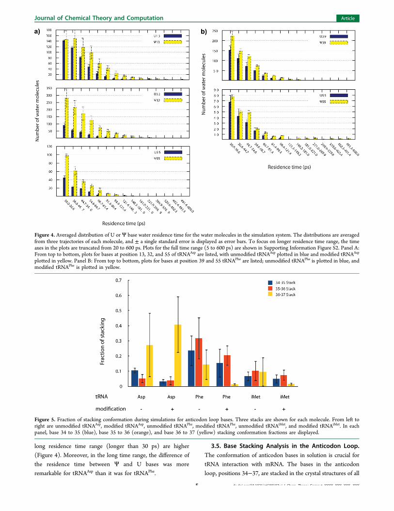
long residence time range (longer than 30 ps) are higher(Figure 4). Moreover, in the long time range, the difference ofthe residence time between Ψ and U bases was moreremarkable for tRNAAsp than it was for tRNAPhe.
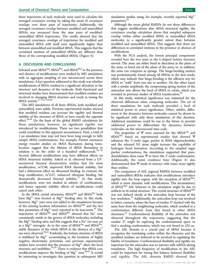
3.5. Base Stacking Analysis in the Anticodon Loop.The conformation of anticodon bases in solution is crucial fortRNA interaction with mRNA. The bases in the anticodonloop, positions 34−37, are stacked in the crystal structures of all
Figure 4. Averaged distribution of U or Ψ base water residence time for the water molecules in the simulation system. The distributions are averagedfrom three trajectories of each molecule, and ± a single standard error is displayed as error bars. To focus on longer residence time range, the timeaxes in the plots are truncated from 20 to 600 ps. Plots for the full time range (5 to 600 ps) are shown in Supporting Information Figure S2. Panel A:From top to bottom, plots for bases at position 13, 32, and 55 of tRNAAsp are listed, with unmodified tRNAAsp plotted in blue and modified tRNAAsp
plotted in yellow. Panel B: From top to bottom, plots for bases at position 39 and 55 tRNAPhe are listed; unmodified tRNAPhe is plotted in blue, andmodified tRNAPhe is plotted in yellow.
Figure 5. Fraction of stacking conformation during simulations for anticodon loop bases. Three stacks are shown for each molecule. From left toright are unmodified tRNAAsp, modified tRNAAsp, unmodified tRNAPhe, modified tRNAPhe, unmodified tRNAiMet, and modified tRNAiMet. In eachpanel, base 34 to 35 (blue), base 35 to 36 (orange), and base 36 to 37 (yellow) stacking conformation fractions are displayed.
Journal of Chemical Theory and Computation Article
dx.doi.org/10.1021/ct500107y | J. Chem. Theory Comput. XXXX, XXX, XXX−XXXF

three tRNAs studied here, with the notable exception thattRNAiMet has t6A37 not stacked. In addition, the bases atposition 37 of all three tRNAs are modified, and themodifications at this position facilitate codon-anticodoninteraction and maintain translational fidelity.17 Therefore, thefraction of time that 34 stacks on 35, 35 stacks on 36, and 36stacks on 37 were determined for all the trajectories (Figure 5)to elucidate the dynamics of stacking. During the simulation,for the stacking of 34 and 35, modified tRNAAsp (0.033 ±0.014) had a significantly (P < 0.05) lower fraction of timestacking than unmodified tRNAAsp (0.105 ± 0.018). Althoughno significant difference was detected except the stacking of 34and 35 in tRNAAsp, in general, modified tRNAs showed lowerfraction of time stacking when compared to correspondingunmodified tRNAs. Only one exception was found for tRNAAsp
at 36 and 37 stacking, modified tRNAAsp (0.408 ± 0.018)showed a higher fraction of time stacking than unmodifiedtRNAAsp (0.273 ± 0.211). These findings correspond to whatwas observed with anticodon RMSDs, i.e. anticodon residueshad more flexibility in conformation in modified tRNAs thanunmodified tRNAs3.6. Principal Component Analysis. To study correlated
motions of the tRNA structure, principal component analysis(PCA) was performed.70,71 In this study, the heavy atoms of thebackbone were used for the analysis and all three trajectories foreach molecule were analyzed together to facilitate thecomparison of modified and unmodified tRNA. The firstprincipal component represents the largest correlated atomicfluctuation in the sampled conformations. The second principalcomponent represents the next largest correlated atomicfluctuation orthogonal to the axis of the previous mode, andso on. For all the molecules in this study, the amplitudes of thefirst three principal components were comparably close.Therefore, in order to summarize the major motions, the firstthree principal components were examined. Furthermore, toavoid losing the correlations within each principal component,the first three principal components were extracted andvisualized individually. The motions observed in animationsof the first three principal components essentially involve themovements of the two L-shaped arms relative to each other.The L-shaped tertiary structure provides the flexibility that
allows the two arms to move relative to each other in threedirections, which are bending the arms as hinge-bendingmotion in the plane of the arms, bending the arms out of theplane of the arms and compressing along the axis of the helix asa spring-like motion.No striking difference was found from the correlated motions
between unmodified and modified tRNAs. In the first principalcomponent, the type of hinge-bending motion was predom-inant among all 18 trajectories; with three trajectories asexceptions: one trajectory of unmodified tRNAAsp (trajectory 2,yellow in Figures 1−3) displayed a combination of motions,with bending of the anticodon arm out of the plane and anacceptor arm spring-like motion; one trajectory of modifiedtRNAAsp (trajectory 1, red in Figures 1−3) displayed a motionbending the arms out of the plane; and one modified tRNAPhe
(trajectory 1, red in Figures 1−3) had a linear spring-likemotion for the acceptor arm and a bending planar motion forthe anticodon arm. The predominant in-plane motion betweenthe two arms of the tRNA was previously noted in normal-mode analysis.72 In the second components, the hinge-bendingmotion decreases and loses its predominance as in the firstprincipal component, and the molecules showed a combinationof all three types of motion for the two arms in the L.Interestingly, in the third principal component, the occurrenceof the spring-like motion increased noticeably as compared tothe second principal component.To assess the similarity in correlated motions between
trajectories, the covariance overlap of the principal componentswas determined.73 When the two trajectories are for identicalmolecules, the covariance overlap tests the convergence ofsampling. The covariance overlap is one only if the sampledsubspaces are identical. It is zero when the sampled subspacesare completely orthogonal. When the two trajectories are ofdifferent molecules, the covariance overlap measures thesimilarity in correlated motions. Another set of PCA analysiswas conducted to measure the covariance overlap betweentrajectories, where all atoms except those on modifications wereselected for the calculation. All-atom selection gives the bestcoverage of the molecule’s dynamics, and the exclusion ofmodification atoms allows the covariance overlap to becalculated between modified and unmodified molecules. The
Figure 6. Averaged covariance overlap between PCA of backbone heavy atoms. From left to right, three sets of bars represent tRNAAsp, tRNAPhe, andtRNAiMet, respectively. The blue bar is the averaged covariance between trajectories of unmodified molecule, the orange bar is the averagedcovariance between trajectories of modified molecules, and the yellow bar is the averaged covariance between trajectories of unmodified moleculeand modified molecule. Mean values are plotted, and a single standard deviation is shown as error bars.
Journal of Chemical Theory and Computation Article
dx.doi.org/10.1021/ct500107y | J. Chem. Theory Comput. XXXX, XXX, XXX−XXXG
three trajectories of each molecule were used to calculate theaveraged covariance overlap by taking the mean of covarianceoverlaps over three pairs of trajectories. Additionally, theaveraged covariance overlap between modified and unmodifiedtRNAs was measured from the nine pairs of modified-unmodified tRNA trajectories. The results showed that theaveraged covariance overlaps of trajectories for modified andunmodified tRNA with itself were significantly higher thanbetween unmodified and modified tRNA. This suggests that thecorrelated motions of unmodified tRNAs are different thanthose of the corresponding modified tRNAs (Figure 6).
4. DISCUSSION AND CONCLUSIONSSolvated yeast tRNAAsp, tRNAPhe, and tRNAiMet in the presenceand absence of modifications were studied by MD simulationswith an aggregate sampling of one microsecond across threesimulations. A key question addressed in this study was whethermodifications of tRNA resulted in a significant alteration in thestructure and dynamics of the molecule. Both functional andstructural studies have demonstrated that modified residues areinvolved in changing tRNA structure and result in tuning thetRNA activity.4−7,11−16,26
The MD simulations of all three tRNAs, both modified andunmodified, were stable. Previous experimental studies showedthat modified residues can increase the rigidity and thermalstability of the structure of tRNA or have exactly the oppositeeffect.18,26 On the basis of the global RMSD calculations forthese simulations, however, no significant rigidity wasintroduced by modifications. There are two possibilities thatcould contribute to this apparent inconsistency. First, a total of1 μs simulation time may not have been long enough to fullysample tRNA conformations. For example, Forster resonanceenergy transfer studies on tRNA fluctuation during trans-location suggest that the lifetime of tRNA fluctuation insolution is on the order of a hundred milliseconds.74−76
Second, different modifications may have different effects ontRNA structural stability. Ashraf et al. observed from a UV-monitored thermal denaturation studies that the stemmodification, m5C40, improved RNA thermal stability, buthad a deleterious effect on ribosomal binding. In contrast, theloop modification, m1G37, enhanced ribosome binding, butdramatically decreased thermal stability.77 In this study,modifications were not studied in subsets of combinations,and hence opposite stability effects of modifications couldcancel each other.In the tRNA crystal structures, tRNAAsp and tRNAPhe both
have Mg2+ ions located at Mg2+ binding sites. In this study,however, Mg2+ ions were not added to the simulations becauseof the missing location information on tRNAiMet and the factthat parameters of Mg2+ were reported to be inaccurate.41 Thetrajectories of tRNAAsp and tRNAPhe showed that Na+ ionsconsistently reside in the groove of tRNA molecules, includingthe Mg2+ binding sites, and this suggests that a Na+ ion may actas a Mg2+ surrogate. As in the cases of tRNAAsp and tRNAPhe,stable dynamics of the whole tRNA in the absence of a Mg2+
ion were observed.36,41 Evidently, the tertiary structure of tRNAis stabilized by Mg2+ accumulating in the locations of highlynegative electrostatic potentials, and previous experimentalstudies have revealed that the presence of Mg2+ alters the localstructure and mobilities.78,79 There is evidence that tRNA coremodifications improve the binding of Mg2+ ions.80,81 It wouldbe interesting to investigate this question in subsequent MD
simulation studies using, for example, recently reported Mg2+
parameters.82
Although the mean global RMSDs do not show differencesthat suggest modifications alter tRNA structural rigidity, thecovariance overlap calculation shows that sampled subspacesoverlap within either modified tRNA or unmodified tRNAmolecules to a significantly greater extent than betweenmodified and unmodified tRNA. This suggests that there aredifferences in correlated motions in the presence or absence ofmodifications.With the PCA analysis, the lowest principal components
revealed how the two arms in the L-shaped tertiary structuremoved. The arms can either bend in directions in the plane ofthe arms, or bend out of the plane of the arms, or, in addition,the arms can compress as springs. The hinge-bending motionwas predominantly found among all tRNAs in the first mode,which may indicate that hinge-bending is the efficient way fortRNA to “walk” from one site to another. On the other hand,with a similar amplitude, the compressing spring motion of theanticodon arm allows the head of tRNA to rotate, which wasrevealed in previous studies of tRNA translocation.83
In this study, statistical significance was estimated for theobserved differences when comparing molecules. The set ofthree simulations for each molecule provided a level ofstatistical power to assess significance. Other, subtler, differ-ences in the dynamics between molecules cannot be shown tobe significant with only three simulations of this duration.Additional simulations could be run in the future to provideadditional power to differentiate the dynamics betweenmolecules on the microsecond time scale.The properties of Ψ were assessed for the tRNAAsp and
tRNAPhe based on experimental studies that showed Ψenhances the 3′-endo sugar pucker in RNA oligonucleotidesand the released N1 atom might increase the capability ofhydrogen bond formation. According to the sampled sugarpucker conformations, the simulations supported that pseu-douridylation does cause Ψ to favor the 3′-endo conformation.Additionally, the water residence time (Figure 4) alsodemonstrated that Ψ tends to interact with water more tightlythan uridine.The comparison of ASL regional RMSDs between modified
and unmodified tRNAs indicates that modifications introducerigidity into the loop region, with the exception of tRNAiMet,which is more dynamic with modifications. The inconsistencyof tRNAiMet ASL behavior in the simulation might be due toartifacts in its initial structure. The crystal structure of tRNAiMet
was not defined clearly at the anticodon loop because of thelow resolution.21 Additionally, the anticodon loop was involvedin lattice contacts, where the base of residue 37 stacked with thesame base from the neighboring molecules, which resulted in aconformation different from that found in other tRNAstructures.21 Conformational flexibility of the anticodon wasobserved throughout the trajectories, suggesting that theresidue 37 might be exploring the conformational space tofind a stacking conformation, which was not found in 333 ns.The ASL domain is a crucial part of tRNA because it
recognizes the translating codon within the ribosome and themodified residues are believed to be involved in regulating thefidelity of translation. Conformational flexibility and rigidity areimportant for the anticodon arm to interact with mRNA duringtranslation. The high frequency of modification in the ASLcould be important for tuning this balance between flexibilityand rigidity. The ASL domain RMSD showed that
Journal of Chemical Theory and Computation Article
dx.doi.org/10.1021/ct500107y | J. Chem. Theory Comput. XXXX, XXX, XXX−XXXH

modifications increased the rigidity of the backbone of the ASLdomain (Figure 2), which was mostly an influence on the loopregion. It was observed (by RMSD) that the stem region forboth modified and unmodified tRNAs are stiff and difficult todifferentiate (data not shown). The all-atom RMSD calculationof the anticodon, however, showed that modifications tended toincrease the dynamics of anticodon residues (Figure 3). Takentogether, these findings suggest an arm and hand model for theASL. The ASL domain of tRNA works as an arm. The stem isstiff for translocation during translation, and the loop domain isa palm that is structured, but can move in relationship to thehelix, as noted in previous coarse-grained studies,72,84 to be ableto position the anticodon. The anticodon residues then work asfingers to recognize the right codon. Flexibility is the keyfeature for the anticodon, and modification strengthens thisproperty by introducing dynamics.Although it was observed that some modified residues
showed distinct properties as compared to unmodified residues,in this study only local tRNA features were found significantlyaltered. One explanation could be that the tRNA moleculeworks as an adaptor, interacting with other molecular devices,such as aminoacyl-tRNA synthetases, ribosomes, and mRNAs.The modified nucleosides may alter the interface betweentRNA and its interactive components, such as the interactionsstudied for tRNA interacting with the ribosome.85 Changes indynamics caused by interactions of modifications with othercomponents would be difficult to capture in the scope of thisstudy. Additional studies that simulated modified andunmodified tRNA interacting with other components oftranslation machinery would be interesting and might elucidatefurther roles for covalent modifications.
■ ASSOCIATED CONTENT
*S Supporting InformationFigures of sugar pucker pseudorotation values as a function oftime for U or Ψ base, figures of the averaged distribution of Uor Ψ base water residence time, table of missing parameterassignments, table of partial charges for 17 nonstandardresidues, AMBER force field files for 17 nonstandard residues,table of svd eigenvectors and eigenvalues, movies for eachtrajectory of first, second, and third principal components. Thismaterial is available free of charge via the Internet at http://pubs.acs.org.
■ AUTHOR INFORMATION
Corresponding Author*E-mail: [email protected].
FundingThis study was supported by National Institutes of HealthGrants R01GM076485 to D.H.M. and R01GM052347 toE.M.P. R.C.W. acknowledges funding from the NationalScience Foundation (NSF) through the Scientific SoftwareInnovations Institutes program NSF SI2-SSE (NSF114876) aswell as a fellowship from NVIDIA Inc. Computer time wasprovided by the San Diego Supercomputer facility and theUniversity of Rochester Center for Integrated ResearchComputing.
NotesThe authors declare no competing financial interest.
■ ACKNOWLEDGMENTSThe authors thank Dejun Lin and Tod D. Romo for help usingLOOS and Alan Grossfield for helpful discussions.
■ REFERENCES(1) Phizicky, E. M.; Alfonzo, J. D. Do all modifications benefit alltRNAs? FEBS Lett. 2010, 584, 265−271.(2) Sprinzl, M.; Vassilenko, K. S. Compilation of tRNA sequencesand sequences of tRNA genes. Nucleic Acids Res. 2005, 33, D139−D140.(3) Cantara, W. A.; Crain, P. F.; Rozenski, J.; McCloskey, J. A.;Harris, K. A.; Zhang, X.; Vendeix, F. A.; Fabris, D.; Agris, P. F. TheRNA modification database, RNAMDB: 2011 update. Nucleic AcidsRes. 2011, 39, D195−D201.(4) Persson, B. C. Modification of tRNA as a regulatory device. Mol.Microbiol. 1993, 8, 1011−1016.(5) Bjork, G. R.; Durand, J.; Hagervall, T. G.; Lundgren, H. K.;Nilsson, K.; Chen, P.; Qian, Q.; Urbonavicius, J. Transfer RNAmodification: Influence on translational frameshifting and metabolism.FEBS Lett. 1999, 452, 47−51.(6) Urbonavicius, J.; Qian, Q.; Durand, J. M.; Hagervall, T. G.; Bjork,G. R. Improvement of reading frame maintenance is a commonfunction for several tRNA modifications. EMBO J. 2001, 20, 4863−4873.(7) Hagervall, T. G.; Tuohy, T. M.; Atkins, J. F.; Bjork, G. R.Deficiency of 1-methylguanosine in tRNA from Salmonella typhimu-rium induces frameshifting by quadruplet translocation. J. Mol. Biol.1993, 232, 756−765.(8) Holley, R. W.; Apgar, J.; Everett, G. A.; Madison, J. T.;Marquisee, M.; Merrill, S. H.; Penswick, J. R.; Zamir, A. Structure of aribonucleic acid. Science 1965, 147, 1462−1465.(9) Kim, S.; Suddath, F.; Quigley, G.; McPherson, A.; Sussman, J.;Wang, A.; Seeman, N.; Rich, A. Three-dimensional tertiary structure ofyeast phenylalanine transfer RNA. Science 1974, 185, 435−440.(10) Robertus, J.; Ladner, J. E.; Finch, J.; Rhodes, D.; Brown, R.;Clark, B.; Klug, A. Structure of yeast phenylalanine tRNA at 3 Åresolution. Nature 1974, 250, 546−551.(11) Sampson, J. R.; Uhlenbeck, O. C. Biochemical and physicalcharacterization of an unmodified yeast phenylalanine transfer RNAtranscribed in vitro. Proc. Natl. Acad. Sci. U. S. A. 1988, 85, 1033−1037.(12) Chu, W. C.; Horowitz, J. 19F NMR of 5-fluorouracil-substitutedtransfer RNA transcribed in vitro: Resonance assignment offluorouracil−guanine base pairs. Nucleic Acids Res. 1989, 17, 7241−7252.(13) Hall, K. B.; Sampson, J. R.; Uhlenbeck, O. C.; Redfield, A. G.Structure of an unmodified tRNA molecule. Biochemistry 1989, 28,5794−5801.(14) Perret, V.; Garcia, A.; Puglisi, J.; Grosjean, H.; Ebel, J.; Florentz,C.; Giege, R. Conformation in solution of yeast tRNAAsp transcriptsdeprived of modified nucleotides. Biochimie 1990, 72, 735−743.(15) Derrick, W. B.; Horowitz, J. Probing structural differencesbetween native and in vitro transcribed Escherichia coli valine transferRNA: Evidence for stable base modification-dependent conformers.Nucleic Acids Res. 1993, 21, 4948−4953.(16) Harrington, K. M.; Nazarenko, I. A.; Dix, D. B.; Thompson, R.C.; Uhlenbeck, O. C. In vitro analysis of translational rate and accuracywith an unmodified tRNA. Biochemistry 1993, 32, 7617−7622.(17) Agris, P. F.; Vendeix, F. A.; Graham, W. D. tRNA’s wobbledecoding of the genome: 40 years of modification. J. Mol. Biol. 2007,366, 1−13.(18) Motorin, Y.; Helm, M. tRNA stabilization by modifiednucleotides. Biochemistry 2010, 49, 4934−4944.(19) Anderson, J.; Phan, L.; Cuesta, R.; Carlson, B. A.; Pak, M.;Asano, K.; Bjork, G. R.; Tamame, M.; Hinnebusch, A. G. The essentialGcd10p−Gcd14p nuclear complex is required for 1-methyladenosinemodification and maturation of initiator methionyl-tRNA. Genes Dev.1998, 12, 3650−3662.
Journal of Chemical Theory and Computation Article
dx.doi.org/10.1021/ct500107y | J. Chem. Theory Comput. XXXX, XXX, XXX−XXXI
(20) Kadaba, S.; Krueger, A.; Trice, T.; Krecic, A. M.; Hinnebusch, A.G.; Anderson, J. Nuclear surveillance and degradation of hypomodifiedinitiator tRNAMet in S. cerevisiae. Genes Dev. 2004, 18, 1227−1240.(21) Basavappa, R.; Sigler, P. B. The 3 A crystal structure of yeastinitiator tRNA: Functional implications in initiator/elongator discrim-ination. EMBO J. 1991, 10, 3105−3111.(22) Auffinger, P.; Louise-May, S.; Westhof, E. Molecular dynamicssimulations of solvated yeast tRNAAsp. Biophys. J. 1999, 76, 50−64.(23) Cabello-Villegas, J.; Winkler, M. E.; Nikonowicz, E. P. Solutionconformations of unmodified and A37 N6-dimethylallyl modifiedanticodon stem-loops of Escherichia coli tRNAPhe. J. Mol. Biol. 2002,319, 1015−1034.(24) Stuart, J. W.; Koshlap, K. M.; Guenther, R.; Agris, P. F.Naturally-occurring modification restricts the anticodon domainconformational space of tRNAPhe. J. Mol. Biol. 2003, 334, 901−918.(25) Durant, P. C.; Bajji, A. C.; Sundaram, M.; Kumar, R. K.; Davis,D. R. Structural effects of hypermodified nucleosides in the Escherichiacoli and human tRNALys anticodon loop: the effect of nucleosidess2U, mcm5U, mcm5s2U, mnm5s2U, t6A, and ms2t6A. Biochemistry 2005,44, 8078−8089.(26) Kawai, G.; Yamamoto, Y.; Kamimura, T.; Masegi, T.; Sekine,M.; Hata, T.; Iimori, T.; Watanabe, T.; Miyazawa, T.; Yokoyama, S.Conformational rigidity of specific pyrimidine residues in tRNA arisesfrom posttranscriptional modifications that enhance steric interactionbetween the base and the 2′-hydroxyl group. Biochemistry 1992, 31,1040−1046.(27) Ge, J.; Yu, Y. T. RNA pseudouridylation: New insights into anold modification. Trends Biochem. Sci. 2013, 38, 210−218.(28) Griffey, R. H.; Davis, D.; Yamaizumi, Z.; Nishimura, S.; Bax, A.;Hawkins, B.; Poulter, C. 15N-Labeled Escherichia coli tRNAfMet,tRNAGlu, tRNATyr, and tRNAPhe. Double resonance and two-dimensional NMR of N1-labeled pseudouridine. J. Biol. Chem. 1985,260, 9734−9741.(29) Davis, D. R.; Poulter, C. D. 1H-15N NMR studies of Escherichiacoli tRNAPhe from HisT mutants: A structural role for pseudouridine.Biochemistry 1991, 30, 4223−4231.(30) Hall, K. B.; McLaughlin, L. W. Properties of a U1/mRNA5′splice site duplex containing pseudouridine as measured bythermodynamic and NMR methods. Biochemistry 1991, 30, 1795−1801.(31) Hall, K. B.; McLaughlin, L. W. Properties of pseudouridine N1imino protons located in the major groove of an A-form RNA duplex.Nucleic Acids Res. 1992, 20, 1883−1889.(32) Davis, D. R. Stabilization of RNA stacking by pseudouridine.Nucleic Acids Res. 1995, 23, 5020−5026.(33) Auffinger, P.; Westhof, E. RNA hydration: three nanoseconds ofmultiple molecular dynamics simulations of the solvated tRNAAsp
anticodon hairpin. J. Mol. Biol. 1997, 269, 326−341.(34) Durant, P. C.; Davis, D. R. Stabilization of the anticodon stem-loop of tRNALys,3 by an A+-C base-pair and by pseudouridine. J. Mol.Biol. 1999, 285, 115−131.(35) Harvey, S. C.; Prabhakaran, M.; Mao, B.; McCammon, J. A.Phenylalanine transfer RNA: Molecular dynamics simulation. Science1984, 223, 1189−1191.(36) Auffinger, P.; Louise-May, S.; Westhof, E. Molecular dynamicssimulations of the anticodon hairpin of tRNAAsp: Structuring effects ofCH···O hydrogen bonds and of long-range hydration forces. J. Am.Chem. Soc. 1996, 118, 1181−1189.(37) Auffinger, P.; Westhof, E. H-bond stability in the tRNA (Asp)anticodon hairpin: 3 ns of multiple molecular dynamics simulations.Biophys. J. 1996, 71, 940−954.(38) McCrate, N. E.; Varner, M. E.; Kim, K. I.; Nagan, M. C.Molecular dynamics simulations of human tRNAUUU
Lys,3 : The role ofmodified bases in mRNA recognition. Nucleic Acids Res. 2006, 34,5361−5368.(39) Westhof, E.; Dumas, P.; Moras, D. Restrained refinement of twocrystalline forms of yeast aspartic acid and phenylalanine transfer RNAcrystals. Acta Crystallogr. A 1988, 44, 112−124.
(40) Shi, H.; Moore, P. B. The crystal structure of yeastphenylalanine tRNA at 1.93 Å resolution: A classic structure revisited.RNA 2000, 6, 1091−1105.(41) Lahiri, A.; Nilsson, L. Molecular dynamics of the anticodondomain of yeast tRNAPhe: Codon−anticodon interaction. Biophys. J.2000, 79, 2276−2289.(42) Salomon-Ferrer, R.; Case, D. A.; Walker, R. C. An overview ofthe Amber biomolecular simulation package. Wiley Interdiscip. Rev.:Comput. Mol. Sci. 2012, DOI: 10.1002/wcms.1121.(43) Case, D.; Darden, T.; Cheatham, T., III; Simmerling, C.; Wang,J.; Duke, R.; Luo, R.; Walker, R.; Zhang, W.; Merz, K. AMBER 11;University of California: San Francisco, CA, 2010.(44) Case, D.; Darden, T.; Cheatham, T., III; Simmerling, C.; Wang,J.; Duke, R.; Luo, R.; Crowley, M.; Walker, R. C.; Zhang, W. AMBER10; University of California: San Francisco, CA, 2008.(45) Cieplak, P.; Cornell, W. D.; Bayly, C.; Kollman, P. A.Application of the multimolecule and multiconformational RESPmethodology to biopolymers: charge derivation for DNA, RNA, andproteins. J. Comput. Chem. 1995, 16, 1357−1377.(46) Hehre, W. J.; Radom, L.; Schleyer, P. v. R.; Pople, J. A. Ab InitioMolecular Orbital Theory; Wiley: New York, 1986; Vol. 33.(47) Hariharan, P. C.; Pople, J. A. The influence of polarizationfunctions on molecular orbital hydrogenation energies. Theor. Chim.Acta 1973, 28, 213−222.(48) Hehre, W. J.; Lathan, W. A. Self-consistent molecular orbitalmethods. XIV. An extended Gaussian-type basis for molecular orbitalstudies of organic molecules. Inclusion of second row elements. J.Chem. Phys. 1972, 56, 5255−5257.(49) Ditchfield, R.; Hehre, W.; Pople, J. A. Self-consistent molecular-orbital methods. IX. An extended Gaussian-type basis for molecular-orbital studies of organic molecules. J. Chem. Phys. 1971, 54, 724−728.(50) Pearlman, D. A.; Case, D. A.; Caldwell, J. W.; Ross, W. S.;Cheatham, T. E., III; DeBolt, S.; Ferguson, D.; Seibel, G.; Kollman, P.AMBER, A package of computer programs for applying molecularmechanics, normal mode analysis, molecular dynamics and free energycalculations to simulate the structural and energetic properties ofmolecules. Comput. Phys. Commun. 1995, 91, 1−41.(51) Wang, J.; Wolf, R. M.; Caldwell, J. W.; Kollman, P. A.; Case, D.A. Development and testing of a general amber force field. J. Comput.Chem. 2004, 25, 1157−74.(52) Case, D. A.; Cheatham, T. E.; Darden, T.; Gohlke, H.; Luo, R.;Merz, K. M.; Onufriev, A.; Simmerling, C.; Wang, B.; Woods, R. J. TheAmber biomolecular simulation programs. J. Comput. Chem. 2005, 26,1668−1688.(53) Bryce, R. AMBER parameter database. http://www.pharmacy.manchester.ac.uk/bryce/amber (accessed September 3, 2010).(54) Jorgensen, W. L.; Chandrasekhar, J.; Madura, J. D.; Impey, R.W.; Klein, M. L. Comparison of simple potential functions forsimulating liquid water. J. Chem. Phys. 1983, 79, 926−935.(55) Ryckaert, J. P.; Ciccotti, G.; Berendsen, H. J. Numericalintegration of the cartesian equations of motion of a system withconstraints: Molecular dynamics of n-alkanes. J. Comput. Phys. 1977,23, 327−341.(56) Darden, T.; York, D.; Pedersen, L. Particle Mesh EwaldAn N·Log(N) method for Ewald sums in large systems. J. Chem. Phys. 1993,98, 10089−10092.(57) Berendsen, H. J.; Postma, J. P. M.; van Gunsteren, W. F.;DiNola, A.; Haak, J. Molecular dynamics with coupling to an externalbath. J. Chem. Phys. 1984, 81, 3684−3690.(58) Loncharich, R. J.; Brooks, B. R.; Pastor, R. W. Langevindynamics of peptides: The frictional dependence of isomerization ratesof N-acetylalanyl-N′-methylamide. Biopolymers 1992, 32, 523−535.(59) Dixit, S. B.; Beveridge, D. L.; Case, D. A.; Giudice, E.; Lankas,F.; Lavery, R.; Maddocks, J. H.; Osman, R.; Sklenar, H.; Thayer, K. M.Molecular dynamics simulations of the 136 unique tetranucleotidesequences of DNA oligonucleotides. II: Sequence context effects onthe dynamical structures of the 10 unique dinucleotide steps. Biophys.J. 2005, 89, 3721−3740.
Journal of Chemical Theory and Computation Article
dx.doi.org/10.1021/ct500107y | J. Chem. Theory Comput. XXXX, XXX, XXX−XXXJ
(60) Roe, D. R.; Cheatham, T. E., III PTRAJ and CPPTRAJ:Software for processing and analysis of molecular dynamics trajectorydata. J. Chem. Theory Comput. 2013, 9, 3084−3095.(61) Romo, T. D.; Grossfield, A. LOOS: An extensible platform forthe structural analysis of simulations. Proceedings of the AnnualInternational Conference of the IEEE Engineering in Medicine and BiologySociety, 2009 (EMBC 2009); Institute of Electrical and ElectronicsEngineers: New York, 2009; pp 21332−2335.(62) Humphrey, W.; Dalke, A.; Schulten, K. VMD: Visual moleculardynamics. J. Mol. Graphics 1996, 14, 33−38.(63) DeLano, W. L. Pymol: An open-source molecular graphics tool.CCP4 Newsl. Protein Crystallogr. 2002, 40 (March), No. Article 11,http://www.ccp4.ac.uk/newsletters/newsletter40.pdf.(64) Altona, C. T.; Sundaralingam, M. Conformational analysis of thesugar ring in nucleosides and nucleotides. New description using theconcept of pseudorotation. J. Am. Chem. Soc. 1972, 94, 8205−8212.(65) Impey, R.; Madden, P.; McDonald, I. Hydration and mobility ofions in solution. J. Phys. Chem. 1983, 87, 5071−5083.(66) Olson, W. K.; Dasika, R. D. Spatial configuration of orderedpolynucleotide chains. 3. Polycyclonucleotides. J. Am. Chem. Soc. 1976,98, 5371−5380.(67) Romo, T.; Clarage, J.; Sorensen, D.; Phillips, G. Automaticidentification of discrete substates in proteins: Singular valuedecomposition analysis of time-averaged crystallographic refinements.Proteins: Struct., Funct., Bioinf. 1995, 22, 311−321.(68) Romo, T. D.; Grossfield, A. Block covariance overlap methodand convergence in molecular dynamics simulation. J. Chem. TheoryComput. 2011, 7, 2464−2472.(69) R Core Team, R Foundation for Statistical Computing. R: Alanguage and environment for statistical computing, 2012. http://cran.r-project.org.(70) Garcia, A. E. Large-amplitude nonlinear motions in proteins.Phys. Rev. Lett. 1992, 68, 2696−2699.(71) Amadei, A.; Linssen, A.; Berendsen, H. J. Essential dynamics ofproteins. Proteins: Struct., Funct., Bioinf. 1993, 17, 412−425.(72) Matsumoto, A.; Tomimoto, M.; Go, N. Dynamical structure oftransfer RNA studied by normal mode analysis. Eur. Biophys. J. 1999,28, 369−379.(73) Hess, B. Convergence of sampling in protein simulations. Phys.Rev. E 2002, 65, 031910.(74) Blanchard, S. C.; Gonzalez, R. L.; Kim, H. D.; Chu, S.; Puglisi, J.D. tRNA selection and kinetic proofreading in translation. Nat. Struct.Mol. Biol. 2004, 11, 1008−1014.(75) Blanchard, S. C.; Kim, H. D.; Gonzalez, R. L.; Puglisi, J. D.; Chu,S. tRNA dynamics on the ribosome during translation. Proc. Natl.Acad. Sci. U. S. A. 2004, 101, 12893−12898.(76) Kim, H. D.; Puglisi, J. D.; Chu, S. Fluctuations of transfer RNAsbetween classical and hybrid states. Biophys. J. 2007, 93, 3575−3582.(77) Ashraf, S. S.; Guenther, R. H.; Ansari, G.; Malkiewicz, A.;Sochacka, E.; Agris, P. F. Role of modified nucleosides of yeasttRNAPhe in ribosomal binding. Cell Biochem. Biophys. 2000, 33, 241−252.(78) Roh, J. H.; Tyagi, M.; Briber, R. M.; Woodson, S. A.; Sokolov, A.P. The dynamics of unfolded versus folded tRNA: the role ofelectrostatic interactions. J. Am. Chem. Soc. 2011, 133, 16406−9.(79) Serebrov, V.; Clarke, R. J.; Gross, H. J.; Kisselev, L. Mg2+-Induced tRNA folding. Biochemistry 2001, 40, 6688−98.(80) Chen, Y.; Sierzputowska-Gracz, H.; Guenther, R.; Everett, K.;Agris, P. F. 5-Methylcytidine is required for cooperative binding ofMg2+ and a conformational transition at the anticodon stem-loop ofyeast phenylalanine tRNA. Biochemistry 1993, 32, 10249−10253.(81) Jones, C. I.; Spencer, A. C.; Hsu, J. L.; Spremulli, L. L.; Martinis,S. A.; DeRider, M.; Agris, P. F. A counterintuitive Mg2+-dependent andmodification-assisted functional folding of mtochondrial tRNAs. J. Mol.Biol. 2006, 362, 771−786.(82) Allner, O.; Nilsson, L.; Villa, A. Magnesium ion-watercoordination and exchange in biomolecular simulations. J. Chem.Theory Comput. 2012, 8, 1493−1502.
(83) Noller, H. F.; Yusupov, M. M.; Yusupova, G. Z.; Baucom, A.;Cate, J. Translocation of tRNA during protein synthesis. FEBS Lett.2002, 514, 11−16.(84) Wang, Y.; Jernigan, R. L. Comparison of tRNA motions in thefree and ribosomal bound structures. Biophys. J. 2005, 89, 3399−3409.(85) Whitford, P. C.; Blanchard, S. C.; Cate, J. H.; Sanbonmatsu, K.Y. Connecting the kinetics and energy landscape of tRNA trans-location on the ribosome. PLoS Comput. Biol. 2013, 9, No. e1003003.
Journal of Chemical Theory and Computation Article
dx.doi.org/10.1021/ct500107y | J. Chem. Theory Comput. XXXX, XXX, XXX−XXXK