invasive validation of antares, a new algorithm to
TRANSCRIPT

Journal of
Clinical Medicine
Article
Invasive Validation of Antares, a New Algorithm toCalculate Central Blood Pressure from OscillometricUpper Arm Pulse Waves
Marcus Dörr 1,2 , Stefan Richter 3, Siegfried Eckert 4, Marc-Alexander Ohlow 3,Fabian Hammer 1, Astrid Hummel 1, Vivien Dornberger 1, Elisabeth Genzel 1
and Johannes Baulmann 5,6,*1 Department of Internal Medicine B, University Medicine Greifswald, Ferdinand-Sauerbruch-Straße,
D-17475 Greifswald, Germany2 German Centre for Cardiovascular Research (DZHK), Partner Site Greifswald, D-17475 Greifswald, Germany3 Zentralklinik Bad Berka GmbH, Herzzentrum, Department of Cardiology, Robert-Koch-Allee 9,
D-99437 Bad Berka, Germany4 Klinik für Allgemeine und Interventionelle Kardiologie/Angiologie, Universitätsklinik der Ruhr-Universität
Bochum, Georgstraße 11, D-32545 Bad Oeynhausen, Germany5 EPC—European Prevention Center, Luise-Rainer-Straße 6-10, D-40235 Düsseldorf, Germany6 Department of Medical Psychology and Psychotherapy, Medical University of Graz, Auenbruggerplatz 2/8,
A-8036 Graz, Austria* Correspondence: [email protected]
Received: 20 June 2019; Accepted: 18 July 2019; Published: 22 July 2019�����������������
Abstract: Background: Antares is an algorithm for pulse wave analysis (PWA) by oscillometricblood pressure (BP) monitors in order to estimate central (aortic) blood pressure (cBP). Antaresaims to enable brachial cuff-based BP monitors to be type II-devices, determining absolute cBPvalues independently of potential peripheral BP inaccuracies. The present study is an invasivevalidation of the Antares algorithm in the custo screen 400. Methods: We followed entirely the2017 ARTERY protocol for validation of non-invasive cBP devices, the 2013 American NationalStandards Institute, Inc./Association for the Advancement of Medical Instrumentation/InternationalOrganization for Standardization (ANSI/AAMI/ISO) 81060-2 and 2018 AAMI/European Society ofHypertension (ESH)/ISO validation standard protocols. In total, 191 patients undergoing cardiaccatheterization were included, of which 145 patients entered analysis. Invasive cBP recordings werecompared to simultaneous non-invasive cBP estimations using the Antares algorithm, integratedinto an oscillometric BP monitor. Results: Mean difference between invasive and non-invasivelyestimated systolic cBP was 0.71 mmHg with standard deviation of 5.95 mmHg, fulfilling highestvalidation criteria. Conclusion: Antares is the first algorithm for estimation of cBP that entirely fulfillsthe 2017 ARTERY and AAMI/ESH/ISO validation protocols. The Antares algorithm turns the custoscreen 400 BP monitor into a type II-device. Integration of Antares into commercially available BPmonitors could make it possible to measure PWA parameters in virtually every practice in future.
Keywords: validation; invasive; central blood pressure; pulse wave analysis; antares
1. Introduction
Central blood pressure (cBP) is not the same as peripheral blood pressure (pBP) [1]. Basically, in ayoung, healthy arterial system cBP is low and pBP much higher [1]. When an individual is gettingolder, cBP rises and may reach the same level or probably even higher cBP levels than pBP [2]. Severalstudies have shown that cBP is more strongly related to hypertension-related organ damage and
J. Clin. Med. 2019, 8, 1073; doi:10.3390/jcm8071073 www.mdpi.com/journal/jcm

J. Clin. Med. 2019, 8, 1073 2 of 15
outcome than pBP [3–5]. According to the current European Society of Hypertension/European Societyof Cardiology (ESH/ESC) hypertension guidelines, measurement of cBP has practical consequencesfor the treatment of young patients with isolated systolic hypertension [6]. For these individuals, nomedication is necessary if cBP is low. Thus, a potentially huge overtreatment can be avoided. A conditiosine qua non should be that cBP is estimated accurately.
For the first time, in 2017 a worldwide consensus paper under the leadership of the ARTERYsociety was published as a task force consensus statement on protocol standardization for the validationof non-invasive central pressure devices [7]. A main issue was that the reference standard againstwhich the device accuracy of central BP estimations is gauged should be intra-arterial (invasive)catheter in the ascending aorta because “currently there are no non-invasive alternatives”. At the sametime, procedures were defined for (a) the proper performance of the invasive pressure recordings andthe technical requirements, (b) non-invasive central BP device measurement standards, (c) samplecharacteristics, and (d) statistical requirements.
The existing devices were categorized as type I or type II devices. Type I means that an estimate ofcentral BP relative to the measured brachial BP is given in order to concentrate on a relatively accuratepressure difference between central and peripheral sites. Type II means that these devices estimatethe intra-arterial central BP, what could be understood as the “true” central BP, despite the knowninaccuracy of peripheral BP measurements compared to invasive measurements [8].
The ANSI/AAMI/ISO 81060-2:2013 (American National Standards Institute, Inc. and Associationfor the Advancement of Medical Instrumentation) was developed by the International Organization forStandardization (ISO)/TC 121/SC 3, the International Electrotechnical Commission (IEC)/SC 62D JointWorking Group (JWG) 7 on non-invasive BP monitoring equipment and the AAMI SphygmomanometerCommittee. The objective of the standard is to provide minimum labeling, performance, and safetyrequirements for the clinical validation of medical electrical equipment used for estimation of thearterial blood pressure by utilizing a cuff. Most, but not all, of the ANSI/AAMI/ISO 81060-2:2013recommendations are included in the ARTERY Society task force consensus statement on protocolstandardization for validation of non-invasive central BP devices [9]. 2018 a statement was publishedthat presents the key aspects of a validation procedure, which were agreed by the AAMI, ESH andISO representatives as the basis for a single universal validation protocol [10]. Because in the 2018statements there are no rules for invasive reference measurements, we did not stop referring to the 2013ANSI/AAMI/ISO protocol with integrated criteria for hemodynamic stability of invasive measurementsand finally followed both protocols.
Antares is an algorithm for calculating central blood pressure values, which can be integrated intoan oscillometric device at the upper arm. Antares aims to enable an oscillometric BP monitor to actas a type II-device, thereby providing “true” invasive central BP values. Antares was developed byRedwave Medical GmbH, Jena, Germany.
The study aims is to invasively validate the central blood pressure calculation of the Antaresalgorithm according to the ARTERY validation protocol as well as the 2013 ANSI/AAMI/ISO 81060-2and the 2018 AAMI/ESH/ISO standard using an oscillometric BP monitor at the upper arm.
2. Material and Methods
We followed entirely the 2017 ARTERY society task force consensus statement on protocolstandardization for the validation of non-invasive central blood pressure devices, the 2013ANSI/AAMI/ISO 81060-2 and the 2018 AAMI/ESH/ISO standard.
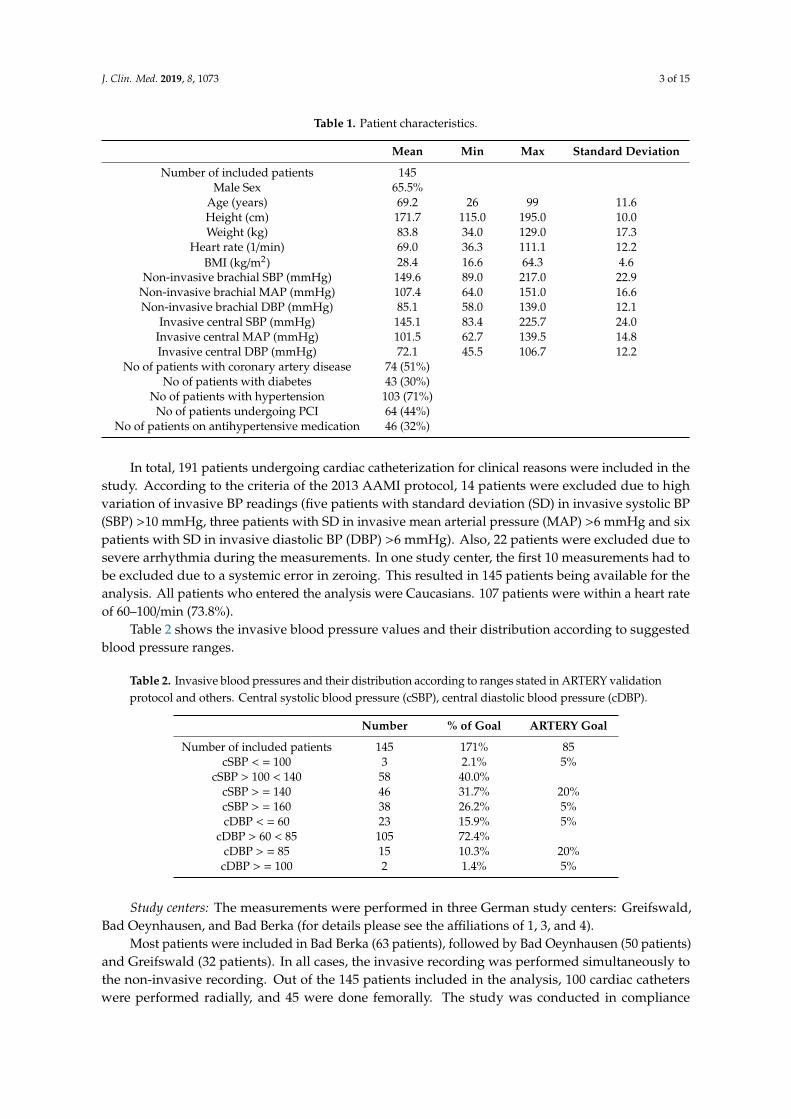
Table 1: Patient characteristics. Blood pressure (BP) range (systolic, diastolic and meanperipheral and invasive blood pressures), body mass index (BMI), systolic blood pressure (SBP),mean arterial pressure (MAP), diastolic blood pressure (DBP), percutaneous coronary intervention(PCI). The non-invasive brachial measurements presented in Table 1 are those recorded at the time ofthe angiogram.
J. Clin. Med. 2019, 8, 1073 3 of 15
Table 1. Patient characteristics.
Mean Min Max Standard Deviation
Number of included patients 145Male Sex 65.5%
Age (years) 69.2 26 99 11.6Height (cm) 171.7 115.0 195.0 10.0Weight (kg) 83.8 34.0 129.0 17.3
Heart rate (1/min) 69.0 36.3 111.1 12.2BMI (kg/m2) 28.4 16.6 64.3 4.6
Non-invasive brachial SBP (mmHg) 149.6 89.0 217.0 22.9Non-invasive brachial MAP (mmHg) 107.4 64.0 151.0 16.6Non-invasive brachial DBP (mmHg) 85.1 58.0 139.0 12.1
Invasive central SBP (mmHg) 145.1 83.4 225.7 24.0Invasive central MAP (mmHg) 101.5 62.7 139.5 14.8Invasive central DBP (mmHg) 72.1 45.5 106.7 12.2
No of patients with coronary artery disease 74 (51%)No of patients with diabetes 43 (30%)
No of patients with hypertension 103 (71%)No of patients undergoing PCI 64 (44%)
No of patients on antihypertensive medication 46 (32%)
In total, 191 patients undergoing cardiac catheterization for clinical reasons were included in thestudy. According to the criteria of the 2013 AAMI protocol, 14 patients were excluded due to highvariation of invasive BP readings (five patients with standard deviation (SD) in invasive systolic BP(SBP) >10 mmHg, three patients with SD in invasive mean arterial pressure (MAP) >6 mmHg and sixpatients with SD in invasive diastolic BP (DBP) >6 mmHg). Also, 22 patients were excluded due tosevere arrhythmia during the measurements. In one study center, the first 10 measurements had tobe excluded due to a systemic error in zeroing. This resulted in 145 patients being available for theanalysis. All patients who entered the analysis were Caucasians. 107 patients were within a heart rateof 60–100/min (73.8%).
Table 2 shows the invasive blood pressure values and their distribution according to suggestedblood pressure ranges.
Table 2. Invasive blood pressures and their distribution according to ranges stated in ARTERY validationprotocol and others. Central systolic blood pressure (cSBP), central diastolic blood pressure (cDBP).
Number % of Goal ARTERY Goal
Number of included patients 145 171% 85cSBP < = 100 3 2.1% 5%
cSBP > 100 < 140 58 40.0%cSBP > = 140 46 31.7% 20%cSBP > = 160 38 26.2% 5%cDBP < = 60 23 15.9% 5%
cDBP > 60 < 85 105 72.4%cDBP > = 85 15 10.3% 20%
cDBP > = 100 2 1.4% 5%
Study centers: The measurements were performed in three German study centers: Greifswald,Bad Oeynhausen, and Bad Berka (for details please see the affiliations of 1, 3, and 4).
Most patients were included in Bad Berka (63 patients), followed by Bad Oeynhausen (50 patients)and Greifswald (32 patients). In all cases, the invasive recording was performed simultaneously tothe non-invasive recording. Out of the 145 patients included in the analysis, 100 cardiac catheterswere performed radially, and 45 were done femorally. The study was conducted in compliance

J. Clin. Med. 2019, 8, 1073 4 of 15
with the Declaration of Helsinki. Ethics approval was obtained from the local ethics committees.All participants gave their informed consent for inclusion before they participated in the study.
Study setting: All measurements were performed in the cardiac catheterization laboratory,with constant temperature, without excessive ambient noise from monitoring devices. The invasiveand non-invasive measurements were performed at the end of each cardiac catheter examinationand were completely simultaneous. Thus, the patient was adapted to the environment and withoutdisturbing influences. Data acquisition was done in a period of undisturbed rest, free from acutehemodynamic interventions, free from acute medication changes and without talking.
Non-invasive central BP device measurement: All non-invasive measurements were performed withthe custo screen 400 device (custo med GmbH, Ottobrunn, Germany), with the integrated Antaresalgorithm to calculate central BP on a connected laptop. The familiarization with the equipmenttook only a few minutes in each study center because it is the process of a normal blood pressuremeasurement. The oscillometric device in its original version is built for 24 h-ambulatory blood pressuremonitoring. The custo screen 400 is validated for peripheral blood pressure measurement accordingto ESH-IP 2010 [11]. Cuff size was chosen according to the directives given by the manufacturerafter measurement of the circumference of the upper arm (small 20–24 cm, medium 24–32 cm, large32–40 cm). The cuff was placed at the left arm, except for two patients (1%) in which the right upperarm was used. After placing the cuff at the upper arm, a first non-invasive measurement was performedbefore the cardiac catheterization in order to test the functioning of the device and to familiarize thepatient with the measurement procedure. This measurement was not included in any calculations.The second measurement was done simultaneously with the invasive measurement on the oppositearm. In 99% of the cases invasive radial access was on the right site and the cuff for non-invasiverecordings was placed on the left arm. It was started exactly after the recording of the catheter in theaorta ascendens was started. Time points of start and end of the non-invasive measurements wereharmonized with the invasive measurement by setting time stamps and marks to definitely be sure ofsimultaneous recordings. When severe arrhythmia occurred during the non-invasive and simultaneousinvasive measurement, a second recording was performed. If the second recording was disturbedby severe arrhythmia again, these recordings were not included in the analysis. After removing theunacceptable invasive measurements (please see below), all non-invasive measurements could beanalyzed without having to discard a single measurement.
The Antares software version 2.0, developed by Redwave Medical GmbH, Jena, Germany, wasapplied in the oscillometric device. Acquisition of the oscillometric pulse waves took place duringthe deflation of the cuff. Cuff deflation speed was 4 mmHg/s with a linear deflation via a regulatedvalve. Redwave Medical is patent holder for pulse wave analysis (PWA) in pulse waves that arerecorded during inflation and deflation of a cuff (patent no DE 10 2017 117 337 B4). Generally speaking,it means that the pulse waves generated during a normal oscillometric BP measurement procedurecan be taken for PWA with no need for altering the standard BP pump operation. The recordedpulse waves were analyzed for non-invasive estimation of central BP using the Antares algorithm.In order to be independent of the potential error of peripheral BP measurement, a recalibration of thebrachial BP waveforms was performed by the Antares algorithm internally. The recalibrated meanarterial pressure and diastolic pressure were used for calibration as an internal preprocessing step.The Antares algorithm receives a cuff pressure signal in deflation stage as input signal and separatesthe pulsatile signal component from the inherent cuff pressure. The single pulse waves are identified.Weighted, multiple transformation of each pulse wave is based on several analytical steps, which couldbe referred to as adaptive transfer function. Grid points are then identified to calculate hemodynamicparameters such as cBP. The residuum, defined as spread between actual and expected deflating cuff
pressure, is calculated. Arrhythmia and other disturbing artifacts are identified based on the residuumand the shape of the pulse wave. The integration of Antares in the software of a blood pressure monitoraims to enable a brachial cuff-based BP monitor to be a type II-device with relatively accurate absolutecentral BP values independently from the peripheral BP measurement.

J. Clin. Med. 2019, 8, 1073 5 of 15
Invasive central BP measurement: The invasive central BP measurements and the non-invasivemeasurements were performed by following exactly the same protocol with one exception: the invasiverecording time in Greifswald was 20 s, while in Bad Oeynhausen and Bad Berka it was 90 s. All invasivemeasurements were performed using fluid-filled catheters. In the majority of measurements (90%),a 5 French Judkins right or left 3.5 standard diagnostic coronary catheter of 100 cm length was used.A mix of multiple catheters was used in the remaining 10%. The test to determine frequency responseinvolves a rapid flush like a rectangular pulse (at least 180 mmHg). After a sudden release, the flushbag pressure decreases rapidly and forces an overshooting of the baseline. The natural frequencywas calculated from the time between 2nd and 3rd oscillation (one cycle). The damping coefficientwas calculated from the ratio of the amplitudes of those two consecutive oscillations. The routinelychecked natural frequency and damping coefficients of the systems were 21.9 Hz (15–29 Hz) and0.21 (0.14–0.29), respectively, which surpasses the recommended guidelines [12]. The dataset used todevelop the Antares algorithm is a different dataset from that used in the present validation. The fulldataset of this study is for validation purposes only.
Flushing was performed before each invasive measurement using sodium chloride 0.9%. At thebeginning of the invasive procedure, zeroing was performed precisely, having in mind that inaccuracyin zeroing will cause severe BP aberration. For calibration of each transducer the zero reference level forpressure measurement was set at midchest height, which was also used for balancing. Both calibrationand balancing were checked before each measurement was performed. In an undefined number of casesrepeated zeroing was performed according to the examiner’s experience. The correct catheter positionwas confirmed by X-ray at the end of the standard procedure, because the invasive measurement wasperformed at the end of each heart catheterization. Sample rates at which waveforms were invasivelyrecorded were 500 Hz in Greifswald, 2000 Hz in Bad Oeynhausen, and 240 Hz in Bad Berka. Waveformdata processing was performed with the use of a Philips Allura Xper FD20 system in Greifswald,Siemens Sensis Axiom system in Bad Oeynhausen and General Electric MacLab IT system in Bad Berka.
The invasive pressure waves were analyzed semi-automatically over the whole period of therecording; meaning 20 s in Greifswald, and 90 s in Bad Oeynhausen and Bad Berka, respectively.To determine the invasive systolic BP, the peak of every recorded pulse wave; for invasive diastolicBP the lowest signal point; and for invasive mean arterial pressure (MAP) the area under the curvewas taken for the calculation. The pulse waves of the invasive recordings were visually checked andin that case cleared if they differed greatly from the mean (e.g., artifacts). As an example please seeFigure 2a where, over 90 s, a total of two pulse waves had to be excluded. All other pulse waveswere included in the analysis of central BP, what means that in the end several extrasystoles still wereincluded. A measurement was considered severe arrhythmia if more than 30% of pulse waves had tobe deleted, and the recording of that patient was withdrawn from the analysis. The values for eachpulse wave were averaged and additionally the standard deviation was calculated. According to theAAMI protocol, recordings with a SD—within the invasive pressure—of more than 10 mmHg forsystolic BP, 6 mmHg for diastolic BP, and 6 mmHg for MAP were withdrawn from the analysis.
Nitroglycerin was injected at the beginning of the heart catheterization with a time difference of atleast 10 minutes until the invasive and non-invasive recordings of the pulse waves were performed.No other medication was given closely prior to the recordings of the pulse waves.
Figures 1–3 show original non-invasive and invasive recordings of a 65-year-old lady to illustratehow data were processed and analyzed.

J. Clin. Med. 2019, 8, 1073 6 of 15J. Clin. Med. 2019, 8, x FOR PEER REVIEW 6 of 15
Figure 1. (a) Original oscillometric recording of a 65-year-old lady showing raw data during inflation
and deflation of the oscillometric cuff. (b) Extracted oscillometric pulse waves of the same 65-year old
lady during deflation of the cuff. The following signals are displayed: cuff pressure (cuff), extracted
oscillometrical pulse waves (osci), and foot points for each identified pulse wave (foot) over time in
seconds (s).
(a)
(b)
(a)
Figure 1. (a) Original oscillometric recording of a 65-year-old lady showing raw data during inflationand deflation of the oscillometric cuff. (b) Extracted oscillometric pulse waves of the same 65-year oldlady during deflation of the cuff. The following signals are displayed: cuff pressure (cuff), extractedoscillometrical pulse waves (osci), and foot points for each identified pulse wave (foot) over time inseconds (s).
J. Clin. Med. 2019, 8, x FOR PEER REVIEW 6 of 15
Figure 1. (a) Original oscillometric recording of a 65-year-old lady showing raw data during inflation
and deflation of the oscillometric cuff. (b) Extracted oscillometric pulse waves of the same 65-year old
lady during deflation of the cuff. The following signals are displayed: cuff pressure (cuff), extracted
oscillometrical pulse waves (osci), and foot points for each identified pulse wave (foot) over time in
seconds (s).
(a)
(b)
(a)
J. Clin. Med. 2019, 8, x FOR PEER REVIEW 7 of 15
Figure 2. (a) Invasive raw data of the same 65-year-old lady. Over 90 s, a total of two pulse waves had
to be excluded (at 18 seconds). All other pulse waves were included in the analysis of central BP. (b)
Enlarged section of the invasive recording of pulse waves of the same 65-year-old lady. The points
mark systolic and diastolic blood pressure.
Figure 3. Distribution of the central blood pressures (BP) of the same 65-year-old lady, extracted from
the invasive recordings with (from left to right) central diastolic BP, central mean arterial pressure,
and central systolic BP.
All patient data and measurement results were stored in a database (Excel 2016, Microsoft Corp.,
Redmond, Washington, USA). Statistical analysis was performed using IBM SPSS 22 software (IBM
Corp., Armonk, New York, USA). Data are presented as means ±standard deviation. The Pearson
correlation coefficient was used to assess the strength of linear correlation between invasive and
estimated central BP. Furthermore, the regression equation y = ax + b was used to create a trend line
in the scatter plots to evaluate the relationship between the pressures. The agreements between
invasive and estimated central BP were compared using Bland–Altman analysis. Statistical
significance was declared at the two-side p < 0.05 level.
3. Results
(b)
Figure 2. (a) Invasive raw data of the same 65-year-old lady. Over 90 s, a total of two pulse waves hadto be excluded (at 18 s). All other pulse waves were included in the analysis of central BP. (b) Enlargedsection of the invasive recording of pulse waves of the same 65-year-old lady. The points mark systolicand diastolic blood pressure.

J. Clin. Med. 2019, 8, 1073 7 of 15
J. Clin. Med. 2019, 8, x FOR PEER REVIEW 7 of 15
Figure 2. (a) Invasive raw data of the same 65-year-old lady. Over 90 s, a total of two pulse waves had to be excluded (at 18 seconds). All other pulse waves were included in the analysis of central BP. (b) Enlarged section of the invasive recording of pulse waves of the same 65-year-old lady. The points mark systolic and diastolic blood pressure.
Figure 3. Distribution of the central blood pressures (BP) of the same 65-year-old lady, extracted from the invasive recordings with (from left to right) central diastolic BP, central mean arterial pressure, and central systolic BP.
All patient data and measurement results were stored in a database (Excel 2016, Microsoft Corp., Redmond, Washington, USA). Statistical analysis was performed using IBM SPSS 22 software (IBM Corp., Armonk, New York, USA). Data are presented as means ±standard deviation. The Pearson correlation coefficient was used to assess the strength of linear correlation between invasive and estimated central BP. Furthermore, the regression equation y = ax + b was used to create a trend line in the scatter plots to evaluate the relationship between the pressures. The agreements between invasive and estimated central BP were compared using Bland–Altman analysis. Statistical significance was declared at the two-side p < 0.05 level.
(b)
Figure 3. Distribution of the central blood pressures (BP) of the same 65-year-old lady, extracted fromthe invasive recordings with (from left to right) central diastolic BP, central mean arterial pressure,and central systolic BP.
All patient data and measurement results were stored in a database (Excel 2016, Microsoft Corp.,Redmond, WA, USA). Statistical analysis was performed using IBM SPSS 22 software (IBM Corp.,Armonk, NY, USA). Data are presented as means ±standard deviation. The Pearson correlationcoefficient was used to assess the strength of linear correlation between invasive and estimated centralBP. Furthermore, the regression equation y = ax + b was used to create a trend line in the scatter plotsto evaluate the relationship between the pressures. The agreements between invasive and estimatedcentral BP were compared using Bland–Altman analysis. Statistical significance was declared at thetwo-side p < 0.05 level.
3. Results
The coefficient of determination of estimated central systolic BP to invasively measured centralsystolic BP was r2 = 0.86. The mean difference was 0.71 mmHg and SD was 5.95 mmHg. For diastolicBP, the correlation was r2 = 0.71, mean difference 2.96 mmHg and SD 5.21 mmHg. For MAP, correlationwas r2 = 0.84, mean difference 0.19 mmHg and SD 3.78 mmHg. The corresponding scatter plots andBland–Altman plots revealed good limits of agreement. The trend lines illustrate no significant over-orunderestimations for systolic BP, MAP or diastolic BP. Figures 4–9 show the results in detail.

J. Clin. Med. 2019, 8, 1073 8 of 15
J. Clin. Med. 2019, 8, x FOR PEER REVIEW 8 of 15
3. Results
The coefficient of determination of estimated central systolic BP to invasively measured central systolic BP was r2 = 0.86. The mean difference was 0.71 mmHg and SD was 5.95 mmHg. For diastolic BP, the correlation was r2 = 0.71, mean difference 2.96 mmHg and SD 5.21 mmHg. For MAP, correlation was r2 = 0.84, mean difference 0.19 mmHg and SD 3.78 mmHg. The corresponding scatter plots and Bland–Altman plots revealed good limits of agreement. The trend lines illustrate no significant over-or underestimations for systolic BP, MAP or diastolic BP. Figures 4–9 show the results in detail.
Figure 4. Scatter plot of estimated versus invasive central systolic blood pressure (SBP). Dashed line: line of identity; dotted line: trend line.
Figure 5. Bland–Altman of estimated versus invasive central systolic blood pressure (SBP). Dashed line (dark): confidence interval/upper and lower limits with mean ±1.96*standard deviation (SD); dashed line (light): mean value; dotted line: trend line of the scatter plot. Correlation of the coefficient
Figure 4. Scatter plot of estimated versus invasive central systolic blood pressure (SBP). Dashed line:line of identity; dotted line: trend line.
J. Clin. Med. 2019, 8, x FOR PEER REVIEW 8 of 15
3. Results
The coefficient of determination of estimated central systolic BP to invasively measured central systolic BP was r2 = 0.86. The mean difference was 0.71 mmHg and SD was 5.95 mmHg. For diastolic BP, the correlation was r2 = 0.71, mean difference 2.96 mmHg and SD 5.21 mmHg. For MAP, correlation was r2 = 0.84, mean difference 0.19 mmHg and SD 3.78 mmHg. The corresponding scatter plots and Bland–Altman plots revealed good limits of agreement. The trend lines illustrate no significant over-or underestimations for systolic BP, MAP or diastolic BP. Figures 4–9 show the results in detail.
Figure 4. Scatter plot of estimated versus invasive central systolic blood pressure (SBP). Dashed line: line of identity; dotted line: trend line.
Figure 5. Bland–Altman of estimated versus invasive central systolic blood pressure (SBP). Dashed line (dark): confidence interval/upper and lower limits with mean ±1.96*standard deviation (SD); dashed line (light): mean value; dotted line: trend line of the scatter plot. Correlation of the coefficient
Figure 5. Bland–Altman of estimated versus invasive central systolic blood pressure (SBP). Dashed line(dark): confidence interval/upper and lower limits with mean ±1.96*standard deviation (SD); dashedline (light): mean value; dotted line: trend line of the scatter plot. Correlation of the coefficient ofdetermination: r2 = 0.86; correlation: r = 0.93; mean: 0.71 mmHg; standard deviation: 5.95 mmHg;confidence interval 95%: −10.95/+12.37 mmHg (mean + 1.96*SD).

J. Clin. Med. 2019, 8, 1073 9 of 15
J. Clin. Med. 2019, 8, x FOR PEER REVIEW 9 of 15
of determination: r2 = 0.86; correlation: r = 0.93; mean: 0.71 mmHg; standard deviation: 5.95 mmHg; confidence interval 95%: −10.95/+ 12.37 mmHg (mean + 1.96*SD).
Figure 6. Scatter plot of estimated versus invasive central mean blood pressure (MAP). Dashed line: line of identity; dotted line: trend line.
Figure 7. Bland–Altman of estimated versus invasive central mean blood pressure (MAP). Dashed line (dark): confidence interval/upper and lower limits with mean ±1.96*SD; dashed line (light): mean value; dotted line: trend line of the scatter plot. Correlation of the coefficient of determination: r2 = 0.84; correlation: r = 0.92; mean: 0.19 mmHg; standard deviation: 3.78 mmHg; confidence interval 95%: −7.21/ + 7.59 mmHg (mean + 1.96*SD).
Figure 6. Scatter plot of estimated versus invasive central mean blood pressure (MAP). Dashed line:line of identity; dotted line: trend line.
J. Clin. Med. 2019, 8, x FOR PEER REVIEW 9 of 15
of determination: r2 = 0.86; correlation: r = 0.93; mean: 0.71 mmHg; standard deviation: 5.95 mmHg; confidence interval 95%: −10.95/+ 12.37 mmHg (mean + 1.96*SD).
Figure 6. Scatter plot of estimated versus invasive central mean blood pressure (MAP). Dashed line: line of identity; dotted line: trend line.
Figure 7. Bland–Altman of estimated versus invasive central mean blood pressure (MAP). Dashed line (dark): confidence interval/upper and lower limits with mean ±1.96*SD; dashed line (light): mean value; dotted line: trend line of the scatter plot. Correlation of the coefficient of determination: r2 = 0.84; correlation: r = 0.92; mean: 0.19 mmHg; standard deviation: 3.78 mmHg; confidence interval 95%: −7.21/ + 7.59 mmHg (mean + 1.96*SD).
Figure 7. Bland–Altman of estimated versus invasive central mean blood pressure (MAP). Dashedline (dark): confidence interval/upper and lower limits with mean ±1.96*SD; dashed line (light): meanvalue; dotted line: trend line of the scatter plot. Correlation of the coefficient of determination: r2 = 0.84;correlation: r = 0.92; mean: 0.19 mmHg; standard deviation: 3.78 mmHg; confidence interval 95%:−7.21/+7.59 mmHg (mean + 1.96*SD).

J. Clin. Med. 2019, 8, 1073 10 of 15J. Clin. Med. 2019, 8, x FOR PEER REVIEW 10 of 15
Figure 8. Scatter plot of estimated versus invasive central diastolic blood pressure (DBP). Dashed line: line of identity; dotted line: trend line.
Figure 9. Bland–Altman of estimated versus invasive central diastolic blood pressure (DBP). Dashed line (dark): confidence interval/upper and lower limits with mean ±1.96*SD; dashed line (light): mean value; dotted line: trend line of the scatter plot. Correlation of the coefficient of determination: r2 = 0.71; correlation: r = 0.84; mean: 2.96 mmHg; standard deviation: 5.21 mmHg; confidence interval 95%: −7.25/+ 13.17 mmHg (mean + 1.96*SD).
The mean standard deviation of the invasive central systolic blood pressure for all 145 patients was 4.08 mmHg (SD 1.62), for central mean arterial pressure 2.83 mmHg (SD 1.19), and for central diastolic blood pressure 2.31 mmHg (SD 1.06 mmHg). As stated above, patients with high invasive blood pressure SD were excluded from the analysis. Table 3 shows the distribution of measurement errors of the Antares algorithm within the ranges of < 5 mmHg, < 10 mmHg and < 15 mmHg.
Figure 8. Scatter plot of estimated versus invasive central diastolic blood pressure (DBP). Dashed line:line of identity; dotted line: trend line.
J. Clin. Med. 2019, 8, x FOR PEER REVIEW 10 of 15
Figure 8. Scatter plot of estimated versus invasive central diastolic blood pressure (DBP). Dashed line: line of identity; dotted line: trend line.
Figure 9. Bland–Altman of estimated versus invasive central diastolic blood pressure (DBP). Dashed line (dark): confidence interval/upper and lower limits with mean ±1.96*SD; dashed line (light): mean value; dotted line: trend line of the scatter plot. Correlation of the coefficient of determination: r2 = 0.71; correlation: r = 0.84; mean: 2.96 mmHg; standard deviation: 5.21 mmHg; confidence interval 95%: −7.25/+ 13.17 mmHg (mean + 1.96*SD).
The mean standard deviation of the invasive central systolic blood pressure for all 145 patients was 4.08 mmHg (SD 1.62), for central mean arterial pressure 2.83 mmHg (SD 1.19), and for central diastolic blood pressure 2.31 mmHg (SD 1.06 mmHg). As stated above, patients with high invasive blood pressure SD were excluded from the analysis. Table 3 shows the distribution of measurement errors of the Antares algorithm within the ranges of < 5 mmHg, < 10 mmHg and < 15 mmHg.
Figure 9. Bland–Altman of estimated versus invasive central diastolic blood pressure (DBP). Dashedline (dark): confidence interval/upper and lower limits with mean ±1.96*SD; dashed line (light): meanvalue; dotted line: trend line of the scatter plot. Correlation of the coefficient of determination: r2 = 0.71;correlation: r = 0.84; mean: 2.96 mmHg; standard deviation: 5.21 mmHg; confidence interval 95%:−7.25/+13.17 mmHg (mean + 1.96*SD).
The mean standard deviation of the invasive central systolic blood pressure for all 145 patientswas 4.08 mmHg (SD 1.62), for central mean arterial pressure 2.83 mmHg (SD 1.19), and for centraldiastolic blood pressure 2.31 mmHg (SD 1.06 mmHg). As stated above, patients with high invasive

J. Clin. Med. 2019, 8, 1073 11 of 15
blood pressure SD were excluded from the analysis. Table 3 shows the distribution of measurementerrors of the Antares algorithm within the ranges of <5 mmHg, <10 mmHg and <15 mmHg.
Table 3. Distribution of measurement errors of the Antares algorithm within the ranges of <5 mmHg,<10 mmHg and <15 mmHg. According all recommended standards, including Association for theAdvancement of Medical Instrumentation (AAMI), ARTERY, European Society of Hypertension (ESH)and British Hypertension Society (BHS), Antares surpasses grade-A-criteria for central systolic bloodpressure (SBP), central mean arterial pressure (MAP), and central diastolic blood pressure (DBP).
Error <5 mmHg <10 mmHg <15 mmHg
Estimated central SBP 89 61.4% 123 84.8% 144 99.3%Estimated central MAP 118 81.4% 143 98.6% 145 100.0%Estimated central DBP 89 61.4% 127 87.6% 141 97.2%
4. Discussion
The study confirmed that the Antares algorithm turns the oscillometric custo screen 400 bloodpressure monitor into a type II-device for non-invasive estimation of true central BP fulfilling entirelythe 2017 ARTERY validation protocol (Table 4) as well as the 2013 ANSI/AAMI/ISO 81060-2 and the2018 AAMI/ESH/ISO validation protocol including the criteria for high-accuracy devices.
This validation study, which is comparing the invasively measured central BP with the non-invasiveestimation using a regular oscillometric BP device, shows a level of agreement that faces the highestrequirements as defined as the mean value of differences within five mmHg and standard deviation ofless than 8 mmHg [7]. We did not show the results of agreement with the invasively measured cBPvalues when the non-invasive pulse waves were calibrated by the invasively measured MAP and DBP.The reason is that we strongly believe in clinical demands. The widespread use of invasive data forcalibration of non-invasive oscillometric devices may lead to over-expectation of the performance ofany oscillometric solution. However, clinically it would not make any sense to calibrate invasivelya non-invasive oscillometric measurement for estimation of central BP. On the other hand, the goodagreement with the invasive BP is achievable only if the peripheral BP is recalibrated to avoid thepotential error of peripheral BP measurement as described earlier [8,12]. For recalibration, the areaunder the curve of every oscillometrically recorded pulse wave is used by Antares. The results ofthe recalibration are not displayed because they are for preprocessing purposes only. In other words:the Antares algorithm must have access to the oscillometric raw data in the deflation of the upperarm cuff to perform pulse wave analysis (PWA) with recalculation of MAP and diastolic BP as well asperforming PWA for calculation of the corresponding central BP. Doing this, for systolic BP a meandifference of 0.71 mmHg is reached in comparison to the simultaneous invasive measurement.
According to the 2013 ANSI/AAMI/ISO 81060-2 protocol, the ranges of the invasively measuredBP as reference blood pressure for comparison of the estimated central BP were for central systolicBP 10 mmHg and for diastolic BP 6 mmHg. In this way, a hemodynamic stability can be assumed.The residual pressure fluctuations may reflect a physiologic pressure range. Patients with arrhythmiain more than 30% of the recorded pulse waves were excluded. Again, this procedure is undertaken inorder to achieve acceptable hemodynamic stability. To set the maximum for arrhythmic pulse waves to30% of all invasively recorded pulse waves means that a relatively high number of patients with acertain degree of arrhythmia were still included in the calculation. Nevertheless, the level of agreementwith a mean difference for estimated systolic BP of 0.71 mmHg and SD of 5.95 mmHg could be reached.Moreover, the data are pooled from three different study sites that could bear another potential error.Still, the measured BP is within the best acceptable range. “Best acceptable range” means for the 2017ARTERY standard that the device tested passes the validation criteria with mean difference < 5 mmHg,SD < 8 mmHg without fail criteria, for the 2017 AAMI protocol that mean difference is below 5 mmHgwith SD below 8 mmHg, for the 2018 AAMI/ESH/ISO protocol if an estimated probability of a tolerableerror (≤ 10 mmHg) is at least 85%, and for the British Hypertension Society (BHS) if it passes grade A

J. Clin. Med. 2019, 8, 1073 12 of 15
criteria. Consequently, for all named protocols, the results of the Antares algorithm to estimate cBP arewithin the best acceptable range. Thus, when implemented into the device, it may be concluded thatthe Antares algorithm is robust and could be used in the real world in order to non-invasively estimatethe central BP.
Table 4. Summary of central blood pressure device validation protocol components and requirementsof the 2017 ARTERYs validation consensus statement applied to the present validation study.
Protocol Section Protocol Item ProtocolRequirement
Protocol Undertakenwith Comment
Study setting Isolated room without disturbinginfluences Should Yes
Non-invasive centralblood pressure device
measurement standards
List manufacturer, model, softwareversion, operating principles, signal
processing step/s, calibrationprocesses
Must Yes
Time for blood pressure measures;time points of brachial and central
blood pressure; cuff deflation speedShould Yes
Define and use appropriate cuff size Must Yes
Dimensions of inflatable bladder forall cuff sizes available; process to
determine cuff sizeShould Yes
Process familiarization withequipment Should Yes
Separate validation studies foradditional or optional features or
functionsMust Yes, here focus on central
blood pressures
Process of quality control; processused to delineate acceptable quality;number of unacceptable readings;
reason/s for exclusion
Must Yes
Invasive (intra-arterial)central blood pressure
reference standard
Micromanometer-tipped catheterused if minor inflection points to be
identifiedShould
Not applicable becauseno waveform features are
topic of this validation
Full description of catheter;frequency response and handling
proceduresMust Yes
Performance comparison offluid-filled catheter with
micromanometer-tipped catheterMay No
Data acquisition at rest Period of undisturbed rest;medications used Should Yes
No talking. Free from acutehemodynamic interventions Must Yes
Test device compared with referenceover time-period matching the test
device deflation cycle; recordedunder stable conditions
Must Yes
Complete description of protocol;time interval between test device
and reference measuresMust Yes
Data acquisition at bloodpressure intervention
Hemodynamic change from restingstate May No
Description of the interventionprocedure Must Not applicable because
no intervention done

J. Clin. Med. 2019, 8, 1073 13 of 15
Limitations
All invasive measurements were performed using fluid-filled catheters. The biggest advantageof using fluid-filled catheters is that they can be easily integrated into everyday clinical practice.This allows achieving a sufficient number of patients included in the invasive measurement,is cost-effective and, furthermore, enhances the probability to show results that can be verifiedby other working groups easily. One disadvantage compared to micromanometer-tipped catheters isthat they are susceptible to false pressures due to damping and altered frequency response. For thegiven fluid-filled catheters, the coefficient of damping and frequency response were within a generallyaccepted range [13]. Another advantage of high-frequency, micromanometer-tipped catheters withhigh-frequency acquisition systems is that they can be used to determine specific waveform features.Thus, the present validation focuses on the invasive validation of cBP and is not a validation of anywaveform feature.
The invasive and non-invasive measurements were performed at the end of each cardiaccatheter simultaneously. There was no lag of time between those measurements. However, 100%synchronization via R-peak was not done. To the best of our knowledge this should be acceptablebecause following the 2013 AAMI protocol, physiologic pressure fluctuations already were taken intoaccount and should be covered by the length of the invasive measurements (20 s in Greifswald, 90 s inBad Oeynhausen and Bad Berka, respectively). It cannot be completely ruled out that this procedureintroduces a systematic error when comparing periods of different length. From a clinical point of viewit may be advantageous to compare the estimated cBP with the invasive measurement procedure thatcomes as close as possible to the “true” central pressure. To average the cBP of an invasive recording of90 s might be closer to the “true” cBP than a shorter period of 20 or 30 s. Therefore we followed the 2013AAMI protocol and used the concept of hemodynamic stability of an entire invasive measurement,which timewise envelops the oscillometric measurement.
Though the development of the recalculated “new” brachial BP was done based on invasivecomparisons (simultaneous invasive and non-invasive brachial BP measurements on both arms),the method of recalculation of the brachial BP is not invasively validated itself, but consideredpreprocessing only. Accordingly, the recalculated values are not displayed as discussed above. Thus,it has to be concluded that an exact estimation of aorta-to-brachial systolic BP amplification cannot begauged using the Antares algorithm. This is partly because it is the nature of a type II-device insteadof providing a type I-device. The decision to have a type II-device was reinforced by findings thattype II-devices have proved better risk stratification for hypertension-related end organ damage andoutcome then type I-devices [14–18].
The AAMI protocols as well the ARTERY protocol suggest that an indicative range for invasivecentral systolic BP may be below/equal 100 mmHg in 5% of the readings and above/equal 160 mmHgin 5% of the readings, and invasive central diastolic BP may be below/equal 60 mmHg in 5% of thereadings and above/equal 85 mmHg in 5% of the readings. In the validation protocols this BP criterionis categorized as “may”, which means that this criterion provides further guidance and is not a “must”or “should” recommendation. Although we have measured invasively in total 191 patients and coulduse data of 145 patients for the analysis, we could include in the extreme BP ranges only three patientsfor central SBP below/equal 100 mmHg and 17 patients for central DBP of above/equal 85 mmHg.The corresponding Bland–Altman plots have a good level of accordance without any significant trend.However, it has to be stated that further invasive comparisons have to show if within these extreme BPranges Antares works to a similarly robust degree as shown for the other ranges.
5. Conclusions
With the integrated Antares algorithm, the oscillometric blood pressure device studied here proofsto serve as a type II-device for estimation of true central BP. Antares fulfills entirely the 2017 ARTERYvalidation protocol as well as the 2013 ANSI/AAMI/ISO and the 2018 AAMI/ESH/ISO validation

J. Clin. Med. 2019, 8, 1073 14 of 15
protocols including the criteria for high accuracy devices for estimation of central BP. Whether Antarescan be integrated reliably with similar robust results in other oscillometric devices has yet to be proven.
The integration of a feature for measuring central blood pressure in commercially available bloodpressure monitors could make it possible to measure these parameters in every practice in the future.The existing knowledge about the significance of central blood pressure could then finally be appliedmore widely in clinical practice.
Author Contributions: The authors contributions are specified as follows: Conceptualization, M.D., S.R., S.E.,J.B.; Methodology, M.D., S.R., S.E., E.G., J.B.; Data Acquisition, M.D., S.R., S.E., M.-A.O., F.H., A.H., V.D., E.G.;Formal Analysis, M.D., S.E., J.B.; Data Curation, M.D., S.R., S.E., E.G., J.B.; Writing—Original Draft Preparation,J.B.; Writing—Review and Editing, M.D., S.R., S.E., M.-A.O., F.H., A.H., V.D., E.G., J.B.; Project Administration, J.B.;Funding Acquisition, J.B.
Funding: The University Medicine Greifswald has received an unrestricted financial support by Redwave MedicalGmbH for carrying out the study. The main parts of the study were financed by the funds of the participatingcenters. Redwave Medical GmbH did not have any influence on design and conduct of the study as well as ondata analyses and writing of the manuscript, except providing detailed information about the algorithm requestedby the reviewer and Figure 1a,b.
Acknowledgments: We are grateful to Chris Stockmann (Redwave Medical GmbH) for providing information ofthe Antares algorithm and Figure 1a,b.
Conflicts of Interest: M.D. has received equipment for research projects from custo med and funding for researchprojects from Redwave Medical GmbH. S.R. has received equipment for research projects from custo med. S.E.has received equipment for research projects from custo med. M.-A.O., F.H., A.H., V.D. and E.G. have no conflictof interest to state. J.B. has interest in Redwave Medical GmbH, has received equipment and lecture fees from BPLab, IEM GmbH, SMT medical GmbH&Co., SOT Medical Systems and Tensiomed. Redwave Medical GmbHhad no role in the design, execution, or interpretation of the study. Redwave Medical GmbH provided detailedinformation about the algorithm requested by the reviewer.
References
1. Schillaci, G.; Grassi, G. Central blood pressure: Getting to the heart of the matter. J. Hypertens. 2010, 28,237–239. [CrossRef] [PubMed]
2. Picone, D.S.; Schultz, M.G.; Peng, X.; Black, J.A.; Dwyer, N.; Roberts-Thomson, P.; Chen, C.H.; Cheng, H.M.;Pucci, G.; Wang, J.G.; et al. Discovery of New Blood Pressure Phenotypes and Relation to Accuracy of Cuff
Devices Used in Daily Clinical Practice. Hypertension 2018, 71, 1239–1247. [CrossRef] [PubMed]3. Kollias, A.; Lagou, S.; Zeniodi, M.E.; Boubouchairopoulou, N.; Stergiou, G.S. Association of Central Versus
Brachial Blood Pressure with Target-Organ Damage: Systematic Review and Meta-Analysis. Hypertension2016, 67, 183–190. [CrossRef] [PubMed]
4. Roman, M.J.; Devereux, R.B.; Kizer, J.R.; Lee, E.T.; Galloway, J.M.; Ali, T.; Umans, J.G.; Howard, B.V. Centralpressure more strongly relates to vascular disease and outcome than does brachial pressure: The StrongHeart Study. Hypertension 2007, 50, 197–203. [CrossRef] [PubMed]
5. Vlachopoulos, C.; Aznaouridis, K.; O’Rourke, M.F.; Safar, M.E.; Baou, K.; Stefanadis, C. Prediction ofcardiovascular events and all-cause mortality with central haemodynamics: A systematic review andmeta-analysis. Eur. Heart J. 2010, 31, 1865–1871. [CrossRef] [PubMed]
6. Williams, B.; Mancia, G.; Spiering, W.; Agabiti Rosei, E.; Azizi, M.; Burnier, M.; Clement, D.; Coca, A.; DeSimone, G.; Dominiczak, A.; et al. List of authors/Task Force m. 2018 Practice Guidelines for the managementof arterial hypertension of the European Society of Hypertension and the European Society of Cardiology:ESH/ESC Task Force for the Management of Arterial Hypertension. J. Hypertens. 2018, 36, 2284–2309.[CrossRef] [PubMed]
7. Sharman, J.E.; Avolio, A.P.; Baulmann, J.; Benetos, A.; Blacher, J.; Blizzard, C.L.; Boutouyrie, P.; Chen, C.H.;Chowienczyk, P.; Cockcroft, J.R.; et al. Validation of non-invasive central blood pressure devices: ARTERYSociety task force consensus statement on protocol standardization. Eur. Heart J. 2017, 38, 2805–2812.[CrossRef] [PubMed]
8. Picone, D.S.; Schultz, M.G.; Otahal, P.; Aakhus, S.; Al-Jumaily, A.M.; Black, J.A.; Bos, W.J.; Chambers, J.B.;Chen, C.H.; Cheng, H.M.; et al. Accuracy of Cuff-Measured Blood Pressure: Systematic Reviews andMeta-Analyses. J. Am. Coll. Cardiol. 2017, 70, 572–586. [CrossRef] [PubMed]

J. Clin. Med. 2019, 8, 1073 15 of 15
9. Association for the Advancement of Medical Instrumentation. American National Standard. ANSI/AAMI/ISO81060-2:2013 Non-invasive Sphygmomanometers—Part 2: Clinical Investigation of Automated MeasurementType. Available online: https://my.aami.org/aamiresources/previewfiles/8106002_1306_preview.pdf (accessedon 17 June 2019).
10. Stergiou, G.S.; Alpert, B.; Mieke, S.; Asmar, R.; Atkins, N.; Eckert, S.; Frick, G.; Friedman, B.; Grassl, T.;Ichikawa, T.; et al. A Universal Standard for the Validation of Blood Pressure Measuring Devices:Association for the Advancement of Medical Instrumentation/European Society of Hypertension/InternationalOrganization for Standardization (AAMI/ESH/ISO) Collaboration Statement. Hypertension 2018, 71, 368–374.[CrossRef] [PubMed]
11. Bramlage, P.; Deutsch, C.; Kruger, R.; Wolf, A.; Muller, P.; Zwingers, T.; Beime, B.; Mengden, T. Validation ofthe custo screen 400 ambulatory blood pressure-monitoring device according to the European Society ofHypertension International Protocol revision 2010. Vasc. Health Risk Manag. 2014, 10, 303–309. [CrossRef][PubMed]
12. Picone, D.S.; Schultz, M.G.; Peng, X.; Black, J.A.; Dwyer, N.; Roberts-Thomson, P.; Qasem, A.; Sharman, J.E.Intra-arterial analysis of the best calibration methods to estimate aortic blood pressure. J. Hypertens. 2019, 37,307–315. [CrossRef] [PubMed]
13. Gardner, R.M. Direct blood pressure measurement—Dynamic response requirements. Anesthesiology. 1981,54, 227–236. [CrossRef] [PubMed]
14. Nakagomi, A.; Okada, S.; Shoji, T.; Kobayashi, Y. Aortic pulsatility assessed by an oscillometric methodis associated with coronary atherosclerosis in elderly people. Blood Press. 2016, 25, 373–380. [CrossRef][PubMed]
15. Negishi, K.; Yang, H.; Wang, Y.; Nolan, M.T.; Negishi, T.; Pathan, F.; Marwick, T.H.; Sharman, J.E. Importanceof Calibration Method in Central Blood Pressure for Cardiac Structural Abnormalities. Am. J. Hypertens.2016, 29, 1070–1076. [CrossRef] [PubMed]
16. Protogerou, A.D.; Argyris, A.A.; Papaioannou, T.G.; Kollias, G.E.; Konstantonis, G.D.; Nasothimiou, E.;Achimastos, A.; Blacher, J.; Safar, M.E.; Sfikakis, P.P. Left-ventricular hypertrophy is associated better with24-h aortic pressure than 24-h brachial pressure in hypertensive patients: The SAFAR study. J. Hypertens.2014, 32, 1805–1814. [CrossRef] [PubMed]
17. Wassertheurer, S.; Baumann, M. Assessment of systolic aortic pressure and its association to all causemortality critically depends on waveform calibration. J. Hypertens. 2015, 33, 1884–1888; discussion 1889.[CrossRef] [PubMed]
18. Zhang, Y.; Kollias, G.; Argyris, A.A.; Papaioannou, T.G.; Tountas, C.; Konstantonis, G.D.; Achimastos, A.;Blacher, J.; Safar, M.E.; Sfikakis, P.P.; et al. Association of left ventricular diastolic dysfunction with 24-h aorticambulatory blood pressure: The SAFAR study. J. Hum. Hypertens. 2015, 29, 442–448. [CrossRef] [PubMed]
© 2019 by the authors. Licensee MDPI, Basel, Switzerland. This article is an open accessarticle distributed under the terms and conditions of the Creative Commons Attribution(CC BY) license (http://creativecommons.org/licenses/by/4.0/).