investigating the effect of high...
TRANSCRIPT
i
INVESTIGATING THE EFFECT OF HIGH
TEMPERATURES AND SUBSTRATES ON
THE DETECTION OF HUMAN BLOOD USING
THE ABACARD® HEMATRACE® KIT
Teaghan MCDONALD
Thesis submitted in fulfillment of the requirements for the degree of Master of Forensic
Science (Professional Practice)
in
The school of Veterinary and Life Sciences
Murdoch University
Supervisors:
Assoc. Prof. James Speers
Dr Mark Reynolds (WA Police Forensic Division)
July, 2017

ii
DECLARATION
I declare that this manuscript does not contain any material submitted previously for the award
of any other degree or diploma at any university or other tertiary institution. Furthermore, to
the best of my knowledge, it does not contain any material previously published or written by
another individual, except where due references has been made in the text. Finally, I declare
that all reported experimentations performed in this research were carried out by myself, except
that any contribution by others, with whom I have worked is explicitly acknowledged.
Signed: Teaghan McDonald
Dated: 05/07/2017
iii
Table of Contents
Title Page............................................................................................................................................... i
Declaration............................................................................................................................................ ii
Part One
Literature Review ......................................................................................................................... 1
Part Two
Manuscript .................................................................................................................................. 30

iv

1
- Part One -
LITERATURE REVIEW
INVESTIGATING THE EFFECT OF HIGH
TEMPERATURES AND SUBSTRATES ON THE
DETECTION OF HUMAN BLOOD USING THE
ABACARD® HEMATRACE® KIT
2
TABLE OF CONTENTS
LIST OF FIGURES…………………………………………………..………………3
LIST OF TABLES……………………………………………………..……………..4
ABSTRACT……………………………………………………………..……….........5
1.0 INTRODUCTION………………………………………………...…………….5
2.0 DISCUSSION……………………………………………………...…………….7
2.1 BLOOD PROPERTIES………………………………………………..…………….7
2.2 HUMAN HAEMOGLOBIN……………………………………………...…………..8
2.2.1 DEGRADATION OF HAEMOGLOBIN……………………………………………..………….10
2.2.2 HUMAN SPECIFIC HAEMOGLOBIN……………………………………………..…………..11
2.3 ABACARD® HEMATRACE® KIT………………………………………………...11
2.4 DRYING OF BLOODSTAINS……………………………………………………....14
2.5 DEGRADATIVE AGENTS……………………………………………………….…17
2.5.1 TEMPERATURE AND THE ABACARD® HEMATRACE® KIT………………………….…17
3.0 EXPERIMENT DESIGN ELEMENTS………………………………….…….20
3.1 AUSTRALIAN WEATHER DATA……………………………………...….………20
3.1.1 PERTH/NORTHERN AUSTRALIA TEMPERATURES…………………………...…………..20
3.2 SUBSTRATE EFFECTS……………………………………………………………..22
3.3 SOLUBILITY OF BLOODSTAINS………………………………………..….……23
3.4 TEMPERATURE EXPOSURE TIME……………………………………….….….24
4.0 EXPERIMENTAL AIMS AND HYPOTHESIS…………………………...…24
5.0 CONCLUSIONS……………………………………………………………..…25.
6.0 REFERENCE LIST…………………………………………………………….27
3
LIST OF FIGURES
Figure 1: The molecular structure of haemoglobin within the red blood cell displaying the four
subunits each with the haeme group, iron atom and globin chains …………………………..……..9
Figure 2: Possible results for the Hematrace® Kit, displaying positive, negative and invalid
results…………………………………………………………………………..................................13
Figure 3: Dried bloodstain displaying the corona, central area and edge (Brutin et al, 2011) ……...15
4
LIST OF TABLES
Table 1: Five stages of blood drop dryness (Brutin et al. 2010) ………………….…..……..16
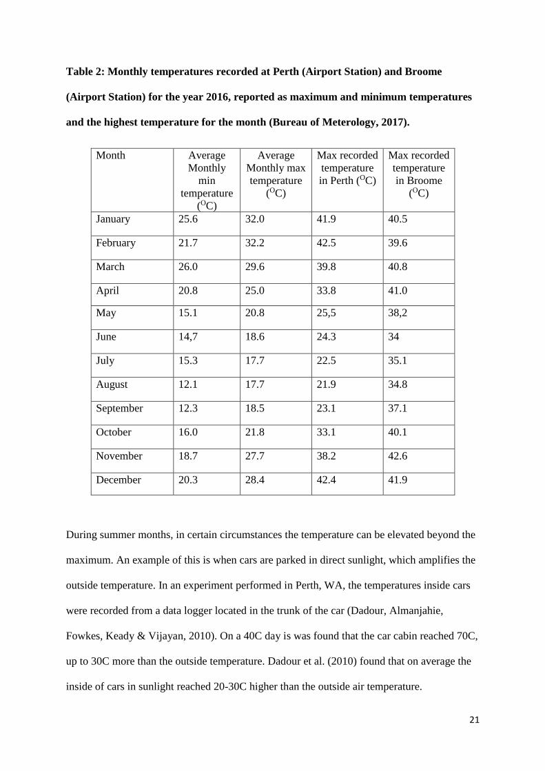
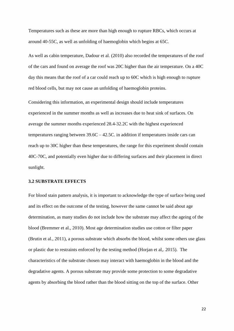
Table 2: : Monthly temperatures recorded at Perth (Airport Station) and Broome (Airport
Station) for the year 2016, reported as maximum and minimum temperatures and the highest
temperature for the month (Bureau of Meterology, 2017) …………………………………..21

5
ABSTRACT
Blood is one of the most commonly encountered types of evidence at a crime scene. The first
step to begin processing is observation and documentation, followed by presumptive testing
which analyses if the substance is likely blood, and if the blood comes from human origin.
This procedure ensures less time and resource wastage of items of evidence which have little
or no forensic value. However this process can be hindered when the presumptive test in
vulnerable to false negative results.
There are many degradative agents which blood can be exposed to at crime scenes. This
ranges from high temperatures, UV light or chemicals such as bleach. This review will focus
on how high temperatures affect blood and more specifically haemoglobin. The ABAcard®
HemaTrace® Kit is an immunochromatographic assay test which relies on antibodies
imbedded within a test strip to detect the presence of human haemoglobin. It is currently
unknown in the literature to what degree haemoglobin is affected by high temperatures and
how this affects the outcome of the HemaTrace® Kit. Denaturation of haemoglobin can
cause structural damage to the protein making is unrecognisable to the antibodies within the
test and therefore producing a false negative result.
The aim of this literature review is to determine the effects of high temperatures on human
haemoglobin as a degradative agent and the effect on the outcome of the HemaTrace® Kit.
The purpose is to assist in future research designing and should hopefully be able to answer
questions regarding further investigations.
1.0 INTRODUCTION
Bloodstains are amongst the most commonly encountered types of biological evidence found
at a crime scene (Schweers, Old, Boonlayangoor & Reich, 2008). Detection and processing
of blood is required in order to reconstruct and sequence events which have occurred, as well

6
as identifying the blood through DNA analysis (Schweers et al., 2008). Correct identification
of blood can be crucial to link a suspect or scene to a crime or dismiss an innocent person.
But before this stage the blood must first be correctly identified as human blood, so resources
and time are not wasted on unrelated stains and DNA profiling. There are many substances
other than blood that also produce similar red-brown stains which can be confused with
evidence from the crime scene, which is why forensic investigators use presumptive tests at
the crime scene.
There are multiple kits available to investigators such as the SERATEC® Hemdirect
Haemoglobin Assay, ABAcard® HemaTrace® Kit, Galatos® Rapid stain Identification of
Human Blood (RSID™-Blood) and Hemastick testing (Horjan, Barbaric, & Mrsic, 2015).
These tests, whilst time efficient often trade specificity for sensitivity, which means that very
small volumes of blood can be detected however there may be some cross reactivity with
blood from other species or other substances. Presumptive tests are also known to give false
results. False positives can result in wasting of resources and the time of the investigator on
irrelevant stains, whereas false negatives can mean critical evidence may get overlooked and
impede the investigation.
The ABAcard® HemaTrace® Kit is an immunochromatographic assay which is used to
identify human haemoglobin within blood. The test is highly sensitive and is used widely by
forensic investigators at crime scenes as it has very low cross reactivity (Abacus Diagnostics,
2001). The test relies on immunochromatography with the target the red blood cell protein,
haemoglobin (Reynolds, 2001). Currently studies have not yet determined the extent to which
degraded haemoglobin has on the test and if the test will work in the present of degraded
forms of the protein. Theories suggest degradation of haemoglobin which changes it structure
may affect the outcome of the test as the protein will not be recognised.

7
It is assumed that the high temperature climate of Western Australia is the reason for the
increase in false negatives from stains at crime scenes despite originating from a human
source (Reynolds, 2017). By testing the temperature variable by itself this may provide
valuable insight into the temperatures which degrade haemoglobin beyond recognition of the
HemaTrace kit.
The purpose of this literature review is to determine if high temperatures are a factor which
influences the degradation of blood and cause it to become unrecognisable to antibodies
within the HemaTrace® Kit, therefore producing a false negative result. The literature review
will also help to design a procedure to test which temperatures cause blood to degrade
beyond detection from the HemaTrace® Kit. This review may also aid investigators to
explain false negative results at crime scenes and in courts, and determine or narrow the
selection of blood stains to avoid false negatives.
2.0 DISCUSSION
This section aims to address current literature available in regards to blood stains and the
properties of haemoglobin. This will include discussions about biological properties of blood,
the degradation process of human haemoglobin and testing using the hematrace presumptive
test to identify human blood. To conclude, the section will discuss the effect that high
temperatures have on blood stains and specifically human haemoglobin.
2.1 BLOOD PROPERTIES
Blood makes up around 8% of the human body weight which roughly equates to 5-6L for
healthy adult males and 4-5L for healthy adult females (Marieb & Hoehn,2010). Blood is part
of the circulatory system which transports nutrients, rids the body of waste products, protects
the body through the immune and inflammatory response, and regulates temperature and pH
control (Marieb & Hoehn, 2010; Schaller, Gerber, Kämper, & Lejon, 2008). Human blood is

8
a complex matrix composed of plasma and formed elements. Plasma, the fluid component of
blood, makes up 55% of the total blood volume (Schaller et al., 2008), however plasma is
primarily made up of water, with the rest composed of nutrients, gases, hormones and waste
products (Marieb & Hoehn, 2010). The formed elements are the cellular components of
blood, the red and white blood cells and platelets. White blood cells (WBC) are made up of
several types of specialised cells which are responsible for the inflammatory response, and
are helped by platelets, which are fragmented pieces of cells which aid in clotting blood
(Marieb & Hoehn, 2010). Red blood cells (RBC) on the other hand are used to transport
oxygen (Marieb & Hoehn, 2010). From a forensic investigator point of view, WBC are useful
to obtain a DNA profile, whereas RBC are used to determine the identity of the origin species
due to the protein known as haemoglobin.
Red blood cells have a unique biconcave disc-like shape and contain no organelles or nucleus
(Marieb & Hoehn, 2010). As RBCs are very flexible in their shape it means they are easily
deformed and able to fit through the small capillaries throughout the body (Schaller, 2008)
The average life span of the cells is around 120 days, after which they are disposed of in the
spleen by phagocytosis (Schaller et al., 2008; Gusev, Govekar, Gedewal & Agalakova, 2017).
RBCs are the carrier cell for the protein haemoglobin, which transports oxygen around the
body. As there are no organelles this means that the RBCs have no need to consume any of
the oxygen they transport making them very efficient and the process of carrying and
transporting oxygen is assisted by the RBCs large surface area (Marieb & Hoehn, 2010).
2.2 HUMAN HAEMOGLOBIN
Haemoglobin is a protein which resides within RBCs and transports oxygen around the body.
It is composed of four globin proteins which are bound in four polypeptide chains, two alpha
9
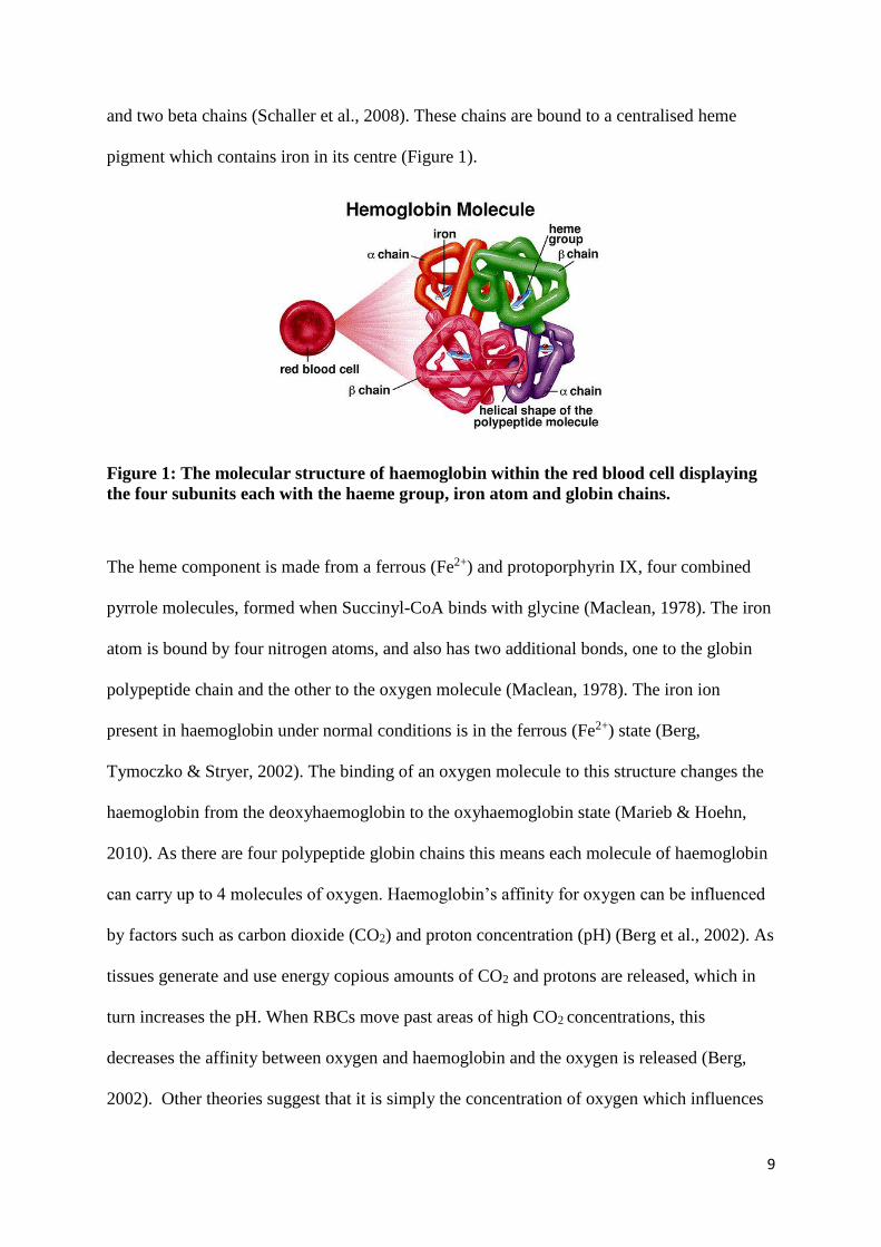
and two beta chains (Schaller et al., 2008). These chains are bound to a centralised heme
pigment which contains iron in its centre (Figure 1).
Figure 1: The molecular structure of haemoglobin within the red blood cell displaying
the four subunits each with the haeme group, iron atom and globin chains.
The heme component is made from a ferrous (Fe2+) and protoporphyrin IX, four combined
pyrrole molecules, formed when Succinyl-CoA binds with glycine (Maclean, 1978). The iron
atom is bound by four nitrogen atoms, and also has two additional bonds, one to the globin
polypeptide chain and the other to the oxygen molecule (Maclean, 1978). The iron ion
present in haemoglobin under normal conditions is in the ferrous (Fe2+) state (Berg,
Tymoczko & Stryer, 2002). The binding of an oxygen molecule to this structure changes the
haemoglobin from the deoxyhaemoglobin to the oxyhaemoglobin state (Marieb & Hoehn,
2010). As there are four polypeptide globin chains this means each molecule of haemoglobin
can carry up to 4 molecules of oxygen. Haemoglobin’s affinity for oxygen can be influenced
by factors such as carbon dioxide (CO2) and proton concentration (pH) (Berg et al., 2002). As
tissues generate and use energy copious amounts of CO2 and protons are released, which in
turn increases the pH. When RBCs move past areas of high CO2 concentrations, this
decreases the affinity between oxygen and haemoglobin and the oxygen is released (Berg,
2002). Other theories suggest that it is simply the concentration of oxygen which influences
10
the haemoglobin-oxygen affinity. If there is a large concentration of oxygen in an area the
haemoglobin is more likely to hold onto to the oxygen for a longer period. The reverse occurs
when a low concentration of oxygen is detected and haemoglobin is more likely to let go of
its oxygen (Anstey, 2003). This process is known as the oxygen-haemoglobin dissociation
curve or as the Haldane dissociation curve (Rosa, Alinovi, Galteri, Scatena & Giardina,
2007).
2.2.1 DEGRADATION OF HAEMOGLOBIN
Red blood cells have a life span of approximately 120 days within the body (Marieb &
Hoehn, 2010). After this, the cells become fragile and are disposed of by phagocytosis,
usually in the spleen (Gusev et al., 2017). This results in the rupturing of the RBC membrane
which means haemoglobin is released from the protection of the cell. Haemoglobin
undergoes several enzymatic processes which degrade it before it can be excreted.
Unfortunately, when encountering blood at crime scenes, there are no regulatory processes
for blood outside the body and it therefore becomes significantly harder to process. In
literature, very few authors discuss what happens to the structure of haemoglobin as it
degrades but the derivatives of this process have been identified. (Seto, Kataoka & Tsuge,
2001; Wood, Hammer, Davis & McNaughton, 2005)
When blood is exposed outside of the body, such as bloodstains at a crime scene, there are no
regulatory processes to control the state of haemoglobin. As such, the haemoglobin becomes
saturated with oxygen which is readily available from the air and all molecules of
haemoglobin become oxy-haemoglobin. When no more oxygen can be taken up the
oxyhaemoglobin eventually degrades to its derivative met-haemoglobin. Usually when inside
the body this is a reversible process and can be undone by an enzyme called cytochrome b5
(Bremmer, Nadort, van Leeuwen, van Gemert & Aalders, 2010). As this enzyme is not
11
present outside the body the change from oxyhaemoglobin to methaemoglobin is irreversible.
At this stage the methaemoglobin may stay in its form or continue to degrade to hemichrome.
Bremmer et al. (2011) found that when exposed to a lab environment blood at time zero
contained only oxyhaemoglobin, but as time passed there was a decrease in oxyhaemoglobin
and an increase in both methaemoglobin and hemichrome with no evidence of any other
derivatives.
2.2.2 HUMAN SPECIFIC HAEMOGLOBIN
Confirmation of the identity of human haemoglobin is important in order to assist
investigators at crime scenes to determine what evidence is relevant. The identification
process is reliant of differentiating human haemoglobin and other animal species using
protein sequences. Studies conducted testing the specificity of the Hematrace® Kit have
found higher primates and ferret haemoglobin also react positively to human haemoglobin
identification tests (Reynolds, 2004; Johnston, Frappier & Newman, 2003). Higher primates
are very closely related phylogenetically to humans and therefore it makes sense that protein
sequences would be shared and that the Hematrace® Kit is specific to humans and primates
(Johnston et al., 2003). Additionally, ferret haemoglobin tested positive, indicating a shared
conserved sequence between ferrets and humans. Although the exact sequence used by the
ABAcard® Hematrace® Kit is unknown, it can be assumed that a highly-conserved sequence
is used to differentiate between human and commonly encountered animal haemoglobin
(Johnston et al., 2003). Johnston et al (2003) discuss the amino acid sequence TNAVAHV is
an optimal sequence which allows for the discrimination needed for differentiating human
haemoglobin from other species.
2.3 ABACARD® HEMATRACE® KIT

12
An understanding of how the HemaTrace® Kit works is essential to interpret how other
influences can affect the outcome of the test. The HemaTrace® kit is based on an
immunochromatographic assay and is able to identify human haemoglobin (Reynolds, 2001).
The test works on the basis of an antigen-antibody reaction complex. When a sample that
contains blood enters the sample well, haemoglobin combines with a mobile monoclonal
antihuman antibody which is imbedded within the test strip. This complex then migrates
down the test strip to the test window, where a polyclonal antihuman haemoglobin antibody
is located (Reynolds, 2004). This antibody catches the moving mobile antibody-antigen
complex and creates an antibody-antigen-antibody sandwich. If human haemoglobin is
present above the minimum detection level, the sandwich complex precipitates a pink dye
forming a visible line on the test strip (Reynolds, 2004). Following this, some of the
antibody-antigen complex continues to migrate further down the test strip to the internal
control area, where it is captured by a second set of polyclonal anti-immunoglobin antibodies
(Reynolds, 2004). This forms a second pink dye line and shows that the test has been
performed as intended (Johnston et al., 2003).
If human haemoglobin is detected, two lines will appear on the test strip, one at the ‘T’ test
region and one at the ‘C’ control region. (Reynolds, 2004; Johnston et al., 2003) Sometimes
false negatives can occur when testing human blood as the Hematrace® Kit is vulnerable to
the high dose hook effect. This occurs when haemoglobin overwhelms the mobile
monoclonal antihuman haemoglobin antibodies, and the unbound haemoglobin migrates
toward the test region. The unbound haemoglobin binds to the stationary antibodies and
prevent the mobile antibodies from binding, therefore a negative result is gained
(Hochmeister et al. 1999). This can be overcome by increasing the volume of the extraction
buffer to blood, or diluting the blood sample before extraction. False negatives can also occur
if the sample is too viscous and physically cannot migrate down the test strip nor bind with
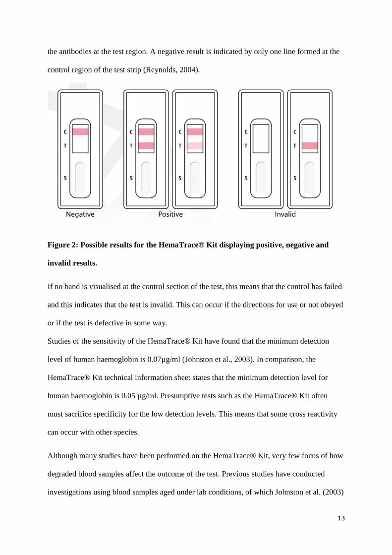
13
the antibodies at the test region. A negative result is indicated by only one line formed at the
control region of the test strip (Reynolds, 2004).
Figure 2: Possible results for the HemaTrace® Kit displaying positive, negative and
invalid results.
If no band is visualised at the control section of the test, this means that the control has failed
and this indicates that the test is invalid. This can occur if the directions for use or not obeyed
or if the test is defective in some way.
Studies of the sensitivity of the HemaTrace® Kit have found that the minimum detection
level of human haemoglobin is 0.07µg/ml (Johnston et al., 2003). In comparison, the
HemaTrace® Kit technical information sheet states that the minimum detection level for
human haemoglobin is 0.05 µg/ml. Presumptive tests such as the HemaTrace® Kit often
must sacrifice specificity for the low detection levels. This means that some cross reactivity
can occur with other species.
Although many studies have been performed on the HemaTrace® Kit, very few focus of how
degraded blood samples affect the outcome of the test. Previous studies have conducted
investigations using blood samples aged under lab conditions, of which Johnston et al. (2003)
14
recorded positive results for stains aged 25-30 years, although the extraction time for the
samples was extended to 30 minutes. Horjan et al. (2015) studied stains aged from 21 to 30
years and received only two positive results, the 21 and 23 year old stains. The authors do not
discuss the reasons why only two stains reacted positively, but it is of note that the sample
size of only 5 stains was severely limited and the results may vary if conducted over a larger
sample size.
2.4 DRYING OF BLOODSTAINS
The drying process of blood is limited in published literature. Often the end result is observed
and discussed but not the process of drying. The drying of a blood drop is a function of both
volume and surface area (Ramsthaler et al., 2012). The rate of drying can also be influenced
by temperature, humidity, air circulation, and vapour pressure as well as the surface that the
blood is on and the viscosity of the drop (Ramsthaler et al., 2012). When blood stains are
deposited onto a surface, RBCs interact with each other and with the edges of the blood stain,
and these interactions are governed by fluid mechanics, biology and chemistry (Brutin,
Sobac, Loquet & Sampol, 2010). When the drop begins to evaporate RBCs and other heavy
colloidal particles are carried by flow motion inside the drop and eventually move to the
edges of the blood drop and create the dark ring called the corona (Brutin et al., 2010). This
can be clearly seen in the below Figure 2 which highlights the main areas of a dried blood
stain.

15
Figure 3: Dried bloodstain displaying the corona, central area and edge (Brutin et al.,
2011)
Crack formation can be seen in larger blood drops and are generally caused by built up stress
on the surface of the blood drop (Brutin et al., 2010). It can also be attributed to RBCs which
adopt hydrophobic behaviour which enables cracks to form particularly through the corona.
Brutin et al. (2010) describe the drying process of blood drops in five stages (Table 1). Each
of the stages is broken down into the percentage of dryness of the blood drop and describes
the main events which the drop.
The stages of dryness can be greatly accelerated when the drop is exposed to increases in
ambient temperature. Ramsthaler et al. (2012) found that the average time to dry a blood drop
to the stage at which the drop is no longer wipeable at 20C was 60 minutes. An increase by
four degrees to 24C found the blood drop dry in just 30 minutes (Ramsthaler et al., 2012).
The increase in just four degrees halved the drying time and indicates temperature is an
obvious factor in the drying process of blood.

16
Table 1: Five stages of blood drop dryness (Brutin et al., 2010)
Stage %
Dryness
Description
1 0-20% Directly after deposition, the RBCs move from the centre out to the
edges of the blood drop. The RBCs then recede away from the
peripheral edge leaving a red deposit along the edges.
2 20-50% Crystallisation occurs at the edge of the drop and begins moving
towards the centre. A dark torus, a high concentration of RBCs, is
observed.
3 50-70% The torus desiccates quickly and at the same time the central part of
drop changes to a lighter red colour. Drop is almost completely
desiccated and the first crack appears between the future corona and
centre of the drop.
4 70-85% Centre of drop is completely desiccated, which produces small plaques.
Corona is also desiccated and drying spots can be observed.
5 85-100% Large plaques of the corona move slightly, until desiccation is complete
and no further movement is observed.
When dealing with dried bloodstains, it is important to rehydrate the blood in order to be able
to form a homogenous solution with the extraction buffer. As it is readily soluble in water, a
moistened swab can assist, as well as the use of cotton as a collection material as it has been
found to be the best capturing material to retain blood. Some studies show successful
recapturing of the bloodstain up to 30 years after deposition and after exposure to different
temperatures. To rehydrate hard dried stains some studies suggest extending the buffer
extraction time and using a moistened swab in order to collect the blood to give it the best

17
chance to rehydrate and for haemoglobin to be extracted (Johnston et al., 2003; Horjan et al.
2015).
2.5 DEGRADATIVE AGENTS
At a crime scene, there are often several factors present which can degrade evidence. This
can include biological material being exposed to sunlight and high temperatures which can
degrade blood and DNA. This can also include suspects using cleaning chemicals such as
bleach in an attempt to hide and destroy evidence of a crime. These are all factors which can
impede investigations and degrade potentially useful evidence. Whilst these factors have been
extensively documented in regard to their effect on biological material, the effect of high
temperatures on RBCs and in particular haemoglobin have not been widely studied. Evans
(2016) studied the effect of 55OC on blood for a period of two weeks and found no change to
the outcome of the HemaTrace® kit, and also investigated the effect of bleach and UV light
on human blood when using the HemaTrace® kit. When exclusively looking at temperatures
most often this will be a factor in outdoor crime scenes or in hot environments. The effect of
high temperatures on haemoglobin specifically should provide an in depth look at how it is
degraded and how this affects the HemaTrace® Kit.
2.5.1 TEMPERATURE AND THE ABACARD® HEMATRACE® KIT
Temperature is a variable factor which impacts crime scenes and potential items of evidence.
It can be difficult to analyse temperatures that blood has been exposed to, especially in indoor
conditions, however weather station records are able to give investigators information on
outdoor weather conditions. It is documented within the literature that increased temperatures
can cause increases in the drying of blood drops and the denaturation of blood proteins,
including haemoglobin (Ramsthaler et al., 2012; Evans, 2016)).
18
When blood is exposed to heating, denaturation of the haemoglobin is accelerated (Seto et al.,
2001). Seto et al. (2001) measured the concentrations of haemoglobin as well as its derivative
methaemoglobin using headspace gas chromatography. When placed overnight at 37C there
was no conversion of haemoglobin and the blood was stable. As the temperature was raised
to 54C, some evidence was seen of methaemoglobin formation such as darkening of the
blood colour. It was found that methaemoglobin concentration increased by 69.2% and the
haemoglobin concentration stayed relatively the same. When the blood was kept at 65C for
one hour, haemoglobin concentration decreased by almost 89% and methaemoglobin
increased by 41%. Lower concentrations of methaemoglobin in the 65C sample can be
explained by the formation of hemichrome, a derivative product of methaemoglobin. The
degradation from methaemoglobin to hemichrome occurs at a lower temperature than the
degradation from haemoglobin, meaning this process may have been accelerated due to the
higher temperature.
Haemoglobin’s functional state is a tetramer protein (Michnik, Drzazga, Kluczewska &
Michalik, 2005). When heated, haemoglobin’s form begins to unfold and the protein
denatures. Drzazga et al. (2001) found that when heating haemoglobin between 40 and 80C
the denaturation process happens in three stages. The first stage is the unfolding of the
tetramer state to a dimer form. The dimer then continues to denature to a monomer form and
finally to individual alpha and beta subunit chains. It was noted that the beta subunits began
to form before the alpha chains. Michnik et al. (2005) found that the unfolding temperature of
haemoglobin was between 63-67C, and that the denaturation of methaemoglobin to
hemichrome was much lower, which explains why often methaemoglobin denatures much
faster than haemoglobin itself.
Wood et al. (2005) describes how haemoglobin denaturation is accompanied by aggregation
of heme moieties, which are the result of excitonic interactions between metalloporphyrins.

19
At temperatures higher than 42C, blood examined by ramen microscopy showed spectra
similar to samples that were subjected to extended laser exposure (Wood et al., 2005). The
increase in band intensity demonstrate the denaturation of haemoglobin as the change in
spectra is caused by excitonic interactions and heme aggregation which are both signs of said
denaturation. Cho and Choy (1979) sought to find out whether the spin state of iron or steric
interactions with ligands were the most responsible for keeping haemoglobin thermally
stable. As haemoglobin contains an iron atom at its centre, it was thought that this may
influence the stability of the whole protein during heating. It was found that there was no
correlation between the stability of haemoglobin and the spin state, yet steric interactions
were found to have a definite effect.
Ivanov (2009) used impedance spectroscopy to examine the effect of heat on the RBC
structural protein, spectrin. Spectrin is the major fibrillar protein of the RBC membrane
skeleton. After heating to 49.5C it as found that spectrin denatures, an irreversible process
which results in a shift of the dimers to tetramers equilibrium as well as irreversible unfolding
od dimers to alpha and beta monomers (Ivanov, 2009). This process is of concern to forensic
investigators because if spectrin denatures this may result in the loss of stability and even
lysing of the RBC membrane which would mean that haemoglobin which is normally
protected inside the cell, is directly exposed to degradative agents. This may dramatically
affect the denaturation process making it harder for investigators to determine the origin
species of the blood when using haemoglobin tests.
There is some inconsistency in the literature when examining whether the denaturation of
haemoglobin is reversible. Some sources state outright that the denaturation is irreversible
and cannot be renatured (Cho & Choy, 1979; Drzazga et al., 2001). Wood et al. (2005) found
that the denaturation of haemoglobin could be reversed if it is not too severe. Michnik et al.
(2005) examined haemoglobin after heating to different temperatures. Wood et al. (2005)
20
found to ensure a completely reversible process haemoglobin should not be heated over 42C.
When only heated to 68C the haemoglobin was partly recovered and the process deemed
partly reversible, as not all of the proteins returned back to the haemoglobin state. When
heated to 90C, it was found that the denaturation process was irreversible and haemoglobin
was not recoverable (Wood et al., 2005). These findings pose problems to investigators using
the ABAcard HemaTrace® Kit on thermally denatured blood. The test relies on specific
binding of antibodies to human haemoglobin however it is still unknown to what degree the
test will still recognise haemoglobin after exposure to high temperatures. If haemoglobin has
become fully denatured it is possible the test will not recognise or bind to the products of said
denaturation giving inaccurate results. This can lead to false negatives from human blood.
3.0 EXPERIMENT DESIGN ELEMENTS
3.1 AUSTRALIAN WEATHER DATA
In the summer months, from December to February, Australia can experience extreme
temperatures and weather conditions. The summer climate consists of hot sinking air, and a
subtropical pressure belt, with average temperatures of high 30s- to 40C in the summer. The
northern parts of Australia can experience temperatures above 40C regularly during summer.
3.1.1 PERTH/NORTHERN AUSTRALIA TEMPERATURES
In the summer months, Perth and northern Australian towns can experience extreme
temperatures with the average maximum temperatures reaching and often exceeding 30C
(Bureau of Meterology, 2017)(Table 2). The following table displays weather data for each
month of 2016 as well as the highest temperatures recording for both Perth and Broome, a
northern town of WA with similar crime statistics.
21
Table 2: Monthly temperatures recorded at Perth (Airport Station) and Broome
(Airport Station) for the year 2016, reported as maximum and minimum temperatures
and the highest temperature for the month (Bureau of Meterology, 2017).
Month Average
Monthly
min
temperature
(OC)
Average
Monthly max
temperature
(OC)
Max recorded
temperature
in Perth (OC)
Max recorded
temperature
in Broome
(OC)
January 25.6 32.0 41.9 40.5
February 21.7 32.2 42.5 39.6
March 26.0 29.6 39.8 40.8
April 20.8 25.0 33.8 41.0
May 15.1 20.8 25,5 38,2
June 14,7 18.6 24.3 34
July 15.3 17.7 22.5 35.1
August 12.1 17.7 21.9 34.8
September 12.3 18.5 23.1 37.1
October 16.0 21.8 33.1 40.1
November 18.7 27.7 38.2 42.6
December 20.3 28.4 42.4 41.9
During summer months, in certain circumstances the temperature can be elevated beyond the
maximum. An example of this is when cars are parked in direct sunlight, which amplifies the
outside temperature. In an experiment performed in Perth, WA, the temperatures inside cars
were recorded from a data logger located in the trunk of the car (Dadour, Almanjahie,
Fowkes, Keady & Vijayan, 2010). On a 40C day is was found that the car cabin reached 70C,
up to 30C more than the outside temperature. Dadour et al. (2010) found that on average the
inside of cars in sunlight reached 20-30C higher than the outside air temperature.
22
Temperatures such as these are more than high enough to rupture RBCs, which occurs at
around 40-55C, as well as unfolding of haemoglobin which begins at 65C.
As well as cabin temperature, Dadour et al. (2010) also recorded the temperatures of the roof
of the cars and found on average the roof was 20C higher than the air temperature. On a 40C
day this means that the roof of a car could reach up to 60C which is high enough to rupture
red blood cells, but may not cause an unfolding of haemoglobin proteins.
Considering this information, an experimental design should include temperatures
experienced in the summer months as well as increases due to heat sink of surfaces. On
average the summer months experienced 28.4-32.2C with the highest experienced
temperatures ranging between 39.6C – 42.5C. in addition if temperatures inside cars can
reach up to 30C higher than these temperatures, the range for this experiment should contain
40C-70C, and potentially even higher due to differing surfaces and their placement in direct
sunlight.
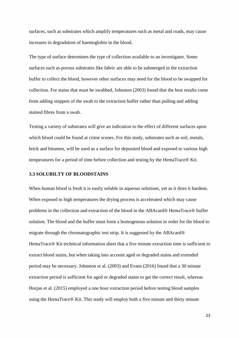
3.2 SUBSTRATE EFFECTS
For blood stain pattern analysis, it is important to acknowledge the type of surface being used
and its effect on the outcome of the testing, however the same cannot be said about age
determination, as many studies do not include how the substrate may affect the ageing of the
blood (Bremmer et al., 2010). Most age determination studies use cotton or filter paper
(Brutin et al., 2011), a porous substrate which absorbs the blood, whilst some others use glass
or plastic due to restraints enforced by the testing method (Horjan et al,. 2015). The
characteristics of the substrate chosen may interact with haemoglobin in the blood and the
degradative agents. A porous substrate may provide some protection to some degradative
agents by absorbing the blood rather than the blood sitting on the top of the surface. Other
23
surfaces, such as substrates which amplify temperatures such as metal and roads, may cause
increases in degradation of haemoglobin in the blood.
The type of surface determines the type of collection available to an investigator. Some
surfaces such as porous substrates like fabric are able to be submerged in the extraction
buffer to collect the blood, however other surfaces may need for the blood to be swapped for
collection. For stains that must be swabbed, Johnston (2003) found that the best results come
from adding snippets of the swab to the extraction buffer rather than pulling and adding
stained fibres from a swab.
Testing a variety of substrates will give an indication to the effect of different surfaces upon
which blood could be found at crime scenes. For this study, substrates such as soil, metals,
brick and bitumen, will be used as a surface for deposited blood and exposed to various high
temperatures for a period of time before collection and testing by the HemaTrace® Kit.
3.3 SOLUBILTY OF BLOODSTAINS
When human blood is fresh it is easily soluble in aqueous solutions, yet as it dries it hardens.
When exposed to high temperatures the drying process is accelerated which may cause
problems in the collection and extraction of the blood in the ABAcard® HemaTrace® buffer
solution. The blood and the buffer must form a homogenous solution in order for the blood to
migrate through the chromatographic test strip. It is suggested by the ABAcard®
HemaTrace® Kit technical information sheet that a five minute extraction time is sufficient to
extract blood stains, but when taking into account aged or degraded stains and extended
period may be necessary. Johnston et al. (2003) and Evans (2016) found that a 30 minute
extraction period is sufficient for aged or degraded stains to get the correct result, whereas
Horjan et al. (2015) employed a one hour extraction period before testing blood samples
using the HemaTrace® Kit. This study will employ both a five minute and thirty minute

24
extraction time for each sample in order to test both tines and ensure the correct result.
comparisons between results will be examined and recommendations made in regards to
extraction times required for heated blood samples.
3.4 TEMPERATURE EXPOSURE TIME
The impact of degradative agents is dependent on the level and period of exposure. Most
degradative agents show a trend of increased degradation over a period of time (Seto et al.
2001; Horjan et al,. 2015). When analysing the effect of temperature, Evans (2016) employed
periods of six hours, one week, and two weeks at 55OC to human blood samples. this resulted
in positive HemaTrace® results indicating the length of time may need to be extended or an
increase in the temperature. For real world applicability, samples should be exposed to levels
and exposure periods similar to that encountered by forensic investigators. However, these
timeframes are highly variable, anywhere between hours to months and potentially years.
Therefore the experimental exposure periods selected may need to be adjusted depending on
the results obtained, in order to replicate real world scenarios.
4.0 EXPERIMENTAL AIMS AND HYPOTHESIS
This literary review has provided research that shows high temperatures can cause
degradation of human haemoglobin in blood which may prevent detection with the
ABAcard® HemaTrace® Kit. The degradation may be in the form of denaturation of the
haemoglobin molecular into its derivative molecules or simply just a change to its structure,
which leads to an inability for the molecule to bind with antibodies in the test thus producing
a false negative result. This may result in investigators dismissing important forensic
evidence because the negative result determined the stain irrelevant. The experiment design
aims to determine if high temperatures can cause false negatives for human blood using the
25
ABAcard® HemaTrace® Kit as well as determining if various substrates amplify the
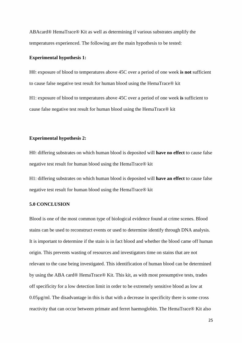
temperatures experienced. The following are the main hypothesis to be tested:
Experimental hypothesis 1:
H0: exposure of blood to temperatures above 45C over a period of one week is not sufficient
to cause false negative test result for human blood using the HemaTrace® kit
H1: exposure of blood to temperatures above 45C over a period of one week is sufficient to
cause false negative test result for human blood using the HemaTrace® kit
Experimental hypothesis 2:
H0: differing substrates on which human blood is deposited will have no effect to cause false
negative test result for human blood using the HemaTrace® kit
H1: differing substrates on which human blood is deposited will have an effect to cause false
negative test result for human blood using the HemaTrace® kit
5.0 CONCLUSION
Blood is one of the most common type of biological evidence found at crime scenes. Blood
stains can be used to reconstruct events or used to determine identify through DNA analysis.
It is important to determine if the stain is in fact blood and whether the blood came off human
origin. This prevents wasting of resources and investigators time on stains that are not
relevant to the case being investigated. This identification of human blood can be determined
by using the ABA card® HemaTrace® Kit. This kit, as with most presumptive tests, trades
off specificity for a low detection limit in order to be extremely sensitive blood as low at
0.05µg/ml. The disadvantage in this is that with a decrease in specificity there is some cross
reactivity that can occur between primate and ferret haemoglobin. The HemaTrace® Kit also
26
is known to produce false negatives, usually as a result of degradative agents such as extreme
temperatures.
The HemaTrace® Kit is an immunochromatographic test which relies on imbedded
antibodies which bind with human haemoglobin to produce visible bands indicating positive
or negative results. Degradative agents such as high temperatures can change the structure of
haemoglobin to the point where it becomes unrecognisable to the antibodies within the test.
Whilst there is extensive research and literature regarding DNA degradation, very little
focuses on haemoglobin and RBC degradation. This literature review aimed to determine
whether high temperatures, beyond 40C, can increase denaturation of haemoglobin and
produce haemoglobins derivatives: methaemoglobin and hemichrome. There are still
disagreements on what exactly causes the degradation of haemoglobin. Ivanov (2009)
attributed the degradation to spectrin denaturation, leading to ruptured RBCs, whereas Cho
and Choy (1979) related the degradation to the spin state of the iron atom within the
haemoglobin molecule.
This literary review has demonstrated the ability of high temperatures to degrade
haemoglobin and damage RBCs. Said degradation may lead to unreliability of presumptive
tests such as the ABAcard® HemaTrace® Kit which tests haemoglobin binding to antibodies.
It is hoped to establish whether once haemoglobin has been structurally damaged, it will no
longer be recognisable by the test antibodies therefore leading to false negative results.
Further study into this subject will be required to definitively state to what degree
haemoglobin can be degraded before the HemaTrace® Kit will no longer detect it. The
findings of this study may assist in explaining why false negatives occur and help forensic
investigators in selecting blood stains to test.

27
6.0 REFERENCE LIST
Abacus Diagnostics. (2001). ABAcard® HemaTrace® test kit – Technical Information
Sheet. (R. 1, Ed.) USA.
Anstey, C. (2003). A new model for the oxyhaemoglobin dissociation curve. Anaesthesia and
Intensive Care, 31(4), 376.
Berg, J., Tymoczko, J. & Stryer, L. (2002). Biochemistry (5th Ed.). New York. W. H.
Freeman.
Bremmer, R. H., Nadort, A., van Leeuwen, T. G., van Gemert, M. J., Aalders, M. C. (2010).
Age Estimation of Blood Stains by Hemoglobin Derivative Determination Using Reflectance
Spectroscopy. Forensic Science
International, 206 (1-3), 166-171.
Bremmer, R. H., de Bruin, K. G., van Gemert, M. J., van Leeuwen, T. G., & Aalders, M. C.
(2012). Forensic Quest for Age Determination of Bloodstains. Forensic Science
International, 216 (1-3), 1-11.
Brutin, D., Sobac, B., Loquet, B., & Sampol, J. (2011). Pattern Formation in Drying
Drops of Blood. Journal of Fluid Mechanics, 667 (1), 85-65.
Bureau of Meterology. (2016). Australian Climate Site Data. Retrieved 26 03 2017
from Australian Government Bureau of Meterology: www.bom.gov.au
Cho, K. C., & Choy, C. L. (1979). Thermal Stability of Hemoglobin and Myoglobin Effect
of Spin States. Biochimica et Biophysica Acta, 622, 320-330.
Dadour, I. R., Almanjahie, I., Fowkes, N. D., Keady, G., & Vijayan, K. (2010).
Temperature Variations in a Parked Vehicle. Forensic Science International, 207
(1), 205-211.
Drzazga, Z., Michnik, A., Bartoszek, M., & Beck, E. (2001). Thermal Stability of
Haemoglobin Solutions Under DC and AC Magnetic Field and UV and IR
Radiation. Journal of Thermal Analysis and Calorimetry, 65, 575-583.
Evans, S. (2016). The Degradative Effects of Temperature, Ultra Violet Radiation and
Sodium Hypochlorite on the Detection of Blood at Crime Scenes Using the ABACard®
HemaTrace® Kit. Unpublished M. For. Sc. Thesis, Murdoch University, Perth, Western
Australia.
Gusev, G., Govekar, R., Gadewal, N & Agalakova, N. (2017). Understanding quasi-apoptosis
of the most numerous enucleated components of blood needs detailed molecular autopsy.
Ageing Research Reviews, 35(1), 46–62.
Horjan, I., Barbaric, L., & Mrsic, G. (2015). Applicability of Three Commercially Available
Kits for Forensic Identification of Blood Stains. Journal of Forensic and Legal
Medicine, 38 (1), 101-105.

28
Ivanov, I. T. (2009). Impendance Spectroscopy of Human Erythrocyte Membrane: Effect of
Frequency at the Spectrin Denaturation Transition Temperature.
Bioelectrochemistry, 78 (2), 181-185.
Johnston, S., Newman, J., & Frappier, R. (2003). Validation Study of the Abacus Diagnostics
ABACard HemaTrace Membrane Test for the Forensic Identification of Human Blood.
Canadian Society of Forensic Science Journal, 36 (3), 173-183.
Maclean, N. (1978). Haemoglobin. London, UK: Edward Ardonld Limited.
Marieb, E. N., & Hoehn, K. (2010). Human Anatomy and Physiology (8th Ed ed.). San
Francisco, CA: Pearson Benjamin Cummings.
Michnik, A., Drzazga, Z., Kluczewska, A., & Muchalik, K. (2005). Differential Scanning
Microcalorimetry Study of the Thermal Denaturation of Haemoglobin.
Biophysical Chemistry, 118 (1), 93-101.
Ramsthaler, F., Schmidt, P., Bux, R., Potente, S., Kaiser, C., & Kettner, M. (2012). Drying
Properties of Bloodstain on Common Indoor Surfaces. International Journal of
Legal Medicine, 126 (1), 739-746.
Reynolds, M. (2004). The ABAcard® HemaTrace®. A Confirmatory Identification of
Human Blood Located at Crime Scenes. International Association of Bloodstain
Pattern Analysis (IABPA) News, 4-10.
Reynolds, M. (2017). Personal communication. Murdoch University, Perth, Western
Australia.
Rosa, M., Alinovi, C., Galteri, A., Scatena, R. & Giardina, B. (2007) The plasma membrane
of erythrocytes plays a fundamental role in the
transport of oxygen, carbon dioxide and nitric oxide and in the maintenance of the reduced
state of the heme iron. Gene, 398 (27), 162-171.
Schaller, J., Gerber, S., Kamper, U., Trachsel, C. & Lejon, S. (2008). Human Blood Plasma
Proteins: Structure and Function. Switzerland. John Wiley & Sons.
Schweers, B., Old, J., Boonlayangoor, P., & Reich, K. (2008). Developmental Validation of a
Novel Lateral Flow Strip Test for Rapid Identification of Human Blood (RSID-Blood).
Forensic Science International: Genetics, 2(3), 243-247.
Seto, Y., Kataoka, M., & Tsuge, K. (2001). Stability of Blood Carbon Monoxide and
Hemoglobins During Heating. Forensic Science International, 121 (1), 144-150.
Wood, B. R., Hammer, L., Davis, L., & McNaughton, D. (2005). Raman Microspectroscopy
and Imaging Provides Insight into Heme Aggregation and Denaturation Within Human
Erythrocytes. Journal of Biomedical Optocs, 10 (1).

29
30
- Part Two -
MANUSCRIPT
INVESTIGATING THE EFFECT OF HIGH
TEMPERATURES AND SUBSTRATES ON THE
DETECTION OF HUMAN BLOOD USING THE
ABACARD® HEMATRACE® KIT

31
TABLE OF CONTENTS
LIST OF TABLES…………………………………………………………………..32
ABSTRACT……………………………………………………………………….....33
INTRODUCTION………………………………………………………………..….33
MATERIALS AND METHODS………………………………………………..….35
RESULTS………………………………………………………………………..…..36
DISCUSSION…………………………………………………………………..……37
CONCLUSION………………………………………………………………..…….38
REFERENCE LIST…………………………………………………………………39

32
LIST OF TABLES
Table 1: ABACard® HemaTrace® results after one day exposure …………………..36
Table 2: ABACard® HemaTrace® results after one week exposure ..……………….37
33
INVESTIGATING THE EFFECT OF HIGH TEMPERATURES AND SUBSTRATES
ON THE DETECTION OF HUMAN BLOOD USING THE ABACARD®
HEMATRACE® KIT.
Teaghan McDonald1, Mark Reynolds2, James Speers1 1Murdoch University, School of Veterinary and Life Sciences, Perth WA 2West Australian Police, Forensic Division
Abstract
The ABAcard® HemaTrace® kit is an immunochromatographic assay which is used by
forensic investigators as a presumptive test for human blood. It is the experience of forensic
investigators within the WA Police that the HemaTrace® kit produces false negatives in high
temperature environments. This study evaluated how high temperatures (from 50OC to 80OC)
and substrates affect human haemoglobin for the purpose of analysis using the ABAcard®
HemaTrace® kit. After one week exposure to 50OC, 60OC and 70OC the ABAcard®
HemaTrace® kit was still able to identify human haemoglobin. After one week at 80OC the
test returned negative results for three of the four substrates tested. This indicates that the
HemaTrace® kit was unable to reliably identify human haemoglobin after exposure to above
80OC.
Keywords
Forensic Science, ABAcard® HemaTrace®, Haemoglobin degradation, Temperature,
Substrate
Introduction
Blood is the most frequent biological evidence type encountered by forensic investigators
(Schweers, Old, Boonlayangoor & Reich, 2008). Its properties mean it can potentially be
used to identify the source of the blood and to reconstruct the blood event (Schweers et al.,
34
2008). The correct identification of the blood as human is important because it ensures time
and resources are not wasted on stains that are not relevant to the crime scene, and therefore
aid in linking persons of interest to the event or exonerating innocent persons (Virkler &
Lednev, 2009). This can become difficult if presumptive testing kits provide false negatives
to human blood stains.
Blood can be exposed to many degradative agents at crime scenes such as chemicals, sunlight
and temperature. In areas where high temperatures are regularly experienced, such as the
northern part of Australia, this may affect the degradation of blood. This especially depends if
the blood is deposited on a surface which may amplify the temperature exposing the blood to
even higher temperatures. The degradation of blood may result in damage to the proteins
within and cause irreversible changes. The structural integrity of certain blood proteins,
mainly haemoglobin, may affect the ability to detect blood using immunochromatographic
assay tests. Without this knowledge, forensic investigators may find it difficult to explain
why false negatives are obtained, especially when these samples may still produce a DNA
result (Coy, Lewis, Fulmer, Hudson & Cruz, 2005).
Test kits such as the ABAcard® HemaTrace® detect the presence of human haemoglobin, on
addition to ferret, and higher primate haemoglobin (Reynolds, 2004; Johnston, Newman &
Frappier, 2003). The kit recognises an amino acid sequence on haemoglobin (Johnston et al.,
2003). This sequence is recognised by antibodies within the test strip, and if the haemoglobin
is degraded, the test may not recognise produce a false negative result for blood.
Erythrocytes have a physical tolerance for temperatures between 25OC and 40OC (Wood,
Hammer, Davis & McNaughton, 2005) beyond this temperature, disruption to the to the
cellular secondary and tertiary structures may occur (Hardin & Bertoni, 2015). Denaturation
of primary structural membrane, spectrin, can occur at 49.5OC (Ivanov, 2009) which causes
the cell wall to ruptured. This process is irreversible at 55C (Wood et al., 2005). The

35
unfolding process of the haemoglobin protein is reported to begin at 63-67OC (Michnik,
Drzazga, Kluczewska & Michalik, 2005) but other studies have reported the degradation of
haemoglobin to methaemoglobin to occur at temperatures as low as 50-54OC (Seto, Kataoka
& Tsuge, 2001).
The surface upon which blood is deposited on affects the drying of the blood stain and may
impact on the temperature that the blood is exposed to. Absorption by a porous surface may
decrease the ability for degradative agents to interact with the blood, thus protecting the
blood (Ramsthaler, Schmidt, Bux, Potente, Kaiser & Kettner, 2012).
This study is based upon the research conducted by Evans (2016) which initiated research on
the problem of false negatives from the HemaTrace® kit. The objective of this study was to
address the effect of temperature and differing substrates on the detection of human blood by
the HemaTrace® kit.
MATERIALS AND METHODS
The bloodstains were deposited onto four different substrates in replicates of three and
subjected to a range of high temperatures. The blood was taken from a healthy 21-year-old
female volunteer and immediately deposited onto the substrates in approximately 50ul
droplets. The substrates were white ceramic tiles, brick pavers, galvanised steel sheet
(approx. 2mm thick), and garden soil.
The substrate on which the blood was deposited was air dried for 30 minutes at room
temperature (temperature?) before being placed into incubator at the appropriate experimental
temperature. The temperatures used for the experiments were 50OC, 60OC, 70OC and 80OC.
For this study only temperature was considered and as such humidity was not controlled nor
recorded. Both positive and negative controls of the HemaTrace® kit were performed and
analysed prior to undergoing experimental procedure.
36
The samples were tested at one day and one week periods using the HemaTrace® kit. Blood
was recovered from the substrate using a sterile cotton swab moistened in the extraction
buffer. A five-minute extraction period was employed for all samples and the results
determined within ten minutes as per the technical information sheet (Abacus Diagnostics,
2001). The presence of two pink lines at the control C and test T region indicate a positive
result for human haemoglobin. A negative result was indicated by only one pink line at the
control C region, and an inconclusive result as indicated by no visible bands at either region.
An inconclusive result sample would be retested .
A 30-minute extraction period was employed for negative and very faint positive results to
determine if a longer extraction period changes the outcome of the test.
RESULTS
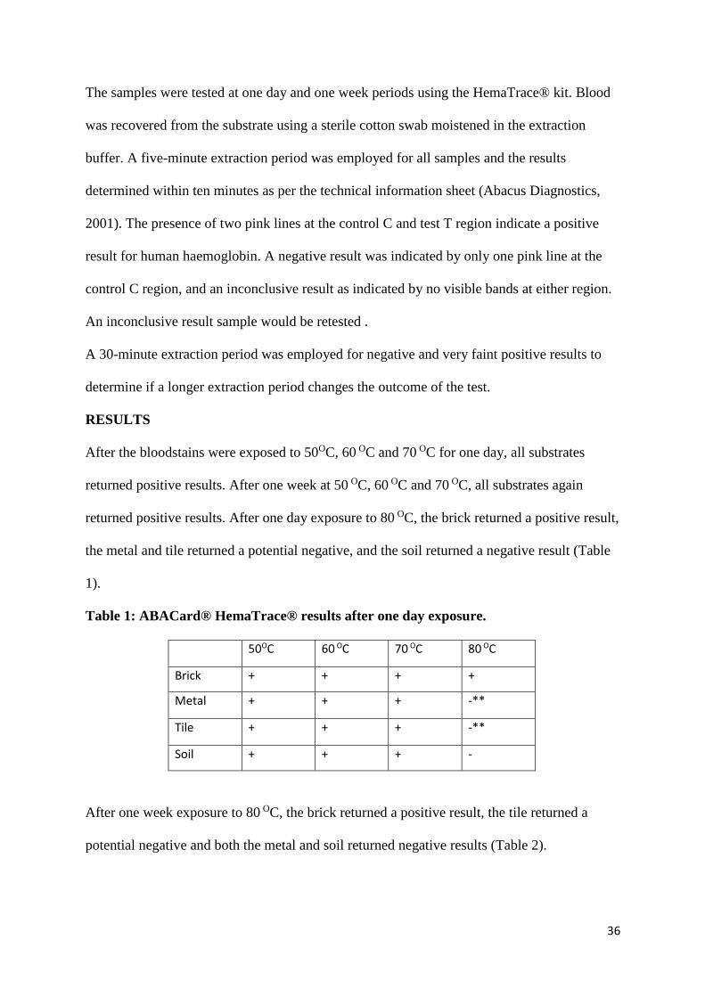
After the bloodstains were exposed to 50OC, 60 OC and 70 OC for one day, all substrates
returned positive results. After one week at 50 OC, 60 OC and 70 OC, all substrates again
returned positive results. After one day exposure to 80 OC, the brick returned a positive result,
the metal and tile returned a potential negative, and the soil returned a negative result (Table
1).
Table 1: ABACard® HemaTrace® results after one day exposure.
50OC 60 OC 70 OC 80 OC
Brick + + + +
Metal + + + -**
Tile + + + -**
Soil + + + -
After one week exposure to 80 OC, the brick returned a positive result, the tile returned a
potential negative and both the metal and soil returned negative results (Table 2).

37
Table 2: ABACard® HemaTrace® results after one week exposure.
ONE WEEK 50OC 60 OC 70 OC 80 OC
Brick + + + +
Metal + + + -
Tile + + + -**
Soil + + + -
For samples which produced faint, potential negative or negative results, a 30-minute
extraction period was applied and the sample retested. At 50 OC two faint positive results
returned the same result after the 30-minute extraction, whilst a potential negative result
returned a faint positive after the longer extraction period. For both 60 OC and 70 OC the
extended extraction period was not employed. At 80OC several extended extraction periods
were employed and the results as follows: of eight potential negatives, 5 returned the same
result, 2 returned negative results and 1 gave a faint positive result; seven negative results
returned one potential negative and six negative results; and of four faint positive results, two
returned the same result and two returned potential negative results after the extended 30-
minute extraction period.
DISCUSSION
In Western Australia, blood samples at crime scenes may be exposed to high temperatures,
especially during the summer months. The increasing amounts of false negative results in
summer environments has been noticed by members of the WA Police forensic BPA division
and. The effect of high temperatures on the integrity of the HemaTrace® kit are currently
Key:
+ Positive; - Negative; ~ Inconclusive; +* Faint Positive; -** Potential Negative: too faint to
confidently class as positive

38
unknown (Wood et al. 2005). This study focused on the effect of higher temperatures and
substrates effects on blood and subsequent analysis by the ABAcard® HemaTrace® kit. the
study is based upon the work of Evans (2016) which found blood exposed to 55 OC for two
weeks could still be correctly identified by the HemaTrace® kit. In depth knowledge of this
would help to explain why immunoassay test kits are able to produce false negative results,
and assist with the analysis procedure and interpretation of results.
After exposure to temperatures from 50C to 70C for up to one week, each blood stained
substrate returned positive results, indicating that the HemaTrace® kit was still able to
correctly identify human blood with five-minute and 30-minute extraction buffer period.
After exposure to 80C for one day, the brick returned a positive result, indicating the test was
still able to correctly identify the blood after one day. The metal and tile returned potential
negative results and the soil returned a negative result. After one week exposure to 80 OC the
brick still returned positive results, the tile returned a potential negative and both the metal
and soil returned negative results. These results indicate that 80 OC is high enough, depending
on the substrate, to denature human blood where it will no longer be recognisable to the
HemaTrace® kit.
Previous studies have concluded that RBC’s begin to degrade at 40 OC (Wood et al. 2005)
and the primary structural protein, spectrin, begins to degrade at 49.5 OC (Ivanov, 2010). This
means that above 49.5 OC, it is expected that the RBC will have ruptured, therefore exposing
its contents to the high temperature. Michnik et. al (2005) indicate that the unfolding of
haemoglobins structure begins at 63--67 OC. The results from this study indicate that the
unfolding temperature occurs somewhere between 70 and 80 OC, as the immunoassay test
could not recognise blood which had been exposed to 80C for one week, yet it could still
positively identify blood exposed to 70 OC.
39
The effect of the type of substrate on which the blood was deposited on the test kit was
investigated. The brick still returned positive results via the HemaTrace® kit after exposure
to 80 OC after both one day and one week may be attributed to the porous nature of the brick.
It is also possible that the large surface area of the brick impacted that amount of heat the
blood was exposed to, as well as acting as heat sink.
Whilst this study is an attempt to mimic conditions experienced in forensic cases, the
experimental conditions require further analysis. This includes exposing the blood samples to
cyclical temperatures to mimic day and night conditions, and placing samples in real
environment conditions to create a more realistic comparison to forensic cases. Additional
variables such as humidity and moisture need to be tested as well as combining variables to
understand further why false negative results are obtained from some forensic samples. In
addition, the degradation of the haemoglobin protein should be examined to determine the
exact nature of the degradation occur when heated and why this produces false negative
results. Four substrates were employed for this study which may need to be expanded on to
give a clearer idea of the effect of the substrate when false negative results are obtained.
These factors should be considered for future research in this area of study.
CONCLUSION
This study found that exposure to 80 OC was able to produce false negative results from the
ABAcard® HemaTrace® kit, when blood is deposited on to tiles, metal, or soil. Positive
results were still obtained when blood was deposited onto brick. It is known that
haemoglobin’s unfolds at 67 OC and it may be possible that 80 OC is sufficient to affect the
ability of the ABAcard® HemaTrace® kit to bind to the haemoglobin molecule. The porous
nature of the brick substrate may have provided some protection to the blood and it was still
tested positive. An extended extraction period was employed on faint positive, partial
40
negative and negative results to determine if this would change the outcome of the test. In
some cases, the outcome of the test changed after the extended buffer period, and therefore it
is recommended to employ an extended extraction buffer period of 30 minutes when negative
results are obtained. The current study has provided some insight into how temperature and
substrates affect the outcome of the ABAcard HemaTrace® kit, however further investigation
in required to confidently explain why false negative results for human blood can be obtained
when using immunochromatographic tests, such as the ABAcard® HemaTrace® kit.
DISCLAIMER
The authors do not endorse any products for the purpose of blood identification, nor
is there a declared conflict of interest within the study conducted.
REFERENCES
Abacus Diagnostics. (2001). ABAcard® HemaTrace® test kit – Technical Information
Sheet. (R. 1, Ed.) USA.
Coy, K. L., Lewis, K. E., Fulmer, A., Hudson, A., & Cruz, T. (2005). Obtaining Typeable
DNA from Bloodstains that Serologically Test Negative. Journal of Forensic
Identification , 55 (5), 633-643.
Evans, S. (2016). The Degradative Effects of Temperature, Ultra Violet Radiation and
Sodium Hypochlorite on the Detection of Blood at Crime Scenes Using the ABACard®
HemaTrace® Kit. Unpublished M. For. Sc. Thesis, Murdoch University, Perth, Western
Australia.
Hardin, J., & Bertoni, G. (2015). Becker's World of the Cell (9th Ed ed.). Fl, USA:
Pearsons.
Ivanov, I. T. (2010). Impendance Spectroscopy of Human Erythrocyte Membrane:
Effect of Frequency at the Spectrin Denaturation Transition Temperature.
Bioelectrochemistry , 78 (2), 181-185.
Johnston, S., Newman, J., & Frappier, R. (2003). Validation Study of the Abacus Diagnostics
ABAcard HemaTrace Membrane Test for the Forensic Identification of Human Blood.
Canadian Society of Forensic Science Journal, 36 (3), 173-183.
Michnik, A., Drzazga, Z., Kluczewska, A., & Muchalik, K. (2005). Differential Scanning
41
Microcalorimetry Study of the Thermal Denaturation of Haemoglobin. Biophysical
Chemistry , 118 (1), 93-101.
Reynolds, M. (2004). The ABAcard® HemaTrace®. A Confirmatory Identification of
Human Blood Located at Crime Scenes. International Association of Bloodstain
Pattern Analysis (IABPA) News, 4-10.
Ramsthaler, F., Schmidt, P., Bux, R., Potente, S., Kaiser, C., & Kettner, M. (2012). Drying
Properties of Bloodstain on Common Indoor Surfaces. International Journal of
Legal Medicine, 126 (1), 739-746.
Schweers, B., Old, J., Boonlayangoor, P., & Reich, K. (2008). Developmental Validation of a
Novel Lateral Flow Strip Test for Rapid Identification of Human Blood (RSID-Blood).
Forensic Science International: Genetics, 2(3), 243-247.
Seto, Y., Kataoka, M., & Tsuge, K. (2001). Stability of Blood Carbon Monoxide and
Hemoglobins During Heating. Forensic Science International , 121 (1), 144-150.
Virkler, K., & Lednev, I. K. (2009). Analysis of Body Fluids for Forensic Purposes: From
Laboratory Testing to Non-Destructive Rapid Confirmatory Identification at a Crime
Scene. Forensic Science International , 188 (1), 1-17.
Wood, B. R., Hammer, L., Davis, L., & McNaughton, D. (2005). Raman
Microspectroscopy and Imaging Provides Insight Into Heme Aggregation and
Denaturation Within Human Erythrocytes. Journal of Biomedical Optocs , 10 (1).