investigating the sperm parameters, oxidative stress and
TRANSCRIPT

lable at ScienceDirect
Theriogenology 144 (2020) 98e106
Contents lists avai
Theriogenology
journal homepage: www.ther iojournal .com
Investigating the sperm parameters, oxidative stress andhistopathological effects of salvia miltiorrhiza hydroalcoholic extractin the prevention of testicular ischemia reperfusion damage in rats
Farshid Davoodi a, Shayan Taheri a, Abbas Raisi a, *, Asghar Rajabzadeh b, c,Hassan Ahmadvand b, d, Mohammad Hassan Hablolvarid e, Amir Zakian a
a Department of Clinical Sciences, Faculty of Veterinary Medicine, Lorestan University, Khorramabad, Iranb Razi Herbal Medicines Research Center, Lorestan University of Medical Sciences, Khorramabad, Iranc Department of Anatomical Sciences, Faculty of Medicine, Lorestan University of Medical Sciences, Khorramabad, Irand Department of Biochemistry, Faculty of Medicine, Lorestan University of Medical Sciences, Khorramabad, Irane Razi Vaccine and Serum Research Institute, Agriculture Research, Education and Extension Organization (AREEO), Karaj, Iran
a r t i c l e i n f o
Article history:Received 1 November 2019Received in revised form22 December 2019Accepted 1 January 2020Available online 3 January 2020
Keywords:Testicular torsion detorsionSalvia miltiorrhizaSperm parametersOxidative stressAntioxidantIschemia reperfusion
* Corresponding author.E-mail addresses: E-mail address: [email protected] (A
com (A. Raisi).
https://doi.org/10.1016/j.theriogenology.2020.01.0020093-691X/© 2020 Elsevier Inc. All rights reserved.
a b s t r a c t
Aims: One of the most common urologic emergencies is spermatic cord torsion, which can damagetesticular tissue and reduce fertility. Salvia miltiorrhiza (SM) hydroalcoholic extract possess high anti-oxidant properties, and its efficacy in ischemia-reperfusion (I/R) injury prevention has been demon-strated in cardiac, renal, and liver tissues. Therefore, the purpose of this study was to assess theprotective mechanism of SM extract on testicular I/R damage.Main methods: 18 mature male Wistar albino rats were randomly divided into 3 groups; with six rats ineach group: Group 1 (Sham) was sham-operated. Group 2 (T-D): torsion was performed, and after2 hours (h) detorsion was done. Group 3 (SM): (200 mg kg�1) SM was intraperitoneally injected thirtyminutes before detorsion. Then testicular and epididymal weight and size alterations, sperm parameters(motility, livability, concentration, and morphology), both plasma and testicular tissue levels of malon-dialdehyde (MDA), catalase (CAT), glutathione peroxidase (GPX), and total antioxidant capacity (TAC)were evaluated. Also, histopathological changes included mean seminiferous tubular diameter (MSTD),testicular capsule thickness (TCT), mean testicular biopsy scoring (MTBS), and germinal epithelial cellthickness (GECT) were examined.Results: Testicular I/R significantly reduced sperm motility, viability, and normality, while SM extractadministration remarkably increased sperm motility, and normality (P < 0.05). Induction of testicular T-Dcaused a significant increment in the level of MDA and notable decline in the levels of GPX, CAT, and TACboth in plasma and testis tissue, whereas administration of SM extract significantly decreased MDA leveland increased GPX, CAT, and TAC levels in plasma and testicular tissue (P < 0.05). Histopathologicalparameters including MSTD, GECT, MTBS, and TCT were significantly lower in the T-D group, whilepretreatment with SM extract remarkably increased MSTD, GECT, and MTBS amounts (P < 0.05).Conclusion: Since the SM extract increased the activity of antioxidant enzymes, improved sperm pa-rameters and reduced the damage to testicular tissue, therefore, its use as a potent antioxidant inreducing testicular I/R damage is suggested.
© 2020 Elsevier Inc. All rights reserved.
. Raisi).dr_abbas_raisi@yahoo.
1. Introduction
Spermatic cord torsion is one of the common urological emer-gencies that can damage testes, the main reproductive organ, andcause infertility [1]. There are two timeframes for testicular torsion,the first occurring in the first year of life and before puberty, and thesecond, more prevalently occurring in the puberty [2]. Subfertility

F. Davoodi et al. / Theriogenology 144 (2020) 98e106 99
and infertility are inevitable, if the testicular torsion is not diag-nosed quickly and surgically corrected [3]. Time elapsed and theintensity of torsion are two crucial factors in the success of surgeryin maintaining fertility [4]. Spermatic cord torsion leads to bloodflow disruption and ischemia and following by detorsion; reper-fusion, generates reactive oxygen species (ROS) [5]. ROS comprisefourmain groups: radical molecules, non-radical molecules, oxygenderivatives and nitrogen derivatives that are produced during thedifferent biological activities of the body [6]. Oxidative stress is acondition that ROS is over generated in the body organs and theantioxidant defense is unable to scavenge and neutralize the extraROS so the balance between ROS production and elimination isdisturbed [6]. Natural enzymatic (superoxide dismutase, gluta-thione peroxidase and catalase) and non-enzymatic antioxidants(zinc, vitamin C, vitamin E, melatonin and cytochrome C) in thebody effectively protect the testes from injury [7]. Oxidative stressdamages all cellular components such as nucleic acids, proteins,lipids, and carbohydrates and the injury rate depends on theamount of the ROS produced and exposure time [8]. Testiculartissue, especially spermatozoa cells, possess high amount of un-saturated fatty acids and spermatogenesis in the germinal layer isperformed at high speed, therefore, it consumes a lot of oxygen andconsequently very sensitive to oxidative damage [8]. As demon-strated in previous studies, oxidative damage and increased lipidperoxide content lead to decreased sperm motility as well as DNAdamage in the genome of spermatozoa and germ cells and inducesapoptosis of testicular tissue [2,9,10].
Medicinal herbs are a group of herbs that are known in manycountries for the treatment of diseases and have certain com-pounds that help improve human health. According to statisticalanalysis, approximately 80% of people in developing countries andabout one-third of people in the United States use herbs to treatdiseases [11]. The exact details of the properties andmechanisms ofmany herbs in traditional medicine are still unclear to patients andphysicians [12].
The genus Salvia belongs to Lamiaceae family knownthroughout the world as a medicinal plant, and it is used as aflavoring agent and in fragrance compounds [13]. The genus Saliviahas about 900 species, of which 58 species are grown in Iran, andabout 17 are endemic to Iran [13]. Salvia miltiorrhiza (SM) is apotent antioxidant with a high ability to scavenger free radicalscomprising diphenyl-2-picrylhydrazyl, hydroxyl, and superoxideanion radicals [14]. The well-known properties of salvia miltior-rhiza are antioxidant, antimicrobial, anti-inflammatory, anti-spas-modic, reduce myocardial infarction and aggregation of platelets[15]. Also, in China, Russia, and Korea, there is a dripping herbal pillknown as Daneshan to cure cardiovascular disease, comprisingatherosclerosis, coronary artery disease, vasculitis, and cerebralinfarction, that one of the main components of which is Salviamiltiorrhiza [14]. Previous studies have shown that the beneficialproperties of SM extract in ischemic injury comprised of vasodila-tation, increased blood flow, and reduced free radicals [16]. To ourknowledge, the effect of SM extract on ischemia-reperfusion (I/R)has been investigated in several organs, including the heart, kidney,and liver, but no studies have been accomplished on the SM extractinfluences on testicular I/R [17e20]. So this study was conducted toassess themechanism that salvia miltiorrhiza protects rat testis in I/R injury induced by torsion/detorsion.
2. Material and methods
2.1. Hydroalcoholic extract preparation
Salvia miltiorrhiza leaves were obtained from the Barij Essenceresearch farm, Kashan, Iran. One sample of the Salvia leaves were
placed at Barij Essence research center, department of agriculture,under herbarium number 186-1. To prepare SM extract, 500 g of theplant was dried at room temperature (25 �C) for ten days. The driedleaves were then thoroughly powdered and dissolved in 1 L of 96%ethanol in a container and left at room temperature for two days(The container was shaken once a day for 15 min). Then the extractwas filtered off, and the extract solvent was removed. Thereafter,the filtered solution was extracted by a vacuum evaporationmethod using a rotary evaporator (IKA RV 05 Rotary Evaporator,Iran). Approximately 100 g extract with high viscosity was obtainedand placed in the oven for 24 hours (h) for further concentration. Atthe end, the resulted powder was dissolved in distilled water toreach the desired concentration and utilized in the experiment.
Salvia miltiorrhiza liquid extract analysis: Also, prior toremoval of the solvent from the extract, the salvia liquid extractanalysis in the central laboratory of the Barij Essence Pharmaceu-tical Co was performed (Code: FCL64-03). The results of this anal-ysis indicate that the Specific Gravity was 0.974 (g ml�1) by USP38-h841i referencemethod, pHwas 5.83 by ISIRI1487-(2-2-5) referencemethod, the Dry residue was 12.83 (%W/W) by BP2015 referencemethod, the Refractive Index was 1.3764 (nD) by ISIRI2274-6reference method, and the Rosmarinic acid amount was 14.28(mg ml�1) by Barij Essence reference method.
Ferric reducing-antioxidant power (FRAP) analysis: The FRAPmethod was used to test the total antioxidant power of thehydroalcoholic extract of Salvia miltiorrhiza [21]. This methodmeasures the reduction ability of the extract to convert Fe3þ toFe2þ. Resulted iron ions (Fe2þ) in acidic pH and the presence oftripyridyl-s-triazine (TPTZ, Fluka, Buchs, Switzerland) as a reagentforms Fe-TPTZ complex which is blue and its intensity wasmeasured by spectrophotometer (JENWAY 6715 UV/Vis Spectro-photometer, Staffordshire, UK) at a wavelength of 593 nm. This is anonspecific reaction, meaning that any molecule that can reducethe ferric ion (Fe3þ) participates in this reaction. The standard usedin this analysis was ferrous sulfate (FeSO4) with concentrations of25e1000 mmol/l. The FRAP of the SM extract was 24.73 � 102 mmolFeSO4 equivalents per liter of sample.
2.2. Animals and groups
Ethical approval was obtained from the Animal Ethics Com-mittee (LU.ACRA.2018.14) at the Veterinary Faculty of LorestanUniversity. The experiments were done using eighteen sexuallymature male Wistar albino rats with an average weight of250e300 g (10e12 weeks old). Standard conditions in terms oftemperature (23 ± 1 �C), humidity (about 65%), and photoperiod12-h were provided for the rats at the animal house. Pellet diet andwater were provided as much as rats need. Prior to experimenta-tion, the animals remained in laboratory conditions for oneweek toget accustomed to the conditions, and were randomly divided into3 groups; with six rats in each group:
1 Group 1 (sham): In this group, the left testis was drawn out, andthen returned to its ordinary scrotal position.
2 Group 2 (T-D): Torsion was performed, and after 2 h detorsionwas done.
3 Group 3 (SM): Testicular torsion/detorsion was induced, andthirty minutes before testicular detorsion rats received Salviamiltiorrhiza (200 mg kg-1) intraperitoneally [22e24].
2.3. Surgical procedure
The surgical procedure of this study was conducted in sterileconditions. Anesthetics include the combination of ketamine 10%

Fig. 2. Eosin stained slide for evaluation of sperm vitality. Black arrows show deadspermatozoa with red or dark pink heads and white arrows show live spermatozoawith white and or pale pink heads. (For interpretation of the references to color in thisfigure legend, the reader is referred to the Web version of this article.)
F. Davoodi et al. / Theriogenology 144 (2020) 98e106100
(80 mg kg-1, Alfasan, Woerden, The Netherlands) and xylazine 2%(10 mg kg-1, Alfasan, Woerden, The Netherlands) were adminis-tered by intraperitoneal injection. After shaving the scrotal area anddisinfection by a 10% povidone-iodine solution, a vertical incision inthe skin of the middle part of the scrotum was made, and the lefttesticle was drawn out and rotated 720� clockwise. Then threesimple interrupted stitches were applied to fix the testis in thetorsion position (5/0 silk non-absorbable, SUPA, Iran). After 2 h,testicular detorsion was performed, and a simple running suturetechnique was carried out in the closure of the scrotal skin (4/0 nylon non-absorbable, SUPA, Iran). Two hours later, thiopentalsodium (250 mg kg-1, i.p. Exipental, Exir, Iran) was injected toeuthanize rats, and 3 ml blood samples were obtained (leftventricle), and they experienced an orchiectomy surgery. Thecaudal part of the epididymis was separated to examine the spermparameters, and with a longitudinal incision in the middle of thetestis, the testis was divided into equal parts. One part was kept in a10% buffered formalin solution, and the second part was frozenat �80 �C (Ultra-low temp freezer, JAL TAJHIZ, Iran) in order todetermine the biochemical parameters. The blood samples werepoured into the EDTA containing tubes (K2 EDTA CBC, FARTEST,Isfahan, Iran) to prevent coagulation, Then, 10 min centrifugation(Hettich ROTOFIX 32 A, Germany) at 3000 rpm was performed toseparate the plasma, and the plasmawas placed in a microtube andfrozen at �80 �C.
2.4. Macroscopic measurements
After orchiectomy, the testes and the epididymis were examinedfor appearance and testicular and epididymal size and weight weremeasured in centimeter and gram (Sartorius BP61S, G€ottingen,Germany) in all experimental groups, respectively (Fig. 1).
2.5. Evaluation of sperm parameters
Sperm collection: Varisli et al. ’s method was used to collectmature sperm stored in the epididymis [25]. Petri dishes were filledwith 5 ml of RPMI 1640 medium (INOCLON, Karaj, Iran), and thecaudal part of the epididymis was separated and inserted in thepetri dishes. Then different cuts were made to allow spermatozoato exit the epididymis more easily, and petri dish containingepididymis was incubated in the incubator at 37 �C for 15 min.Sperm parameters were then measured according to the methods
Fig. 1. Macroscopic evaluation of testis and epididymis sizes. Column A shows the sham g
proposed by the World Health Organization (WHO) in 2010 [26].Sperm motility: Diluted sperm samples in 5 ml RPMI medium
were utilized to evaluate sperm motility. A phase-contrast micro-scope (Olympus IMT-2, Japan) was used to observe the samples at400� magnification. According to the WHO method, about twohundred spermatozoa in each sample were examined for differenttypes of sperm motions, including progressive (PR), non-progressive (NP), and Immotile (IM) [26].
Sperm viability: The second parameter after sperm motilitythat should be evaluated quickly is the viability of the sperms.Based onWHO sperm analysis methods, there are three methods toassess sperm vitality, including the eosin-nigrosin staining method,eosin staining method, and hypo-osmotic swelling method. In thisstudy, we used the eosin staining method as follows: 100 ml so-dium chloride 0.9% solutionwas prepared, and 0.5 g eosin Y (Merck,Germany) was dissolved in it. Then 5 mL of 0.5% eosin Y solutionwasmixed with 5 mL of the sperm sample, and a thin smear was placedon the slide and incubated at 37 �C for 30 s. Subsequently, the lightmicroscope (CX21, Olympus, Japan) was used to determine theviability of spermatozoa. In each slide, approximately 200 sper-matozoa were evaluated for viability to minimize test error. In thisstaining method, live sperm’s heads are stained in white or faintpink, and dead sperm’s heads are stained in red or dark pink [26](Fig. 2).
roup, Column B shows the T-D group, and Column C shows the SM treatment group.

F. Davoodi et al. / Theriogenology 144 (2020) 98e106 101
Sperm concentration: To count and determine sperm concen-tration, sperm motilities must be stopped (in order to ease count-ing) therefore, spermatozoa should be fixed using a fixativesolution. The fixative solution was made from 0.5 g sodium bicar-bonate (NaHCO3) and 1 ml of 35% formalin solution dissolved in100 ml of purified water. Then 50 mL of the sample was mixed with200 mL of the fixative solution to achieve a 1:5 dilution rate, and a10 mL aliquot of the diluted sample was loaded to Neubauer he-mocytometer (Brand, Germany). Then, according to the WHOmethod, the sperm concentration as sperm/ml was calculated bycounting 200 spermatozoa with the phase-contrast microscope at400� magnification [26].
Sperm morphology: Sperm morphology was determinedfollowing these steps: First, an aliquot of sperm suspension (10 mL)was located on a slide, and a smear was prepared. The slide was air-dried and then fixed with 70% ethanol and finally stained withhematoxylin Papanicolaou’s solution. (Padtan Teb, Iran). About 200spermatozoa were examined for normal and abnormalities.Abnormal sperms were classified into categories of short tail, coiledtail, bent tail, cytoplasmic droplet, elongated head, and detachedhead with a very short tail (Fig. 3).
2.6. Biochemical assays
Preparing testis tissue for enzyme analysis: Testis tissuespecimens stored in �80 �C, were defrosted and separated intominor pieces and placed in phosphate buffer solution (PH ¼ 7.4).The samples were then homogenized with homogenizer (Cole-Parmer LabGEN 700 Homogenizer, U.S) and centrifuged at 3000 gfor ten minutes to separate the supernatants. We used the super-natants to ascertain oxidative stress.
Oxidative stress biomarkers assessment: In order to deter-mine total protein concentrations, malondialdehyde (MDA), cata-lase (CAT), glutathione peroxidase (GPx), and total antioxidantcapacity (TAC) levels, the colorimetrical commercial biochemicalkits (Asan, Khorramabad, Iran) were used.
2.7. Histopathologic evaluations
In this study, two types of tissue staining methods, includinghematoxylin and eosin (H&E) (Fig. 4 a, b), and Masson’s trichrome(Fig. 4 c, d), were performed. H&E staining protocols were carriedout as follows: tissue samples were removed from the fixative (10%neutral buffered formalin) and embedded in paraffin wax forpreparation of tissue blocks. Subsequently, thin sections (4 mm
Fig. 3. Hematoxylin Papanicolaou’s solution stained spermatozoa with normal and abnorma(e) abnormal (elongated) head; (f) free head with a very short tail.
thickness) were made from all parts of the testis and after paraffinremoval, staining was performed. The pathology slides wereappraised by a skilled pathologist, unknowing the previous steps ofthe experiment. Microscope slides were observed using a lightmicroscope (CX21, Olympus, Japan) with different magnificationsof 100�, 200�, and 400�. Histopathological changes and testistissue necrosis were examined. A Microscope Eyepiece Reticle wasused to investigate germinal epithelial cell thickness (GECT) in twocases according to micron thickness and the number of cell layers,mean seminiferous tubular diameter (MSTD), and mean testicularbiopsy scoring (MTBS) according to Johnson’s method (Table 1)[27]. Masson’s trichrome staining method was performed to mea-sure testicular capsule thickness (TCT).
2.8. Analytical approach
The statistical analysis was carried out by MedCalc software(Ver. 14.8.1, Ostend, Belgium) and Analyse-it software (Ver. 4.80.8,Leeds, UK) in order to examine the effects of different treatmentson sperm parameters, oxidative damage markers, and histopatho-logical alterations with final significance declared at a P-value�0.05. Descriptive statistics of each variable were calculated forsham, T-D and SM groups. Distributions of data in each experi-mental groups were evaluated relying on one-sample Kolmogorov-Smirnov test, then data with statistically normal distribution wereshown as mean ± standard deviation, and data with non-normaldistribution were reported as median and interquartile range.Groups with normal distribution data were evaluated using one-way ANOVA with Tukey-Kramer’s post hoc, and non-normaldistributed data were compared using the non-parametric Krus-kal-Wallis test.
3. Results
3.1. Testicular, epididymal weight and size measurements
Testis and epididymis weight, as well as size (Fig. 1), are pre-sented in Table 2. There was no significant difference betweengroups (p > 0.05).
3.2. Sperm parameters
The results of the sperm motility parameter are shown inFig. 5(a). In the T-D group, the PRmotility is significantly lower thanthe sham group, and the NPR, and IM motilities were significantly
l morphology. (400x) (a) normal spermatozoa; (b) coiled tail; (c) bent tail; (d) short tail;

Fig. 4. Hematoxylin and eosin (H&E) and Masson’s trichrome stained slides of seminiferous tubules for histopathological evaluation. Column a shows H&E with 200X magnifi-cation, column b shows H&E with 400X magnification, column c shows Masson’s trichrome with 200X magnification, and column d shows Masson’s trichrome with 400Xmagnification. Black arrows indicate extensive coagulation necrosis in the T-D group. White arrows indicate interstitial haemorrhage in the SM group.
Table 1Mean testicular biopsy scoring (MTBS) according to Johnson’s method.
Score Spermatogenesis level
10 Full spermatogenesis along with the presence of numerous spermatozoa9 Slightly impaired spermatogenesis, many late spermatids, disorganized epithelium8 Less than five spermatozoa per tubule, few late spermatids7 Absence of spermatozoa and late spermatids, many early spermatids6 Absence of spermatozoa and late spermatids, few early spermatids5 Absence of spermatozoa and spermatids, many spermatocytes4 Absence of spermatozoa and spermatids, few spermatocytes3 Spermatogonia only2 Absence of germ cells, Sertoli cells only1 Absence of seminiferous epithelial cells
F. Davoodi et al. / Theriogenology 144 (2020) 98e106102
higher than the sham group (p < 0.05). The SM administrationcould significantly increase the PR motility compared to the T-Dgroup and reduced the NPR motility in comparison with the T-Dgroup (p < 0.05). It can be seen from the data in Fig. 5(b) that spermviability was significantly lower in the T-D and SM groupscompared with the sham group, but no significant difference wasobserved between the T-D and SM groups (p > 0.05)(Fig. 2). Theresults obtained from the preliminary analysis of sperm concen-tration are presented in Fig. 5(c). It is apparent from this figure thatsperm concentration in the SM group is significantly higher thanthe T-D and sham groups (p < 0.05). Table 3 presents the summarystatistics for spermmorphology parameters. In the sham group thenormal morphology percentage of spermatozoa was significantlygreater than the T-D group (p < 0.05). SM administration couldsubstantially increase the normal morphology percentagecomparedwith the T-D group (p< 0.05). In the othermorphologicalsperm parameters, there was a significant difference just for thecoiled tail percentage among the T-D group and sham group(p < 0.05).
3.3. Oxidative stress parameters
The results of the testicular tissue and plasma oxidative stress
biomarkers evaluation are set out in Table 4. A significant incre-ment in the MDA levels of the testicular tissue and plasma in the T-D groupwere observed compared to the sham group (p < 0.05), andboth testicular tissue and plasma levels of theMDA in the SM groupwere significantly lower than the T-D group (p < 0.05). As it can beobserved in Table 4, testicular torsion detorsion significantlyreduced both testicular tissue and plasma levels of the GPX, CATand TAC in comparison with the sham group (p < 0.05), also SMpretreatment notably increased all the mentioned parameters(p < 0.05) except for the testis tissue CAT that its change was notnotable (p > 0.05).
3.4. Histopathological parameters
The histopathological parameters are illustrated in Table 5.Experimental testicular torsion detorsion (T-D group) significantlydecreased all histopathological parameters including MSTD, GECT,GECT based on cell layer, MTBS, and TCT in comparison with thesham group (p < 0.05). The MSTD, GECT, GECT based on cell layer,and MTBS values were significantly increased in the SM group thanthe T-D group (p < 0.05). The difference between the SM group andthe T-D group for the TCT parameter was not notable (p > 0.05).
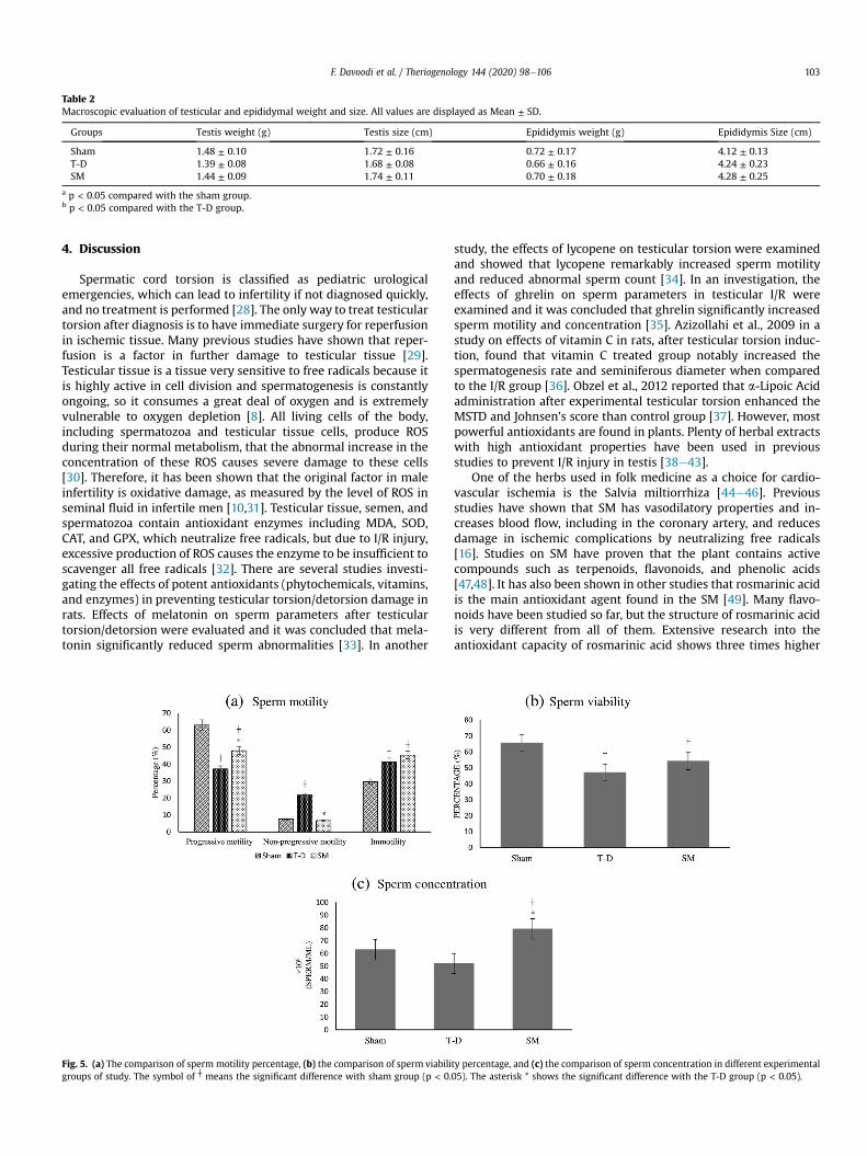
Table 2Macroscopic evaluation of testicular and epididymal weight and size. All values are displayed as Mean ± SD.
Groups Testis weight (g) Testis size (cm) Epididymis weight (g) Epididymis Size (cm)
Sham 1.48 ± 0.10 1.72 ± 0.16 0.72 ± 0.17 4.12 ± 0.13T-D 1.39 ± 0.08 1.68 ± 0.08 0.66 ± 0.16 4.24 ± 0.23SM 1.44 ± 0.09 1.74 ± 0.11 0.70 ± 0.18 4.28 ± 0.25
a p < 0.05 compared with the sham group.b p < 0.05 compared with the T-D group.
F. Davoodi et al. / Theriogenology 144 (2020) 98e106 103
4. Discussion
Spermatic cord torsion is classified as pediatric urologicalemergencies, which can lead to infertility if not diagnosed quickly,and no treatment is performed [28]. The only way to treat testiculartorsion after diagnosis is to have immediate surgery for reperfusionin ischemic tissue. Many previous studies have shown that reper-fusion is a factor in further damage to testicular tissue [29].Testicular tissue is a tissue very sensitive to free radicals because itis highly active in cell division and spermatogenesis is constantlyongoing, so it consumes a great deal of oxygen and is extremelyvulnerable to oxygen depletion [8]. All living cells of the body,including spermatozoa and testicular tissue cells, produce ROSduring their normal metabolism, that the abnormal increase in theconcentration of these ROS causes severe damage to these cells[30]. Therefore, it has been shown that the original factor in maleinfertility is oxidative damage, as measured by the level of ROS inseminal fluid in infertile men [10,31]. Testicular tissue, semen, andspermatozoa contain antioxidant enzymes including MDA, SOD,CAT, and GPX, which neutralize free radicals, but due to I/R injury,excessive production of ROS causes the enzyme to be insufficient toscavenger all free radicals [32]. There are several studies investi-gating the effects of potent antioxidants (phytochemicals, vitamins,and enzymes) in preventing testicular torsion/detorsion damage inrats. Effects of melatonin on sperm parameters after testiculartorsion/detorsion were evaluated and it was concluded that mela-tonin significantly reduced sperm abnormalities [33]. In another
Fig. 5. (a) The comparison of sperm motility percentage, (b) the comparison of sperm viabiligroups of study. The symbol of ┼ means the significant difference with sham group (p < 0.
study, the effects of lycopene on testicular torsion were examinedand showed that lycopene remarkably increased sperm motilityand reduced abnormal sperm count [34]. In an investigation, theeffects of ghrelin on sperm parameters in testicular I/R wereexamined and it was concluded that ghrelin significantly increasedsperm motility and concentration [35]. Azizollahi et al., 2009 in astudy on effects of vitamin C in rats, after testicular torsion induc-tion, found that vitamin C treated group notably increased thespermatogenesis rate and seminiferous diameter when comparedto the I/R group [36]. Obzel et al., 2012 reported that a-Lipoic Acidadministration after experimental testicular torsion enhanced theMSTD and Johnsen’s score than control group [37]. However, mostpowerful antioxidants are found in plants. Plenty of herbal extractswith high antioxidant properties have been used in previousstudies to prevent I/R injury in testis [38e43].
One of the herbs used in folk medicine as a choice for cardio-vascular ischemia is the Salvia miltiorrhiza [44e46]. Previousstudies have shown that SM has vasodilatory properties and in-creases blood flow, including in the coronary artery, and reducesdamage in ischemic complications by neutralizing free radicals[16]. Studies on SM have proven that the plant contains activecompounds such as terpenoids, flavonoids, and phenolic acids[47,48]. It has also been shown in other studies that rosmarinic acidis the main antioxidant agent found in the SM [49]. Many flavo-noids have been studied so far, but the structure of rosmarinic acidis very different from all of them. Extensive research into theantioxidant capacity of rosmarinic acid shows three times higher
ty percentage, and (c) the comparison of sperm concentration in different experimental05). The asterisk * shows the significant difference with the T-D group (p < 0.05).

Table 3Morphology of cauda epididymal spermatozoa. Data with normal distribution are displayed as Mean ± SD; Non-normal distribution data are expressed as median andinterquartile range.
Groups Normal morphology (%) Bent tail (%) Coiled tail (%) Distal cytoplasmic droplet (%) Short tail (%) Abnormal head (%)
Sham 82.33 ± 3.44 14.48 ± 2.42 2.35(1.20,3.00) 0.21 ± 0.53 0 ± 0.00 1.01 ± 1.29T-D 71.41 ± 3.15a 19.37 ± 5.28 8.06(3.98,14.24)a 0.83 ± 1.35 0.39 ± 0.61 0 ± 0.00SM 80.92 ± 4.52b 14.06 ± 3.78 5.00(2.70,5.96) 0.20 ± 0.48 0.16 ± 0.40 0.16 ± 0.40
a p < 0.05 compared with the sham group.b p < 0.05 compared with the T-D group.
Table 4Malondialdehyde (MDA), glutathione peroxidase (GPX), catalase (CAT), and total antioxidant capacity (TAC) activities in testis tissue and plasma.
Groups Testis tissue Plasma
MDA (mmol/mg-pr)
GPX (Unit/mg-pr)
CAT (Unit/mg-pr)
TAC (nmol Trolox equivalent/mg-protein)
MDA (mmol/mg-pr)
GPX (Unit/mg-pr)
CAT (Unit/mg-pr)
TAC (nmol Trolox equivalent/mg-protein)
Sham 0.24(0.17,0.33) 20.62 ± 1.92 0.41(0.34,0.51) 0.43 ± 0.08 0.15(0.15,0.16) 10.16 ± 0.04 4.53(3.47,7.72) 1.21 ± 0.19T-D 0.98(0.93,1.01)a 8.24 ± 2.57a 0.13(0.10,0.18)a 0.15 ± 0.04a 0.52(0.48,0.59)a 8.07 ± 0.51a 0.44(0.33,0.64)a 0.45 ± 0.08a
SM 0.21(0.19,0.32)b 19.8 ± 2.99b 0.33(0.21,0.34) 0.50 ± 0.03b 0.29(0.18,0.40)a,b 11.67 ± 0.55a,b 5.73(4.94,6.13)b 1.44 ± 0.12b
Data with normal distribution are displayed as Mean ± SD; Non-normal distribution data are expressed as median and interquartile range.a p < 0.05 compared with the sham group.b p < 0.05 compared with the T-D group.
F. Davoodi et al. / Theriogenology 144 (2020) 98e106104
antioxidant activity than torolox, prevention of xanthine oxidaseactivity, and scavenging excessive free radicals. It has also beendemonstrated that rosmarinic acid is able to reduce molybdenumblue (VI) to (V), which reduces the free radicals produced by metals[50]. In addition to the antioxidant properties, protection againstHIV type 1, anticarcinogenic, liver protection, and anti-hepatitis,preventing coagulation and the formation of blood clots and anti-inflammatory are also other properties of rosmarinic acid [50]. Asmentioned above, in our study, the SM extract was comprising14.28 (mg ml�1) rosmarinic acid.
ROS generation is biphasic in testicular I/R. The onset of phase Iis associated with tissue reperfusion and lasts for short hours and isalso associated with oxidative stress, but damage to cells in thisphase may be repaired. Phase II occurs when oxidative damagepersists for several days, and damage to the testicular tissue in thisphase is irreversible [1,40,51]. The results reported in this studywere determined for the first phase and for a short period ofreperfusion.
Our macroscopic results included testicular and epididymalweight, and size did not have any significant difference betweenthe T-D group and the sham group, and these results were differentfrom previous findings that reported a significant reduction intesticular weight and size following I/R [52,53]. A possible expla-nation for this might be that our results were indicated in a shortreperfusion time and only in the first phase of ROS generation.
As the amount of ROS in the tissue increases, plasma membranelipids are damaged and peroxidated, and the intracellular compo-nents are destroyed, and the marker for lipid peroxidation is MDA.Also, in the male reproductive system, GPX is one of the majorenzymes involved in reducing free radicals [54]. In an investigation,
Table 5Histopathological evaluations. MSTD, mean seminiferous tubular diameter; GECT, germcapsule thickness. All values are displayed as Mean ± SD.
Groups MSTD (mm) GECT (mm)
Sham 294.83 ± 22.01 78.16 ± 7.83T-D 195.16 ± 26.97a 32.66 ± 5.34a
SM 272.00 ± 10.71b 61.00 ± 6.38a,b
a p < 0.05 compared with the sham group.b p < 0.05 compared with the T-D group.
the oxidative stress and histopathological effects of SM extractpretreatment in the prevention of renal I/R damage in rats havebeen evaluated and indicated that SM prescription considerablyreduced renal MDA levels and increased the levels of GSH, SOD,CAT, and GPX [20]. In another study of the oxidative stress inhibi-tion impacts of Salvia militiorihiza mixed with Radix Puerariae onvascular damage in diabetic rats, it has been concluded that theextract could ameliorate the oxidative stress by a significantreduction in the level of MDA and increment in the serum levels ofthe SOD and CAT [24]. In the present study, in the T-D group theplasma and testicular tissue level of MDA significantly increasedand levels of GPX, CAT, and TAC reduced considerably, and the SMpretreatment could significantly reduce the MDA level and elevatethe levels of GPX, CAT, and TAC both in the plasma and testis tissuecompared to the T-D group which is in keeping with previousobservational studies [20,24,29,54].
Previously, it was thought that ischemia/reperfusion-inducedcell damage were only caused by necrosis. It has now beenshown that changes in ion exchange in the plasma membrane andthe loss of cell membrane integrity cause secretion of toxic mole-cules and inflammatory reactions that both damage and cause celldeath [55]. This is the mechanism that can damage cells, includingspermatozoa, in I/R injury. In 2015, Shen et al. published a paper inwhich they described that application of the SM polysaccharides infreezing and thawing of boar sperms prevented oxidative damageto spermatozoa and increased the percentage of motile sperms[56]. Jasem et al. (2010) carried out an investigation on salviahypoleuca (one of the species of salvia) effects on rat spermato-genesis and found that the extract could significantly increase themotility of spermatozoa, epididymal sperm concentration, and
inal epithelial cell thickness; MTBS, mean testicular biopsy scoring; TCT, testicular
GECT (Cell Layer) MTBS (Score) TCT (mm)
8.66 ± 0.51 9.33 ± 0.51 37.66 ± 4.714.00 ± 0.63a 6.66 ± 0.51a 28.16 ± 4.53a
6.5 ± 0.83a,b 8.50 ± 0.54a,b 33.00 ± 3.03

F. Davoodi et al. / Theriogenology 144 (2020) 98e106 105
testicular weights [57]. The beneficial impacts of rosmarinic acid insex hormones and testicular apoptosis induced by the electro-magnetic field in male rats were evaluated by Khaki et al. (2012),and an increment in the testosterone level of the serum and posi-tive effects on histology of the testis was observed in the rosmarinicacid treatment group [58]. In accordance with previous studies, ourresults proved that I/R could damage the epididymal stored sper-matozoa, and the SM extract pretreatment could markedly increaseprogressive motile spermatozoa, normal morphological sperma-tozoa, sperm concentration, and significant reduction in non-progressive motile spermatozoa was observed when compared tothe T-D group.
5. Conclusion
In this investigation, the aimwas to assess the protective effectsof Salvia miltiorrhiza hydroalcoholic extract pretreatment in pre-venting the ischemia/reperfusion damage caused by torsiondetorsion of testis. The findings of the present study suggest thatSalvia miltiorrhiza is a potent antioxidant that can reduce oxidativestress and improve sperm parameters as well as reduce the rate oftesticular tissue injury. One limitation of this study was that theprecise timing of the administration of the extract and the appro-priate dose to achieve the best effect of the extract were not exactlyknown, and the dose and time of injection in our study were basedon previous documentations. Further research is required tocompletely understand the suitable dose and administration timeto achieve the best treatment efficiency.
Declaration of competing interest
Authors declare no conflict of interest.
CRediT authorship contribution statement
Farshid Davoodi: Conceptualization, Methodology, Investiga-tion, Writing - original draft. Shayan Taheri: Conceptualization,Methodology, Investigation. Abbas Raisi: Project administration,Writing - original draft, Writing - review & editing. AsgharRajabzadeh: Writing - original draft, Writing - review & editing.Hassan Ahmadvand: Visualization, Resources. Mohammad Has-san Hablolvarid: Methodology, Validation. Amir Zakian: Datacuration, Formal analysis, Writing - review & editing.
Acknowledgment
This paper has been extracted from a DVM thesis of FarshidDavoodi and Shayan Taheri carried out in Lorestan University andthe authors want to appreciate the vice-chancellor of research ofLorestan University, Razi Herbal Medicines Research Center ofLorestan Medical University, Khorramabad, Iran, and Razi Vaccineand Serum Research Institute, Karaj, Iran.
Funding: This work was supported by the Lorestan Universitygrant number: [LU-9211501009-2020]
References
[1] Abdel-Gaber SA, Mohammed RK, Refaie MM. Mechanism mediating the pro-tective effect of diacerein in ischemia-reperfusion-induced testicular injury inrats. Life Sci 2018;209:57e62.
[2] Wilhelm Filho D, Torres MA, Bordin AL, Crezcynski-Pasa TB, Boveris A. Sper-matic cord torsion, reactive oxygen and nitrogen species andischemiaereperfusion injury. Mol Asp Med 2004;25:199e210.
[3] Bozlu M, Acar D, Cayan S, Aktas S, Tunckiran A. Protective effect of trapidil onlong-term histologic damage in a rat model of testicular ischemia-reperfusioninjury. World J Urol 2009;27:117.
[4] G€okçe A, Oktar S, Koc A, Gonenci R, Yalcinkaya F, Yonden Z, et al. Protective
effect of thymoquinone in experimental testicular torsion. Urol Int 2010;85:461e5.
[5] Bilommi R, Nawas BA, Kusmayadi DD, Diposarosa R, Chairul A, Hernowo BS.The effects of glutathione on malondialdehyde expression and seminiferoustubule damage in experimental testicular torsionedetorsion in Wistar rats.J Pediatr Urol 2013;9:1059e63.
[6] Agarwal A, Prabakaran SA. Mechanism, measurement, and prevention ofoxidative stress in male reproductive physiology 2005;43:963e74.
[7] Aitken RJ, Roman SD. Antioxidant systems and oxidative stress in the testes.Oxid Med Cell Longevity 2008;1:15e24.
[8] Agarwal A, Makker K, Sharma R. Clinical relevance of oxidative stress in malefactor infertility: an update. Am J Reprod Immunol 2008;59:2e11.
[9] Sawyer DE, Mercer BG, Wiklendt AM, Aitken RJ. Quantitative analysis of gene-specific DNA damage in human spermatozoa. Mutat Res Fundam Mol MechMutagen 2003;529:21e34.
[10] Aitken RJ, Baker MA. Oxidative stress, sperm survival and fertility control. MolCell Endocrinol 2006;250:66e9.
[11] Mikaili P, Shayegh J, Asghari MH, Sarahroodi S, Sharifi M. Currently usedtraditional phytomedicines with hot nature in Iran. Biol Res 2011;2:56e68.
[12] O’hara M, Kiefer D, Farrell K, Kemper K. A review of 12 commonly used me-dicinal herbs. Arch Fam Med 1998;7:523e36.
[13] Abad ANA, Nouri MHK, Tavakkoli F. Effect of Salvia officinalis hydroalcoholicextract on vincristine-induced neuropathy in mice. Chin J Nat Med 2011;9:354e8.
[14] Zhao G-R, Xiang Z-J, Ye T-X, Yuan Y-J, Guo Z-X. Antioxidant activities of Salviamiltiorrhiza and Panax notoginseng. Food Chem 2006;99:767e74.
[15] Gordon M, Weng X. Antioxidant properties of extracts from tanshen (Salviamiltiorrhiza Bunge). Food Chem 1992;44:119e22.
[16] Anversa P, Olivetti G, Meggs L, Sonnenblick E, Capasso J. Cardiac anatomy andventricular loading after myocardial infarction. MonogrAm Heart Assoc1993;87. VII. 22-VII. 7.
[17] Sun J, Huang SH, Tan BK-H, Whiteman M, Zhu YC, Wu YJ, et al. Effects ofpurified herbal extract of Salvia miltiorrhiza on ischemic rat myocardium afteracute myocardial infarction. Life Sci 2005;76:2849e60.
[18] Jia Y, Huang F, Zhang S, Leung S-w. Is danshen (Salvia miltiorrhiza) drippingpill more effective than isosorbide dinitrate in treating angina pectoris? Asystematic review of randomized controlled trials. Int J Cardiol 2012;157:330e40.
[19] Nan JX, Park EJ, Kang HC, Park PH, Kim JY, Sohn DH. Anti-fibrotic effects of ahot-water extract from Salvia miltiorrhiza roots on liver fibrosis induced bybiliary obstruction in rats. J Pharm Pharmacol 2001;53:197e204.
[20] Chen G, Fu Y, Wu X. Protective effect of Salvia miltiorrhiza extract againstrenal ischemia-reperfusion-induced injury in rats. Molecules 2012;17:1191e202.
[21] Benzie IF, Strain JJ. The ferric reducing ability of plasma (FRAP) as a measure of“antioxidant power”: the FRAP assay. Anal Biochem 1996;239:70e6.
[22] Wang X, Lee WY, Zhou X, Or PM, Yeung JH.A pharmacodynamicepharmacokinetic (PDePK) study on the effects ofDanshen (Salvia miltiorrhiza) on midazolam, a model CYP3A probe substrate,in the rat. Phytomedicine 2010;17:876e83.
[23] Wang X, Yeung JH. Effects of the aqueous extract from Salvia miltiorrhizaBunge on caffeine pharmacokinetics and liver microsomal CYP1A2 activity inhumans and rats. J Pharm Pharmacol 2010;62:1077e83.
[24] Zhao W, Yuan Y, Zhao H, Han Y, Chen X. Aqueous extract of Salvia miltiorrhizaBunge-Radix Puerariae herb pair ameliorates diabetic vascular injury byinhibiting oxidative stress in streptozotocin-induced diabetic rats. Food ChemToxicol 2019;129:97e107.
[25] Varisli O, Uguz C, Agca C, Agca Y. Various physical stress factors on rat spermmotility, integrity of acrosome, and plasma membrane. J Androl 2009;30:75e86.
[26] World Health Organization DoRHaR. WHO laboratory manual for the exami-nation and processing of human semen. fifth ed. Geneva: Switzerland: WorldHealth Organization; 2010. ed. Switzerland, 2010.
[27] Johnsen SG. Testicular biopsy score countea method for registration ofspermatogenesis in human testes: normal values and results in 335 hypo-gonadal males. Hormone Res Paediatr 1970;1:2e25.
[28] Lee J-W, Kim J-I, Lee Y-A, Lee D-H, Song C-S, Cho YJ, et al. Inhaled hydrogengas therapy for prevention of testicular ischemia/reperfusion injury in rats.J Pediatr Surg 2012;47:736e42.
[29] Ameli M, Hashemi MS, Moghimian M, Shokoohi M. Protective effect of tada-lafil and verapamil on testicular function and oxidative stress after torsion/detorsion in adult male rat. Andrologia 2018;50:e13068.
[30] Dokmeci D, Inan M, Basaran UN, Yalcin O, Aydogdu N, Turan FN, et al. Pro-tective effect of L-carnitine on testicular ischaemiaereperfusion injury in rats.Cell Biochem Funct: Cellular biochemistry and its modulation by active agentsor disease 2007;25:611e8.
[31] Allamaneni SS, Agarwal A, Nallella KP, Sharma RK, Thomas Jr AJ, Sikka SC.Characterization of oxidative stress status by evaluation of reactive oxygenspecies levels in whole semen and isolated spermatozoa. Fertil Steril 2005;83:800e3.
[32] Dogan C, Halici Z, Topcu A, Cadirci E, Karakus E, Bayir Y, et al. Effects ofamlodipine on ischaemia/reperfusion injury in the rat testis. Andrologia2016;48:441e52.
[33] Kurcer Z, Hekimoglu A, Aral F, Baba F, Sahna E. Effect of melatonin onepididymal sperm quality after testicular ischemia/reperfusion in rats. Fertil

F. Davoodi et al. / Theriogenology 144 (2020) 98e106106
Steril 2010;93:1545e9.[34] Hekimoglu A, Kurcer Z, Aral F, Baba F, Sahna E, Atessahin A. Lycopene, an
antioxidant carotenoid, attenuates testicular injury caused by ischemia/reperfusion in rats. Tohoku J Exp Med 2009;218:141e7.
[35] Taati M, Moghadasi M, Dezfoulian O, Asadian P, Kheradmand A, Abbasi M,et al. The effect of ghrelin pretreatment on epididymal sperm quality andtissue antioxidant enzyme activities after testicular ischemia/reperfusion inrats. J Physiol Biochem 2012;68:91e7.
[36] Azizollahi S, Babaei H, Derakhshanfar A, Oloumi M. Effects of co-administra-tion of dopamine and vitamin C on ischaemia-reperfusion injury afterexperimental testicular torsion-detorsion in rats. Andrologia 2011;43:100e5.
[37] Ozbal S, Ergur BU, Erbil G, Tekmen I, Bagrıyanık A, Cavdar Z. The effects of a-lipoic acid against testicular ischemia-reperfusion injury in rats. Sci World J2012;2012.
[38] Aktas C, Erboga M, Fidanol Erboga Z, Bozdemir Donmez Y, Topcu B, Gurel A.Protective effects of Urtica dioica L. on experimental testicular ischaemiareperfusion injury in rats. Andrologia 2017;49:e12636.
[39] Bayatli F, Akkus D, Kilic E, Saraymen R, S€onmez MF. The protective effects ofgrape seed extract on MDA, AOPP, apoptosis and eNOS expression in testiculartorsion: an experimental study. World J Urol 2013;31:615e22.
[40] Kim Y-H, Kim G-H, Shin J-H, Kim K-S, Lim J-S. Effect of Korean red ginseng ontesticular tissue injury after torsion and detorsion. Korean J Urol 2010;51:794e9.
[41] Shokoohi M, Shoorei H, Soltani M, Abtahi-Eivari SH, Salimnejad R,Moghimian M. Protective effects of the hydroalcoholic extract of Fumariaparviflora on testicular injury induced by torsion/detorsion in adult rats.Andrologia 2018;50:e13047.
[42] Uyeturk U, Terzi EH, Gucuk A, Kemahli E, Ozturk H, Tosun M. Prevention oftorsion-induced testicular injury by Rhodiola rosea. Urology 2013;82:254. e1-.e6.
[43] Dursun R, Zengin Y, Gündüz E, _Içer M, Durgun HM, Da�ggulli M, et al. Theprotective effect of goji berry extract in ischemic reperfusion in testis torsion.Int J Clin Exp Med 2015;8:2727.
[44] Ji X, Tan BK-H, Zhu YC, Linz W, Zhu YZ. Comparison of cardioprotective effectsusing ramipril and DanShen for the treatment of acute myocardial infarctionin rats. Life Sci 2003;73:1413e26.
[45] Ji X. Effects of Salvia miltiorrhiza in ischemic myocardium in experimental
rats. Perspective. In: Tan B, Bay BH, Zhu YZ, editors. Novel compounds fromnatural products in new millenniums. London: World Scientific Press; 2004.p. 183e95.
[46] Zhu Y, Huang S, Tan B, Sun J, Whiteman M, Zhu Y-C. Antioxidants in Chineseherbal medicines: a biochemical perspective. Nat Prod Rep 2004;21:478e89.
[47] Bisio A, Romussi G, Ciarallo G, De Tommasi N. Flavonoide und triterpenoideaus Salvia blepharophylla Brandegee ex Epling. Pharmazie 1997;52:330e1.
[48] G€okdil G, Topcu G, S€onmez U, Ulubelen A. Terpenoids and flavonoids fromSalvia cyanescens. Phytochemistry 1997;46:799e800.
[49] Lu Y, Foo LY. Antioxidant activities of polyphenols from sage (Salvia offici-nalis). Food Chem 2001;75:197e202.
[50] Petersen M, Simmonds MS. Rosmarinic acid. Phytochemistry 2003;62:121e5.[51] Cutrìn JC, Boveris A, Zingaro B, Corvetti G, Poli G. In situ determination by
surface chemiluminescence of temporal relationships between evolvingwarm ischemia-reperfusion injury in rat liver and phagocyte activation andrecruitment. Hepatology 2000;31:622e32.
[52] Dejban P, Rahimi N, Takzare N, Jahansouz M, Dehpour AR. Protective effects ofsumatriptan on ischaemia/reperfusion injury following torsion/detorsion inipsilateral and contralateral testes of rat. Andrologia 2019:e13358.
[53] Mogilner JG, Lurie M, Coran AG, Nativ O, Shiloni E, Sukhotnik I. Effect ofdiclofenac on germ cell apoptosis following testicular ischemia-reperfusioninjury in a rat. Pediatr Surg Int 2006;22:99e105.
[54] Asghari A, Akbari G, Meghdadi A, Mortazavi P. Protective effect of metforminon testicular ischemia/reperfusion injury in rats. Acta Cir Bras 2016;31:411e6.
[55] Kalogeris T, Baines CP, Krenz M, Korthuis RJ. Ischemia/reperfusion. Compre-hensive Physiology 2016;7:113e70.
[56] Shen T, Jiang Z-L, Liu H, Li Q-W. Effect of Salvia miltiorrhiza polysaccharideson boar spermatozoa during freezingethawing. Anim Reprod Sci 2015;159:25e30.
[57] Jasem E, Nasim J, Gholamreza M, Naser S, Nader M, Maryam SL, et al. Evalu-ation of the effects of Salvia hypoleuca on the cAMP-responsive elementmodulator (CREM) gene expression and spermatogenesis in rat. Middle EastFertil Soc J 2010;15:274e7.
[58] Khaki A, Imani S, Golzar FS. Effects of rosmarinic acid on male sex hormones(testosterone-FSH-LH) and testis tissue apoptosis after exposure to electro-magnetic field (EMF) in rats. Afr J Pharm Pharmacol 2012;6:248e52.