investigation of the antiurolithiatic...
TRANSCRIPT

RESEARCH ARTICLE e-ISSN: 2454-7867
Krishnaveni et al. Int J Trends in Pharm & Life Sci. 2015: 1(5); 566- 586. 566
Available online at www.ijtpls.com
International Journal of Trends in Pharmacy and Life Sciences Vol. 1, Issue: 5, 2015: 566-586
INVESTIGATION OF THE ANTIUROLITHIATIC ACTIVITY OF Ixora pavetta ON
SODIUM OXALATE INDUCED UROLITHIASIS IN ALBINO RATS Kalasani Krishna Veni, K. Sravanth & D. Rama brahma Reddy
Nalanda institute of Pharmaceutical Sciences, Siddharth Nagar, Kantepudi(Village),
Sattenapalli(Mandal), Guntur(Dist)-522438 E.Mail: [email protected]
ABSTRACT Ixora pavetta (torch tree) is a plant in the Rubiaceae family most widely used by the tribals of
andhrapradesh and Karnataka for the various purposes and having traditional values. preliminary
phytochemical screening of methanolic root extract of Ixora pavetta revealed the presence of Carbohydrates,
alkaloids, flavonoids, triterpenes, tannins ,phenols, saponins, phytosterols, proteins, aminoacids. In group of
Male wister Albino rats weighing 200-250g treated with sodium oxalate (0.164 ml, IP, single dose) a
significant increase (P<0.05) in serum biochemical parameters sodium, potassium, chloride, uric acid
,creatinine, urea levels (136.5±5.071,72±2.67,91.3±3.86,1.945±0.17,31.1±2.79,50.3±2.9) was observed
when compared to normal group (80.3±±±±±) and
urine parameters like oxalate , uric acid, creatinine (4.101±0.433,34.92±2.841,4.793±0.222) was observed
when compared to normal group(1.90±0.309,15.78±1.512,1.535±0.167) This indicates NaOx induces
urolithiasis in rats. In group that received standard drug (Cystone, 500 mg/kg, p.o, once daily) there was
significant decrease (P<0.05) in the serum sodium, potassium, chloride, uric acid, creatinine, urea and urine
oxalate, uric acid, creatinine levels when compared to NaOx treated group. On administration of MEIP
(200mg/kg, 400 mg/kg, p.o, once daily) there was a significant decrease (P<0.05) in the serum sodium,
potassium, chloride, uric acid ,creatinine, urea and urine oxalate, uric acid, creatinine levels (338.71 8.79)
when compared to negative control group. A dose related effect was not observed in groups treated with the
test drug. These results suggest that MEIP has better Anti urolithiatic activity.
*Corresponding Author:
K.Krishna Veni,
Nalanda institute of Pharmaceutical Sciences, Siddharth Nagar,
Kantepudi(Village),
Sattenapalli(Mandal), Guntur(Dist)-522438 Ph: 9441810344
INTRODUCTION
Many medicinal plants provide relief of symptoms comparable to that obtained from allopathic
medicines. Ixora pavetta (torch tree) is a plant in the Rubiaceae family commonly seen in Ballari district of
Karnataka and Tirumala hills, Andhrapradesh, India. It is called as Goravi in Telugu. People use the
branches of this tree for making walls and paste with mud for their thatched huts in villages, but now this
practice has become obsolete owing to modern housing materials. Summer is the fruiting season and the
fruits are globose, 2-seeded, become black when ripened. Indian Sloth Bears eat the fruits and the seeds are
dispersed through its scat. Urolithiasis, which is referred to as the process of formation of calculi (singular
calculus) in the urinary system. By extensive literature review, we have concluded that, the above said plant
was traditionally used for the treatment of gall stones and the aim of our study is to investigate the
antiurolithiatic activity of Ixora pavetta on sodium oxalate induced urolithiasis in albino rats. [1, 2]
Received: 28/10/2015
Revised: 20/11/2015
Accepted: 30/11/2015

RESEARCH ARTICLE e-ISSN: 2454-7867
Krishnaveni et al. Int J Trends in Pharm & Life Sci. 2015: 1(5); 566- 586. 567
Materials and Method
Collection and Authentication of plant material:
Ixora pavetta roots were collected from thirupati hills. The plant was identified and authenticated by
Dr.K.Madhava chetty, Department of Pharmacognosy, S.V.university,thirupati.
Preparation of extract:
Roots are collected, washed properly and dried under shade for few days. They are powdered coarsely and
subjected for cold maceration with pet ether for 48hrs and filtered. The dried mass obtained were subjected
for soxhlet extraction with methanol at 65ºc for 6hrs.the extract was
Made to dried under reduced vacuum. The yield of root in 42gm/kg.
Preliminary Phytochemical screening:
The methanolic root extract of Ixora pavetta were subjected to phytochemical screening for identification of
various phyto conctituents present in it. [3]
ANTI UROLITHIATIC ACTIVITY
Experimental Animals
Male Wistar albino rats of weighing between (200-250 g) used for the experimentation.animals were
obtained from the animal house attached to our institute. Six animals were housed in each Cage made up of
poly-propylene with stainless steel top grill. After randomization into various groups, animals were
acclimatized for period of 7 days under standard husbandry conditions. Room temperature 27 ± 30C,
Relative humidity 65 ± 10%, 12 hrs light/dark cycle. All the animals were fed with rodent pellet diet (Malla
Reddy institute of pharmaceutical sciences, hydearbad) and water was allowed ad-libitum under strict
hygienic condition. Ethical clearance for performing experiments on animals was obtained from Institutional
Animal Ethical Committee (Regd No:1662/PO/a/CPCSEA,2013) and were in accordance with the
guidelines of CPCSEA[4].
Acute Toxicity Study
The acute oral toxicity procedure was followed by using OECD 423 guidelines. The acute toxic class
method is a step wise procedure with 6 animals of a single sex per step (OECD,2001). Depending on the
mortality and /or moribund status of the animals, on the average 2-4 steps may be necessary to allow
judgment on the acute toxicity of the test substance. This procedure results in the use of minimal number of
animals while allowing for acceptable data based scientific conclusion. The method used defined doses (5,
50, 300, 2000 mg/kg b.wt.) and the results allow a substance to be ranked and classified according to the
Globally Harmonized System for the classification of chemicals which cause acute toxicity.
After oral administration of the test, animals were observed individually at least once during the first 30
minutes and periodically during the first 24 hrs, with special attention given during the first 4 hrs and daily
thereafter for total of 14 days. Body weight of the rats before and after treatment were noted and any
changes in the skin, fur, eyes, mucous membranes, salivation, nasal discharge, urination, behavioural,

RESEARCH ARTICLE e-ISSN: 2454-7867
Krishnaveni et al. Int J Trends in Pharm & Life Sci. 2015: 1(5); 566- 586. 568
neuromuscular, cardiovascular, lethargy, sleep and coma were noted for 14 days. Test compounds were not
mortal for rats even at 200 mg/kg dose hence, 1/20th
of this dose was selected for further study.
EXPERIMENTAL DESIGN
Evaluation of Antiurolithiatic activity in Sodium oxalate induced urolithiasis
Male wister Albino rats weighing 200-250g were selected and divided into five groups consisting of six
animals each and they are grouped as follow.
Group I - Animal received 2% v/v Tween 80, p.o. vehicle for 10 days and served as as Normal group.
Group II - Animal received Sodium oxalate 70mg/kg,i.p. for 10 days and served as disease control group.
Group III - Cystone 500mg/kg, p.o.suspended in 2%v/v Tween 80 and Sodium Oxalate 70mg/kg,i.p. for 10
days and served as standard group
Group IV - Methanolic extract of Ixora pavetta (MEIP) 200mg/kg,p.o. suspended in 2%v/v Tween 80
and sodium oxalate(NaOx) 70mg/kg,i.p. for 10 days and served as Test group-I.
Group V - Methanolic extract of Ixora pavetta (MEIP)400mg/kg,p.o.suspended in 2% Tween 80 and
Sodium oxalate(NaOx) 70mg/kg,i.p. for 10 days and served as Test Group-II.
Collection of blood and urine samples:
At the end of 10th
day blood was collected from Retro orbital plexus and the serum was separated by
centrifuge at 800rpm for 20 min and serum was subjected to biochemical parameters. after collected blood
sample from each group placed in individual cages and the Urine was collected for 24hrs.the volume is
measured in ml. the urine was subjected for estimation of urinary parameters.
ESTIMATION OF PARAMETERS [5-10]
Estimation of sodium by colorimetric method
The sodium and the proteins are precipitated simultaneously by means of reagent containing magnesium
uranyl acetate containing alcohol. The precipitate is separated by centrifugation. The content of sodium is
calculated from the loss in concentration of magnesium uranyl acetate in the reagent solution in comparison
to standard sodium solution treated similarly. The residual amount of magnesium uranyl acetate is estimated
by forming brown (dark) ferrous uranyl acetate, which is read in a colorimeter.
Reagents:
1. Sodium Precipitating Reagent - 33ml
2. Standard Sodium/Potassium - 3ml
3. Sodium colour Reagent - 10ml
Reaction type End point with std.
Reaction Slope Increasing
Wave length 540nm
Incubation temperature Room temperature
Incubation time 10min
Standard conc 150m.Mol/L
Linearity 200m.Mol/L
Unit m.Mol/L

RESEARCH ARTICLE e-ISSN: 2454-7867
Krishnaveni et al. Int J Trends in Pharm & Life Sci. 2015: 1(5); 566- 586. 569
Procedure:
Pipette into clean dry test tubes, labeled standard(S) and test (T).
Standard Test
Standard PPT Reagent 1.0ml 1.0ml
Standard sodium/potassium 0.02ml -
Serum - 0.02ml
Mix well on vortex for one minute and wait for 5minutes at room temperature. Centrifuge for one minute at
3000 rpm.
Blank Standard Test
Distilled water 3ml 3ml 3ml
Supernatant from step-1 - 0.05ml 0.05ml
Sodium PPT Reagent 0.05ml - -
Sodium Colour Reagent 0.2ml 0.2ml 0.2ml
Pipette into 3 clean dry test tubes labelled Blank (B) Standard (S) and Test (T).
Mix well and allow it to stand at room temperature for 5minutes.then measure absorbance of Blank,
Standard and Test against distilled water on photocolorimeter at 540nm within 10minutes
Calculations:
Sodium in m.Mol/L= Absorbance of B-T ×150(Standard concentration)
Absorbance of B-S
Estimation of potassium by colorimetric method
Principle:
Potassium can be determined by a number of different methods. It can be directly estimated by flame
photometry, colorimetry. It can also be measurement of turbidity of the reaction mixture Containing Sodium
Tetra phenyl Boron, alkaline EDTA, formaldehyde and sample containing potassium (or) standard
potassium salt. The method accurate within concentration of 2.0 to 7.0 m.M/L, there is a good agreement
with flame photometry.
Reagents:
1.Potassium Reagent - 45ml
2.Standard Sodium/Potassium – 3ml
Reaction type End point
Reaction slope Increasing
Wave length 620nm
Incubation temperature Room temoerature
Incubation time 5min
Standard concentration 5m.Mol/L
Linearity 7m.Mol/L

RESEARCH ARTICLE e-ISSN: 2454-7867
Krishnaveni et al. Int J Trends in Pharm & Life Sci. 2015: 1(5); 566- 586. 570
Unit m.Mol/L
Procedure:
Pipette into two clean dry test tubes labelled Standard (S) and test (T).
Standard Blank
Potassium Reagent 1.0ml 1.0ml
Standard Sodium/Potassium 0.05ml -
Serum - 0.05ml
Mix gently wait for 5minutes at room temperature read the absorbance of standard and test against
distilled water on photo colorimeter at 620nm within 10minutes.
Calculation:
Potassium in mMol/L= Absorbance of T × 5 (standard concentration)
Absorbance of S
Estimation of chloride by Colorimetric method
Principle:
Chloride ions form a coloured complex when reacting with mercury (II) thiocynate solution. The intensity of
the colour is proportional to chloride concentration.
Reagents:
1. Chloride Reagent
2. Standard chloride
Reaction type End point with std.
Reaction Slope Increasing
Wavelength 505nm
Incubation temperature Room temperature
Incubation time 2min.
Satandard concentration 100m.Mol/L
Linearity 130m.Mol/L
Unit m.Mol/L
Chloride Assay:
Pipette into three clean dry test tubes labelled Blank (B), Standard(S) and Test (T).
Blank Standard Test
Chloride Reagent 1.0ml 1.0ml 1.0ml
Standard chloride - 0.005ml -
Serum - - 0.005ml
Mix well wait for 2 minutes at room temperature and read the absorbance of Blank,Standard and Test
against distilled water on a photocolorimeter at 505nm

RESEARCH ARTICLE e-ISSN: 2454-7867
Krishnaveni et al. Int J Trends in Pharm & Life Sci. 2015: 1(5); 566- 586. 571
Calculation:
Chloride in m.Mol/L= Absorbance of T-B × 100(standard concentration)
Absorbance of S-B
Estimation of Creatinine by Jaffes’s method :
Principle: Creatinine reacts with alkaline picrate to produce a red coloured complex; the rate of red
coloured complex formation is directly proportional to the creatinine concentration52
.
Reagents Composition:
1) Picric acid Reagent
2) Alkaline Buffer Reagent
3) Standard Creatinine (2mg %)
4) Acid Reagent
Preparation of working reagent: Mix equal volumes of picric acid reagent & alkaline buffer reagent.
Sample: Serum, plasma, urine
Dilute urine specimen 1:100 using distilled water before use.
Parameter Values
Reaction Type Kinetic with std
Wavelength 520 nm (green filter)
Temperature Room Temperature
Incubation Time 30 sec
Read Time 60 sec
No of Readings 2
Sample Volume 100µl
Reagent Volume 1000µl
Standard Concentration 2 mg%
Linearity 20 mg%
Unit mg%
Procedure: KINETIC METHOD
1. Pipette into a clean dry cuvette
Working reagent 1ml
Serum /dilute urine/standard 0.1ml
2. Mix well and start stopwatch. Read intial absorbance Aₒ exactly after 30 seconds for test and standard.
3. Read another absorbance A1 for test and standard exactly 60 secs later.
4. Calculate change in absorbance for test and standard.
For test Δ AT= A1T-A0 T
For standard Δ AS = A 1S – A 0S
Calculation:

RESEARCH ARTICLE e-ISSN: 2454-7867
Krishnaveni et al. Int J Trends in Pharm & Life Sci. 2015: 1(5); 566- 586. 572
a) Serum creatinine in mg% =Δ𝐴𝑇
Δ𝐴𝑆 x 2 std conc
b) Urine creatinine in gm/lit = Δ𝐴𝑇
Δ𝐴𝑆 x 2 std conc
c) Urine creatinine in gm/24 hr =(b) x 24 hrs urine volume in liters
SI conversion factor
µ/L= mg% x 88.4
1. Mix well and allow it to stand at RT for 5 mins.
2. Read absorbance for standard As0 and Test At0 against distilled water at 520 nm (or) green filter in
spectrophotometer.
3. Read absorbance for standard As1 and Test At1 against distilled water at 520nm (or) green filter in
spectrophotometer.
Calculation
Δ AS = AS0 – AS1
Δ AT = AT0 – AT1
a) Serum Creatinine =Δ𝐴𝑇
Δ𝐴𝑆 × 2(Std. Conc)
Estimation of uric acid by end point assay
Principle:
Uric acid is oxidized to allantoin & hydrogen peroxide by the enzyme uricase. In presence of
peroxidase, released hydrogen peroxide is coupled with aniline derivative and 4-amino antipyrine (4-AAP)
to form colored chromogen complex. Absorbance of colored dye is measured at 550nm and is proportional
to uric acid concentration in the sample.
Uricase
Uric acid +2 H20 Allatonin +CO2 +H2O2
POD
H2O2 +Alanine derivative + 4-AAP chromogen complex + H2O2
Standard Test
Working Reagent 1.0 ml 1.0 ml
Standard 0.1ml ---
Serum/Diluted urine --- 0.1ml
Acid Reagent 0.05ml 0.05ml

RESEARCH ARTICLE e-ISSN: 2454-7867
Krishnaveni et al. Int J Trends in Pharm & Life Sci. 2015: 1(5); 566- 586. 573
Pipette into tube marked Blank Standard Test
Serum/plasma/urine - - 20µL
Reagent 2 - 20µL -
Reagent 1 1000 µL 1000 µL 1000 µL
Mix well and incubate at 37ºc for 5 minutes
1. Blank the analyzer with reagent blank
2. Measure the absorbance of standard followed by test
3. Calculate results
Mode
End point
Wavelength 550 nm
Flow-cell temperature 37ºc
Optical path length 1 cm
Blanking Reagent blank
Sample volume 20 µL
Reagent volume 1000 µL
Incubation time 5 minutes
Concentration of standard 6 mg/dL
Stability of final colour 15 minutes
Permissible reagent blank absorbance <0.4 AU
Linearity 25 mg/dL
Units mg/dL
Calculation55,56,57
:
Serum/plasma uric acid mg/dL = Absorbance of test
---------------------------- x 6
Absorbance of standard
Urine uric acid (mg/day) = Absorbance of test
---------------------------- x 6 x dilution
Absorbance of standard x 24 hrs urine volume in dL
Conversion factor in mmol/L= uric acid concentration in mg/dL x 0.059
Estimation of Urea by Berthelot method
Principle:
Urease catalyses the conversion of Urea to Ammonia and Carbon dioxide. The Ammonia released reacts
with a mixture of salicylate, hypochlorite and Nitroprusside to yield a blue-green colored compound

RESEARCH ARTICLE e-ISSN: 2454-7867
Krishnaveni et al. Int J Trends in Pharm & Life Sci. 2015: 1(5); 566- 586. 574
(Indophenol). The intensity of color produced is proportional to the concentration of urea in the Sample and
is measured photometrically at 570nm or with yellow filter.
Reaction:
Urea + H2O 2NH3 + CO2
Urease
Nitroprusside
NH3 + salicylate 2-2-Dicarboxy Indophenol
Hypochloride
Reagents composition:
2×50ml 2×100ml
1) Urease Reagent 1×50ml 1×100ml
2) Enzyme Concentrate 1Vial 1Vial
3) Color Reagent 1×50ml 1×100ml
4) Urea Standard (40 mg%) 1×2ml 1×2ml
Parameter Values
Reaction Type End point with std
Wavelength 578 nm (yellow filter)
Incubation Temp 37oC
Incubation Time 10 mins
Standard 40 mg%
Linearity 300 mg%
Unit mg%
Procedure:
Working Reagent Preparation: Transfer the entire enzyme Concentrate (2) into Urease Reagent (1) with
dropper or micro tip, rinse the Enzyme Concentrate Vial with little Urease Reagent and transfer the residual
enzyme to ensure better reconstitution.
Blank Standard Test
Urease Reagent
(working)
1.0 ml 1.0 ml 1.0 ml
Urea Standard --- 0.01ml ---
Specimen --- --- 0.01ml
Incubate for 5min at 37oC/10 min at RT
Colour reagent 1.0 ml 1.0 ml 1.0 ml
RESEARCH ARTICLE e-ISSN: 2454-7867
Krishnaveni et al. Int J Trends in Pharm & Life Sci. 2015: 1(5); 566- 586. 575
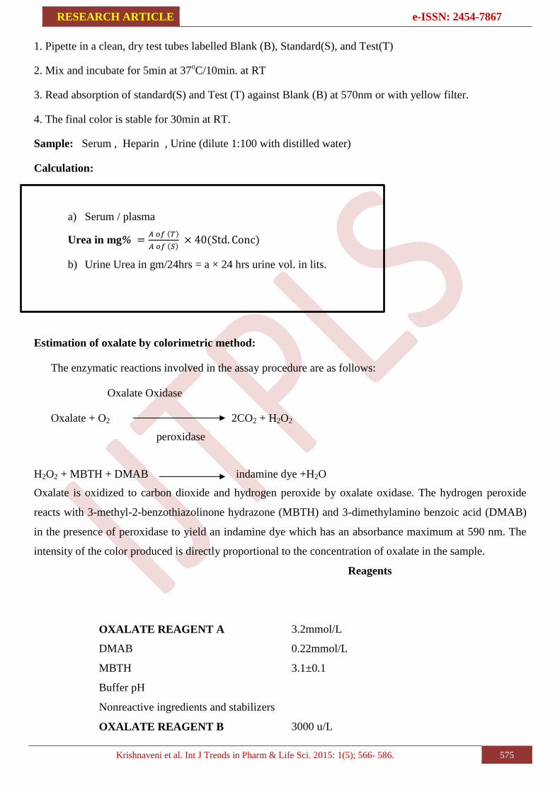
1. Pipette in a clean, dry test tubes labelled Blank (B), Standard(S), and Test(T)
2. Mix and incubate for 5min at 37oC/10min. at RT
3. Read absorption of standard(S) and Test (T) against Blank (B) at 570nm or with yellow filter.
4. The final color is stable for 30min at RT.
Sample: Serum , Heparin , Urine (dilute 1:100 with distilled water)
Calculation:
a) Serum / plasma
Urea in mg% =𝐴 𝑜𝑓 (𝑇)
𝐴 𝑜𝑓 (𝑆) × 40(Std. Conc)
b) Urine Urea in gm/24hrs = a × 24 hrs urine vol. in lits.
Estimation of oxalate by colorimetric method:
The enzymatic reactions involved in the assay procedure are as follows:
Oxalate Oxidase
Oxalate + O2 2CO2 + H2O2
peroxidase
H2O2 + MBTH + DMAB indamine dye +H2O
Oxalate is oxidized to carbon dioxide and hydrogen peroxide by oxalate oxidase. The hydrogen peroxide
reacts with 3-methyl-2-benzothiazolinone hydrazone (MBTH) and 3-dimethylamino benzoic acid (DMAB)
in the presence of peroxidase to yield an indamine dye which has an absorbance maximum at 590 nm. The
intensity of the color produced is directly proportional to the concentration of oxalate in the sample.
Reagents
OXALATE REAGENT A
DMAB
MBTH
Buffer pH
Nonreactive ingredients and stabilizers
OXALATE REAGENT B
3.2mmol/L
0.22mmol/L
3.1±0.1
3000 u/L

RESEARCH ARTICLE e-ISSN: 2454-7867
Krishnaveni et al. Int J Trends in Pharm & Life Sci. 2015: 1(5); 566- 586. 576
Oxalate Oxidase (Barley)
Peroxidase (Horseradish)
SAMPLE DILUENT
EDTA
Buffer pH
100.000 u/L
10 mmol/L
7.6 ± 0.1
1. Warm oxalate reagents to assay temperature (any temperature between ambient and 37°C).
2. Label tubes for Reagent Blank, Standard, urine Control and urine Sample. Pipette 1 ml Oxalate
Reagent A into each tube.
3. Pipette 50 μl of Supernatants or Filtrates ("Sample Preparation" section, Step 9), to
respective tubes. Add 50 μl deionized water to Reagent Blank tube and 50 μl standard to
to tube labeled standard.
4. Pipette 0.1 ml of Oxalate Reagent B into each tube and immediately mix by gentle.
5.Incubate the tubes at desired temperature (18 - 37°C) for 5 minutes.
6. Read absorbances (A) of Blank, Standard, Control and urine Sample at 590 nm.
7. Determine the corrected absorbances (∆A) of Standard, Control and Sample by subtracting Reagent
Blank absorbance from the absorbance readings of Standard, Control and urine Sample.
8. To determine oxalate concentration in urine Sample, refer to "Calculations" section.
Calculations
Determine oxalate concentration in sample as follows:
Oxalate (mmol/L) = ΔA Sample x 0.5 x 2
ΔA Standard
Where: 0.5 = Concentration (mmol/L) of oxalate in standard
2 = Dilution factor
Quantity of Oxalate Excreted During 24-Hour Period = Oxalate (mmol/L) x Volume of Urine Voided
during 24 hours (L)
ESTIMATION OF INVIVO ANTI OXIDANT ACTIVITY
Preparation of mitochondrial supernatant (PMS):
The kidney were perfused with ice cold saline (0.9% sodium chloride) and homogenized in cold potassium
chloride(1.17%)using a homogenizer.the homogenates were centrifuged at 800rpm for 5 minutes at 4ºc to
separate the nuclear debris the supernatant so obtained was centrifuged at 10,500rpm for 20min at 4ºc to get
the PMS.
Invivo antioxidant activity
The anti-oxidant activity of ixora pavetta root extract has been determined by subjecting the PMS to the
following invivo methods.
1. Lipid peroxidation method (LPM)

RESEARCH ARTICLE e-ISSN: 2454-7867
Krishnaveni et al. Int J Trends in Pharm & Life Sci. 2015: 1(5); 566- 586. 577
2. Catalase method (CAT)
Lipid peroxidation method [11]
Principle
Oxidative stress is associated with peroxidation of cellular lipids,which is determined by measurement of
thiobarbituric acid reacting substance(TBARS).the concentration of LPO products may reflect the degree of
oxidative stress. the increased level of TBARS results in increase of OFR’s,which attacks the poly
unsaturated fatty acids in cell membranes and cause LPO. The malandialdehyde (MDA) content, a measure
of lipid peroxidation was assayed in the form of TBARS.
Reagents used
1.0.1M Tris HCl buffer
2.10%w/v Tris chloro acetic acid
3.0.67%w/v Thio barbituric acid
Procedure
0.5ml of PMS was taken and to it 0.5ml of Tris hydrogen chloride buffer was added and incubated at 37ºc
for 2hrs and then 1ml of ice cold Trichloro acetic acid was added and centrifuged of 1000rpm for
10min.from the above, 1ml of supernatant was take and added to 1ml of thiobarbituric acid and the tubes
were kept in boiling water bath for 10min. the tubes were removed and brought to room temperature and
1ml of distilled water was added .absorbance
Was measured at 532nm by using spectrophotometer.
Blank: it was prepared without tissue homogenate.
Calculation
3 X Absorbance of sample
-------------------------------------- = µM/ mg tissue
50.156X mg of tissue taken
2.Catalase method(CAT) [12]
Catalase is hemoprotein localized in the microperoxisomes.it reduces hydrogen peroxide produced by
dismutation reaction and prevents generation of hydroxyl radicals,there by protecting the cellular
constituents from oxidative damage in peroxisome. the enzyme catalyses the decomposition of H2O2 to
water and oxygen and thus protects the cell from oxidative damage by H2O2.
Reagents used
1.50mM phosphate buffer (pH7.0)
2.12.5mM H2O2 in phosphate buffer
Procedure
Catalase activity was assayed by the method of claiborne et al. the assy mixture consisted of
1.95ml.phosphate buffer(0.05M,pH 7.0),1.0ml hydrogen peroxide (H2O2)(0.019), and 0.05mL of PMS(10%)

RESEARCH ARTICLE e-ISSN: 2454-7867
Krishnaveni et al. Int J Trends in Pharm & Life Sci. 2015: 1(5); 566- 586. 578
in a final volume of 3mL, changes in absorbance were recorded at 240nm.catalase activity was calculated in
terms of k.
HISTOPATHOLOGICAL STUDIES:
Processing of isolated kidneys:
At the end of the study, the animals were sacrificed and the kidneys of each animal was isolated and
was cut into small pieces, preserved and fixed in 10% formalin for two days. Then the kidneys piece was
washed in running water for about 12 hours to remove the formalin and was followed by dehydration with
isopropyl alcohol of increasing strength (70%, 80% and 90%) for 12 hours each. Then finally dehydration is
done using absolute alcohol with about three changes for 12 hours each.
Dehydration was performed to remove all traces of water. Further alcohol was removed by using
chloroform and chloroform is removed by paraffin infiltration. The clearing was done by using chloroform
with two changes for 15 to 20 minutes each. After paraffin infiltration the pancreas pieces were subjected to
automatic tissue processing unit.
Embedding in paraffin vacuum:
Hard paraffin was melted and the hot paraffin was poured into L-shaped blocks. The kidney pieces
were then dropped into the molten paraffin quickly and allow it to cool.
Sectioning:
The blocks were cut using microtome to get sections of thickness of 5µ. The sections were taken on a
micro slide on which egg albumin i.e., sticking substance was applied. The sections were allowed to remain
in an oven at 600C for 1 hour. Paraffin melts and egg albumin denatures, thereby fixing tissue to slide.
Staining:
Eosin is an acid stain, hence it stains all the cell constituents pink which are basic in nature i.e.,
cytoplasm. Haematoxylin, a basic stain which stains all the acidic cell components blue i.e. DNA in the
nucleus.
Statistical analysis:
Results were expressed as mean SEM, (n=6). Statistical analyses were performed with one way analysis of
variance (ANOVA) followed by Dunnet t- test using Graph pad Instat software. P < 0.05 were considered
to be statistically significant.
RESULTS AND DISCUSSION
Phytochemical screening
It was observed from the preliminary phytochemical screening of methanolic root extract of Ixora pavetta
revealed the presence of Carbohydrates, alkaloids, flavonoids, triterpenes, tannins, phenols, saponins,
phytosterols, proteins,aminoacids.

RESEARCH ARTICLE e-ISSN: 2454-7867
Krishnaveni et al. Int J Trends in Pharm & Life Sci. 2015: 1(5); 566- 586. 579
Serum parameters
In group treated with sodium oxalate (0.164 ml, IP, single dose) a significant increase (P<0.05) in serum
biochemical parameters sodium, potassium, chloride, uric acid ,creatinine, urea levels
(136.5±5.071,72±2.67,91.3±3.86,1.945±0.17,31.1±2.79,50.3±2.9) was observed when compared to normal
group (80.3±±±±±) and urine parameters like
oxalate , uric acid, creatinine (4.101±0.433,34.92±2.841,4.793±0.222) was observed when compared to
normal group(1.90±0.309,15.78±1.512,1.535±0.167) This indicates NaOx induces urolithiasis in rats. In
group that received standard drug (Cystone, 500 mg/kg, p.o, once daily) there was significant decrease
(P<0.05) in the serum sodium, potassium, chloride, uric acid, creatinine, urea and urine oxalate, uric acid,
creatinine levels when compared to NaOx treated group.
On administration of MEIP (200mg/kg, 400 mg/kg, p.o, once daily) there was a significant decrease
(P<0.05) in the serum sodium, potassium, chloride, uric acid ,creatinine, urea and urine oxalate, uric acid,
creatinine levels (338.71 8.79) when compared to negative control group. A dose related effect was not
observed in groups treated with the test drug. These results suggest that MEIP has better Anti urolithiatic
activity. The effect of MEIP on serum and urine parameters levels is given in table-17, 18 and graph-
Table 17: Effect of MEIP on Serum parameters in Sodium oxalate induced urolithiasis
Treatment
Group
Sodium
(meq/l)
Potassium
(meq/l)
Chloride
(meq/l)
Creatinine
(mg/dl)
Uricacid
(mg/dl)
Urea
(mg/dl)
Normal
Group
80.3±3.621
29.5±3.43 78.6±3.15 1.505±0.107 11.1±1.125 12.5±0.87
Disease
Control
Group
136.5±5.071a
72.0±2.67a
91.3±3.86a
3.968±0.18a
31.1±2.79a
31.1±2.7a
Standard
Group
92.5±4.080b
31.8±2.26b
76.1±3.89b
1.901±0.20b
13.6±1.52b
13.1±0.9b
Drug
treated
group 1
106.6±3.765b
49.9±2.98b
83.1±2.48b
2.926±0.22b
17.3±1.85b
17.3±1.8b
Drug
treated
group 2
101.6±4.006b
41.4±2.43b
80.6±3.40b
2.105±0.23b 14.3±1.33
b 15.8±1.1
b
Note: Statistical significance test wa done by ANOVA followed by Dunnest’s‘t’-test;
a = P<0.05 compared to normal group.
b= P<0.05 compared to disease control group.
Values are expressed as Mean±SEM of 6 animals.
MEIP-Methanolic extracts of Ixora pavetta roots.

RESEARCH ARTICLE e-ISSN: 2454-7867
Krishnaveni et al. Int J Trends in Pharm & Life Sci. 2015: 1(5); 566- 586. 580
No
rmal g
rou
p
dis
ease c
on
tro
l
Sta
nd
ard
gro
up
Test
gro
up
-I
Test
gro
up
-II
0
5 0
1 0 0
1 5 0
N o rm a l g ro u p
d is e a s e c o n tro l
S ta n d a rd g ro u p
T e s t g ro u p -I
T e s t g ro u p - II
a
b
bb
Graph 1: Effect of MEIP on serum Sodium parameters
No
rmal g
rou
p
Co
ntr
ol g
rou
p
Sta
nd
ard
gro
up
Test
gro
up
-I
Test
gro
up
-II
0
2 0
4 0
6 0
8 0
N o rm a l g ro u p
C o n tro l g ro u p
S ta n d a rd g ro u p
T e s t g ro u p -I
T e s t g ro u p -II
a
b
b
b
Graph 2: Effect of MEIP on serum potassium parameters
No
rmal g
rou
p
Dis
ease c
on
tro
l g
rou
p
Sta
nd
ard
gro
up
Test
gro
up
-I
Test
gro
up
-II
0
2 0
4 0
6 0
8 0
1 0 0
N o rm a l g ro u p
D is e a s e c o n tro l g ro u p
S ta n d a rd g ro u p
T e s t g ro u p -I
T e s t g ro u p -II
a
b
b b
Graph No-3: Effect of MEIP on serum chloride
parameters
No
rmal g
rou
p
Dis
ease c
on
tro
l g
rou
p
Sta
nd
ard
gro
up
Dru
g t
reate
d g
rou
p 1
Dru
g t
reate
d g
rou
p 2
0
1
2
3
4
5
N o rm a l g ro u p
D is e a s e c o n tro l g ro u p
S ta n d a rd g ro u p
D ru g tre a te d g ro u p 1
D ru g tre a te d g ro u p 2
a
b
b
b
Graph No-4: Effect of MEIP on serum creatinine
parameters
No
rmal g
rou
p
Dis
ease c
on
tro
l
Sta
nd
ard
gro
up
Test
gro
up
-I
Test
gro
up
-II
0
1 0
2 0
3 0
4 0
N o rm a l g ro u p
D is e a s e c o n tro l
S ta n d a rd g ro u p
T e s t g ro u p -I
T e s t g ro u p -II
a
b
b
b
Graph No-5: Effect of MEIP on serum uric acid
parameters
No
rmal g
rou
p
Co
ntr
ol g
rou
p
Sta
nd
ard
gro
up
Test
gro
up
-I
Test
gro
up
II
0
2 0
4 0
6 0
N o rm a l g ro u p
C o n tro l g ro u p
S ta n d a rd g ro u p
T e s t g ro u p -I
T e s t g ro u p II
a
b
b
b
Graph No-6: Effect of MEIP on serum Urea parameters
Note: a = P<0.05 compared to normal group
b = P<0.05 compared to disease control group
Table No-18: Effect of MEIP on urine parameters of Sodium oxalate induced urolithiasis

RESEARCH ARTICLE e-ISSN: 2454-7867
Krishnaveni et al. Int J Trends in Pharm & Life Sci. 2015: 1(5); 566- 586. 581
Treatment Group Oxalate(mg/dl) Uric acid
(mg/dl)
Creatinine
(mg/dl)
Urine volume (ml) Urine pH
Normal Group 1.90±0.309 15.782±1.512 1.535±0.167 4.10±0.355 7.53±0.297
Disease Control Group 4.101±0.433a
34.929±2.841a
4.793±0.222a
1.10±0.243a
5.17±0.406a
Standard Group 1.702±0.314b
16.152±1.679b
1.920±0.180b
3.54±0.308b
7.98±0.213b
Drug treated group 1 2.365±0.383b
23.713±1.363b
2.413±0.377b
3.14±0.313b
7.74±0.346b
Drug treated group 2 1.737±0.338b
19.450±1.363b
2.041±0.267b
3.41±0.267b
7.85±0.356b
Note: Statistical significance test wa done by ANOVA followed by Dunnest’s‘t’-test;
a = P<0.05 compared to normal group.
b= P<0.05 compared to disease control group.
Values are expressed as Mean±SEM of 6 animals.
MEIP-Methanolic extracts of Ixora pavetta roots.
No
rmal g
rou
p
Dis
ease c
on
tro
l
Sta
nd
ard
gro
up
Test
gro
up
I
Test
gro
up
II
0
1
2
3
4
5
N o rm a l g ro u p
D is e a s e c o n tro l
S ta n d a rd g ro u p
T e s t g ro u p I
T e s t g ro u p II
a
b
b
b
Graph No-7: Effect of extract of MEIP on urine oxalate
parameters
No
rmal g
rou
p
Dis
ease c
on
tro
l
Sta
nd
ard
gro
up
Test
gro
up
-I
Test
gro
up
-II
0
1 0
2 0
3 0
4 0
N o rm a l g ro u p
D is e a s e c o n tro l
S ta n d a rd g ro u p
T e s t g ro u p -I
T e s t g ro u p -II
a
b
b
b
Graph No-8 Effect of MEIP on urine uric acid parameters
No
rmal g
rou
p
Dis
ease c
on
tro
l g
rou
p
Sta
nd
ard
gro
up
Test
gro
up
I
Test
gro
up
II
0
2
4
6
N o rm a l g ro u p
D is e a s e c o n tro l g ro u p
S ta n d a rd g ro u p
T e s t g ro u p I
T e s t g ro u p II
a
b
b
b
Graph No- 9: Effect of MEIP on urine creatinine
parameters
No
rmal g
rou
p
Dis
ease C
on
tro
l g
rou
p
Sta
nd
ard
gro
up
Test
gro
up
I
Test
gro
up
II
0
1
2
3
4
5
N o rm a l g ro u p
D is e a s e C o n tro l g ro u p
S ta n d a rd g ro u p
T e s t g ro u p I
T e s t g ro u p II
a
b
bb
Graph No-10 :Effect of MEIP on urine volume
No
rmal g
rou
p
Dis
ease C
on
tro
l g
rou
p
Sta
nd
ard
gro
up
Test
gro
up
I
Test
gro
up
II
0
2
4
6
8
1 0
N o rm a l g ro u p
D is e a s e C o n tro l g ro u p
S ta n d a rd g ro u p
T e s t g ro u p I
T e s t g ro u p II
a
b
b b
Graph No-11: Effect of extract of MEIP on urine pH
INVIVOANTI-OXIDENT PARAMETERS

RESEARCH ARTICLE e-ISSN: 2454-7867
Krishnaveni et al. Int J Trends in Pharm & Life Sci. 2015: 1(5); 566- 586. 582
In the present study various anti-oxidant parameters were assessed in the kidneys of NaOx induced
urolithiatic rats at the end of the study on10th day.
Estimation of Malondialdehyde by lipid peroxidase:
Rats treated with only NaOx (disease control group) had MDA levels of (1.7±0.068µmoles/mg tissue) when
measured on day 10. This was significantly higher (p<0.05) when compared to MDA levels in normal group
(0.51±0.05µmoles /mg tissue)
Rats treated with standard drug (cystone, 500mg/kg, orally, once daily) had MDA levels of
(0.39±0.03µmoles/gm tissue) when measured on day 10. This was significantly lower (p<0.05) when
compared to the disease control group.The groups treated with different doses of MEIP (200mg/kg and
400mg/kg, orally, once daily) also exhibited a significant decrease (p<0.05) in the MDA levels (0.47±0.034
and 0.43±0.04µmoles/gm tissue) when compared to the disease control group respectively.
Estimation of Catalase: A significant decrease in the levels of catalase was observed in the disease control
group (0.37±0.020µmoles/gm tissue) when compared to the normal group (0.52±0.03µmoles/gm tissue).The
group III receiving standard drug (cystone, 500mg/kg) had significant (p<0.05) increase in the catalase
levels (0.89±0.023µmoles/gm tissue) when compared to the disease control group. The groups treated with
different doses (200mg/kg and 400 mg/kg , orally, once daily) of MEIP also exhibited a significant increase
(p<0.05) in the catalase levels (0.52±0.03and 0.62±0.03 µmoles/gm tissue) when compared to the disease
control group respectively. The results are given in Table no 5 and Graph no.12 & 13.
Table No-5: Catalase and lipid peroxidation method
S.No Treatment groups In vivo anti-oxidant parameters (mean± SEM)
Catalase method Lipid peroxidation method
1 Normal group 0.52 ± 0.03 0.51±0.05
2 Disease control group 0.37 ±0.02 1.7±0.06
3 Standard group 0.89 ± 0.023 0.39±0.03
4 Test group 1 0.52 ±0.03 0.47 ± 0.03
5 Test group 2 0.62 ±0.03 0.43±0.04
No
rmal g
rou
p
Dis
ease C
on
tro
l g
rou
p
Sta
nd
ard
gro
up
Test
gro
up
I
Test
gro
up
II
0 .0
0 .2
0 .4
0 .6
0 .8
1 .0
N o rm a l g ro u p
D is e a s e C o n tro l g ro u p
S ta n d a rd g ro u p
T e s t g ro u p I
T e s t g ro u p IIa
b
b
b
Graph No- 12: Catalase method
No
rmal g
rou
p
Dis
ease C
on
tro
l g
rou
p
Sta
nd
ard
gro
up
Test
gro
up
I
Test
gro
up
II
0 .0
0 .5
1 .0
1 .5
2 .0
N o rm a l g ro u p
D is e a s e C o n tro l g ro u p
S ta n d a rd g ro u p
T e s t g ro u p I
T e s t g ro u p II
a
bb b
Graph No-13:Lipid peroxidation method
Note:

RESEARCH ARTICLE e-ISSN: 2454-7867
Krishnaveni et al. Int J Trends in Pharm & Life Sci. 2015: 1(5); 566- 586. 583
a = P<0.05 compared to normal group.
b= P< 0.05 compared to disease control group.
Histopathologcal studies
Histopathological studies of kidneys of normal group (Group-I) animals showed normal glomerulus,
collecting tubules ,and particular interstitium, while disease control group (Group-II) showed tubules
applied focally ecstatic and are surrounded by inflammatory infiltration, focal hyperplasia, flattened
epithelium with focal vascular degeneration and single cell necrosis and bordered the tubules which focally
contained hyelin casts. Irregular crystals were present inside the tubules and in the peritubular interstitium
along with nephron at papillary level. Group treated with standard drug ( G-3) showed normal glomeruli
mild inflammation of nephrons, more prominent epithelial recovery and group treated with MEIP at
200mg/kg showed normal glomeruli ,edema of tubular cells slight epithelial recovery compare to the disease
control group while MEIP treated at the dose of 400mg/kg showed normal glomeruli, prominent epithelial
recovery slight oedema of the tubular cells. Histopathological studies revealed treatment by MEIP reduced
kidney damage caused by sodium oxalate. Results were shown in fig 3,4,5,6,7.
Fig. 3: Normal group focally ecstatic and surrounded by inflammatory infiltration
Fig. 4: Disease control Group Normal glomeruli, normal tubule

RESEARCH ARTICLE e-ISSN: 2454-7867
Krishnaveni et al. Int J Trends in Pharm & Life Sci. 2015: 1(5); 566- 586. 584
Fig. 5: Standard group
Fig.6: Oedema of tubular cells More protein epithelial recovery Normal glomeruli
Fig. 7: Drug treated group Slight epithelial recovery Normal glomeruli
DISCUSSION
Previous reports showed that sodium oxalate administration results in hyper oxoluria in untreated group
[13,14] since it is accepted that hyperoxoluria is a major risk factor in the pathogenesis of renal stones
[15].The observation that oxalate levels were significantly decreased by MEIP at 400mg/kg dose.
In urolithiasis, the glomerular filtration rate decreases due to the obstruction of out flow of the urine by
stones in kidney. The waste products, particularly creatinine and uric acid, accumulate in the blood [16]. In
NaOx alone group urine and serum creatinine were increased, which indicated that there was a marked renal
damage; however, in MREP-treated group urine creatinine was significantly decreased(P<0.05) and also
serum creatinine was lowered, which indicated that there was arrest of breakdown of protein due to renal
damage in MREP group.hyperoxaluria is a more significant risk factor in the pathogenesis of renal stone.it

RESEARCH ARTICLE e-ISSN: 2454-7867
Krishnaveni et al. Int J Trends in Pharm & Life Sci. 2015: 1(5); 566- 586. 585
has been reported that oxalte plays an important role in stone formation and has about 15-fold greater effect
than urinary calcium [17]. In earlier studies cystone was used as standard drug for composition of
antiurolithiatic activity of plant Crataeva magna Lour. Bark, Mimosops elengi and Bergenia ciliate
leaves [18-19].clinically study on cystone described the efficacy and safety in the management of
urolithiasis in human[20]. In this study,cystone was used as a standard drug. There was increase in urinary
oxalate after NaOx administration decreased level of oxalate
In cystone and MREP group was seen. This effect may be due to the inhibition of formation of oxalte by the
MREP treatment increased excretion of uric acid has been reported in stone formers and hyperoxaluric
rats.uric acid interferes with CaOx solubility and it binds and reduces
The inhibitory activity of glycosaminoglycans [21].the urine and serum uric acid levels of NaOx group
increased, which indicated that there was a renal damage; however, in MREP-treated group urine and serum
uric acid were significantly lower, which confirm arrest of renal damage.
Phytochemical constituents, as triterpenes [22-24] and c-glycosyl flavonoids containing plants showed anti
urolithiatic effect. The mechanism underlying this effect is still unknown, but it is apparently related to its
diuretic properties and lowering of urinary concentrations of stone forming constituents, which may be
attributed to the presence of triterpenoids and flavonoid.
It is thus apparent that the flavonoids, triterpenes present in MREP might have been responsible for
reduction of CaOx crystal aggregation and stone formation in kidney as observed in present study. The
results support the use of MREP as an effective alternative in treating NaOx-induced urolithiasis.
CONCLUSION
In conclusion, the results indicate that administration of MREP reduced and prevented the
growth of urinary stones. The underlying mechanism could be due to its diuretic activity, nephron protective
effect and lowering the concentration of urinary stone forming constituents. Further experimental and
clinical studies are required to elucidate the chemical constituents of extract with potent antiurolithiatic
activity.
REFERENCES
1. https://en.wikipedia.org/wiki/Ixora_pavetta
2. http://indiabiodiversity.org/species/show/31460/?max=8&offset=0&classification=265799&taxon=2
9693&view=grid
3. Khandelwal KR. Practical Pharmacognosy, Techniques and experiments,Nirali Prakasam, 17 th Edn.
2007; p.149-161.
4. Prema Veeraraghavan. Expert consultant, CPCSEA, OECD guideline No. 420; Oct. 2000.
5. Terri AE and Sesin PG. Determination of serum potassium by using sodium tetraphenylboron. Am.
J. Clin. Path. 1958: 29; 86- 90.
6. William Sunderman FW and Sunderman AMJ. Determination of serum Sodium .Clin Path.1958: 29;
95-103.

RESEARCH ARTICLE e-ISSN: 2454-7867
Krishnaveni et al. Int J Trends in Pharm & Life Sci. 2015: 1(5); 566- 586. 586
7. Fouweather FS and Anderson WN. Electrolyte test kit. J Clin Path. 1948: 1; 177.
8. Trinder. Electrolyte test kit Anal Chem.1951: 76; 596.
9. Schoenfeld RG et al. Electrolyte test kit. Clin Chem. 1964: 10; 533.
10. Henry JB.Clinical Diagnosis and Management by Laboratory Method, 16th ed. Saunders,
Philadelphia PA, 1974. p. 263.
11. Jollow D, Mitchell L, Zampaglione N and Gillete J. Bromobenzene induced liver necrosis:
protective role of glutathione and evidence for 3,4-bromobenzenoxide as the hepatotoxic
intermediate.Pharmacol.1974: 11;151-169.
12. Claiborne A. Handbook of Methods for Oxygen Radical Research. Edited by: Boca Raton FL, CRC
Press. 1985: 283-284.
13. Fan Impact of ammonium chloride administration on a rat urolithiasis model. Scanning microscopy.
1999: 13; 299-306.
14. Parks JH, Fredric LC, Asplin JR. The Pathogenesis and Treatment of kidney stones: N Engl J Med.
1992: 327; 1141-52.
15. Tiselius HG.Risk formulas in calcium ocalte urolithiasis. World J Urol. 1997:15; 176-85.
16. Divakar K, Pawar AT , Chandrasekhar S , Dighe S, Divakar G. Protective effect of hydroalcoholic
extract of Rubia cardifolia against ethylene glycol induced urolithiasis in rats. Food chem.
Toxicol. 2010: 48; 1013-8.
17. Karadi RV, Gadge N, Alagawadi KR, Savadi RV.Effect of Moringa oleifera Lam. Root-wood on
ethylene glycol induced urolithiasis in rats. J Ethnopharmacol. 2006:105; 306-11.
18. Mekap SK, Mishra S, Sahoo S, Panda P. Antiurolithiatic activity of Crataeva magna Lour. Bark.
Indian J Nat Prod Res. 2011: 2; 28-33.
19. Ashok P, Koti BC, Vishwanathswamy AH. Antiurolithiatic and antioxidant activity of Mimosops
elengi on ethylene glycol-induced urolithiasis in rats. Indian J Pharmacol. 2010; 42; 380-383.
20. Byahatti VV, Pai KV, D’ Souza MG. Effect of phenolic compounds from Bergenia ciliate
leaves on experimental kidney stones. Anc Sci Life. 2010: 30; 1.
21. Karamakar D, Patki PS. Evaluation of efficacy and safety of herbal formulation cystone in the
management of urolithiasis: Meta-analysis of 50 clinical studies.Int J Altern Med. 2010; 8; 1-18.
22. Vyas BA, Vyas RB, Joshi SV, Santai DD. Antiurolithiatic activity of whole-plant
hydroalcoholic extract of Pergularia daemia in rats. J Young Pharm 2011: 3; 36-40.
23. Vidya L, Lenin M, Varalakshmi V. Evaluation of the Effect of Triterpenes on Urinary Risk
Factors of stone Formation in Pyrdoxine Deficient Hyperoxaluric Rats. Phytother Res.
2002:16; 514-518.
24. Mayee R, Thosar A. Evaluation of Lantana camara Linn.(Verbenaceae) for Antiurolithiatic
and Antioxidant activities in rats. Int J Pharm Clin Res 2011:3;10-14.