investigation of the material properties of alginate for
TRANSCRIPT

J O U R N A L O F T H E M E C H A N I C A L B E H AV I O R O F B I O M E D I C A L M A T E R I A L S 4 ( 2 0 1 1 ) 1 6 – 3 3
available at www.sciencedirect.com
journal homepage: www.elsevier.com/locate/jmbbm
Research paper
Investigation of the material properties of alginate for thedevelopment of hydrogel repair of dura mater
Elizabeth A. Nunamakera, Kevin J. Ottob,∗,1, Daryl R. Kipkea
aDepartment of Biomedical Engineering, University of Michigan, 1107 Gerstacker, 2200 Bonisteel Blvd., Ann Arbor,MI 48109-2099, USAbDepartment of Otolaryngology, University of Michigan, Kresge Hearing Research Institute, 1150 W. Medical Center Drive,Room 4605 Med Sci II, Ann Arbor, MI 48109-5616, USA
A R T I C L E I N F O
Article history:
Received 6 March 2010
Received in revised form
22 August 2010
Accepted 27 August 2010
Published online 21 September 2010
Keywords:
Dural sealant
Dural patch
Alginate
Calcium chloride
Calcium carbonate
Material properties
A B S T R A C T
The collagenous dura mater isolates the brain from the external environment and
requires a secure closure following invasive neurosurgery. This is typically accomplished
by approximation of the dura mater via sutures and adhesives. In selected cases, however,
large portions of dura mater require excision, necessitating a tissue replacement patch.
The mild reaction conditions and long-term biocompatibility of alginate evince strong
candidacy for these applications. This study investigates the potential of diffusion and
internally gelled alginates for these applications. Specifically, we quantified the viscosity,
gel rate, syneresis level, compressive strength, compressive modulus, complex modulus
and loss angle in the context of dura mater repair. The ideal sealant would have a rapid
cross-link speed, while the ideal dura mater replacement would have a low level of
syneresis. Both applications require a compressive modulus of 20–100 kPa and a complex
modulus of 1–24 kPa. The data collected in this study suggests that the use of 1.95 wt%
43 mPa s alginate with 200 mM CaCl2 is sufficient for approximating the dural membrane
for closure alone or in conjunction with suture. Alternatively, the use of 1.95 wt% 43 mPa s
alginate with 100 mM CaCO3 is sufficient for tissue replacement in large dural defects.
Published by Elsevier Ltd
t
E
1. Introduction
Invasive neurosurgery often requires a method to close thecollagenousmeninges that functionally isolate the brain fromthe external environment. Typically, adhesive materials canbe used as sealants for minor dural defects, while large duraldefects require patches. Several different materials have been
∗ Corresponding author. Tel.: +1 765 496 1012; fax: +1 765 496 1912.E-mail addresses: [email protected] (E.A. Nunamaker), kjot
(D.R. Kipke).1 Present address: Purdue University, Weldon School of Biomedical
IN 47907-2032, USA.
1751-6161/$ - see front matter. Published by Elsevier Ltddoi:10.1016/j.jmbbm.2010.08.006
[email protected], [email protected] (K.J. Otto), [email protected]
ngineering, 206 S. Martin Jischke Drive, West Lafayette,
evaluated for both sized dural defects; however, sufficientdrawbacks exist limiting the efficacy of the state-of-the-artapproaches.
In the case of minor dural defects, sutures or adhesivesare used as dural sealants. Even for minor defect closure,there are sufficient drawbacks to these approaches. Suturesalone are quick but do not provide adequate sealing of

J O U R N A L O F T H E M E C H A N I C A L B E H AV I O R O F B I O M E D I C A L M A T E R I A L S 4 ( 2 0 1 1 ) 1 6 – 3 3 17
approximated tissue (Cain et al., 1990). Fibrin glue, the mostcommonly used adhesive, has been shown to cause bothseizures and a significant inflammatory response (Agarwalet al., 1998; Schlag et al., 2000; Schwartz et al., 1973). Thesedrawbacks drive the need for an alternative dural sealantmaterial for minor defects.
Recently, the implantation of neural prosthetic devicessuch as deep brain stimulators and penetrating cortical elec-trodes for motor and sensory prostheses have necessitatedthe creation of large dural defects (Maynard et al., 2000).The remaining tissue surrounding the large defects cannotbe approximated, and thus requires a patch to replace thelost tissue and re-isolate the brain from the external envi-ronment. Historically, a variety of materials have been usedfor this application including metal foils (Beach, 1890), var-ious polymers (Barbolt et al., 2001; Cain et al., 1988; Collinset al., 1991; Friedman et al., 2002; Kumar et al., 2003; Parkand Tator, 1998; Vinas et al., 1999), cadaveric human tissues(Abbott and Dupree, 1971; Costantino et al., 2000; Thadaniet al., 1988), and xenografts (Anson and Marchand, 1996; Fil-ippi et al., 2001; Parizek et al., 1989; Xu et al., 1988; Zemanet al., 1993). These materials have all had deleterious effects,including leaks (Cain et al., 1990; Sawamura et al., 1999; Ze-man et al., 1993), seizure activity (Abbe, 1895; Schlag et al.,2000), hematomas (Ohbayashi et al., 1994; Robertson andMenezes, 1997; Schwartz et al., 1973), Creutzfeldt–Jakob dis-ease (Bernoulli et al., 1977; Budka et al., 1995; Thadani et al.,1988), and significant inflammatory response (Agarwal et al.,1998; Barbolt et al., 2001; Cain et al., 1988; Collins et al., 1991;De Vries et al., 2002; Narotam et al., 1995; Park and Tator,1998). Thus, engineered biocompatible materials need to bedeveloped for dural replacement patches.
Recently, the feasibility of using hydrogels for both ap-plications (dural sealant and dural replacement patch) hasbeen demonstrated in animal models (Alleyene et al., 1998;Preul et al., 2003; Vetter et al., 2003; Williams et al., 2003).Of these, alginate shows promise due to its rapid gel forma-tion, biocompatibility, and mild reaction conditions that al-low hydrogel cross-linking while in direct contact with thesurrounding meningeal and neural tissue (Aydelotte et al.,1998; Becker et al., 2001, 2005; Le Tallec et al., 1997; Nguyenet al., 2003; Williams et al., 2003). In the presence of diva-lent cations (e.g. Ca2+, Ba2+, and Sr2+) alginate participatesin an accelerated cross-linking reaction that allows for quicksealing of any size dural defect with an alginate-based hydro-gel. Furthermore, the alginate-hydrogel components do notinduce seizure activity (Becker and Kipke, 2003) or a signifi-cant inflammatory response (Becker et al., 2001, 2002, 2005;Draget et al., 1997). Minimal inflammatory response or riskof seizure activity is especially important considering the im-minent and prolonged contact of the hydrogel componentswith the meninges and underlying cortex. Due to these ben-efits and documented preliminary results (Vetter et al., 2003;Williams et al., 2003), alginate demonstrates promise as ei-ther a dural sealant or a dural patch. The goal of these currentstudies is to determine the optimal alginate properties neces-sary to maximize the mechanical and chemical viability forboth dural sealants and dura mater replacement.
The mechanism of gelation may dictate alginate’s successas a dural sealant or patch. Twomethods of gelation using cal-cium to cross-link alginate have been extensively described
and used to create alginate hydrogels: diffusion gelling andinternal gelling (Becker et al., 2001; Haug and Smidsrod, 1965;Ishikawa et al., 1999; Kuo and Ma, 2001; Liu et al., 2003; Mam-marella and Rubiolo, 2003; Shchipunov et al., 2002; Smid-srod and Haug, 1965, 1972; Wang et al., 1993, 1994; Yamagiwaet al., 1995). Calcium chloride (CaCl2) is a commonly used cal-cium source that relies on diffusion to cross-link alginate ver-sus calcium carbonate (CaCO3), which utilizes internal gellingthrough the release of calcium ions in situ.
In diffusion-based gelling using a CaCl2 solution, disso-ciated Ca2+ cross-links with the first carboxyl groups withwhich it comes into contact. Due to this rapid reaction, CaCl2cannot be easily mixed with the alginate volume; rather,when the two fluids are exposed to each other, the Ca2+ ionsdiffuse into the alginate volume. This diffusion and subse-quent reaction creates a skin of cross-linked alginate aroundthe volume of liquid alginate. Subsequently, the calcium ionsdiffuse inward, increasing the skin’s thickness as the alginatebolus is cross-linked (Bienaime et al., 2003; Blandino et al.,1999). This makes diffusion an ideal method for approximat-ing minor dural defects where rapid closure is of utmost im-portance. However, the resulting hydrogel structure is highlyvariable (Aydelotte et al., 1998; Bienaime et al., 2003) and maylack stability due localized inhomogeneities and potential re-gions of gel failure, making this approach less ideal for use inrepairing large dural defects.
Alternatively, with internal gelling, poorly soluble Ca2+ inthe form of CaCO3 is homogeneously mixed with alginateand then the Ca2+ is solubilized by the addition of a catalyst,such as D-glucono-δ-lactone (GDL). GDL slowly acidifies thealginate:CaCO3 solution, driving the release of Ca2+ ions(Draget et al., 1991; Shchipunov et al., 2002). Calcium ions bindto the nearest available carboxyl groups, cross-linking thehydrogel in a spatially uniform manner. This method can betime intensive due to the time required for the hydrolysis ofGDL and subsequent calcium release; however, the resultanthydrogel has a more uniform and reproducible structure(Draget et al., 1991; Kuo andMa, 2001; Shchipunov et al., 2002).This may make in situ gelling a better method for creating amore stable patch for repairing larger dural defects.
In this study, we manipulated the conditions for diffu-sion and in situ-based gelation of alginate (concentration andmolecular weight of ingredients) and measured (1) the rateof gel formation, (2) the syneresis levels, (3) the compressivemodulus, (4) the complex modulus, and (5) the loss angle.Based on our hypotheses, these measurands are important inspecific manifestations. The rate of gel formation should besufficient to rapidly close a dural defect (small or large), thusminimizing cerebrospinal fluid leaking and brain swelling.The syneresis level is an indicator of howwell the alginate hy-drogel will maintain its original volume. A low syneresis levelis important such that a dural patch will maintain its originalvolume, preserving the seal with the surrounding meningealtissue. After application of the dural sealant or dura materreplacement, the gel is subjected to a constant intracranialpressure (approximately 1 kPa) as well as pulsation of thebrain with heart rate and respiratory rate that compressesthe gel between the brain and skull. Thus, both the compres-sive and complex moduli of alginate and the underlying brain
18 J O U R N A L O F T H E M E C H A N I C A L B E H AV I O R O F B I O M E D I C A L M A T E R I A L S 4 ( 2 0 1 1 ) 1 6 – 3 3
tissue should match in order to decrease the occurrence oftrauma due to a mechanical mismatch.
The goals of this study were to investigate gel formationand the material properties of alginate hydrogels createdby diffusion and in situ-based gelling in the context ofdura mater repair. Additionally, we sought to elucidate howthese properties are influenced by alginate molecular weight,alginate concentration, and CaCl2 or CaCO3 concentration.More specifically, the goal was to determine the combinationsof these components that (1) most closely approximatedthe material properties of the tissues (meningeal and brain)which the alginate will contact in vivo, and (2) meet the designrequirements, as previously discussed, for use as a duralsealant or dural replacement patch. The resulting data wereused to determine the individual roles of each componentand the relationships between the components.
2. Materials and methods
2.1. Alginate preparation
Batches of sodium alginate, having average molecular weig-hts of 125–205 kDa, and apparent viscosities of 43–200 mPa s,were acquired from Novamatrix (Pronova UP LVG, Drammen,Norway). The apparent viscosity (mPa s) of each batch ofalginate was determined by creating a 1 weight % (wt%)solution of alginate dissolved in water and measuring itsviscosity at 20 ◦C. The apparent viscosity is highly dependenton the molecular weight of the alginate sample (Haug, 1964;Smidsrod, 1970), and Novamatrix supplies the alginate basedon apparent viscosity; thus, this unit is used synonymouslywith molecular weight in these experiments. All of thealginates used were of a high G content (65%–68 %) asdetermined through NMR conducted by the manufacturer(Grasdalen, 1983). The purified, high G content alginatesused in these experiments came in a range of molecularweights, which affected the usable concentration range andfinal viscosity of the liquid alginate solution. Different unitswere utilized to denote the difference between the apparentviscosity and final liquid viscosity. The apparent viscosity,which is used as the name of the alginate samples, isdependent on the molecular weight and is quantified inmPa s. The liquid viscosity is dependent on the concentration,and is quantified in cP. Unreacted alginate was tested forliquid viscosity changes with respect to concentration of thesample and shear using a 2◦ conical rheometer shear plate(AR1000 Rheometer, TA Instruments, New Castle, DE) at roomtemperature (25 ◦C). The rheometric testing was repeated fourtimes at four concentrations of alginate: 0.5, 1, 1.5, and 2 wt%.Averages and standard errors were determined.
We used exponential regression to model the relation-ship of each alginate’s liquid viscosity relationship to algi-nate molecular weight and concentration. These equationsallowed for interpolation of equivalent liquid viscositymatch-ing with respect to concentration of the alginates of differentmolecular weight in subsequent experiments. The liquid vis-cosities of 1.5 wt% 43 mPa s, 1.5 wt% 116 mPa s, and 1.5 wt%200 mPa s alginate were chosen for viscosity matching of thefour different alginates.
Each of the four alginate types were mixed at three dif-ferent concentrations and reacted with one of three differ-ent CaCl2 concentration solutions (50, 200, and 680 mM) orthree different CaCO3 concentrations (20, 50, or 100 mM). Thisresulted in 72 testable combinations of alginate molecularweight, alginate concentration, and CaCl2 or CaCO3 concen-tration. These combinations can be seen in Table 1 (CaCl2)and Table 2 (CaCO3).
There was variation in the conditions for diffusion and insitu-gelled alginate due to the differences in gelation mech-anism. CaCO3 can be thoroughly mixed with alginate be-fore gelation begins but CaCl2 cannot due to the rapid cross-link reactions. The diffusion-gelled alginate conditions wereas follows: (1) gels used for mechanical testing and synere-sis were created by reacting 10 ml of alginate solution with10 ml of CaCl2 solution in a 60 mm Petri dish for 24 h; (2)gels used for viscoelastic testing were created by reacting 1mlalginate solution with 1 ml CaCl2 solution in a 30 mm diam-eter glass beaker for 24 h. The in situ-gelled alginate condi-tions were as follows: (1) gels used for mechanical testing andsyneresis were created by mixing 10 ml of alginate solutionwith CaCO3 and GDL powder at the specified concentrationin a 60 mm Petri dish and allowed to set for 24 h; (2) gels usedfor viscoelastic testing were created by mixing 1 ml alginatesolution with CaCO3 and GDL powder at the specified concen-tration in a 30 mm glass beaker and allowed to set for 24 h.
2.2. Gel rate — diffusion-based gelation
To initiate gelation, alginate samples were reacted with cal-cium chloride dihydrate (Sigma, St. Louis, MO) at concentra-tions of 50–680 mM. The gel rate studies were all conductedat room temperature, since there is no significant differencein gelation rate due to a temperature change from 25 to37 ◦C (Kuo and Ma, 2001). The gel rate was characterized byrecording the time for murexide, a calcium:alginate complexindicator, to change from dark-red to yellow in 50% of thehydrogel (Bienaime et al., 2003). Briefly, murexide was addedto the alginate solution at a final concentration of 0.01 wt%.A volume of 200 µl of the alginate:murexide solution was re-acted with 500 µl of CaCl2 to maintain an excess of calciumions and to mimic potential conditions of surgical use. Algi-nate was pipetted onto a Petri dish and CaCl2 was droppedonto the alginate bolus. The gelation phenomenon was visu-ally monitored and images recorded at 10 s intervals using acolor camera (Coolsnap-Pro cf, Roper Scientific, Photometrics,Tucson, AZ) on a stereomicroscope (Leica MZFLIII, Leica Mi-croscopy Systems Ltd., Heerbrugg, Switzerland). Images wereprocessed offline using custom software that utilized the im-age analysis toolbox in Matlab 7.0 (Mathworks, Natick, MA)to identify the regions of cross-linked (yellow) and unreacted(red) alginate in each image based on a colorimetric scale. Afull factorial design was employed to determine the effect ofCaCl2 concentration and alginate molecular weight and con-centration. Five trials of each combination were completed,and the average gel times and standard errors were calcu-lated.

J O U R N A L O F T H E M E C H A N I C A L B E H AV I O R O F B I O M E D I C A L M A T E R I A L S 4 ( 2 0 1 1 ) 1 6 – 3 3 19
Tabl
e1
–S
um
mar
yof
exp
erim
enta
lfac
tori
ald
esig
nan
dth
ere
sult
ant
aver
age
mea
sure
men
tsfo
rea
chm
easu
ran
d.D
iffu
sion
-bas
edge
lati
onof
algi
nat
e.
Alginate
molec
ular
weigh
t(m
Pas)
Alginate
conc.
(wt%
)CaC
l 2co
nc.
(mM)
Gel
rate
(min)
Syneres
isleve
l(%)
10%
Com
pressive
strength(kPa
)60
%Com
pressive
strength(kPa
)Com
pressive
mod
ulus(kPa
)Com
plex
mod
ulus(kPa
)Lo
ssan
gle
(deg
rees
)
431.5
5020
.23
±1.03
41.25
±1.89
2.26
±1.08
48.17
±6.78
22.95
±11
.48
22.50
±3.30
9.87
±0.22
431.5
200
6.63
±0.46
55.00
±1.08
4.74
±1.51
85.49
±10
.44
46.64
±14
.79
14.55
±2.70
9.29
±0.24
431.5
680
3.10
±1.36
58.00
±1.14
6.32
±1.24
108.10
±10
.13
63.33
±11
.82
16.10
±2.66
9.62
±0.32
431.95
5022
.97
±4.28
27.00
±1.47
2.74
±0.85
25.13
±7.01
32.01
±12
.13
25.37
±1.55
9.38
±0.08
431.95
200
8.23
±1.74
52.25
±1.03
5.42
±1.64
111.30
±12
.54
54.37
±16
.32
21.62
±3.97
9.30
±0.24
431.95
680
4.13
±0.73
57.13
±2.47
8.72
±2.85
145.38
±31
.67
88.73
±29
.22
25.78
±2.76
9.75
±0.20
432.27
5057
.57
±6.17
17.06
±3.72
1.78
±0.30
18.79
±2.24
17.57
±3.06
28.30
±3.37
9.41
±0.15
432.27
200
15.73
±3.50
50.80
±1.66
6.01
±1.39
112.48
±19
.01
59.02
±12
.84
25.91
±3.95
9.56
±0.31
432.27
680
4.87
±0.41
57.20
±1.03
6.80
±2.03
183.78
±26
.92
68.29
±19
.30
45.67
±15
.29
10.55
±0.88
641.36
5024
.13
±2.39
48.88
±1.59
2.34
±0.74
41.08
±7.15
23.87
±7.99
15.17
±2.18
9.47
±0.15
641.36
200
6.47
±0.71
60.63
±0.80
3.55
±0.84
74.47
±8.15
36.56
±9.24
15.05
±1.08
9.35
±0.10
641.36
680
2.80
±0.31
63.00
±0.71
4.57
±0.54
81.63
±10
.48
46.72
±5.97
12.90
±0.41
9.50
±0.10
641.79
5038
.03
±3.62
38.38
±2.84
5.46
±1.27
48.86
±7.26
57.19
±14
.05
23.93
±1.62
9.58
±0.17
641.79
200
11.00
±1.97
56.00
±0.41
6.46
±1.94
112.37
±6.40
65.24
±19
.95
25.47
±3.00
9.55
±0.18
641.79
680
3.60
±0.36
57.83
±0.59
9.07
±2.38
150.03
±43
.12
89.21
±23
.14
19.20
±2.69
9.63
±0.29
642.11
5039
.67
±6.24
25.20
±3.09
2.13
±0.67
19.80
±3.41
21.94
±6.81
33.00
±3.53
9.79
±0.19
642.11
200
9.07
±1.05
56.70
±1.18
5.48
±1.04
115.48
±5.06
54.76
±9.91
37.73
±5.27
10.18
±0.33
642.11
680
3.37
±0.37
57.40
±2.58
5.30
±0.97
141.72
±12
.45
53.71
±9.64
42.22
±4.81
10.26
±0.24
116
1.08
5023
.23
±5.43
53.38
±1.14
2.73
±0.71
51.71
±7.32
27.16
±7.03
10.70
±2.07
9.26
±0.25
116
1.08
200
5.63
±1.04
63.50
±1.67
1.44
±0.40
43.54
±8.27
14.48
±3.83
7.55
±1.11
9.08
±0.18
116
1.08
680
2.30
±0.39
65.88
±2.11
3.62
±0.46
71.02
±4.67
36.58
±5.56
8.88
±2.35
9.28
±0.35
116
1.5
5042
.07
±4.28
43.25
±2.99
5.57
±1.20
55.74
±10
.20
56.90
±11
.95
16.32
±2.82
9.55
±0.17
116
1.5
200
10.13
±1.65
57.75
±1.36
7.50
±1.89
118.34
±14
.91
76.37
±18
.49
14.84
±3.11
9.52
±0.29
116
1.5
680
3.57
±0.34
58.88
±0.66
6.85
±1.63
114.78
±16
.57
71.02
±15
.91
14.12
±2.37
9.42
±0.22
116
1.8
5036
.53
±2.72
38.30
±3.92
2.88
±1.43
30.02
±3.62
29.32
±15
.31
18.21
±2.68
9.50
±0.18
116
1.8
200
10.57
±1.61
59.10
±1.19
5.04
±1.11
106.33
±16
.49
53.50
±10
.22
17.25
±2.67
9.40
±0.18
116
1.8
680
4.03
±0.19
62.10
±0.75
7.43
±1.78
145.64
±13
.84
75.60
±17
.96
20.74
±3.63
9.72
±0.25
200
0.9
5017
.90
±1.97
62.88
±1.48
1.83
±0.31
49.91
±11
.15
18.02
±2.79
6.44
±1.81
9.00
±0.25
200
0.9
200
3.77
±0.56
67.00
±1.22
3.71
±0.47
61.04
±4.07
37.31
±4.87
6.10
±1.67
8.98
±0.28
200
0.9
680
2.30
±0.32
66.88
±2.13
3.57
±0.44
50.50
±2.21
35.93
±6.34
6.04
±1.68
9.23
±0.35
200
1.25
5030
.40
±3.69
55.13
±1.76
6.72
±1.49
74.49
±18
.19
67.91
±15
.00
8.68
±2.29
9.06
±0.22
200
1.25
200
8.10
±0.77
61.83
±0.82
7.64
±2.00
121.36
±15
.59
77.86
±20
.13
7.35
±1.43
9.12
±0.27
200
1.25
680
3.40
±0.33
62.13
±0.97
7.08
±1.00
95.45
±6.72
74.74
±9.67
7.10
±1.54
9.20
±0.22
200
1.5
5026
.80
±0.55
51.80
±1.93
4.87
±1.93
60.37
±13
.77
48.71
±19
.58
11.79
±2.57
9.15
±0.35
200
1.5
200
7.70
±1.19
60.80
±0.78
5.04
±1.63
88.28
±13
.10
51.21
±15
.83
9.29
±1.92
9.29
±0.33
200
1.5
680
4.30
±0.30
64.50
±1.64
5.09
±1.06
104.35
±10
.49
53.60
±10
.69
10.15
±2.26
9.28
±0.29

20 J O U R N A L O F T H E M E C H A N I C A L B E H AV I O R O F B I O M E D I C A L M A T E R I A L S 4 ( 2 0 1 1 ) 1 6 – 3 3
Tabl
e2
–S
um
mar
yof
exp
erim
enta
lfac
tori
ald
esig
nan
dth
ere
sult
ant
aver
age
mea
sure
men
tsfo
rea
chm
easu
ran
d.Insitu
-bas
edge
lati
onof
algi
nat
e.
Alginate
molec
ular
weigh
t(m
Pas)
Alginate
conc.
(wt%
)CaC
O3
conc.
(mM)
Gel
rate
(min)
Syneres
isleve
l(%)
10%
Com
pressive
strength(kPa
)60
%co
mpressive
strength(kPa
)Com
pressive
mod
ulus(kPa
)Com
plex
mod
ulus
(kPa
)
Loss
angle
(deg
rees
)
431.5
2030
.02
±1.85
0.02
±0.02
1.92
±0.22
19.08
±1.70
17.07
±1.14
4.24
±0.80
5.31
±0.32
431.5
5019
.94
±1.28
2.84
±0.96
5.70
±0.88
94.86
±17
.46
59.32
±6.30
15.91
±2.72
8.34
±0.26
431.5
100
13.38
±0.19
10.75
±0.95
7.32
±1.81
148.02
±13
.42
92.63
±4.64
22.75
±5.04
8.99
±0.49
431.95
2040
.96
±3.87
0.00
±0.00
2.83
±1.13
20.93
±3.64
15.07
±3.02
4.58
±1.14
4.99
±0.29
431.95
5030
.08
±0.36
1.26
±0.17
6.50
±1.02
102.50
±8.16
65.37
±9.59
19.96
±3.13
7.88
±0.40
431.95
100
19.28
±0.84
3.55
±0.68
7.86
±1.36
132.21
±4.90
85.34
±7.50
29.60
±4.45
8.88
±0.42
432.27
2046
.86
±1.95
0.00
±0.00
2.93
±1.77
22.05
±0.88
11.80
±3.30
3.96
±0.76
4.68
±0.21
432.27
5036
.62
±1.46
0.24
±0.07
7.86
±1.24
123.86
±7.88
79.41
±12
.61
25.30
±5.58
8.07
±0.42
432.27
100
24.34
±1.33
0.91
±0.34
11.64
±1.20
140.35
±9.92
129.47
±15
.57
29.01
±4.36
8.50
±0.36
641.36
2031
.56
±1.34
0.63
±0.47
2.71
±0.30
30.24
±3.72
26.28
±3.82
6.95
±1.26
6.51
±0.32
641.36
5021
.12
±1.61
11.83
±1.14
6.20
±1.02
103.85
±15
.16
55.55
±10
.55
16.77
±3.43
8.34
±0.38
641.36
100
12.56
±0.75
16.83
±1.24
8.58
±0.48
154.50
±16
.87
84.77
±4.41
22.17
±3.63
9.18
±0.30
641.79
2033
.48
±2.36
0.00
±0.00
2.57
±0.43
43.73
±1.41
22.15
±1.58
5.19
±1.82
5.05
±0.46
641.79
5023
.88
±1.21
6.19
±1.14
7.32
±0.42
117.19
±12
.51
77.94
±0.91
19.68
±3.50
8.34
±0.37
641.79
100
16.18
±0.75
8.11
±1.12
6.91
±1.19
137.62
±8.95
72.65
±13
.15
26.68
±3.92
9.19
±0.20
642.11
2038
.72
±1.57
0.00
±0.00
2.45
±0.65
47.87
±4.51
17.95
±0.80
6.01
±0.86
5.18
±0.24
642.11
5027
.50
±1.26
3.31
±0.80
9.01
±0.32
139.77
±7.96
85.66
±1.87
18.92
±3.03
7.85
±0.21
642.11
100
20.92
±2.01
6.37
±1.34
9.27
±0.62
180.82
±9.22
93.64
±10
.73
23.47
±3.80
8.26
±0.28
116
1.08
2027
.86
±2.52
7.25
±0.48
4.20
±1.37
55.32
±3.34
30.57
±5.62
7.83
±1.53
7.19
±0.17
116
1.08
5015
.96
±1.24
18.88
±1.83
6.55
±0.84
101.5
±12
.29
59.65
±3.44
14.59
±2.83
8.82
±0.29
116
1.08
100
8.26
±0.89
24.88
±1.09
8.02
±0.96
136.00
±8.07
70.93
±3.18
16.89
±2.97
9.66
±0.38
116
1.5
2035
.16
±1.74
1.25
±0.25
3.22
±0.18
65.18
±3.10
33.75
±3.64
8.43
±1.33
6.90
±0.42
116
1.5
5021
.60
±2.09
12.75
±1.11
10.09
±2.35
125.97
±7.21
80.04
±4.81
16.46
±2.59
8.97
±0.30
116
1.5
100
12.08
±0.49
18.50
±0.65
6.07
±1.65
161.10
±19
.41
76.27
±12
.46
21.59
±4.58
9.54
±0.50
116
1.8
2035
.88
±1.73
0.05
±0.03
3.69
±0.95
61.08
±2.77
24.16
±3.22
6.32
±0.87
5.54
±0.22
116
1.8
5027
.36
±2.03
8.55
±0.87
7.06
±1.91
150.17
±9.62
88.32
±3.96
21.75
±4.52
8.91
±0.34
116
1.8
100
17.50
±1.26
10.40
±1.08
8.97
±0.19
185.69
±12
.38
89.60
±4.61
24.73
±4.61
8.88
±0.20
200
0.9
2017
.98
±1.47
18.25
±2.72
3.95
±1.58
49.31
±5.97
24.52
±5.62
6.01
±1.70
7.08
±0.25
200
0.9
5011
.76
±0.97
27.50
±1.55
3.88
±0.98
83.90
±8.41
45.36
±9.78
10.57
±2.61
9.51
±0.44
200
0.9
100
7.52
±0.53
30.50
±1.94
4.55
±1.37
100.68
±6.71
58.15
±9.29
11.80
±2.64
9.67
±0.25
200
1.25
2026
.34
±0.77
4.50
±0.96
5.66
±2.23
67.70
±1.59
34.52
±1.63
6.39
±1.30
6.27
±0.53
200
1.25
5019
.12
±2.27
20.25
±2.29
6.97
±0.52
119.81
±13
.24
69.99
±6.51
17.15
±3.09
9.21
±0.23
200
1.25
100
10.16
±0.90
23.25
±0.95
7.08
±0.36
150.46
±15
.31
79.87
±6.78
18.41
±2.97
9.54
±0.19
200
1.5
2036
.72
±1.56
1.29
±0.31
3.08
±0.40
73.42
±4.73
31.53
±3.49
8.05
±1.43
5.85
±0.53
200
1.5
5024
.50
±1.86
15.00
±1.02
8.12
±0.27
134.06
±9.77
75.52
±3.66
26.27
±2.71
9.22
±0.15
200
1.5
100
13.22
±0.59
15.74
±1.31
6.95
±0.56
160.52
±3.95
66.84
±5.00
25.60
±2.15
9.23
±0.10
J O U R N A L O F T H E M E C H A N I C A L B E H AV I O R O F B I O M E D I C A L M A T E R I A L S 4 ( 2 0 1 1 ) 1 6 – 3 3 21
2.3. Gel time — in situ-based gelation
Alginate samples were reacted with 20–100 mM calciumcarbonate (Sigma, St. Louis, MO) and 80 mM GDL (Sigma,St. Louis, MO) to initiate gelation. The gel time wasdetermined by reacting 0.75 ml of alginate with the additionof CaCO3 and GDL powder at the specified concentration.Measurements were collected with a rheometer (RA 550, TAInstruments, New Castle, DE) via a time sweep program usinga 40 mm 2◦ steel cone oscillating at a frequency of 1 Hz anda 1% strain at 25 ◦C. The gel times were determined as thetime required for the storage modulus (G′) to exceed the lossmodulus (G′′) after adding both the CaCO3 and GDL to thealginate solution as described by Shchipunov et al. (2002).The gel time studies were conducted at room temperatureto maintain consistency with the other experiments in thisstudy. A full factorial design was employed to determine theeffect of CaCO3 concentration, alginate molecular weight,and alginate concentration. Five specimens of each conditionwere tested, and the averages and standard errors werecalculated.
2.4. Syneresis
Syneresis is macroscopically characterized by a slow, time-dependent de-swelling of a gel, resulting in an exudationof liquid (Draget et al., 2001). The percentage of syneresiswas determined by measuring the amount of fluid remainingin the gel mold 24 h after mixing the alginate and CaCl2solutions or CaCO3 and GDL powders at room temperatureusing Eq. (1):
Syneresis = 100 − 100 ×Vr
Vt, (1)
where Vr is the volume of fluid removed and Vt is the totalinitial volume of alginate and CaCl2 combined (Becker et al.,2001). A full factorial design was employed to determine theeffect of CaCl2 or CaCO3 concentration, alginate molecularweight, and alginate concentration. Four specimens of eachcondition were tested, and the averages and standard errorswere calculated.
2.5. Mechanical properties: compressive strength andcompressive modulus
Themechanical properties of the alginate gels were tested us-ing uniaxial compression (Instron 4502, Instron Corporation,Canton, MA). The compressive strength and modulus weretested at room temperature, as it has previously been shownthat there is no significant difference in either measurementover the range 25–37 ◦C (Andresen and Smidsrod, 1977). Com-pressive testing was completed on 8 mm thick gels using acrosshead speed of 4.8 mm/min, compressing the samplesto 95%. Individual compressions did not exceed a maximumforce of 2200 N (6800 kPa) (Becker et al., 2001; Kuo and Ma,2001). All samples had a surface area greater than that ofthe cylindrical load cell so that a resistive pressure could becalculated by dividing the force reading by the load cell sur-face area. The diameter of the load cell was 31.7 mm, andthe diameter of the samples was a minimum of 32 mm. The
compressive strength was determined graphically from theresistive pressure versus compression (%) for each sample todirectly compare the various alginate samples (Becker et al.,2001). The compression was calculated via Eq. (2):
Compression (%) = 100 ×
ti − tfti
, (2)
where ti is the initial gel thickness and tf is the gel thicknessafter compression (Becker et al., 2001).
In this study, 10% and 60% compression were chosenas the values for comparison. These compression valueswere chosen because: (1) alginate has a high elasticity andtypically exceeds intracranial pressure (1 kPa) at the 10%compression point without degrading, (2) although alginatedoes not exhibit a pronounced fracture point due its highelasticity and water content, 60% compression was found tobe at or near the elastic limit of most alginates. A full factorialexperimental design was used to test all of the gellingconditions thoroughly. Four specimens were tested for eachcondition combination. The average compressive strengths at10% and 60%, and respective standard errors were calculated.
The compressive modulus of the alginate was calculatedfrom the slope of the linear regions of the compressivestrength curves for each gel condition. A full factorial experi-mental design was used to test all of the gelling conditionsthoroughly. Four specimens were tested for each conditioncombination. Average compressive moduli and respectivestandard errors were calculated.
2.6. Viscoelasticity: complex modulus and loss angle
The viscoelastic behavior of the cross-linked alginatewas tested with a parallel plate rheometer (AR 550, TAInstruments, New Castle, DE) using a 25 mm plate at 25 ◦C.The alginate samples were compressed to 60% and subjectedto a 1% strain across a frequency sweep of 1–100 rad/s at eachlevel. Sandpaper (150-grit) was placed on the plate surfaces tominimize the occurrence of slip at the plate/sample interface.The storage (G′) and loss moduli (G′′) were calculated by therheometer and recorded for further analysis.
The complex modulus (G∗), which represents the frequ-ency-dependent stiffness of the hydrogel, was obtained from(G′) and (G′′) as seen in Eq. (3):
G∗=
G′2 + G′′2. (3)
The loss angle, δ, which provides a relativemeasure of viscouseffects to elastic effects in a material, was obtained fromEq. (4):
δ = tan−1 G′′/G′
. (4)
Low values of δ indicate minimal internal damping, a result ofenergy dissipation and internal friction in deformation cycles(δ = 0◦, elastic solid; δ = 90◦, Newtonian viscous fluid) (LeRouxet al., 1999). Alginate’s internal cross-links and entrappedentanglements likely contribute to the elastic behavior of thegel while other physical mechanisms, such as slippage at thegel–plate interface, can contribute to viscous behaviors. Thestorage and loss moduli, and therefore G∗ and δ, typicallydepend on frequency (LeRoux et al., 1999). For application asa dural sealant, the frequency range of 1–5 Hz was chosen to
22 J O U R N A L O F T H E M E C H A N I C A L B E H AV I O R O F B I O M E D I C A L M A T E R I A L S 4 ( 2 0 1 1 ) 1 6 – 3 3
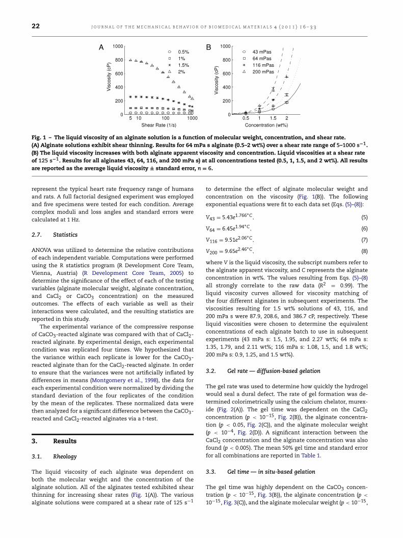
Fig. 1 – The liquid viscosity of an alginate solution is a function of molecular weight, concentration, and shear rate.(A) Alginate solutions exhibit shear thinning. Results for 64 mPa s alginate (0.5–2 wt%) over a shear rate range of 5–1000 s−1.(B) The liquid viscosity increases with both alginate apparent viscosity and concentration. Liquid viscosities at a shear rateof 125 s−1. Results for all alginates 43, 64, 116, and 200 mPa s) at all concentrations tested (0.5, 1, 1.5, and 2 wt%). All resultsare reported as the average liquid viscosity ± standard error, n = 6.
represent the typical heart rate frequency range of humansand rats. A full factorial designed experiment was employedand five specimens were tested for each condition. Averagecomplex moduli and loss angles and standard errors werecalculated at 1 Hz.
2.7. Statistics
ANOVA was utilized to determine the relative contributionsof each independent variable. Computations were performedusing the R statistics program (R Development Core Team,Vienna, Austria) (R Development Core Team, 2005) todetermine the significance of the effect of each of the testingvariables (alginate molecular weight, alginate concentration,and CaCl2 or CaCO3 concentration) on the measuredoutcomes. The effects of each variable as well as theirinteractions were calculated, and the resulting statistics arereported in this study.
The experimental variance of the compressive responseof CaCO3-reacted alginate was compared with that of CaCl2-reacted alginate. By experimental design, each experimentalcondition was replicated four times. We hypothesized thatthe variance within each replicate is lower for the CaCO3-reacted alginate than for the CaCl2-reacted alginate. In orderto ensure that the variances were not artificially inflated bydifferences in means (Montgomery et al., 1998), the data foreach experimental condition were normalized by dividing thestandard deviation of the four replicates of the conditionby the mean of the replicates. These normalized data werethen analyzed for a significant difference between the CaCO3-reacted and CaCl2-reacted alginates via a t-test.
3. Results
3.1. Rheology
The liquid viscosity of each alginate was dependent onboth the molecular weight and the concentration of thealginate solution. All of the alginates tested exhibited shearthinning for increasing shear rates (Fig. 1(A)). The variousalginate solutions were compared at a shear rate of 125 s−1
to determine the effect of alginate molecular weight andconcentration on the viscosity (Fig. 1(B)). The followingexponential equations were fit to each data set (Eqs. (5)–(8)):
V43 = 5.43e1.766∗C, (5)
V64 = 6.45e1.94∗C, (6)
V116 = 9.51e2.06∗C, (7)
V200 = 9.65e2.46∗C, (8)
where V is the liquid viscosity, the subscript numbers refer tothe alginate apparent viscosity, and C represents the alginateconcentration in wt%. The values resulting from Eqs. (5)–(8)all strongly correlate to the raw data (R2 = 0.99). Theliquid viscosity curves allowed for viscosity matching ofthe four different alginates in subsequent experiments. Theviscosities resulting for 1.5 wt% solutions of 43, 116, and200 mPa s were 87.9, 208.6, and 386.7 cP, respectively. Theseliquid viscosities were chosen to determine the equivalentconcentrations of each alginate batch to use in subsequentexperiments (43 mPa s: 1.5, 1.95, and 2.27 wt%; 64 mPa s:1.35, 1.79, and 2.11 wt%; 116 mPa s: 1.08, 1.5, and 1.8 wt%;200 mPa s: 0.9, 1.25, and 1.5 wt%).
3.2. Gel rate — diffusion-based gelation
The gel rate was used to determine how quickly the hydrogelwould seal a dural defect. The rate of gel formation was de-termined colorimetrically using the calcium chelator, murex-ide (Fig. 2(A)). The gel time was dependent on the CaCl2concentration (p < 10−15, Fig. 2(B)), the alginate concentra-tion (p < 0.05, Fig. 2(C)), and the alginate molecular weight(p < 10−4, Fig. 2(D)). A significant interaction between theCaCl2 concentration and the alginate concentration was alsofound (p < 0.005). The mean 50% gel time and standard errorfor all combinations are reported in Table 1.
3.3. Gel time — in situ-based gelation
The gel time was highly dependent on the CaCO3 concen-tration (p < 10−15, Fig. 3(B)), the alginate concentration (p <
10−15, Fig. 3(C)), and the alginatemolecular weight (p < 10−15,

J O U R N A L O F T H E M E C H A N I C A L B E H AV I O R O F B I O M E D I C A L M A T E R I A L S 4 ( 2 0 1 1 ) 1 6 – 3 3 23
A
C
B
D
Fig. 2 – The gel time is dependent on the CaCl2 concentration, the alginate concentration, and the alginate molecularweight. (A) Murexide is an indicator of an alginate:calcium complex and can be used to determine the gel rate as seen inthe micrograph demonstrating murexide as an indicator of alginate cross-linked with calcium ions. The unreacted alginateis a dark-red color that changes to yellow as the carboxyl groups interact with the free calcium ions. The scale barrepresents 1 mm. (B) The gel time is dependent on the CaCl2 concentration (p < 10−15). Resulting gel curves for 1.25 wt%200 mPa s alginate reacted with CaCl2 (50, 200, and 680 mM). The lines above and below each curve represents the standarderror, n = 5. (C) The gel time is dependent on the alginate concentration (p < 0.05). Results for 64 mPa s alginate (1.36, 1.79,and 2.11 wt%) reacted with CaCl2 (50, 200, and 680 mM). (D) The gel time is dependent on the alginate molecular weight(p < 10−4). Results for all alginates (43, 64, 116, and 200 mPa s) at concentrations that viscosity match the samples at 87.9,208.6, and 386.7 cP and reacted with 200 mM CaCl2. All results are reported as the average 50% gel time ± standard error,n = 5. (For interpretation of the references to colour in this figure legend, the reader is referred to the web version of thisarticle.)
Fig. 3(D)). The interaction between the CaCO3 concentrationand the alginate concentration was significant (p < 0.05), aswell as the three-way interaction between the alginatemolec-ular weight, the alginate concentration, and the CaCO3 con-centration (p < 0.05). Fig. 3(A) shows a representative gelcurve. The gel times for each experimental combination of al-ginate molecular weight, alginate concentration, and CaCO3concentration are reported in Table 2.
3.4. Syneresis
Diffusion-gelled alginate hydrogels exhibited high and vary-ing levels of syneresis dependent on the experimental pa-rameters. The syneresis was dependent on the CaCl2 con-centration (p < 10−15, Fig. 4(A)), the alginate concentration(p < 10−14, Fig. 4(B)), and the alginate molecular weight(p < 10−8, Fig. 4(C)). Significant interactions between the algi-natemolecular weight and the CaCl2 concentration (p < 10−4)and the CaCl2 concentration and the alginate concentration(p < 0.001) were also found. The average syneresis level andstandard error for all diffusion-gelled hydrogel experimentalcombinations are reported in Table 1.
In situ-gelled alginate showed a low but varying amountof syneresis dependent upon the various alginate and CaCO3parameters. The syneresis was dependent on the CaCO3 con-centration (p < 10−15, Fig. 5(A)), the alginate concentration(p < 10−15, Fig. 5(B)), and the alginate molecular weight (p <
10−15, Fig. 5(C)). We found significant interactions betweenthe alginate molecular weight and the CaCO3 concentration(p < 10−4), the alginate molecular weight and the alginateconcentration (p < 10−6), and the CaCO3 concentration andthe alginate concentration (p < 10−4), as well as a three-wayinteraction between the alginate molecular weight, the algi-nate concentration, and the CaCO3 concentration (p < 0.001).The average syneresis level and standard error for all in situ-gelled hydrogel experimental combination are reported inTable 2.
3.5. Mechanical properties: compressive strength andcompressive modulus
The dependences of the compressive strength of diffusion-gelled alginate on the CaCl2 concentration, alginate concen-tration, and alginate molecular weight are seen in Fig. 6.

24 J O U R N A L O F T H E M E C H A N I C A L B E H AV I O R O F B I O M E D I C A L M A T E R I A L S 4 ( 2 0 1 1 ) 1 6 – 3 3
A B
C D
Fig. 3 – The gel time is dependent on the CaCO3 concentration, the alginate concentration, and the alginate molecularweight. (A) The gel time was determined as the point when G′ exceeds G′′ under a shear rate of 1 Hz and 1% strain as seenin the representative gelation curve for 1.5 wt% 200 mPa s alginate with 50 mM CaCO3. (B) The gel time decreases withincreasing CaCO3 concentration (p < 10−15). Results for all alginate molecular weights (43, 64, 116, and 200 mPa s) viscositymatched at 87.9 cP and reacted with CaCO3 (20, 50, and 100 mM). (C) The gel time increases with increasing alginateconcentration (p < 10−15). Results for 200 mPa s alginate (0.9, 1.25, and 1.5 wt%) reacted with CaCO3 (20, 50, and 100 mM).(D) The gel time decreases with increasing alginate molecular weight (p < 10−15). Results for all alginates (43, 64, 116, and200 mPa s) at concentrations that viscosity match the samples at 87.9, 208.6, and 386.7 cP and reacted with 50 mM CaCO3.All results are reported as the average gel time ± standard error, n = 5.
Fig. 4 – The syneresis of diffusion-gelled alginate hydrogels is dependent on the CaCl2 concentration, the alginateconcentration, and the alginate molecular weight. (A) The syneresis is dependent on the CaCl2 concentration (p < 10−15).Results for all alginate molecular weights (43, 64, 116, and 200 mPa s) viscosity matched at 87.9 cP and reacted with CaCl2(50, 200, and 680 mM). (B) The syneresis is dependent on the alginate concentration (p < 10−14). Results for 200 mPa salginate (0.9, 1.25, and 1.5 wt%) reacted with CaCl2 (50, 200, and 680 mM). (C) The syneresis is dependent on the molecularweight (p < 10−8). Results for all alginates (43, 64, 116, and 200 mPa s) viscosity matched at 87.9, 208.6, and 386.7 cP andreacted with 200 mM CaCl2. All results are reported as the average syneresis level ± standard error, n = 4.
The gel strength was dependent on the CaCl2 concentration
(p < 10−4, Fig. 6(A) and (D)) and the alginate concentration
(p < 0.01, Fig. 6(B) and (E)), but the molecular weight did not
have a statistically significant effect (p = 0.89, Fig. 6(C), and
(F)). A significant interaction between the alginate molecu-
lar weight and the CaCl2 concentration (p < 0.05) was found

J O U R N A L O F T H E M E C H A N I C A L B E H AV I O R O F B I O M E D I C A L M A T E R I A L S 4 ( 2 0 1 1 ) 1 6 – 3 3 25
Fig. 5 – The syneresis of in situ-gelled alginate hydrogels is dependent on the CaCO3 concentration, the alginateconcentration, and the alginate molecular weight. (A) The syneresis increases with increasing CaCO3 concentration(p < 10−15). Results for all alginate molecular weights (43, 64, 116, and 200 mPa s) viscosity matched at 87.9 cP and reactedwith CaCO3 (20, 50, and 100 mM). (B) The syneresis decreases with increasing alginate concentration (p < 10−15). Results for200 mPa s alginate (0.9, 1.25, and 1.5 wt%) reacted with CaCO3 (20, 50, and 100 mM). (C) The syneresis increases withincreasing molecular weight (p < 10−15). Results for all alginates (43, 64, 116, and 200 mPa s) viscosity matched at 87.9,208.6, and 386.7 cP and reacted with 50 mM CaCO3. All results are reported as the average syneresis level ± standard error,n = 4.
A B C
D E F
Fig. 6 – The compressive strength of diffusion-gelled alginate is dependent on the CaCl2 concentration and the alginateconcentration. (A)–(C) Results are reported as the average compressive strengths at 10% compression ± standard error, n = 4.(A) The gel strength is dependent on the CaCl2 concentration (p < 10−4). Results for all alginates (43, 64, 116, and 200 mPa s)viscosity matched at 87.9 cP and reacted with CaCl2 (50, 200, and 680 mM). (B) The compressive strength is dependent onthe alginate concentration (p < 0.01). Results for 200 mPa s alginate (0.9, 1.25, and 1.5 wt%) reacted with CaCO3 (50, 200, and680 mM). (C) The alginate molecular weight has no significant effect on the compressive strength (p = 0.89). Results for allalginates (43, 64, 116, and 200 mPa s) viscosity matched at 87.9, 208.6, and 386.7 cP and reacted with 200 mM CaCl2.(D)–(F) Results are reported as the average compressive strengths at 60% compression ± standard error, n = 4. (D) The gelstrength is dependent on the CaCl2 concentration (p < 10−15). Results for all alginates (43, 64, 116, and 200 mPa s) viscositymatched at 87.9 cP and reacted with CaCl2 (50, 200, and 680 mM). (E) The compressive strength is dependent on the alginateconcentration (p < 10−5). Results for 200 mPa s alginate (0.9, 1.25, and 1.5 wt%) reacted with CaCl2 (50, 200, and 680 mM).(F) The alginate molecular weight has no significant effect on the compressive strength (p = 0.066). Results for all alginates(43, 64, 116, and 200 mPa s) viscosity matched at 87.9, 208.6, and 386.7 cP and reacted with 200 mM CaCl2.
26 J O U R N A L O F T H E M E C H A N I C A L B E H AV I O R O F B I O M E D I C A L M A T E R I A L S 4 ( 2 0 1 1 ) 1 6 – 3 3
Fig. 7 – The compressive strength of in situ-gelled alginate is dependent on the CaCO3 concentration and the alginateconcentration. (A)–(C) Results are reported as the average compressive strengths at 10% compression ± standard error, n = 4.(A) The gel strength increases with increasing CaCO3 concentration (p < 10−12). Results for all alginates (43, 64, 116, and 200mPa s) viscosity matched at 87.9 cP and reacted with CaCO3 (20, 50, and 100 mM). (B) The compressive strength increaseswith increasing alginate concentration (p < 0.05). Results for 200 mPa s alginate (0.9, 1.25, and 1.5 wt%) reacted with CaCO3(20, 50, and 100 mM). (C) The alginate molecular weight has no significant effect on the compressive strength (p = 0.39).Results for all alginates (43, 64, 116, and 200 mPa s) viscosity matched at 87.9, 208.6, and 386.7 cP and reacted with 50 mMCaCO3. (D)–(F) Results are reported as the average compressive strengths at 60% compression ± standard error, n = 4. (D) Thegel strength increases with increasing CaCO3 concentration (p < 10−15). Results for all alginates (43, 64, 116, and 200 mPa s)viscosity matched at 87.9 cP and reacted with CaCO3 (20, 50, and 100 mM). (E) The compressive strength increases withincreasing alginate concentration (p < 10−4). Results for 200 mPa s alginate (0.9, 1.25, and 1.5 wt%) reacted with CaCO3 (20,50, and 100 mM). (F) The alginate molecular weight has no significant effect on compressive strength (p = 0.052). Results forall alginates (43, 64, 116, and 200 mPa s) viscosity matched at 87.9, 208.6, and 386.7 cP and reacted with 50 mM CaCO3.
at both compression levels. Additionally, a significant inter-action between the alginate concentration and the CaCl2concentration (p < 10−4) was also found at the 60% compres-sion level. The average compressive strength and standard er-ror for all diffusion-gelled combinations at both 10% and 60%compression are reported in Table 1.
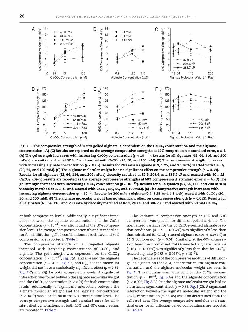
The compressive strength of in situ-gelled alginateincreased with increasing concentrations of CaCO3 andalginate. The gel strength was dependent on the CaCO3concentration (p < 10−12, Fig. 7(A) and (D)) and the alginateconcentration (p < 0.05, Fig. 7(B) and (E)), but the molecularweight did not have a statistically significant effect (p = 0.39,Fig. 7(C) and (F)) for both compression levels. A significantinteraction was found between the alginate molecular weightand the CaCO3 concentration (p < 0.01) for both compressionlevels. Additionally, a significant interaction between thealginate molecular weight and the alginate concentration(p < 10−6) was also found at the 60% compression level. Theaverage compressive strength and standard error for all insitu-gelled combinations at both 10% and 60% compressionare reported in Table 2.
The variance in compression strength at 10% and 60%compression was greater for diffusion-gelled alginate. Thenormalized variance for the 36 CaCO3-reacted alginate reac-tion conditions (0.367 ± 0.067%) was significantly less thanthat calculated for CaCl2-reacted alginate (0.504 ± 0.031%) at10 % compression (p < 0.01). Similarly, at the 60% compres-sion level the normalized CaCO3-reacted alginate variance(0.163 ± 0.006%) was significantly less than that for CaCl2-reacted alginate (0.282 ± 0.015%, p < 10−5).
The dependences of the compressivemodulus of diffusion-gelled alginate on the CaCl2 concentration, the alginate con-centration, and the alginate molecular weight are seen inFig. 8. The modulus was dependent on the CaCl2 concen-tration (p < 10−4, Fig. 8(A)) and the alginate concentration(p < 0.005, Fig. 8(B)), but the alginate molecular weight had nostatistically significant effect (p = 0.82, Fig. 8(C)). A significantinteraction between the alginate molecular weight and theCaCl2 concentration (p < 0.05) was also determined from thecollected data. The average compressive modulus and stan-dard error for all diffusion-gelled combinations are reportedin Table 1.
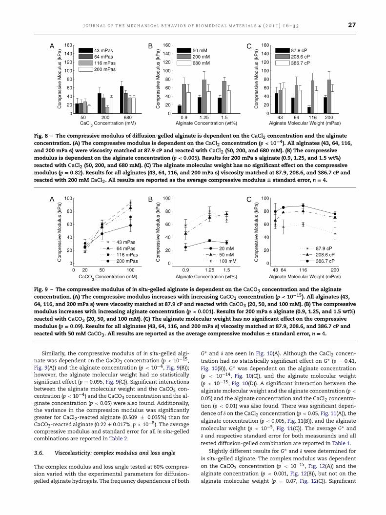
J O U R N A L O F T H E M E C H A N I C A L B E H AV I O R O F B I O M E D I C A L M A T E R I A L S 4 ( 2 0 1 1 ) 1 6 – 3 3 27
A B C
Fig. 8 – The compressive modulus of diffusion-gelled alginate is dependent on the CaCl2 concentration and the alginateconcentration. (A) The compressive modulus is dependent on the CaCl2 concentration (p < 10−4). All alginates (43, 64, 116,and 200 mPa s) were viscosity matched at 87.9 cP and reacted with CaCl2 (50, 200, and 680 mM). (B) The compressivemodulus is dependent on the alginate concentration (p < 0.005). Results for 200 mPa s alginate (0.9, 1.25, and 1.5 wt%)reacted with CaCl2 (50, 200, and 680 mM). (C) The alginate molecular weight has no significant effect on the compressivemodulus (p = 0.82). Results for all alginates (43, 64, 116, and 200 mPa s) viscosity matched at 87.9, 208.6, and 386.7 cP andreacted with 200 mM CaCl2. All results are reported as the average compressive modulus ± standard error, n = 4.
A B C
Fig. 9 – The compressive modulus of in situ-gelled alginate is dependent on the CaCO3 concentration and the alginateconcentration. (A) The compressive modulus increases with increasing CaCO3 concentration (p < 10−15). All alginates (43,64, 116, and 200 mPa s) were viscosity matched at 87.9 cP and reacted with CaCO3 (20, 50, and 100 mM). (B) The compressivemodulus increases with increasing alginate concentration (p < 0.001). Results for 200 mPa s alginate (0.9, 1.25, and 1.5 wt%)reacted with CaCO3 (20, 50, and 100 mM). (C) The alginate molecular weight has no significant effect on the compressivemodulus (p = 0.09). Results for all alginates (43, 64, 116, and 200 mPa s) viscosity matched at 87.9, 208.6, and 386.7 cP andreacted with 50 mM CaCO3. All results are reported as the average compressive modulus ± standard error, n = 4.
Similarly, the compressive modulus of in situ-gelled algi-nate was dependent on the CaCO3 concentration (p < 10−15,Fig. 9(A)) and the alginate concentration (p < 10−4, Fig. 9(B));however, the alginate molecular weight had no statisticallysignificant effect (p = 0.095, Fig. 9(C)). Significant interactionsbetween the alginate molecular weight and the CaCO3 con-centration (p < 10−4) and the CaCO3 concentration and the al-ginate concentration (p < 0.05) were also found. Additionally,the variance in the compression modulus was significantlygreater for CaCl2-reacted alginate (0.509 ± 0.035%) than forCaCO3-reacted alginate (0.22 ± 0.017%, p < 10−8). The averagecompressive modulus and standard error for all in situ-gelledcombinations are reported in Table 2.
3.6. Viscoelasticity: complex modulus and loss angle
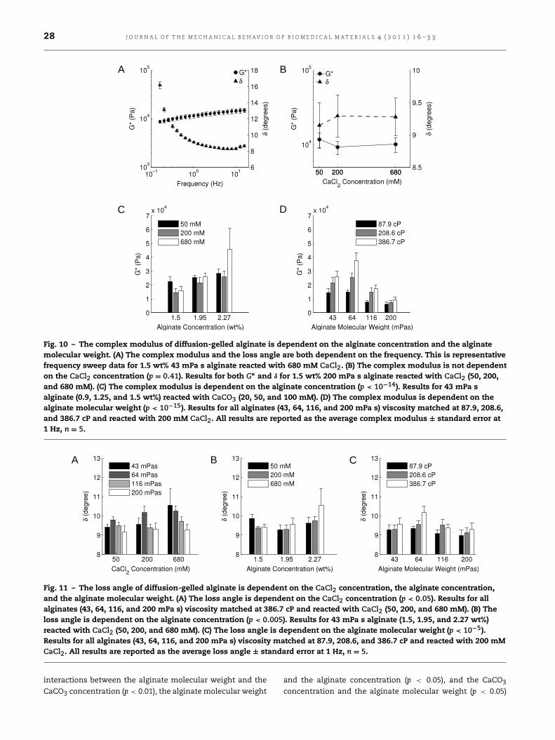
The complex modulus and loss angle tested at 60% compres-sion varied with the experimental parameters for diffusion-gelled alginate hydrogels. The frequency dependences of both
G∗ and δ are seen in Fig. 10(A). Although the CaCl2 concen-tration had no statistically significant effect on G∗ (p = 0.41,Fig. 10(B)), G∗ was dependent on the alginate concentration(p < 10−14, Fig. 10(C)), and the alginate molecular weight(p < 10−15, Fig. 10(D)). A significant interaction between thealginatemolecular weight and the alginate concentration (p <
0.05) and the alginate concentration and the CaCl2 concentra-tion (p < 0.01) was also found. There was significant depen-dence of δ on the CaCl2 concentration (p < 0.05, Fig. 11(A)), thealginate concentration (p < 0.005, Fig. 11(B)), and the alginatemolecular weight (p < 10−5, Fig. 11(C)). The average G∗ andδ and respective standard error for both measurands and alltested diffusion-gelled combination are reported in Table 1.
Slightly different results for G∗ and δ were determined forin situ-gelled alginate. The complex modulus was dependenton the CaCO3 concentration (p < 10−15, Fig. 12(A)) and thealginate concentration (p < 0.001, Fig. 12(B)), but not on thealginate molecular weight (p = 0.07, Fig. 12(C)). Significant
28 J O U R N A L O F T H E M E C H A N I C A L B E H AV I O R O F B I O M E D I C A L M A T E R I A L S 4 ( 2 0 1 1 ) 1 6 – 3 3
A B
C D
Fig. 10 – The complex modulus of diffusion-gelled alginate is dependent on the alginate concentration and the alginatemolecular weight. (A) The complex modulus and the loss angle are both dependent on the frequency. This is representativefrequency sweep data for 1.5 wt% 43 mPa s alginate reacted with 680 mM CaCl2. (B) The complex modulus is not dependenton the CaCl2 concentration (p = 0.41). Results for both G∗ and δ for 1.5 wt% 200 mPa s alginate reacted with CaCl2 (50, 200,and 680 mM). (C) The complex modulus is dependent on the alginate concentration (p < 10−14). Results for 43 mPa salginate (0.9, 1.25, and 1.5 wt%) reacted with CaCO3 (20, 50, and 100 mM). (D) The complex modulus is dependent on thealginate molecular weight (p < 10−15). Results for all alginates (43, 64, 116, and 200 mPa s) viscosity matched at 87.9, 208.6,and 386.7 cP and reacted with 200 mM CaCl2. All results are reported as the average complex modulus ± standard error at1 Hz, n = 5.
A B C
Fig. 11 – The loss angle of diffusion-gelled alginate is dependent on the CaCl2 concentration, the alginate concentration,and the alginate molecular weight. (A) The loss angle is dependent on the CaCl2 concentration (p < 0.05). Results for allalginates (43, 64, 116, and 200 mPa s) viscosity matched at 386.7 cP and reacted with CaCl2 (50, 200, and 680 mM). (B) Theloss angle is dependent on the alginate concentration (p < 0.005). Results for 43 mPa s alginate (1.5, 1.95, and 2.27 wt%)reacted with CaCl2 (50, 200, and 680 mM). (C) The loss angle is dependent on the alginate molecular weight (p < 10−5).Results for all alginates (43, 64, 116, and 200 mPa s) viscosity matched at 87.9, 208.6, and 386.7 cP and reacted with 200 mMCaCl2. All results are reported as the average loss angle ± standard error at 1 Hz, n = 5.
interactions between the alginate molecular weight and the
CaCO3 concentration (p < 0.01), the alginatemolecular weight
and the alginate concentration (p < 0.05), and the CaCO3
concentration and the alginate molecular weight (p < 0.05)

J O U R N A L O F T H E M E C H A N I C A L B E H AV I O R O F B I O M E D I C A L M A T E R I A L S 4 ( 2 0 1 1 ) 1 6 – 3 3 29
Fig. 12 – The complex modulus of in situ-gelled alginate is dependent on the CaCO3 concentration and the alginateconcentration. (A) The complex modulus increases with increasing CaCO3 concentration (p < 10−15). Results for all alginates(43, 64, 116, and 200 mPa s) viscosity matched at 386.7 cP and reacted with CaCO3 (20, 50, and 100 mM). (B) The complexmodulus increases with increasing alginate concentration (p < 0.001). Results for 43 mPa s alginate (1.5, 1.95, and 2.27 wt%)reacted with CaCO3 (20, 50, and 100 mM). (C) The complex modulus was not significantly dependent on the alginatemolecular weight (p = 0.07). Results for all alginates (43, 64, 116, and 200 mPa s) viscosity matched at 87.9, 208.6, and 386.7cP and reacted with 50 mM CaCO3. All results are reported as the average complex modulus ± standard error at 1 Hz, n = 5.
A B C
Fig. 13 – The loss angle of in situ-gelled alginate is dependent on the CaCO3 concentration, the alginate concentration, andthe alginate molecular weight. (A) The loss angle increases with increasing CaCO3 concentration (p < 10−15). Results for allalginates (43, 64, 116, and 200 mPa s) viscosity matched at 386.7 cP and reacted with CaCO3 (20, 50, and 100 mM). (B) Theloss angle increases with increasing alginate concentration (p < 0.001). Results for 43 mPa s alginate (1.5, 1.95, and 2.27wt%) reacted with CaCO3 (20, 50, and 100 mM). (C) The loss angle increases with increasing alginate molecular weight(p < 10−6). Results for all alginates (43, 64, 116, and 200 mPa s) viscosity matched at 87.9, 208.6, and 386.7 cP and reactedwith 50 mM CaCO3. All results are reported as the average loss angle ± standard error at 1 Hz, n = 5.
were found for G∗ at the 60% level. The loss angle wasdependent on the CaCO3 concentration (p < 10−15, Fig. 13(A)),the alginate concentration (p < 0.001, Fig. 13(B)), and thealginate molecular weight (p < 10−11, Fig. 13(C)). The averageG∗ and δ and respective standard error for both measurandsand all tested in situ-gelled combination are reported inTable 2.
4. Discussion
To further investigate alginate as a potential dural sealantor dura mater replacement, the effects and interactionsof alginate molecular weight, alginate concentration, andCaCl2 or CaCO3 concentration were identified. Specifically,the effects of these experimental parameters on the gelrate, syneresis, gel strength, compressive modulus, complex
modulus, and loss angle were determined for gels inequilibrium.
The results aid in the identification of an ideal combina-tion of alginate and CaCl2 that produce a gel with ideal prop-erties for small applications, such as a dural sealant. Basedon the data collected in this study, we would suggest theuse of 1.95 wt% 43 mPa s alginate with 200 mM CaCl2 forapproximation of minor dural defects with or without addi-tional sutures. This combination results in a gel that forms in8.23 ± 1.74 min, has a syneresis level of 52.25 ± 1.03%, a com-pressive modulus of 54.37 ± 16.32 kPa, a complex modulus of21.62 ± 3.97 kPa, and a loss angle of 9.30 ± 0.24 degrees.
These results also suggest that specific mixing conditionsof alginate and CaCO3 result in a material that is sufficientfor closing large dural defects. Furthermore, internally cross-linked hydrogels should be preferentially pursued overdiffusion cross-linked gels for use as a dural replacement due

30 J O U R N A L O F T H E M E C H A N I C A L B E H AV I O R O F B I O M E D I C A L M A T E R I A L S 4 ( 2 0 1 1 ) 1 6 – 3 3
to lower levels of syneresis, higher levels of homogeneity, andlower loss angle values. Based on the data collected in thisstudy, we would suggest the use of 1.95 wt% 43 mPa s alginatewith 100 mM CaCO3 and 80 mM GDL. This combinationresults in a gel that forms in 19.28 ± 1.87 min, has asyneresis level of 3.55 ± 0.68%, a compressive modulus of85.35 ± 7.5 kPa, a complex modulus of 11.88 ± 1.52 kPa, anda loss angle of 6.82 ± 0.20 degrees.
To date, a number of groups have studied or modeledthe gelation rate of alginate under a variety of conditionsand methods (Bienaime et al., 2003; Blandino et al., 1999;Kuo and Ma, 2001; Liu et al., 2003; Mikkelsen and Elgsaeter,1995; Smidsrod and Haug, 1972; Stokke et al., 2000; Wang andSpencer, 1998; Wang et al., 1995, 1993, 1994). We employeda recently described and relatively simple method from theliterature to quantify the rate of gel formation (Bienaimeet al., 2003). Murexide, a calcium chelator, indicates cross-link formation based on color, and it indicates the rate ofCaCl2 penetration into the liquid alginate volume. An excessof CaCl2 solution was used to (1) mimic in vivo applicationof a dural sealant, and (2) accommodate any small changesin gel rate that could be attributed to the interaction ofcalcium with murexide instead of the carboxyl groups onthe alginate chains. Although 200 ml of alginate was cross-linkedwith 500ml CaCl2 to simulate surgical use, it is possiblethat less alginate and CaCl2 volume would be indicated,depending on the length of the dural interface requiringapproximation. Less volume would thereby decrease thecross-link time, further decreasing the time to create aseal between the approximated tissues. These rapid cross-link times are highly desirable in dural repair because theyreestablish the barrier with the external environment quickly.Our results indicate that both the CaCl2 concentration andthe alginate concentration have a significant effect on thegel rate (p < 10−4), which supports the findings of previousinvestigations (Blandino et al., 1999). We identified that thealginate molecular weight, as well as the interaction betweenthe CaCl2 concentration and the alginate concentrationboth contribute significantly to the gel time. This findinghas not been previously reported, and current theoreticalmodels for diffusion-based cross-linking of alginate donot accommodate these parameters (Blandino et al., 1999;Mikkelsen and Elgsaeter, 1995). Further investigation mayresult in experimentally derived models of the mathematicalrelationship between the alginate molecular weight, alginateconcentration, CaCl2 concentration, and the interaction of theCaCl2 concentration and the alginate concentration.
In situ gelation of alginate can have a highly variable geltime (minutes to days) (Kuo and Ma, 2001); thus, the gel timeis a potentially important limitation in the development ofan alginate dural replacement patch. Previously, Shchipunovet al. employed rheology to investigate the relationshipbetween GDL concentration, pH, and gel rate with alginate,and they developed a pseudophase diagram based on GDLand CaCO3 concentration (Shchipunov et al., 2002). Basedon this model, we were able to choose a range of CaCO3concentrations and an ideal GDL concentration, and measuregel times of 8–55 min for combinations of alginate and CaCO3with 80 mM GDL (Table 2). These results not only corroboratethe findings of several research groups (Draget et al., 1991;
Kuo and Ma, 2001; Shchipunov et al., 2002; Smidsrod andHaug, 1972), but additionally identify the effects due toalginate molecular weight and concentration. Furthermore,several of the gelation times for CaCO3 are rapid enoughto use in select dura mater repair applications. Althoughthe gelation rate of alginate with CaCO3 is slower than thatof CaCl2, the components can be mixed and then appliedas a single viscous solution to a large dural defect. Site-specific application can be further improved by waiting forthe alginate:CaCO3-reacted mixture to reach a high, but stilluseable, viscosity (∼400 cP) before filling the dural defect.
Hydrogel syneresis is an indicator of the level of gel shrink-age that could result in a malformed seal between the gel andthe surrounding meningeal tissue. A defective seal could al-low gaps to form between the dura mater and the alginate, al-lowing a pathway for pathogen entry and cerebrospinal fluidleakage. As discussed previously, increasing the calcium con-centration expedites the cross-linking process and leads toa decreased gelling time and an increased cross-link den-sity (Draget et al., 2001, 2003). However, it is this cross-linkdensity that is responsible for the occurrence of syneresis, aslow time-dependent exudation of liquid from the hydrogel(Draget et al., 2001; Shchipunov et al., 2002). CaCO3-reactedalginate demonstrated low levels of syneresis (<10%) for mostof the tested combinations, indicating good long-term sealingpotential as a dural replacement. CaCl2, however, demon-strated high levels of syneresis (>25%) for all tested combina-tions, indicating low sealing potential. Based on these results,we would suggest the use of CaCl2 below 200 mM or alterna-tively utilizing CaCO3 to minimize syneresis levels.
Alginate gel behavior under compression is important inthe development of a dura mater repair material. After ap-plication of the alginate, the skull is sealed and the gel isthen subjected to a constant intracranial pressure (approxi-mately 1 kPa) that compresses the gel between the brain andthe skull. Immediately following surgery, intracranial pres-sure is increased due to trauma and edema. Our results in-dicate that the compressive strength of the gel far exceedsthe pressures that the alginate would be subjected to in vivo.This suggests that alginate used alone or with sutures to ap-proximate tissue or replace portions of the tissue would notbe compressed to failure by intracranial pressure and wouldthereby be able to maintain a seal or fill a large defect. Thecompressive strength near the elastic limit of alginate, 60%compression, indicates that alginate can withstand pressuresgreater than 100 kPa. Even after significant neural trauma, thehighest pressure a dural sealant would likely be exposed to is3 kPa (Czosnyka et al., 2005). Alginate is thereby expected towithstand intracranial pressure under any physiological cir-cumstance without failure.
The compressive modulus is an ideal measure for com-parison of alginate with brain tissue. It is desirable to matchthe modulus of alginate to that of the underlying brain tissueto decrease the occurrence of trauma due to a mechanicalmismatch. Brain tissue has been shown to have a compres-sive modulus of 20–100 kPa, depending on the tissue sourceand measurement technique (Sarron et al., 2000; Walsh andSchettini, 1976). Most of the alginate combinations tested fitin this range (Figs. 8 and 9, Tables 1 and 2), and the compres-sive modulus was not a major contributor to identifying theoptimal combination of alginate and CaCl2 or CaCO3.

J O U R N A L O F T H E M E C H A N I C A L B E H AV I O R O F B I O M E D I C A L M A T E R I A L S 4 ( 2 0 1 1 ) 1 6 – 3 3 31
Hydrogel homogeneity is important in the development ofa dural patch. The hydrogel should have the same response tocompression throughout to avoid localized mechanical fail-ure of the patch. Overall, the hydrogel response to compres-sion is more consistent for alginate reacted with CaCO3 thanwith CaCl2. The normalized variance for 10% compressivestrength, 60% compressive strength, and compressive mod-ulus all indicate that CaCO3-reacted alginate is significantlyless variable. This suggests that CaCO3 results in more homo-geneous and uniform gels than CaCl2, and is thereby moreappropriate for use as a dural patch. These results corrobo-rate an earlier study comparing alginate reacted with CaSO4and CaCO3, which indicated that the slow gelation kinetics ofCaCO3 were responsible for increased hydrogel homogeneity(Kuo and Ma, 2001).
Due to the pulsation of the brain with heart rate and res-piratory rate, the alginate will be subject to oscillatory strain.Thus, alginate’s response to shear is an important design fac-tor for a dural sealant as it is a good indicator of the fatigue re-sistance and how it will perform in vivo. Brain tissue typicallyexhibits a complex modulus ranging from 1 to 24 kPa (Don-nelly and Medige, 1997). Based on our results, alginates havea complex modulus range of 6–186 kPa. Therefore, the oscilla-tory behavior of intracranial pressure should not adversely af-fect the fatigue resistance of alginate, and alginate is expectedto remain stable in vivo. Additionally, the complex modulus ofalginate can be tailored to match that of the brain, reducingmechanical mismatch at the alginate–dura interface.
The final gel parameter investigated, δ, addresses the elas-tic behavior of the gel due to internal cross-linkages. While itis clear that the alginate must exhibit low values of δ, the ex-act δ cannot be identified at this time. The hydrogel must beable to retain its shape and orientation to the tissue while ac-commodating some motion of the dura, yet the magnitude ofsuchmotion in vivo is not known. The ideal dural patch wouldexhibit elastic solid behavior (low loss angle values), enablingthe hydrogel to maintain its original form. CaCO3-reacted hy-drogels resulted in more densely cross-linked gels (lower lossangle) than did those gels made with CaCl2. This increasedcross-link density potentially indicates a superior stability forinternally gelled hydrogels versus diffusion-gelled hydrogelsin vivo. Therefore, CaCO3 is potentially more appropriate touse to cross-link alginate when used as a dural patch be-cause of an increased need for long-term stability. However,the ideal value for δ is currently unknown. The gel shouldkeep its original shape but still be able to move with the duraand underlying brain. Previous in vitro studies indicate thatthe loss angle will become more important when measuringthe elastic behavior of the gels over time (LeRoux et al., 1999;Nunamaker et al., 2007; Wang et al., 1994). We expected that,after prolonged exposure to low concentrations of inert elec-trolytes in vivo, the calcium ions that are holding the hydrogeltogether may be displaced, resulting in a less densely cross-linked gel and ultimately a higher loss angle. However, by thisextended time point we postulate that significant meningealreplacement would have occurred beneath the dural sealant.Further in vivo testing is necessary to answer these questions.
In summary, we have investigated gel formation and thematerial properties of alginate hydrogels created by diffusionand in situ-based gelling in the context of dura mater repair.
We elucidated how these properties are influenced by thealginate molecular weight, alginate concentration, and CaCl2or CaCO3 concentration. The data collected in this studysuggest that the use of 1.95 wt% 43 mPa s alginate with 200mM CaCl2 is sufficient for approximating small dural defectsfor closure alone or in conjunction with suture. Alternatively,the use of 1.95 wt% 43 mPa s alginate with 100 mM CaCO3 issufficient for tissue replacement in large dural defects.
Acknowledgements
This work was supported by the Michigan EconomicDevelopment Corporation. The authors gratefully thankDavid Pinkney for skillful technical assistance, Drs. TimothyBecker and Edward Ionides for insightful discussions, MattJohnson and Erin Purcell for manuscript discussions, NeuralIntervention Technologies and Dr. Soloman for the useof their rheometers, and the Van Vlack Material ScienceLaboratory for the use of the Instron mechanical tester.
R E F E R E N C E S
Abbe, R., 1895. Rubber tissue for meningeal adhesions. Transac-tions of American Surgical Association 13, 490–491.
Abbott, W.M., Dupree Jr., E.L., 1971. Clinical results of lyophilizedhuman cadaver dura transplantation. Journal of Neurosurgery34 (6), 770–773.
Agarwal, A., Varma, A., et al., 1998. Histopathological changesfollowing the use of biological and synthetic glue for duralgrafts: an experimental study. British Journal of Neurosurgery12 (3), 213–216.
Alleyene Jr., C.H., Cawley, C.M., et al., 1998. Efficacy andbiocompatibility of a photopolymerized, synthetic, absorbablehydrogel as a dural sealant in a canine craniotomy model.Journal of Neurosurgery 88 (2), 308–313.
Andresen, I.L., Smidsrod, O., 1977. Temperature-dependence ofelastic properties of alginate gels. Carbohydrate Research 58(2), 271–279.
Anson, J.A., Marchand, E.P., 1996. Bovine pericardium for duralgrafts: clinical results in 35 patients. Neurosurgery 39 (4),764–768.
Aydelotte, M.B., Thonar, E.J.M.A., et al., 1998. Culture ofchondrocytes in alginate gel: variations in conditions ofgelation influence the structure of the alginate gel, and thearrangement andmorphology of proliferating chondrocytes. InVitro Cellular & Developmental Biology-Animal 34 (2), 123–130.
Barbolt, T.A., Odin, M., et al., 2001. Biocompatibility evaluationof dura mater substitutes in an animal model. NeurologicalResearch 23 (8), 813–820.
Beach, H., 1890. Compound comminuted fracture of the skull;epilepsy for five years, operation, recovery. BostonMedical andSurgical Journal 122 (14), 313–315.
Becker, T.A., Kipke, D.R., 2003. ALGEL R⃝ as a dural sealant:determination of effects on the sensori-motor cortex in rats.In: 25th Annual International Conference of the IEEE/EMBS,Cancun, Mexico.
Becker, T.A., Kipke, D.R., et al., 2001. Calcium alginate gel: a bio-compatible and mechanically stable polymer for endovascularembolization. Journal of Biomedical Materials Research 54 (1),76–86.
32 J O U R N A L O F T H E M E C H A N I C A L B E H AV I O R O F B I O M E D I C A L M A T E R I A L S 4 ( 2 0 1 1 ) 1 6 – 3 3
Becker, T.A., Kipke, D.R., et al., 2002. In vivo assessment ofcalcium alginate gel for endovascular embolization of acerebral arteriovenous malformation model using the swinerete mirabile. Neurosurgery 51 (2), 453–458.
Becker, T.A., Preul, M.C., et al., 2005. Calcium alginate gelas a biocompatible material for endovascular arteriovenousmalformation embolization: six-month results in an animalmodel. Neurosurgery 56 (4), 793–801.
Bernoulli, C., Siegfried, J., et al., 1977. Danger of accidentalperson-to-person transmission of Creutzfeldt–Jakob diseaseby surgery. Lancet 1 (8009), 478–479.
Bienaime, C., Barbotin, J.N., et al., 2003. How to build an adaptedand bioactive cell microenvironment? A chemical interactionstudy of the structure of Ca-alginate matrices and theirrepercussion on confined cells. Journal of Biomedical MaterialsResearch Part A 67A (2), 376–388.
Blandino, A., Macias, M., et al., 1999. Formation of calciumalginate gel capsules: influence of sodium alginate and CaCl2concentration on gelation kinetics. Journal of Bioscience andBioengineering 88 (6), 686–689.
Budka, H., Aguzzi, A., et al., 1995. Tissue handling in suspectedCreutzfeldt–Jakob disease (CJD) and other human spongiformencephalopathies (prion diseases). Brain Pathology 5 (3),319–322.
Cain Jr., J.E., Dryer, R.F., et al., 1988. Evaluation of dural closuretechniques. Suture methods, fibrin adhesive sealant, andcyanoacrylate polymer. Spine 13 (7), 720–725.
Cain Jr., J.E., Rosenthal, H.G., et al., 1990. Quantification of leakagepressures after durotomy repairs in the canine. Spine 15 (9),969–970.
Collins, R.L., Christiansen, D., et al., 1991. Use of collagen filmas a dural substitute: preliminary animal studies. Journal ofBiomedical Materials Research 25 (2), 267–276.
Costantino, P.D., Wolpoe, M.E., et al., 2000. Human duralreplacement with acellular dermis: clinical results and areview of the literature. Head Neck 22 (8), 765–771.
Czosnyka, M., Balestreri, M., et al., 2005. Age, intracranialpressure, autoregulation, and outcome after brain trauma.Journal of Neurosurgery 102 (3), 450–454.
De Vries, J., Menovsky, T., et al., 2002. Histological effects offibrin glue on nervous tissue: a safety study in rats. SurgicalNeurology 57 (6), 415–422.
Donnelly, B.R., Medige, J., 1997. Shear properties of human braintissue. Journal of Biomechanical Engineering-Transactions ofthe ASME 119 (4), 423–432.
Draget, K.I., Gaserod, O., et al., 2001. Effects of molecular weightand elastic segment flexibility on syneresis in Ca-alginate gels.Food Hydrocolloids 15 (4–6), 485–490.
Draget, K.I., Ostgaard, K., et al., 1991. Homogeneous alginate gels:a technical approach. Carbohydrate Polymers.
Draget, K.I., SkjakBraek, G., et al., 1997. Alginate based newmaterials. International Journal of Biological Macromolecules21 (1–2), 47–55.
Draget, K.I., Stokke, B.T., et al., 2003. Small-angle X-ray scatteringand rheological characterization of alginate gels. 3. Alginicacid gels. Biomacromolecules 4 (6), 1661–1668.
Filippi, R., Schwarz, M., et al., 2001. Bovine pericardium forduraplasty: clinical results in 32 patients. NeurosurgicalReview 24 (2–3), 103–107.
Friedman, J.A., Windebank, A.J., et al., 2002. Biodegradablepolymer grafts for surgical repair of the injured spinal cord.Neurosurgery 51 (3), 742–751.
Grasdalen, H., 1983. High-Field, H-1-Nmr spectroscopy ofalginate — sequential structure and linkage conformations.Carbohydrate Research 118 (JUL), 255–260.
Haug, A., 1964. Composition and properties of alginates.Norwegian Institute of Seaweed Research 1–123.
Haug, A., Smidsrod, O., 1965. Effect of divalent metals onproperties of alginate solutions. 2. Comparison of differentmetal ions. Acta Chemica Scandinavica 19 (2), 341–351.
Ishikawa, K., Ueyama, Y., et al., 1999. Self-setting barriermembrane for guided tissue regeneration method: initialevaluation of alginate membrane made with sodium alginateand calcium chloride aqueous solutions. Journal of BiomedicalMaterials Research 47 (2), 111–115.
Kumar, A., Maartens, N.F., et al., 2003. Evaluation of the use ofBioGlue (R) in neurosurgical procedures. Journal of ClinicalNeuroscience 10 (6), 661–664.
Kuo, C.K., Ma, P.X., 2001. Ionically crosslinked alginate hydrogelsas scaffolds for tissue engineering: part 1. Structure, gelationrate and mechanical properties. Biomaterials 22 (6), 511–521.
Le Tallec, L.P., Korwin-Zmijowska, C., et al., 1997. Limitations ofalginate gels as a culture model for the study of the effects ofUVA radiation on human dermal fibroblasts. Cell Biology andToxicology 13 (2), 95–102.
LeRoux, M.A., Guilak, F., et al., 1999. Compressive and shearproperties of alginate gel: effects of sodium ions and alginateconcentration. Journal of Biomedical Materials Research 47 (1),46–53.
Liu, X.X., Qian, L.Y., et al., 2003. Rheology characterization ofsol–gel transition in aqueous alginate solutions induced bycalcium cations through in situ release. Polymer 44 (2),407–412.
Mammarella, E.J., Rubiolo, A.C., 2003. Crosslinking kinetics ofcation-hydrocolloid gels. Chemical Engineering Journal 94 (1),73–77.
Maynard, E.M., Fernandez, E., et al., 2000. A technique to preventdural adhesions to chronically implanted microelectrodearrays. Journal of Neuroscience Methods 97 (2), 93–101.
Mikkelsen, A., Elgsaeter, A., 1995. Density distribution of calcium-induced alginate gels — a numerical study. Biopolymers 36 (1),17–41.
Montgomery, D., Runger, G., et al., 1998. Engineering Statistics.John Wiley & Sons, Inc.
Narotam, P.K., van Dellen, J.R., et al., 1995. A clinicopathologicalstudy of collagen sponge as a dural graft in neurosurgery.Journal of Neurosurgery 82 (3), 406–412.
Nguyen, H., Qian, J.J., et al., 2003. Enhanced cell attachment andosteoblastic activity by P-15 peptide-coated matrix in hydro-gels. Biochemical and Biophysical Research Communications311 (1), 179–186.
Nunamaker, E.A., Purcell, E.K., et al., 2007. In vivo stability andencapsulation of subcutaneously implanted alginate disks.Journal of Biomedical Materials Research: Part A 83 (4),1128–1137.
Ohbayashi, N., Inagawa, T., et al., 1994. Complication of silasticdural substitute 20 years after dural plasty. Surgical Neurology41 (4), 338–341.
Parizek, J., Mericka, P., et al., 1989. Xenogeneic pericardium as adural substitute in reconstruction of suboccipital dura materin children. Journal of Neurosurgery 70 (6), 905–909.
Park, Y.K., Tator, C.H., 1998. Prevention of arachnoiditis andpostoperative tethering of the spinal cord with Gore-Tex surgical membrane: an experimental study with rats.Neurosurgery 42 (4), 813–823.
Preul, M.C., Bichard, W.D., et al., 2003. Toward optimal tissuesealants for neurosurgery: use of a novel hydrogel sealant in acanine durotomy repair model. Neurosurgery 53 (5), 1189–1198.
R Development Core Team. 2005. R: a language and environmentfor statistical computing, Vienna, Austria, R Foundation forStatistical Computing.
Robertson, S.C., Menezes, A.H., 1997. Hemorrhagic complicationsin association with silastic dural substitute: pediatric andadult case reports with a review of the literature. Neurosurgery40 (1), 201–205.

J O U R N A L O F T H E M E C H A N I C A L B E H AV I O R O F B I O M E D I C A L M A T E R I A L S 4 ( 2 0 1 1 ) 1 6 – 3 3 33
Sarron, J.C., Blondeau, C., et al., 2000. Identification of linearviscoelastic constitutive models. Journal of Biomechanics 33(6), 685–693.
Sawamura, Y., Asaoka, K., et al., 1999. Evaluation of applicationtechniques of fibrin sealant to prevent cerebrospinal fluidleakage: a new device for the application of aerosolized fibringlue. Neurosurgery 44 (2), 332–337.
Schlag, M.G., Hopf, R., et al., 2000. Convulsive seizures followingsubdural application of fibrin sealant containing tranexamicacid in a rat model. Neurosurgery 47 (6), 1463–1467.
Schwartz, F.T., Sartawi, M.A., et al., 1973. Unusual hematomasoutside the spinal cord. Report of two cases. Journal ofNeurosurgery 39 (2), 249–251.
Shchipunov, Y.A., Koneva, E.L., et al., 2002. Homogeneous alginategels: Phase behavior and rheological properties. PolymerScience Series A 44 (7), 758–766.
Smidsrod, O., 1970. Solution properties of alginate. CarbohydrateResearch 13 (3), 359–372.
Smidsrod, O., Haug, A., 1965. Effect of divalent metals onproperties of alginate solutions. I. Calcium ions. Acta ChemicaScandinavica 19 (2), 329.
Smidsrod, O., Haug, A., 1972. Dependence upon gel–sol stateof ion-exchange properties of alginates. Acta ChemicaScandinavica 26 (5), 2063.
Stokke, B.T., Draget, K.I., et al., 2000. Small-angle X-ray scatteringand rheological characterization of alginate gels. 1. Ca-alginategels. Macromolecules 33 (5), 1853–1863.
Thadani, V., Penar, P.L., et al., 1988. Creutzfeldt–Jakob diseaseprobably acquired from a cadaveric dura mater graft. Casereport. Journal of Neurosurgery 69 (5), 766–769.
Vetter, R.J., Becker, T.A., et al. 2003. The use of ALGEL as an artificialdura for chronic cortical implant neuroprosthetics. In: FirstInternational IEEE EMBS Conference on Neural Engineering,Capri Island, Italy.
Vinas, F.C., Ferris, D., et al., 1999. Evaluation of expandedpolytetrafluoroethylene (ePTFE) versus polydioxanone (PDS)for the repair of dura mater defects. Neurological Research 21(3), 262–268.
Walsh, E.K., Schettini, A., 1976. Elastic behavior of brain-tissue invivo. American Journal of Physiology 230 (4), 1058–1062.
Wang, X.W., Spencer, H.G., 1998. Calcium alginate gels: formationand stability in the presence of an inert electrolyte. Polymer 39(13), 2759–2764.
Wang, Z.Y., White, J.W., et al., 1995. A small-angle X-ray-scatteringstudy of alginate solution and its sol–gel transition by additionof divalent-cations. Biopolymers 35 (2), 227–238.
Wang, Z.Y., Zhang, Q.Z., et al., 1993. Sol–gel transition of alginatesolution by addition of calcium-ions— alginate concentration-dependence of gel point. Journal De Physique Ii 3 (1), 1–7.
Wang, Z.Y., Zhang, Q.Z., et al., 1994. Sol–gel transition of alginatesolution by the addition of various divalent-cations — arheological study. Biopolymers 34 (6), 737–746.
Williams, J.C., Becker, T.A., et al. 2003. ALGEL as a dural sealant:effects on implantable microelectrodes in the sensori-motorcortex of rats, Society for Neuroscience 2003, New Orleans,Louisiana, USA.
Xu, B.Z., Pan, H.X., et al., 1988. Study and clinical application of aporcine biomembrane for the repair of dural defects. Journal ofNeurosurgery 69 (5), 707–711.
Yamagiwa, K., Kozawa, T., et al., 1995. Effects of alginatecomposition and gelling conditions on diffusional andmechanical properties of calcium-alginate gel beads. Journalof Chemical Engineering of Japan 28 (4), 462–467.
Zeman, A.Z., Maurice-Williams, R.S., et al., 1993. Lymphocyticmeningitis following insertion of a porcine dermis dural graft.Surgical Neurology 40 (1), 75–80.