investigation of the mechanical effects of targeted drugs
TRANSCRIPT

AnalyticalMethods
PAPER
Ope
n A
cces
s A
rtic
le. P
ublis
hed
on 2
8 M
ay 2
021.
Dow
nloa
ded
on 1
2/11
/202
1 11
:08:
53 P
M.
Thi
s ar
ticle
is li
cens
ed u
nder
a C
reat
ive
Com
mon
s A
ttrib
utio
n-N
onC
omm
erci
al 3
.0 U
npor
ted
Lic
ence
.
View Article OnlineView Journal | View Issue
Investigation of t
aSchool of Engineering, University of Warwic
[email protected]; [email protected] Research Centre for Nano
Changchun University of Science and TechncMinistry of Education Key Laboratory for Cr
Changchun University of Science and Techn
Cite this: Anal. Methods, 2021, 13, 3136
Received 15th April 2021Accepted 27th May 2021
DOI: 10.1039/d1ay00649e
rsc.li/methods
3136 | Anal. Methods, 2021, 13, 3136–
he mechanical effects of targeteddrugs on cancerous cells based on atomic forcemicroscopy
Jiajing Zhu, a Yanling Tian, *a Zuobin Wang, bc Ying Wang, bc
Wenxiao Zhang, bc Kaige Qu, bc Zhankun Weng bc and Xianping Liu *a
Cancer is currently drawing more and more attention as the leading factor in death worldwide. However,
little research has been directed towards investigating the micro/nanoscale mechanical properties of
cancer cells treated by targeted drugs to evaluate the model systems of targeted drugs using atomic
force microscopy (AFM) nano-indentation, especially in light of the multiple drugs targeting various
cancerous cells. This paper aims to compare the mechanical effects of sorafenib tosylate and osimertinib
mesylate on hepatoma carcinoma cells and lung cancerous cells using atomic force microscopy from
the perspective of a model system based on nano-indentation at the micro/nanoscale, which has rarely
been investigated. The Sneddon model is applied to fit the force–distance curves, and the mechanical
properties, i.e., Young's moduli, can then be calculated. For the SMMC-7721 cells, osimertinib mesylate is
a more effective inhibitor than sorafenib tosylate. For the A549 cells, osimertinib mesylate and sorafenib
tosylate both have an obvious inhibitory effect. The experimental results may make possible
contributions to the diagnosis and treatment of early-stage cancers.
1. Introduction
Cancer is currently considered to be one of the leading causes ofdeath worldwide. Among the various cancers, lung cancer hasbeen recognized as having the highest incidence (11.6%) andmortality (18.4%), followed by liver cancer with a 8.2% deathrate.1 Therefore, the detection of cancer and identication ofdrugs have become promising and urgent work in a variety ofdisciplines, especially the biomechanics eld. Atomic forcemicroscopy (AFM) has played an important role in measure-ment, because it represents a milestone in nanotechnologybased on its powerful visualization andmultifunction operationfor measuring various materials, especially biological samples,in liquids or in the atmosphere at ambient temperature with(sub-)nanometre resolution.2–8
There are many antineoplastic agents and propaedeuticpotential drugs for the treatment of carcinomas, includingresveratrol, paclitaxel, oridonin, docetaxel, colchicine, taxol,disulram, Celebrex, Totamine, TPA, valproic acid, amongothers. Some studies have focused on highlighting the effects ofa single drug on a single cell type. Iturri et al. mainly quantied
k, Coventry, CV4 7AL, UK. E-mail: Y.Tian.
Handling and Manufacturing of China,
ology, Changchun, 130022, China
oss-Scale Micro and Nano Manufacturing,
ology, Changchun 130022, China
3146
the temporal cytomechanical changes in cells exposed toa 50 mM concentration of resveratrol and concluded that boththe Young's modulus and adhesion force decreased at 3 h,increased at 24 h and nally decreased at 48 h.9 In addition, theanticancer activity of oridonin toward living esophageal cancerKYSE-150 cells was evaluated using AFM-based single moleculeforce spectroscopy (AFM-SMFS).10 Another study combinedmultiple techniques to evaluate the effects of a single drug ona single cell type. Docetaxel was applied to stimulate HeLa cells,and the microtubule network in the HeLa cells was found toincrease due to the drug effects via atomic force microscopy(AFM), transmission electron microscopy (TEM) and chiasmaltargeted blotting techniques.11 In addition, comparisonsbetween the effects of a single drugs on various cell types havebeen carried out. The effect of paclitaxel on the morphology,biophysical properties and apoptosis of Ishikawa and HeLa cellswas evaluated using AFM, and the results showed that theIshikawa cells exhibited more apparent changes compared toHeLa cells aer paclitaxel treatment.12 Moreover, comparisonsbetween the effects of different drugs on a single cell type havebeen outlined in previous research. In one investigation, U937cancer cells were measured using AFM before and aer thetreatment with colchicine and Taxol, and it was found that theirYoung's moduli followed the order Ecolchicine < Econtrol < Etaxol.13
Another study based on AFM also obtained the result that theYoung's modulus of PC-3 cells increased aer the administra-tion of eight drugs, including disulram, paclitaxel, MK-2206,Celebrex, BAY, Totamine, TPA, and valproic acid.14
This journal is © The Royal Society of Chemistry 2021

Paper Analytical Methods
Ope
n A
cces
s A
rtic
le. P
ublis
hed
on 2
8 M
ay 2
021.
Dow
nloa
ded
on 1
2/11
/202
1 11
:08:
53 P
M.
Thi
s ar
ticle
is li
cens
ed u
nder
a C
reat
ive
Com
mon
s A
ttrib
utio
n-N
onC
omm
erci
al 3
.0 U
npor
ted
Lic
ence
.View Article Online
In general, sorafenib and osimertinib are universal drugs foranticancer therapy in actual clinical treatment. Sorafenib hasbeen regarded as a blocker that obstructs vascular endothelialgrowth factor (VEGF) ligands or vascular endothelial growthfactor receptor (VEGFR) signalling pathways.15,16 Osimertinib,an FDA-approved epidermal growth factor receptor (EGFR)inhibitor, has been reported to have the potential function ofinhibiting the MAPK pathway.17 However, little research hasbeen directed towards the mechanical properties of cancer cellstreated by targeted drugs to evaluate the model systems of tar-geted drugs using AFM nano-indentation at the micro/nanoscale. It is worth noting that an increasing amount ofresearch focuses on the effect of only one drug on one type ofcancerous cells which lacks the comparison of multiple targeteddrugs on various types of cancerous cells. Therefore, it isessential to investigate the mechanical effects of targeted drugson cancerous cells using AFM. This paper aims to compare theeffects of two targeted drugs on the micro/nanoscale biome-chanical properties of hepatoma carcinoma cells and lungcancer cells using atomic force microscopy nano-indentation inorder to provide guidance for evaluating the effects of distinctdrugs on different types of cancerous cells from the perspectiveof the model system.
2. Experimental details2.1 Chemicals
RPMI1640 medium and fetal bovine serum (FBS) were suppliedby Hyclone Laboratories Inc. (Canada). Dulbecco's modiedEagle medium (DMEM), trypsin and phosphate buffered saline(PBS) were obtained from Gibco (USA). Sorafenib tosylate waspurchased from Bayer Pharma (Germany) and osimertinibmesylate was obtained from Astrazeneca (Sweden).
2.2 Cell culture
The hepatoma carcinoma cells (SMMC-7721 cells) and lungcancer cells (A549 cells) were obtained from the Cell Bank ofType Culture Collection of Chinese Academy of Sciences(Shanghai, China). The SMMC-7721 cells were cultured in RPMI1640 medium supplemented with 10% FBS while the A549 cellswere cultivated in DMEM with 10% FBS. The prepared cellswere grown in an incubator (IP610, Yamato, Japan) at 37 �C with5% CO2. Before the experiment, diluted live cells were dispersedon thin glass sheets in the culture dish with cell densities of 1.0� 104 cells for another 24 h of cultivation aer centrifuging. Inorder to eliminate impurities and dead cells, PBS was used towash the experimental samples three times, and fresh mediumsolutions were added.
2.3 Cell viability assay
The inhibition rates of the targeted drugs on cell growth weredetermined using the MTT assay. A microplate spectropho-tometer (Epoch 2, BioTek, USA) was used to detect the apoptosisof SMMC-7721 cells and A549 cells treated with the targeteddrugs at an optical density (OD) value of 492 nm. Cells in thelogarithmic growth phase were seeded into 96-well plates
This journal is © The Royal Society of Chemistry 2021
(Kangning Co., USA) with a density of 1.0 � 104 cells witha volume of 100 mL per well and placed in an incubator for 24 hat 37 �C with 5% CO2. 100 mL aliquots of the two targeted drugswith increasing concentrations (0, 3.90625, 7.8125, 15.625,31.25, 62.5, 125, 250, 500, 1000 mmol mL�1) were added toindividual wells for another 24 h of cultivation in the incubator.Therefore, the total volume of the cell culture medium con-taining the targeted drugs was 200 mL in each well. Aer 24 h ofstimulation with the targeted drugs, 20 mL of MTT (Sigma-Aldrich, America) was added to each well for another 4 h ofincubation. Following the removal of the supernatant liquid,150 mL of dimethylsulfoxide (DMSO) was added to each well,and the inhibition rates of cell proliferation were examined byMTT assay.
2.4 AFM analysis
A JPK AFM system (NanoWizard®3, Germany), was employed torecord the morphological and mechanical properties of the livecells. QI mode was used to measure the morphology of the cellswith 256 � 256 pixels, while force spectroscopy mode wasapplied to obtain the force–distance curves in the nano-indentation experiments. In order to remove the inuence ofdifferent culture media and serum concentrations, PBS wasapplied as the medium solution during the measurementsusing the JPK system. Before measurements, the samples werewashed with PBS three times in order to eliminate dead cellsand oating impurities, and then new PBS solutions were addedfor the following measurement. To ensure the accuracy of theexperimental results, it was also essential to avoid rupture orcontamination of the probe. All experiments were carried out atroom temperature on a super-clean bench.
A Bruker MLCT A probe with a triangular silicon nitride tip(Bruker Corporation, Santa Barbara, CA) was utilized for theAFM nano-indentation experiments. The Bruker MLCT A probehas a nominal spring constant of 0.07 N m�1 and nominalradius of 20 nm. The sharp tip is suitable for closely tracking themorphological changes of the cells. The deection sensitivityand spring constant were optimized by pushing on the sameglass base in liquid without cells for calibration of the probeusing thermal tuning. The deection sensitivity and springconstant for the MLCT A probe were calculated to be17.82 nm V�1 and 0.072 N m�1, respectively.
Using the JPK force spectroscopy mode, the force–distancecurves were obtained by applying a stable force on the centralarea of cells in order to investigate the internal microstructuralchanges of the cells. The force–distance curves were measuredusing targeted drug concentrations of 0 mmol mL�1,31.25 mmol mL�1, 62.5 mmol mL�1 and 125 mmol mL�1. Basedon the AFM morphology results above, the stimulation time ofthe targeted drugs was nally set to 10 h for the nano-indentation test. Moreover, in order to exclude the inuenceof other measurement parameters, the measurement parame-ters of indentation force, indentation velocity, extend time,extract time and Z length were set to 1.0 nN, 2.0 mm s�1, 0.0 s,0.0 s and 5.0 mm, respectively. The samples were cleaned withfresh PBS three times and nally, fresh PBS solutions were
Anal. Methods, 2021, 13, 3136–3146 | 3137

Analytical Methods Paper
Ope
n A
cces
s A
rtic
le. P
ublis
hed
on 2
8 M
ay 2
021.
Dow
nloa
ded
on 1
2/11
/202
1 11
:08:
53 P
M.
Thi
s ar
ticle
is li
cens
ed u
nder
a C
reat
ive
Com
mon
s A
ttrib
utio
n-N
onC
omm
erci
al 3
.0 U
npor
ted
Lic
ence
.View Article Online
added at room temperature for the nano-indentation test. Inorder to ensure the validity and consistency of the experimentalresults, at least ve force–depth curves were obtained for eachcell and at least ve cells were detected for each sample. Theforce–distance curves were simplied by selecting only vecurves for drawing aer removing the curves with random error.
2.5 Theoretical contact models
Essentially, the analysis and characterization of mechanicalproperties are closely related to the theoretical contact models.The tting results of the mechanical properties may be inu-enced by the application of different theoretical contact models.Therefore, the selection of suitable theoretical models playsa signicant role in the experimental results of the tting.Theoretical contact models have been developed for decadessince the Hertz model was proposed in 1881,18 such as theSneddon,19 Bradley,20 JKR,21 DMT,22 M-D,23 CSLC,24–26 SLS,27–29
poroelastic30 and t-shell models.31
The mechanical properties and behaviour of cells deter-mined based on the AFM nano-indentation offer the possibilityto explore the potential interior microstructure and characterinside the cells. The force spectroscopy mode of the JPKinstrument was applied in this experiment to characterize themechanical properties of the cells, and their Young's moduliwere calculated from the experimental force–depth curves bytting them with the Sneddon model in case of the triangularprobe. The equation of Sneddon model is expressed as:
F ¼ 2Ed2 tan q
pð1� y2Þ (1)
Fig. 1 Evaluation of the proliferation of cells with various concentratiostimulation with sorafenib tosylate for SMMC-7721 cells; (b) cell viability aviability after stimulation with sorafenib tosylate for A549 cells; (d) cell v
3138 | Anal. Methods, 2021, 13, 3136–3146
where F, d, E, n and q denote the indentation force, indentationdepth, Young's modulus, Poisson's ratio and half angle of theconical probe, respectively. Poisson's ratio is assumed to be 0.5for incompressible materials, which is generally applied forcells.32,33
2.6 Data analysis
JPK SPM Data Processing soware was implemented forbaseline correction of the force–distance curves before thenano-indentation experiments. The morphology, height,length, width, adhesion and roughness results were obtainedin QI mode (peak force tapping mode for quick imaging)during the scanning process using AFM. The Young's moduliwere derived from the force–distance curves using the JPKforce spectroscopy mode. The experimental results obtainedfrom the AFM indentation were processed and extracted usingJPK SPMData Processing soware. In the case of the triangularprobe, the Hertz–Sneddon model was implemented for theanalysis of the approach curve. Statistical analysis andgraphical presentation were carried out using the sowareOrigin Pro 9.0 and Microso Visio 2010. The soware MatlabR2014a was used to t the force curves obtained from the AFMnano-indentation experiments.
3. Results and discussion3.1 Cell viability
The inhibition rates of sorafenib tosylate and osimertinibmesylate on cell growth were determined by MTT assay. Aercell cultivation with a density of 1.0 � 104 cells with 100 mL perwell in 96 well plates for 24 h, 100 mL of sorafenib tosylate or
ns of the targeted drugs using the MTT assay. (a) Cell viability afterfter stimulation with osimertinib mesylate for SMMC-7721 cells; (c) celliability after stimulation with osimertinib mesylate for A549 cells.
This journal is © The Royal Society of Chemistry 2021

Fig. 2 Morphology and adhesion characterization of SMMC-7721 cells based on AFM with 256 � 256 pixel imaging: A, a, and a0 are the heightmap image, adhesion map image and cross-section image of the control group without drug stimulation for SMMC-7721 cells; (B–D, b–d andb0–d0) are the height map images, adhesion map images and cross-section images after the stimulation of the SMMC-7721 cells with sorafenibtosylate; (E–G, e–g and e0–g0) are the height map images, adhesion map images and cross-section images after the stimulation of SMMC-7721cells with osimertinib mesylate. The scale bar is 10 mm.
Paper Analytical Methods
Ope
n A
cces
s A
rtic
le. P
ublis
hed
on 2
8 M
ay 2
021.
Dow
nloa
ded
on 1
2/11
/202
1 11
:08:
53 P
M.
Thi
s ar
ticle
is li
cens
ed u
nder
a C
reat
ive
Com
mon
s A
ttrib
utio
n-N
onC
omm
erci
al 3
.0 U
npor
ted
Lic
ence
.View Article Online
osimertinib mesylate with increasing concentrations (0,3.90625, 7.8125, 15.625, 31.25, 62.5, 125, 250, 500, and 1000mmol mL�1) were added to the individual wells with for another
This journal is © The Royal Society of Chemistry 2021
24 hours of cultivation in an incubator. Finally, the inhibitionrates of the targeted drugs on cell growth were measured usinga microplate spectrophotometer an OD value of 492 nm.
Anal. Methods, 2021, 13, 3136–3146 | 3139

Analytical Methods Paper
Ope
n A
cces
s A
rtic
le. P
ublis
hed
on 2
8 M
ay 2
021.
Dow
nloa
ded
on 1
2/11
/202
1 11
:08:
53 P
M.
Thi
s ar
ticle
is li
cens
ed u
nder
a C
reat
ive
Com
mon
s A
ttrib
utio
n-N
onC
omm
erci
al 3
.0 U
npor
ted
Lic
ence
.View Article Online
The MTT experimental results (shown in Fig. 1) reect theeffects of sorafenib tosylate and osimertinib mesylate on theSMMC-7721 cells and A549 cells aer stimulation for 24 h.Because the inhibition rates of the targeted drugs on cell growthwere almost relatively stable above a concentration of250 mmol mL�1, the results for concentrations of 500 mmol mL�1
and 1000 mmol mL�1 are omitted in Fig. 1. It can be observed thatthe survival rates of the SMMC-7721 cells and A549 cells bothdecrease with increasing concentrations of the targeted drugs.Thus, the inhibitory effects of sorafenib tosylate and osimertinibmesylate on the SMMC-7721 cells and A549 cells are demonstratedfrom the MTT assay results.
Additionally, the apparent inection point of the decreaseof the survival rate of the SMMC-7721 cells is 21.96% at 62.5mmol mL�1 for sorafenib tosylate and 6.95% at 125 mmol mL�1
for osimertinib mesylate. For the A549 cells, the apparentinection points of the decrease of the survival rate are 6.28%at 62.5 mmol mL�1 for sorafenib tosylate and 4.02% at 125mmol mL�1 for osimertinib mesylate. Thus, sorafenib tosylatehas a more inhibitory effect on A549 cells and a somewhatinhibitory effect on SMMC-7721 cells. Osimertinib mesylatehas an obvious inhibitory effect on both SMMC-7721 cells andA549 cells. Hence, it can be concluded from the MTT assayresults that osimertinib mesylate is more effective in inhibit-ing SMMC-7721 cells than sorafenib tosylate. However, osi-mertinib mesylate and sorafenib tosylate both have obviousinhibitory effects on A549 cells. The obtained knowledge willprovide guidelines for specifying the effects of different drugson different cancerous cell types in scientic research andclinical applications.
Furthermore, concentrations of 0 mmol mL�1, 31.25 mmolmL�1, 62.5 mmol mL�1 and 125 mmol mL�1 were selected for thefollowing AFM measurements in order to analyse the morpho-logical and mechanical changes before and aer the irritationof SMMC-7721 cells and A549 cells with sorafenib tosylate andosimertinib mesylate.
3.2 Morphological and mechanical properties of cells
A JPK system (NanoWizard®3, Germany) was also utilized tocharacterize the effects of sorafenib tosylate and osimertinibmesylate on the morphological and mechanical properties ofthe cells. Based on the MTT assay results, four concentrations,namely, 0 mmol mL�1, 31.25 mmol mL�1, 62.5 mmol mL�1 and125 mmol mL�1 were selected to analyse the morphological andadhesion changes before and aer the irritation of the SMMC-7721 and A549 cells with sorafenib tosylate and osimertinibmesylate using the QI mode of AFM with 256 � 256 pixelimaging. In order to maintain consistency with the MTT assayresults, the same concentrations of the targeted drugs wereselected. Preliminary experiments with stimulation times of10 h, 12 h, 24 h and 48 h were carried out in the early stage.However, the cells were almost in a state of suspension andbecame unsuitable for measurement at times longer than 10hours according to the prior experimental results. For thesereasons, the stimulation time was nally set to 10 h for the AFMtest.
3140 | Anal. Methods, 2021, 13, 3136–3146
Fig. 2–4 demonstrate the effects of the different concentra-tions of sorafenib tosylate and osimertinib mesylate on themorphology and adhesion characterizations based on the AFMimages for the SMMC-7721 cells and A549 cells. The height mapimages, adhesion map images and cross-section images for thestimulation of the SMMC-7721 cells and A549 cells with sor-afenib tosylate and osimertinib mesylate (with concentrationsof 0 mmol mL�1, 31.25 mmol mL�1, 62.5 mmol mL�1 and 125mmol mL�1) are presented in Fig. 2 and 3. The morphology andmechanical characterization results of over ve samples aresummarized in Fig. 4 based on the AFM images acquired byscanning in QI mode, which include the adhesion force, height,roughness, length and width changes.
As shown in Fig. 2, 3 and 4a, the SMMC-7721 cells exhibit anelliptical shape, whereas the A549 cells are spindle shaped inappearance. Aer stimulation with sorafenib tosylate and osi-mertinib mesylate, the lamellipodium on the edges of the cellswere destroyed and gradually shrivelled and disappeared. As theconcentrations of the drugs were increased, the lamentousand disordered ridge-like microstructure also became blurredand almost invisible. The shapes of the SMMC-7721 cells andA549 cells were more round in the drug groups with drugstimulation. Particularly for the SMMC-7721 cells, themorphology of the cells became oval under stimulation bysorafenib tosylate, while the morphology of cells became morecircular with increased concentration of osimertinib mesylate.It can be speculated that osimertinib mesylate is more effectivein inhibiting SMMC-7721 cells than sorafenib tosylate.
As the concentrations of sorafenib tosylate and osimertinibmesylate is increased, the length and width of the SMMC-7721cells and A549 cells decrease. The highest length values areexhibited in the control groups, with values of 54.473� 6.667 mmfor the SMMC-7721 cells and 86.880 � 1.649 mm for the A549cells, respectively. As the concentration of sorafenib tosylate isincreased, the length values change to 52.162� 1.816 mm, 46.635� 0.625 mm, and 44.383 � 4.023 mm for SMMC-7721 cells and81.277 � 7.255 mm, 71.990 � 5.781 mm, and 61.387 � 10.118 mmfor the A549 cells. As the concentrations of osimertinib mesylateare increased, the length values change to 49.005 � 4.859 mm,48.405 � 4.943 mm, and 45.012 � 5.886 mm for SMMC-7721 cellsand 77.925 � 1.131 mm, 52.340 � 0.269 mm, and 53.580 � 7.389mm for A549 cells, respectively. Likewise, an increasing trend alsoemerges for the width values with increasing concentrations ofsorafenib tosylate and osimertinib mesylate. As the concentra-tions of sorafenib tosylate are increased, the width values changefrom 40.218 � 1.004 mm for 0 mmol mL�1 to 35.520 � 4.295 mmfor 31.25 mmolmL�1, 35.067� 1.031 mm for 62.5 mmolmL�1, and31.998 � 3.481 mm for 125 mmol mL�1 for the SMMC-7721 cellswhile the width values vary from 40.445 � 1.482 mm for 0 mmolmL�1 to 29.905� 4.732 mm for 31.25 mmol mL�1, 27.563� 1.216mm for 62.5 mmol mL�1, and 27.560 � 0.204 mm for 125 mmolmL�1 for the A549 cells, respectively. Moreover, the width valueschange to 41.105 � 2.701 mm, 36.812 � 5.163 mm, and 33.246 �3.310 mm for the SMMC-7721 cells and 25.930� 0.743 mm, 25.590� 2.458 mm, and 22.907 � 1.523 mm for the A549 cells as theconcentration of osimertinib mesylate is increased from 31.25mmol mL�1 to 125 mmol mL�1.
This journal is © The Royal Society of Chemistry 2021
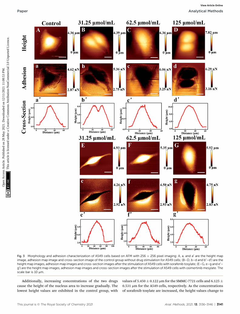
Fig. 3 Morphology and adhesion characterization of A549 cells based on AFM with 256 � 256 pixel imaging: A, a, and a0 are the height mapimage, adhesion map image and cross-section image of the control group without drug stimulation for A549 cells; (B–D, b–d and b0–d0) are theheightmap images, adhesionmap images and cross-section images after the stimulation of A549 cells with sorafenib tosylate; (E–G, e–g and e0–g0) are the height map images, adhesion map images and cross-section images after the stimulation of A549 cells with osimertinib mesylate. Thescale bar is 10 mm.
Paper Analytical Methods
Ope
n A
cces
s A
rtic
le. P
ublis
hed
on 2
8 M
ay 2
021.
Dow
nloa
ded
on 1
2/11
/202
1 11
:08:
53 P
M.
Thi
s ar
ticle
is li
cens
ed u
nder
a C
reat
ive
Com
mon
s A
ttrib
utio
n-N
onC
omm
erci
al 3
.0 U
npor
ted
Lic
ence
.View Article Online
Additionally, increasing concentrations of the two drugscause the height of the nucleus area to increase gradually. Thelowest height values are exhibited in the control group, with
This journal is © The Royal Society of Chemistry 2021
values of 5.450� 0.122 mm for the SMMC-7721 cells and 6.125�0.531 mm for the A549 cells, respectively. As the concentrationsof sorafenib tosylate are increased, the height values change to
Anal. Methods, 2021, 13, 3136–3146 | 3141

Fig. 4 Morphology andmechanical characterization results for SMMC-7721 cells and A549 cells based on AFMwith 256� 256 pixels imaging: (a)and (b) are morphological images of SMMC-7721 cells and A549 cells, respectively; (c–g) mechanical results from AFM images: (c) adhesionforce; (d) height; (e) roughness; (f) length; (g) width. In images (c–g), group A is the control group without drug stimulation; groups (B–D) werestimulated with 31.25 mmol mL�1, 62.5 mmol mL�1 and 125 mmol mL�1 of sorafenib tosylate, respectively; and groups (E and F) were stimulatedwith 31.25 mmol mL�1, 62.5 mmol mL�1 and 125 mmol mL�1 of osimertinib mesylate, respectively. The scale bar is 10 mm.
Analytical Methods Paper
Ope
n A
cces
s A
rtic
le. P
ublis
hed
on 2
8 M
ay 2
021.
Dow
nloa
ded
on 1
2/11
/202
1 11
:08:
53 P
M.
Thi
s ar
ticle
is li
cens
ed u
nder
a C
reat
ive
Com
mon
s A
ttrib
utio
n-N
onC
omm
erci
al 3
.0 U
npor
ted
Lic
ence
.View Article Online
5.750 � 0.122 mm, 6.275 � 0.132 mm, and 6.333 � 0.147 mm forthe SMMC-7721 cells and 6.387 � 0.500 mm, 6.632 � 0.794 mm,and 7.490 � 0.544 mm for the A549 cells. As the concentrationsof osimertinib mesylate are increased, the height values changeto 6.447 � 0.288 mm, 6.960 � 0.317 mm, and 8.810 � 1.094 mmfor the SMMC-7721 cells and 6.950 � 0.500 mm, 7.500 � 0.283mm, and 8.300 � 0.874 mm for the A549 cells.
Additionally, it can be observed that the roughness andadhesion force of the SMMC-7721 cells and A549 cells bothshow increasing trends with increasing concentrations of sor-afenib tosylate and osimertinib mesylate. The lowest roughnessvalues are exhibited in the control groups for both the SMMC-7721 cells and A549 cells. As the concentration of sorafenibtosylate is increased, the roughness values change from 1.592�0.005 mm for 0 mmol mL�1 to 1.732 � 0.177 mm for 31.25 mmol
3142 | Anal. Methods, 2021, 13, 3136–3146
mL�1, 1.773 � 0.111 mm for 62.5 mmol mL�1, and 1.970 � 0.042mm for 125 mmol mL�1 for the SMMC-7721 cells while the valueschange from 1.596� 0.179 mm for 0 mmol mL�1 to 1.858� 0.246mm for 31.25 mmol mL�1, 1.866� 0.233 mm for 62.5 mmol mL�1,and 2.044 � 0.359 mm for 125 mmol mL�1 for the A549 cells. Asthe concentration of osimertinib mesylate is increased, theroughness values change to 1.935 � 0.081 mm, 2.028 � 0.140mm, and 2.436� 0.269 mm for the SMMC-7721 cells and 2.288�0.137 mm, 2.503 � 0.047 mm, and 2.693 � 0.218 mm for the A549cells at concentrations of 31.25 mmol mL�1, 62.5 mmol mL�1,and 125 mmol mL�1. Moreover, the lowest adhesion force valuesare exhibited in the control group, with values of 2.497 � 0.119nN for the SMMC-7721 cells and 2.462 � 0.001 nN for the A549cells, respectively. As the concentration of sorafenib tosylate isincreased, the adhesion force values change to 2.557 � 0.107
This journal is © The Royal Society of Chemistry 2021

Fig. 5 Force–distance curves for the SMMC-7721 cells based on the JPK force spectroscopy mode: (a) force–distance curves of the controlgroup of SMMC-7721 cells without drug stimulation; (b–d) force distance curves for SMMC-7721 cells after stimulation with 31.25 mmol mL�1,62.5 mmol mL�1 and 125 mmol mL�1 of sorafenib tosylate, respectively; (e–g) force–distance curves for SMMC-7721 cells after stimulation with31.25 mmol mL�1, 62.5 mmol mL�1 and 125 mmol mL�1 of osimertinib mesylate, respectively.
Paper Analytical Methods
Ope
n A
cces
s A
rtic
le. P
ublis
hed
on 2
8 M
ay 2
021.
Dow
nloa
ded
on 1
2/11
/202
1 11
:08:
53 P
M.
Thi
s ar
ticle
is li
cens
ed u
nder
a C
reat
ive
Com
mon
s A
ttrib
utio
n-N
onC
omm
erci
al 3
.0 U
npor
ted
Lic
ence
.View Article Online
nN, 3.038� 0.142 nN, and 3.062� 0.093 nN for the SMMC-7721cells. For the A549 cells, the values change to 2.905 � 0.171 nN,2.92 � 0.166 nN, and 3.190 � 0.033 nN with increasingconcentration of sorafenib tosylate. As the concentration ofosimertinib mesylate is increased, the adhesion force valueschange to 3.004 � 0.049 nN, 3.127 � 0.049 nN, and 3.168 �0.162 nN for the SMMC-7721 cells. For the A549 cells, the valueschange to 2.466 � 0.135 nN, 2.502 � 0.048 nN, and 2.631 �0.174 nN with increasing concentration of osimertinibmesylate.
Based on previous research, the shrivelled morphology,membrane roughness and adhesion force are considered to be
Fig. 6 Force–distance curves for A549 cells based on the JPK force speccells without drug stimulation; (b–d) force–distance curves for A549 cellmL�1 of sorafenib tosylate, respectively; (e–g) force–distance curves for125 mmol mL�1 of osimertinib mesylate, respectively.
This journal is © The Royal Society of Chemistry 2021
signals of cellular apoptosis.34–36 It is assumed that the inhibi-tion effects of sorafenib tosylate and osimertinib mesylateinduce the acceleration of the cell apoptosis process.
3.3 Fitting results
Nano-indentation tests are generally considered to be a populartool to reveal the effects of drugs on the biophysical propertiesof cancerous cells. A probe with a triangular silicon nitride tip(MLCT-A, with a nominal spring constant of 0.07 N m�1) wasutilized for the AFM nano-indentation experiments. Sinusoidaland the triangular waveforms are the most common waveformsused to modulate the tip-sample displacement, which might
troscopy mode: (a) force–distance curves of the control group of A549s after stimulation with 31.25 mmol mL�1, 62.5 mmol mL�1 and 125 mmolA549 cells after stimulation with 31.25 mmol mL�1, 62.5 mmol mL�1 and
Anal. Methods, 2021, 13, 3136–3146 | 3143

Fig. 7 Young's moduli results derived from the force–distance curves based on the JPK force spectroscopy mode: (a) after stimulation of theSMMC-7721 cells with sorafenib tosylate; (b) after stimulation of the SMMC-7721 cells with osimertinib mesylate; (c) after stimulation of the A549cells with sorafenib tosylate; (d) after stimulation of the A549 cells with osimertinib mesylate.
Analytical Methods Paper
Ope
n A
cces
s A
rtic
le. P
ublis
hed
on 2
8 M
ay 2
021.
Dow
nloa
ded
on 1
2/11
/202
1 11
:08:
53 P
M.
Thi
s ar
ticle
is li
cens
ed u
nder
a C
reat
ive
Com
mon
s A
ttrib
utio
n-N
onC
omm
erci
al 3
.0 U
npor
ted
Lic
ence
.View Article Online
involve pre-loading steps and dwell times.38 Sinusoidal wave-forms were applied for the force–distance curve measurementswith a modulation frequency of 50 Hz using JPK force spec-troscopy mode.
The force–distance curves before and aer the stimulation ofthe SMMC-7721 cells and A549 cells with sorafenib tosylate andosimertinib mesylate are presented in Fig. 5 and 6, respectively.In the case of the tip sharpness of triangular probes, theSneddon model is applied to t the approach force–distancecurves and employed for the calculation of the Young's moduli.The Young's moduli were evaluated from the approach curves(shown as blue lines) in Fig. 5 and 6. As the concentrations ofsorafenib tosylate and osimertinib mesylate were increased, theslopes of the force curves changed simultaneously. Thisphenomenon can be explained by the external stimulation withtargeted drugs, which has been suggested to inuence the cellstiffness with potential distribution variation of the actin la-ments and cytoskeleton. For instance, chemotherapy exposurehas been revealed to be a factor that increases leukemia cellstiffness.37 Although the force–distance graphs are intuitive, it isstill difficult to compare the large number of force–distancecurves. Thus, the nal calculation results were extracted and aresummarized in Fig. 7.
The Young's moduli derived from the force–distance curvesbased on the JPK force spectroscopy mode for the SMMC-7721cells and A549 cells are shown in Fig. 7. The gure illustratesthat the average Young's modulus values for the control groupsof the SMMC-7721 cells and A549 cells are 1.276 � 0.174 kPaand 2.122 � 0.298 kPa, respectively. It can be observed that the
3144 | Anal. Methods, 2021, 13, 3136–3146
Young's modulus of the SMMC-7721 cells increases withincreasing concentrations of sorafenib tosylate and osimertinibmesylate. Aer the stimulation of the SMMC-7721 cells withsorafenib tosylate, their average Young's modulus varies from1.276 � 0.174 kPa for 0 mmol mL�1 to 1.53942 � 0.3089 kPa for31.25 mmol mL�1, 1.66058 � 0.60221 kPa for 62.5 mmol mL�1,and 2.00379� 0.64333 kPa for 125 mmol mL�1, respectively. Theeffects of osimertinib mesylate on the SMMC-7721 cells result ina change in the average Young's modulus from 1.276 � 0.174kPa for 0 mmol mL�1 to 1.42821 � 0.38798 kPa for 31.25 mmolmL�1, 1.7063 � 0.49526 kPa for 62.5 mmol mL�1, and 2.41239 �0.75401 kPa for 125 mmol mL�1, respectively. The results indi-cate that the Young's modulus of the SMMC-7721 cells changedmore signicantly under stimulation with osimertinib mesy-late. Thus, it can be speculated that the osimertinib mesylate ismore effective in inhibiting SMMC-7721 cells than sorafenibtosylate, which is consistent with the MTT assay results.
For the A549 cells, the Young's modulus increases withincreasing concentration of sorafenib tosylate, while theYoung's modulus rst decreases and then increases withincreasing concentration of osimertinib mesylate. Aer stimu-lation of the A549 cells with sorafenib tosylate, the averageYoung's modulus varies from 2.122 � 0.298 kPa for 0 mmolmL�1 to 2.78293 � 1.40221 kPa for 31.25 mmol mL�1, 2.94556 �0.90315 kPa for 62.5 mmol mL�1, and 5.17427 � 0.85226 kPa for125 mmol mL�1, respectively. As the concentration of osimerti-nib mesylate is increased, the average Young's modulus of theA549 cells changes from 2.122 � 0.298 kPa for 0 mmol mL�1 to1.64879 � 0.82043 kPa for 31.25 mmol mL�1, 1.1395 � 0.0786
This journal is © The Royal Society of Chemistry 2021

Paper Analytical Methods
Ope
n A
cces
s A
rtic
le. P
ublis
hed
on 2
8 M
ay 2
021.
Dow
nloa
ded
on 1
2/11
/202
1 11
:08:
53 P
M.
Thi
s ar
ticle
is li
cens
ed u
nder
a C
reat
ive
Com
mon
s A
ttrib
utio
n-N
onC
omm
erci
al 3
.0 U
npor
ted
Lic
ence
.View Article Online
kPa for 62.5 mmol mL�1, and 3.01264 � 1.16897 kPa for 125mmol mL�1, respectively. The results demonstrate the inuenceof osimertinib mesylate and sorafenib tosylate on the Young'smoduli of the A549 cells. Thus, osimertinib mesylate and sor-afenib tosylate both have an obvious inhibitory effect on A549cells, which is consistent with the MTT assay results. TheYoung's moduli of different cells under stimulation withdifferent targeted drugs stimulation have different trends,which may because sorafenib has been regarded as a VEGF/VEGFR inhibitor while osimertinib is generally considered tobe an EGFR inhibitor.15–17
4. Conclusions
In this paper, we focused mainly on comparing the mechanicaleffects of sorafenib tosylate and osimertinib mesylate onhepatoma carcinoma cells and lung cancer cells using atomicforce microscopy in order to evaluate the model system of thetargeted drugs based on nano-indentation experiments, whichhas rarely been discussed to date. The Sneddon model wasapplied to t the force–distance curves, and then the mechan-ical properties, i.e., Young's moduli, could be calculated. Theinhibitory effects of sorafenib tosylate and osimertinib mesylateon the SMMC-7721 cells and A549 cells are consistent with theMTT assay and AFM measurements. With increasing concen-trations of the targeted drugs, the survival rates of both types ofcancerous cells decrease. For the SMMC-7721 cells, osimertinibmesylate is more effective at inhibition than sorafenib tosylate.The Young's moduli of the SMMC-7721 cells increased withincreasing concentrations of sorafenib tosylate and osimertinibmesylate. For the A549 cells, osimertinib mesylate and sor-afenib tosylate both have an obvious inhibitory effect. TheYoung's moduli increase with increasing concentration of sor-afenib tosylate, while the Young's moduli rst decreases andthen increases with increasing concentration of osimertinibmesylate. These results may make contributions to the earlydiagnosis and treatment of cancer.
Abbreviations
AFM
This journal is © Th
Atomic force microscopy
VEGF Vascular endothelial growth factor VEGFR Vascular endothelial growth factor receptor EGFR Epidermal growth factor receptor SMMC-7721 Hepatoma carcinoma cells A549 Lung cancer cells PBS Phosphate buffered saline FBS Fetal bovine serum DMSO Dimethylsulfoxide JKR model Johnson–Kendall–Roberts model DMT model Derjaguin–Muller–Toporov model M–D model Maugis–Dugdale model CSLC model Cortical shell liquid core model SLS model Standard linear solid modele Royal Society of Chemistry 2021
Conflicts of interest
There are no conicts of interest to declare.
Acknowledgements
This research was supported by National Key R&D Program ofChina (No. 2017YFE0112100 and No. 2017YFB1104700), EUH2020 Program (MNR4SCell No. 734174), Program of Interna-tional S&T Cooperation (No. 2016YFE0112100), Jilin ProvincialScience and Technology Program (Nos. 20180414002GH,20180414081GH, 20180520203JH, 20190702002GH and20200901011SF) and “111 Project” of China (D17017).
References
1 F. Bray, J. Ferlay, I. Soerjomataram, R. L. Siegel, L. A. Torreand A. Jemal, Ca-Cancer J. Clin., 2018, 68, 394–424.
2 G. Binnig, C. F. Quate and C. Gerber, Phys. Rev. Lett., 1986,56, 930–934.
3 Y. F. Dufrene, T. Ando, R. Garcia, D. Alsteens, D. Martinez-Martin and A. Engel, Nat. Nanotechnol., 2017, 12, 295–307.
4 A. Alessandrini and P. Facci,Meas. Sci. Technol., 2005, 16, 65–92.
5 T. G. Kuznetsova, M. N. Starodubtseva, N. I. Yegorenkov,S. A. Chizhik and R. I. Zhdanov, Micron, 2007, 38, 824–833.
6 S. Moreno-Flores and J. L. Toca-Herrera, Hybridizing SurfaceProbe Microscopies Toward a Full Description of the Meso-and Nanoworlds, CRC Press, Boca Raton, 2012.
7 G. Massimiliano, G. L. Tang, S. B. Chandra, J. L. Zhao,S. J. Chen and J. S. Florian, Nat. Commun., 2018, 9, 3584.
8 R. Garcia, Chem. Soc. Rev., 2020, 49, 5850–5884.9 J. Iturri, A. Weber, A. Moreno-Cencerrado, M. D. Vivanco,B. Rafael and S. Leporatti, Int. J. Mol. Sci., 2019, 20, 13.
10 J. Pi, H. Jin, J. Jiang, F. Yang, H. Cai and P. Yang, Pharmacol.Res., 2017, 119, 479–489.
11 X. Yun, M. Tang, Z. Yang, J. J. Wilksch, P. Xiu and H. Gao,RSC Adv., 2017, 7, 43764–43771.
12 K. K. Sook, C. C. Hoon, P. E. Kuk, J. Min-Hyung, Y. Kyung-Sikand P. Hun-Kuk, PLoS One, 2012, 7, e30066.
13 M. S. Hung and M. F. Tsai, BioNanoScience, 2015, 5, 156–161.14 J. Ren, H. Huang, Y. Liu, X. Zheng and Q. Zou, PLoS One,
2015, 10, 1–14.15 S. F. Razavi, F. F. Bamoharram, T. Hashemi,
K. Shahrokhabadi and A. Davoodnia, Toxicol. In Vitro,2020, 68, 104917.
16 T. Kamba and D. M. McDonald, Br. J. Cancer, 2007, 96, 1788–1795.
17 C. Chen, C. D. Cheng, H. Wu, et al., Acta Pharmacol. Sin.,2021, 42, 108–114.
18 H. Hertz, Journal fur die reine und angewandte Mathematik,1881, 92, 156–171.
19 I. N. Sneddon, Int. J. Eng. Sci., 1865, 3(1), 47–57.20 R. S. Bradley, The London, Edinburgh, and Dublin
Philosophical Magazine and Journal of Science, 1932, 7, 853–862.
Anal. Methods, 2021, 13, 3136–3146 | 3145

Analytical Methods Paper
Ope
n A
cces
s A
rtic
le. P
ublis
hed
on 2
8 M
ay 2
021.
Dow
nloa
ded
on 1
2/11
/202
1 11
:08:
53 P
M.
Thi
s ar
ticle
is li
cens
ed u
nder
a C
reat
ive
Com
mon
s A
ttrib
utio
n-N
onC
omm
erci
al 3
.0 U
npor
ted
Lic
ence
.View Article Online
21 K. L. Johnson, K. Kendall and A. D. Roberts, Proc. R. Soc.London, Ser. A, 1971, 324, 301–313.
22 B. V. Derjaguin, V. M. Muller and Y. P. Toporov, J ColloidInterf Sci, 1975, 53, 314–326.
23 D. Maugis, J Colloid Interf Sci, 1992, 150, 243–269.24 M. Krieg, Y. Arboleda-Estudillo, P. H. Puech, J. Kafer,
F. Graner, D. J. Muller and C. P. Heisenberg, Nat. CellBiol., 2008, 10, 429–436.
25 E. B. Lomakina, C. M. Spillmann, M. R. King andR. E. Waugh, Biophys. J., 2004, 87, 4246–4258.
26 M. J. Rosenbluth, W. A. Lam and D. A. Fletcher, Biophys. J.,2006, 90, 2994–3003.
27 Y. M. Efremov, W. H. Wang, S. D. Hardy, R. L. Geahlen andA. Raman, Sci. Rep., 2017, 7, 1541.
28 J. S. De Sousa, J. A. C. Santos, E. B. Barros, L. M. R. Alencar,W. T. Cruz, M. V. Ramos and J. M. Filho, J. Appl. Phys., 2017,121, 034901.
29 E. M. Darling, S. Zauscher and F. Guilak, Osteoarthritis andCartilage, 2006, 14, 571–579.
3146 | Anal. Methods, 2021, 13, 3136–3146
30 E. Moeendarbary, L. Valon, M. Fritzsche, A. R. Harris,D. A. Moulding, A. J. Thrasher, E. Stride, L. Mahadevanand G. T. Charras, Nat. Mater., 2013, 12, 253–261.
31 I. L. Ivanovska, P. J. De-Pablo, B. Ibarra, G. Sgalari,F. C. MacKintosh, J. L. Carrascosa, C. F. Schmidt andG. J. L. Wuite, Proc. Natl. Acad. Sci. U. S. A., 2004, 101,7600–7605.
32 C. Rianna and M. Radmacher, AIP Conf. Proc., 2016, 1760,020057.
33 D. Kirmizis and S. Logothetidis, Int. J. Nanomed., 2010, 5,137–145.
34 D. C. Wang, K. Y. Chen, C. H. Tsai, G. Y. Chen andC. H. Chen, J. Biomech., 2011, 44(16), 2790–2794.
35 P. Lazar, S. Zhang, S. Klara, Q. Li, J. P. Froning, J. Granatier,P. Hobza, R. Zboril, F. Besenbacher, M. D. Dong andM. Otyepka, ACS Nano, 2013, 7(2), 1646–1651.
36 S. Xu, M. Dong, X. Liu, K. A. Howard, J. Kjems andF. Besenbacher, Biophys. J., 2007, 93(3), 952–959.
37 W. A. Lam, M. J. Rosenbluth and D. A. Fletcher, Blood, 2007,109(8), 3505–3508.
38 R. Garcia, Chem. Soc. Rev., 2020, 49, 5850–5884.
This journal is © The Royal Society of Chemistry 2021