invited paper: animal toxins of asia and australia
TRANSCRIPT

Clinical and Experimental Pharmacology and Physiology
(2002)
29
,
815–822
Invited Paper: Animal Toxins of Asia and Australia
MOLECULAR MOULDS WITH MULTIPLE MISSIONS: FUNCTIONAL SITES IN THREE-FINGER TOXINS
R Manjunatha Kini
Department of Biological Sciences, Faculty of Science, National University of Singapore, Singapore and Department of Biochemistry and Molecular Biophysics, Medical College of Virginia, Virginia Commonwealth
University, Richmond, Virginia, USA
SUMMARY
1. Snake venoms are complex mixtures of pharmaco-logically active peptides and proteins.
2. These protein toxins belong to a small number of super-families of proteins. The present review describes structure–function relationships of three-finger toxins.
3. All toxins share a common structure of three
�
-strandedloops extending from a central core. However, they bind todifferent receptors/acceptors and exhibit a wide variety ofbiological effects.
4. Thus, the structure–function relationships of this groupof toxins are complicated and challenging.
5. Studies have shown that the functional sites in these‘sibling’ toxins are located on various segments of the molec-ular surface.
Key words: calciseptine, cardiotoxin, cytotoxin, fasciculin,functional site, muscarinic toxin, post-synaptic neurotoxin,protein–protein interaction, snake venom, toxin evolution.
INTRODUCTION
Snake venoms are complex mixtures of pharmacologically activeproteins and polypeptides. Some of these proteins exhibit lethaland debilitating effects as a consequence of neurotoxic, cardiotoxicand tissue necrotizing effects, whereas others induce variouspharmacological effects, but are of a lower order of toxicity. Allthese protein toxins attack various physiological processes atspecific sites. The study of snake venoms and toxins by scientistswith diverse backgrounds and expertise has focused on one or moreof the following objectives: (i) to determine the mode and mech-anism of action of the toxins; (ii) to find ways and means toneutralize the toxicity and adverse effects of snake bites; (iii) todevelop specific research tools that are useful in understandingnormal physiological processes at both cellular and molecular
levels; and (iv) to develop prototypes of pharmaceutical agentsbased on the structure of toxins. Important lessons can be learnt,particularly from the latter two objectives, as to how simplemolecular templates have been used in nature to design a widearsenal of proteins that exhibit diverse (toxic) functions.
A large number of protein toxins has been purified andcharacterized from snake venoms.
1–5
Many early efforts weredirected towards the isolation and characterization of eitherproteins that are found in abundance or the most toxic componentsof the venom. The advent of more sophisticated purificationtechniques has resulted in the study of more interesting proteinsfound in smaller quantities. It is now known that snake venomscontain over 100 protein toxins. However, these toxins belong to avery small number of superfamilies of proteins. Some of the well-recognized families of venom proteins are: (i) three-finger toxinfamily; (ii) proteinase inhibitor family; (iii) lectin family; (iv)phospholipase A
2
(PLA
2
) family; (v) serine proteinase family; and(vi) metalloproteinase family. The members in a single familyshow remarkable similarities in their primary, secondary andtertiary structures. At times, however, they differ from each otherin their biological targeting and, hence, their pharmacologicaleffects. That is, each family of protein toxins has a similarmolecular scaffold but exhibits multiple functions. Thus, structure–function relationships and the mechanisms of action of snakevenom proteins are intriguing and pose exciting challenges toscientists. So far, we have understood structure–function relation-ships of only a small number of toxins in some of these families.The theme of ‘molecular moulds with multiple missions’ is wellillustrated by the structure–function relationships of the family ofthree-finger toxins, which is concisely covered in the presentreview.
THREE-FINGER TOXIN FAMILY
This is a family of non-enzymatic polypeptides containing 60–74amino acid residues.
2, 6
This family of proteins is found only in thevenoms of elapids (cobras, kraits and mambas) and hydrophid (seasnakes) and not those of vipers and crotalids (rattlesnakes). Similarto other snake venom proteins, three-finger toxins are also rich indisulphide bonds. They contain four or five disulphide bridges, ofwhich four are conserved in all members.
2
Consequently, allproteins of this family show a similar pattern of protein folding:three
�
-stranded loops extending from a central core containing the
Correspondence: R Manjunatha Kini, Department of BiologicalSciences, Faculty of Science, National University of Singapore, 10 KentRidge Crescent, Singapore 119260. Email: [email protected]
This is an invited paper, subjected to peer review.Received 17 April 2002; accepted 19 April 2002.
816
RM Kini
four conserved disulphide bridges.
7, 8
Because of this appearance,this family of proteins is called the three-finger toxin family.
DIVERSITY IN BIOLOGICAL PROPERTIES
Despite the overall similarity in structure, these polypeptides differfrom each other in their biological activities. Members of thisfamily include
�
-neurotoxins, which antagonize muscle nicotinicacetylcholine receptors (nAChR),
8–9
�
-bungarotoxins, whichrecognize neuronal nicotinic receptors,
10
muscarinic toxins withselectivity towards distinct types of muscarinic receptors,
11
fasci-culins that inhibit acetylcholinesterase,
2
calciseptins that block theL-type calcium channels,
12,13
cardiotoxins (cytotoxins) that exerttheir toxicity by forming pores in cell membranes
17
and dendro-aspins, which are antagonists of various cell-adhesion processes.
15
Interestingly, the three-fingered fold is not restricted to snakevenom toxins because several other non-venom proteins andpolypeptides also belong to this superfamily of proteins.
16–20
Neurotoxins
A large number of members of this family of toxins are neuro-toxins. These neurotoxins interfere with cholinergic transmission atvarious post-synaptic sites in the peripheral and central nervoussystems.
9
Based on their receptor selectivity, they can be broadlyclassified as curaremimetic or
�
-neurotoxins,
�
-toxins and musca-rinic toxins that target muscle nAChR, neuronal nAChR andvarious subtypes of muscarinic receptors, respectively. Over theyears, curaremimetic toxins have contributed significantly to iso-lation and characterization of muscle (
�
1) AChR, making it one ofthe best characterized receptors today.
21
Similarly, muscarinic and
�
-toxins have also helped us understand molecular details ofvarious muscarinic and neuronal AChR subtypes and their role inneurotransmission.
Curaremimetic toxins
Curaremimetic toxins (or
�
-neurotoxins) bind to muscle (
�
1)nAChR and inhibit acetylcholine from binding to the receptor,
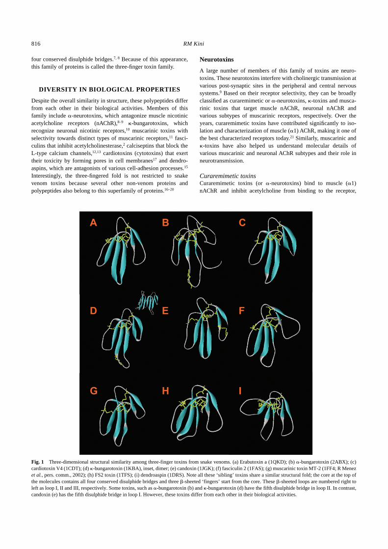
Fig. 1
Three-dimensional structural similarity among three-finger toxins from snake venoms. (a) Erabutoxin a (1QKD); (b)
�
-bungarotoxin (2ABX); (c)cardiotoxin V4 (1CDT); (d)
�
-bungarotoxin (1KBA), inset, dimer; (e) candoxin (1JGK); (f) fasciculin 2 (1FAS); (g) muscarinic toxin MT-2 (1FF4; R Menez
et al.
, pers. comm., 2002); (h) FS2 toxin (1TFS); (i) dendroaspin (1DRS). Note all these ‘sibling’ toxins share a similar structural fold; the core at the top ofthe molecules contains all four conserved disulphide bridges and three
�
-sheeted ‘fingers’ start from the core. These
�
-sheeted loops are numbered right toleft as loop I, II and III, respectively. Some toxins, such as
�
-bungarotoxin (b) and
�
-bungarotoxin (d) have the fifth disulphide bridge in loop II. In contrast,candoxin (e) has the fifth disulphide bridge in loop I. However, these toxins differ from each other in their biological activities.

Three-finger toxins from snake venoms
817
thereby impairing neuromuscular transmission.
1,9
In this respect,they imitate the effects of the alkaloid curare (hence the name).They are further classified as short-chain neurotoxins (60–62amino acid residues and four disulphides) and long-chain neuro-toxins (66–74 amino acid residues and five disulphides). Despitetheir difference in size, they share similarity in structural folding(Fig. 1a,b). However, the presence of a fifth unconserved disul-phide bridge at the tip of the second loop, as well as a longercarboxy terminal tail, constitute two significant differences seen inlong-chain neurotoxins.
2,7,8
These differences notwithstanding,both long- and short-chain neurotoxins bind to the same site on themuscle nAChR with equal affinity, competing with each other forbinding.
2
In addition, long-chain but not short-chain neurotoxinsbind to neuronal
�
7 nAChR with high affinity.
22–24
Thus, the long-and short-chain neurotoxins appear to have differences in theirtargeting and this subtle difference has been correlated to thepresence of the fifth disulphide in the second loop.
22
Muscarinic toxins
Muscarinic
toxins
bind
specifically
and
with
high
selectivity
tovarious
subsets
of
muscarinic
AChR.
25
These
toxins
arestructurally related to short-chain neurotoxins (Fig. 1c). However,unlike
�
-neurotoxins, some muscarinic toxins act as agonists,whereas others act as antagonists on muscarinic AChR.
�
-Toxins
�
-Toxins bind specifically to neuronal (
�
3
�
4) nAChR.
10
Like long-chain
�
-neurotoxins,
�
-toxins have five disulphide bridges, withthe fifth disulphide bridge located in the second loop (Fig. 1d).However, unlike any other member of the three-finger toxin family,
�
-toxins exist as dimmers.
10
Short- and long-chain neurotoxinshave poor affinity for neuronal (
�
3
�
4) nAChR. Similarly,
�
-toxinsdo not bind to
�
1 nAChR.
Other toxins
Other toxins with a unique cysteine scaffold different from those ofshort- and long-chain neurotoxins have recently been character-ized. These toxins, in addition to the four conserved disulphidebridges, have a fifth disulphide bond in the first loop.
8,26–28
Basedon their toxicity, they were classified as weak neurotoxins.
29,30
Oneof the members, WTX from cobra (
Naja kaouthia
) venom, hasbeen shown to bind to both muscle (
�
1) and neuronal (
�
7) AChR,albeit at micromolar concentrations.
30
In contrast, candoxin fromthe venom of the Malayan krait (
Bungarus candidus
; Fig. 1e) bindsto muscle (
�
1) and neuronal (
�
7) AChR with nanomolar affinity.Interestingly, its binding to muscle (
�
1) nAChR is easily reversible,in contrast with its binding to neuronal
�
7 nAChR, which is onlypartially reversible.
31
Acetylcholinesterase inhibitors
This class of three-finger toxins interferes with neuromusculartransmission by inhibiting the enzyme acetylcholinesterase (AChE)present at the neuromuscular junction. Thus, these toxins inducefasciculation in muscle due to accumulation of acetylcholine at thesynapse and are aptly named as fasciculins.
2
Fasciculins have beenisolated from mamba (
Dendroaspis
) snake venoms. They arestructurally
similar
to
short-chain
neurotoxins
(Fig. 1f).
Theybind to the peripheral site of AChE and block the entry of acetyl-
choline into the active site of the enzyme, thereby preventing itsbreakdown.
32
Cardiotoxins
This group of polypeptides is found only in cobra venoms and isthe second-largest group of three-finger toxins. Structurally, cardio-toxins resemble short-chain neurotoxins: they have 59–62 aminoacid residues and four conserved disulphide bonds (Fig. 1g).
6,14
Atlower concentrations, cardiotoxins increase heart rate and, at higherconcentrations, kill the animal by cardiac arrest.
6
However, theprotein target of cardiotoxins in cardiac myocytes has not yet beenidentified. A large number of this group of toxins also exhibitsgeneral cytolytic effects (i.e. form ion pores in the lipidmembranes)
and,
therefore,
they
are
also
referred
to
ascytolysins.
6, 33
In addition, poorly characterized cardiotoxin-likebasic polypeptides (CLBP) have also been described.
34,35
Thesepolypeptides do not act as cardiotoxins or cytotoxins, their nomen-clature notwithstanding, and may have entirely different biologicalactivity.
Other three-finger toxins
A group of three-finger toxins, such as calciseptine and FS2(Fig. 1h), specifically block L-type calcium channels.
12,13
Thesepolypeptides are structurally similar to short-chain neurotoxins,with 60 amino acid residues and four conserved disulphide bridges.They bind to the 1,4-dihydropyridine binding site of the L-typecalcium channels and physically block the calcium currents.
36
Another toxin, named dendroaspin (or mambin) and isolated from
Dendroaspis jamesoni
venom, is a potent inhibitor of plateletaggregation.
15
This protein is also structurally similar to short-chain neurotoxins, with 60 amino acid residues and four conserveddisulphide bridges (Fig. 1i). Dendroaspin contains an Arg-Gly-Asptripeptide sequence, which is involved in the adhesive function ofseveral proteins. As expected, dendroaspin interferes with theinteraction
between
fibrinogen
and
its
receptor glycoproteinIIB-IIIa (�IIb�3) complex and, hence, platelet aggregation. Inaddition, there are several other three-finger toxins, includingsynergistic toxins and angusticeps toxins,2 that have not been wellcharacterized functionally and, hence, will not be dealt in anyfurther detail in the present review.
FUNCTIONAL SITES
As is clearly evident, three-finger toxins share similar protein foldsand three-dimensional structures, but exhibit diverse biologicalproperties. Therefore, understanding their structure–functionrelationships and identifying their functional sites is a subtle,complicated and challenging task. Using a combination oftheoretical and experimental approaches, we and others havesuccessfully identified some of the functional sites in a number ofthree-finger toxins.
Neurotoxins
Earlier studies on neurotoxins were based on the chemical modifi-cation of specific amino acid residues from which some criticalresidues have been identified to be important for binding to the

818 RM Kini
muscle (or Torpedo) nAChR. More recently, Menez et al., usingsystematic and well-targeted site-directed mutagenesis, havedelineated the functional sites of erabutoxin a,37,38 a short-chainneurotoxin, and �-cobratoxin,24,39 a long-chain neurotoxin (Fig. 2).These studies reveal that both short- and long-chain neurotoxinsuse a number of structurally equivalent residues, including Lys23/Lys27, Asp27/Asp31, Arg33/Arg33 and Lys49/Lys4, as well asTrp25/Trp29 and Phe29/Phe32, in binding to Torpedo receptor. Inaddition, the tip of the first loop in erabutoxin a and, in contrast, thecarboxy terminal tail in �-cobratoxin also play important bindingroles. Antil-Delbeke et al.24 and Antil et al.39 have identified theresidues in �-cobratoxin that are involved in the recognition of andbinding to neuronal �7 nAChR. Interestingly, �-cobratoxin bindsto both Torpedo (�1) and �7 receptors using some commonresidues (Trp25, Asp27 and Arg33). In addition, it also usesreceptor-specific residues: Ala28, Lys35 and Cys26-Cys30 forrecognition of the �7 receptor and Lys23 and Lys49 for theTorpedo receptor. Moreover, the cyclic structure formed by the fifthdisulphide bridge at the tip of the second loop of �-cobratoxin hasbeen reported to be essential for its binding to the �7 receptor.24
Therefore, neurotoxins appear to use a common core of criticalresidues for binding and additional residues to determine thespecificity of their molecular target.
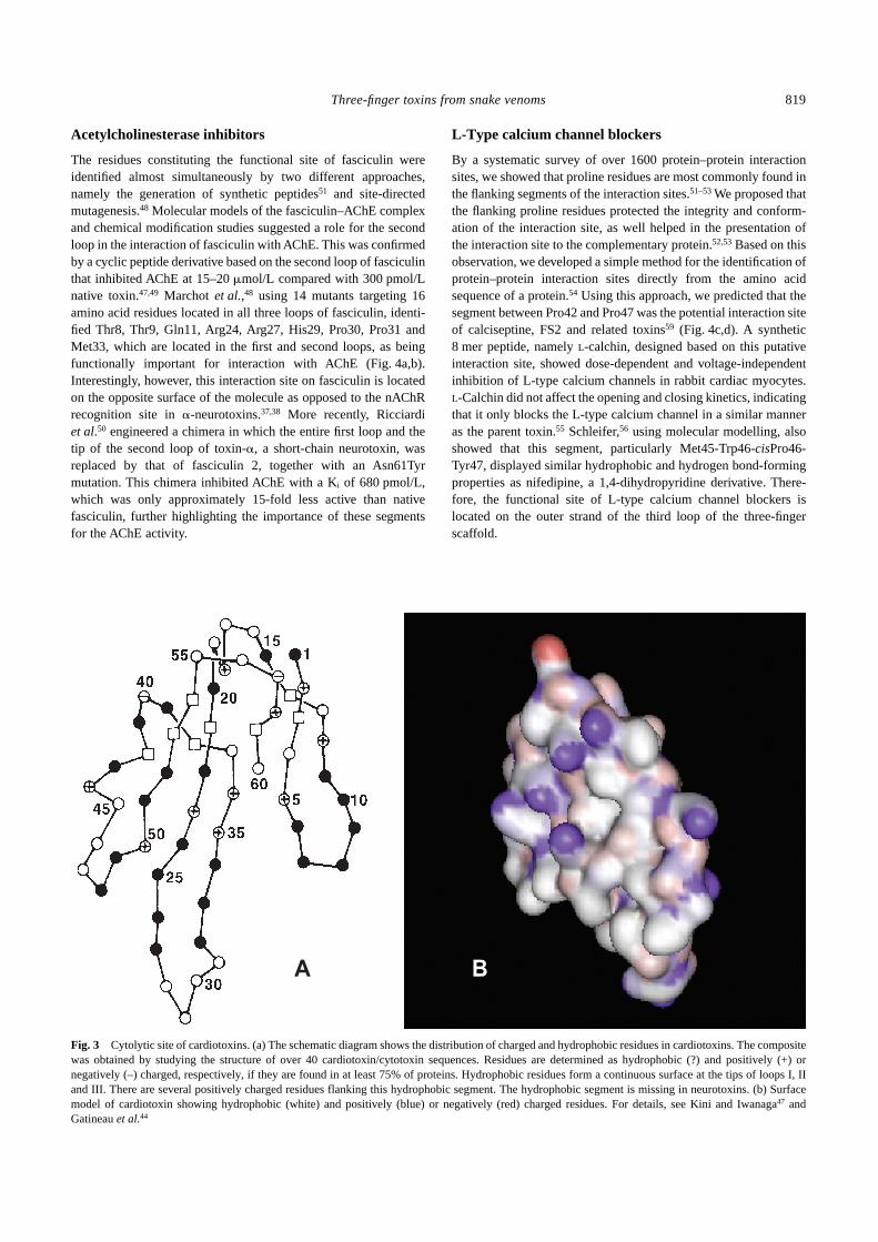
Cardiotoxins
The cytolytic site of cardiotoxins was identified in our laboratory,essentially by a combination of theoretical44 and chemicalmodification45 methods. We had previously predicted theneurotoxic46 and myotoxic47 sites in venom phospholipases. Ofsignificance was the finding that the myotoxic site of venomphospholipases and the non-enzymatic myotoxins contained acationic segment flanking a hydrophobic region.43 Because myo-toxic activity is a reflection of the cytolytic effects of these proteins,we extended this observation to cytolytic regions of proteins fromother sources, including bacteria, plants, insects, amphibians,snakes and even humans. Interestingly, all cytolytic proteins, inde-pendent of their target cells, showed the presence of hydrophobicand cationic sites flanking each other either in the primary, second-ary or tertiary structure.40 Using this approach, we showed that thecytolytic region in cardiotoxins is spread on all three loops: thereis a significant hydrophobic patch extending from the middle to thebottom end of all loops, whereas there is a row of positivelycharged lysine residues located at the top end (Fig. 3). We modifiedthe positive charges on lysine residues to negative, neutral andpositive charges using chemical methods of succinylation,carbamylation and guanidination, respectively.41 The native andguanidinated derivative showed cytolytic activity, whereas thesuccinylated and carbamylated derivatives did not. However, allderivatives showed similar protein folding, as shown by circulardichroism, and retained their ability to bind to phospholipids.41
These experiments clearly showed the importance of cationicresidues for cytolytic activity. In addition, we also oxidized the twomethionine residues present in the middle of the hydrophobicsegment to methionine sulphoxide, thereby disrupting the hydro-phobic site. This derivative failed to show any cytolytic activity andfirmly supported the importance of the hydrophobic site in cytolyticregion of cardiotoxins (RM Kini and HJ Evans, unpubl. obs.,1989). Our results were corroborated by the demonstration of the
role of lysine48 and methionine49 residues by monoacetylation andoxidation, respectively. Nuclear magnetic resonance studiesindicate that cardiotoxin interacts with phospholipid membranesthrough its hydrophobic face.46
Fig. 2 Molecular determinants of neurotoxins. Interaction site residues of(a–d) erabutoxin a involved in binding to muscle nicotinic acetylcholinereceptors (nAChR) and (e–h) �-cobratoxin involved in binding to musclenAChR (e,f) and neuronal �7 nAChR (g,h). Space-filling models of toxinsare shown. The residues on the ‘concave’ surface (a,b) that interacts withnAChR are shown. Corey–Pauling–Koltun (CPK) models (c–h) show thefunctionally important residues. The molecules are rotated 90° in (d,f,h) toshow the surface as seen by the receptor. Site-directed mutation studieswere used to determine the residues involved in interaction. The residuesshown in white are either not mutated or do not affect the affinity of inter-action. The residues in yellow, brown and red, when mutated, decrease theaffinity by at least five-, 10- or 100-fold, respectively. For details seeServent et al.,22,23 Antil-Delbeke et al.,24 Harvey,2 Pillet et al.37 andTremaeau et al.38
Three-finger toxins from snake venoms 819
Acetylcholinesterase inhibitors
The residues constituting the functional site of fasciculin wereidentified almost simultaneously by two different approaches,namely the generation of synthetic peptides51 and site-directedmutagenesis.48 Molecular models of the fasciculin–AChE complexand chemical modification studies suggested a role for the secondloop in the interaction of fasciculin with AChE. This was confirmedby a cyclic peptide derivative based on the second loop of fasciculinthat inhibited AChE at 15–20 �mol/L compared with 300 pmol/Lnative toxin.47,49 Marchot et al.,48 using 14 mutants targeting 16amino acid residues located in all three loops of fasciculin, identi-fied Thr8, Thr9, Gln11, Arg24, Arg27, His29, Pro30, Pro31 andMet33, which are located in the first and second loops, as beingfunctionally important for interaction with AChE (Fig. 4a,b).Interestingly, however, this interaction site on fasciculin is locatedon the opposite surface of the molecule as opposed to the nAChRrecognition site in �-neurotoxins.37,38 More recently, Ricciardiet al.50 engineered a chimera in which the entire first loop and thetip of the second loop of toxin-�, a short-chain neurotoxin, wasreplaced by that of fasciculin 2, together with an Asn61Tyrmutation. This chimera inhibited AChE with a Ki of 680 pmol/L,which was only approximately 15-fold less active than nativefasciculin, further highlighting the importance of these segmentsfor the AChE activity.
L-Type calcium channel blockers
By a systematic survey of over 1600 protein–protein interactionsites, we showed that proline residues are most commonly found inthe flanking segments of the interaction sites.51–53 We proposed thatthe flanking proline residues protected the integrity and conform-ation of the interaction site, as well helped in the presentation ofthe interaction site to the complementary protein.52,53 Based on thisobservation, we developed a simple method for the identification ofprotein–protein interaction sites directly from the amino acidsequence of a protein.54 Using this approach, we predicted that thesegment between Pro42 and Pro47 was the potential interaction siteof calciseptine, FS2 and related toxins59 (Fig. 4c,d). A synthetic8 mer peptide, namely L-calchin, designed based on this putativeinteraction site, showed dose-dependent and voltage-independentinhibition of L-type calcium channels in rabbit cardiac myocytes.L-Calchin did not affect the opening and closing kinetics, indicatingthat it only blocks the L-type calcium channel in a similar manneras the parent toxin.55 Schleifer,56 using molecular modelling, alsoshowed that this segment, particularly Met45-Trp46-cisPro46-Tyr47, displayed similar hydrophobic and hydrogen bond-formingproperties as nifedipine, a 1,4-dihydropyridine derivative. There-fore, the functional site of L-type calcium channel blockers islocated on the outer strand of the third loop of the three-fingerscaffold.
Fig. 3 Cytolytic site of cardiotoxins. (a) The schematic diagram shows the distribution of charged and hydrophobic residues in cardiotoxins. The compositewas obtained by studying the structure of over 40 cardiotoxin/cytotoxin sequences. Residues are determined as hydrophobic (?) and positively (+) ornegatively (–) charged, respectively, if they are found in at least 75% of proteins. Hydrophobic residues form a continuous surface at the tips of loops I, IIand III. There are several positively charged residues flanking this hydrophobic segment. The hydrophobic segment is missing in neurotoxins. (b) Surfacemodel of cardiotoxin showing hydrophobic (white) and positively (blue) or negatively (red) charged residues. For details, see Kini and Iwanaga47 andGatineau et al.44
820 RM Kini
Platelet aggregation inhibitor
As described previously, the amino acid sequence of dendroaspin(or mambin) contains the Arg-Gly-Asp tripeptide sequence.15 Thistripeptide sequence is flanked by two proline residues and isinvolved in adhesive function (Fig. 4e,f). Recently, Lu et al.57
evaluated the role of the two flanking prolines by substituting bothwith alanine, with a resulting five- to eightfold loss in the ability toinhibit platelet aggregation. Moreover, Wattam et al.58 have alsoreplaced the Arg-Gly-Asp sequence by Arg-Tyr-Asp and Arg-Cys-Asp tripeptide sequences and have demonstrated that thesemutations promote selective inhibition of �1 and �3 integrins,respectively. Thus, the functional site of dendroaspin is located atthe tip of its third loop.
Hannalgesin
Hannalgesin, isolated from Ophiophagus hannah (King cobra)venom by our group, exhibited potent analgesic as well as neuro-toxic effects in mice.59 Using the proline bracket method, weidentified the functional site of this protein to be located at the
carboxy terminal end of the toxin (RM Kini and P Gopalakrishna-kone, unpubl. obs., 1996). A short peptide synthesized on the basisof the predicted site showed significant selective analgesic effectin vivo in the absence of neurotoxicity.60 Thereby, we were able toidentify the analgesic site of hannalgesin and isolate and developthe beneficial effect of this toxin.
CONCLUSIONS AND FUTURE PROSPECTS
The compact structure of three-finger proteins has been exploitedby nature for developing ligands that perform a wide variety offunctions. In elapid and hydrophid snakes, the ancestral gene(s)encoding for three-finger protein(s) were duplicated several timesand a wide array of offensive weapons in their armamentariumresulted through accelerated evolution in their exons.61–63 Duringevolution, most features that are essential for protein folding andstructural integrity were preserved, leading to ‘sibling’ toxins thatresembled each other in their overall structural features but differedin their missions of targeting various vital physiological processes.This is clearly evident in three-finger toxins, because the core,consisting of the four disulphide bridges, is highly conserved andthe functional sites are located on either surface of the molecule onthe different loops and/or the carboxy terminal tail. In fact, there isno single designated location for the functional sites. As is thecase with other superfamilies of toxins, snakes have used therobust three-finger protein mould to construct a group of toxinswith wide variations in function involving just a few subtlechanges in the functional sites. This capacity of adaptation nodoubt offers them the flexibility to effectively capture any speciesof prey at their disposal. Their intended interests notwithstanding,snake venom toxins provide us with ample challenging oppor-tunities to decipher the subtleties in their functional sites such thatwe may better understand the plasticity of protein structure andfunction. Moreover, they will also tremendously enhance ourpotential to use their molecular architecture to design and developmini proteins with novel functions of scientific and therapeuticinterest.
ACKNOWLEDGEMENTS
I thank Professors André Ménez and Renée Menez (Department ofProtein Engineering, CEA Saclay, France) for providing the crystalstructure data of muscarinic toxin before its publication. I alsothank my colleagues Drs Bryan Fry, S Nirthanan and T Sivaraman(Department of Biological Sciences, National University ofSingapore, Singapore) for their critical reading of the manuscriptand suggestions. This work was supported by several researchgrants from the Academic Research Fund of the NationalUniversity of Singapore and a research grant from DefenseScience and Technology Agency of Singapore.
REFERENCES
1. Lee CY. Snake Venoms. Handbook of Experimental Pharmacology,Vol. 52. Springer-Verlag, Berlin. 1979.
2. Harvey AL. Snake Toxins. Pergamon Press, New York. 1991.3. Bailey GS. Enzymes from Snake Venom. Alaken, Fort Collins. 1998.4. Tu AT. Reptile Venoms and Toxins. Marcel Decker, New York. 1991.5. Kini RM. Venom Phospholipase A2 Enzymes: Structure, Function and
Mechanism. John Wiley and Sons, Chichester. 1997.
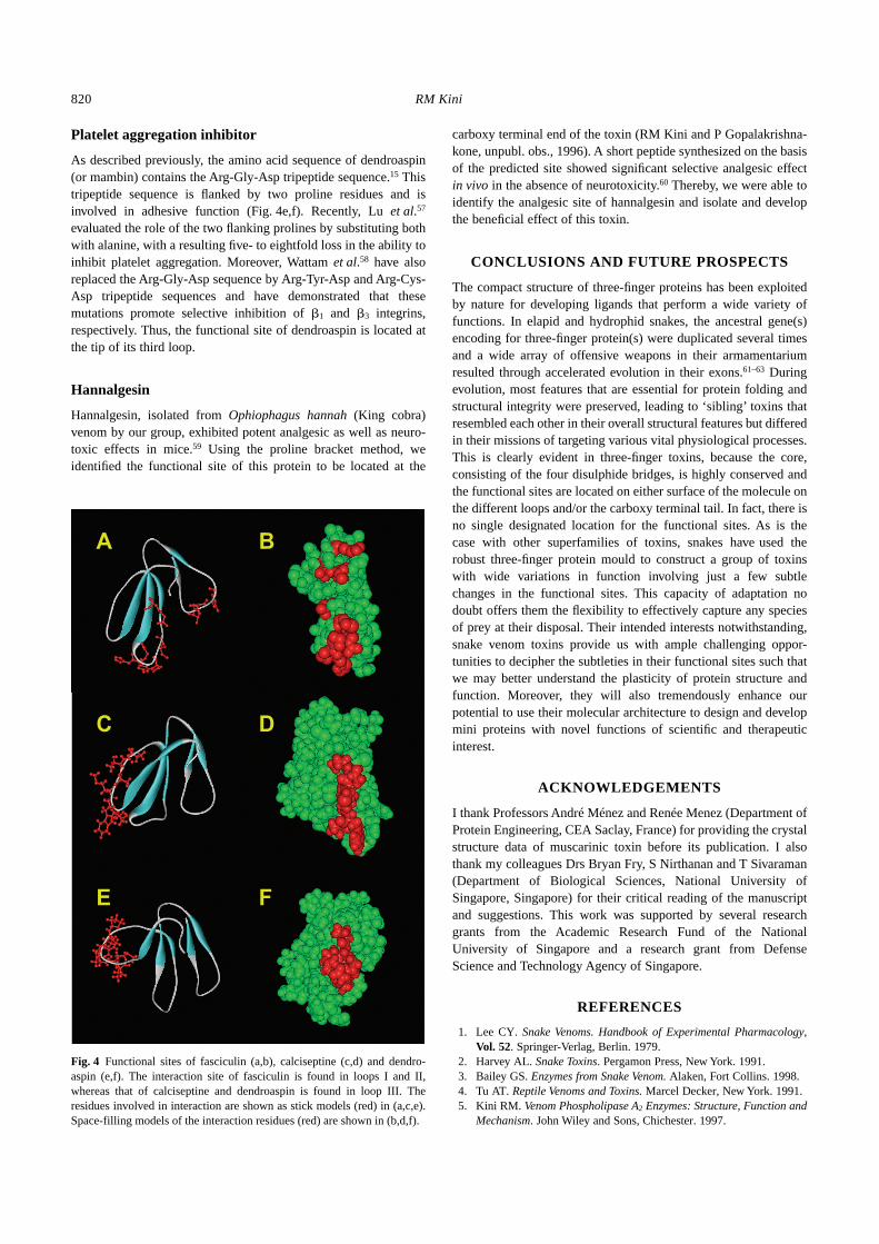
Fig. 4 Functional sites of fasciculin (a,b), calciseptine (c,d) and dendro-aspin (e,f). The interaction site of fasciculin is found in loops I and II,whereas that of calciseptine and dendroaspin is found in loop III. Theresidues involved in interaction are shown as stick models (red) in (a,c,e).Space-filling models of the interaction residues (red) are shown in (b,d,f).

Three-finger toxins from snake venoms 821
6. Dufton MT, Hider RC. Structure and pharmacology of elapid cyto-toxins. Pharmacol. Ther. 1988; 36: 1–40.
7. Menez A. Functional architectures of animal toxins: A clue to drugdesign? Toxicon 1998; 36: 1557–72.
8. Tsetlin V. Snake venom alpha-neurotoxins and other ‘three-finger’proteins. Eur. J. Biochem. 1999; 264: 281–6.
9. Changeux JP. The TiPS Lecture. The nicotinic acetylcholine receptor.An allosteric protein prototype of ligand-gated ion channels. TrendsPharmacol. Sci. 1990; 11: 485–92.
10. Grant GA, Chiappinelli VA. �-Bungarotoxin: Complete amino acidsequence of a neuronal nicotinic receptor probe. Biochemistry 1985;24: 1532–7.
11. Jerusalinsky D, Harvey AL. Toxins from mamba venoms: Smallproteins with selectivities for different subtypes of muscarinic acetyl-choline receptors. Trends Pharmacol. Sci. 1994; 15: 424–30.
12. De Weille JR, Schweitz H, Maes P, Tartar A, Lazdunski M. Calci-septine, a peptide isolated from black mamba venom, is a specificblocker of the L-type calcium channel. Proc. Natl Acad. Sci. USA1991; 88: 2437–40.
13. Albrand JP, Blackledge MJ, Pascaud F, Hollecker M, Marion D. NMRand restrained molecular dynamics study of the three-dimensionalsolution structure of toxin FS2, a specific blocker of the L-typecalcium channel, isolated from black mamba venom. Biochemistry1995; 34: 5923–37.
14. Bilwes A, Rees B, Moras D, Menez R, Menez A. X-Ray structure at1.55 A of toxin gamma, a cardiotoxin from Naja nigricollis venom.Crystal packing reveals a model for insertion into membranes. J. Mol.Biol. 1994; 239: 122–36.
15. McDowell RS, Dennis MS, Louie A, Shuster M, Mulkerrin MG,Lazarus RA. Mambin, a potent glycoprotein IIb–IIIa antagonist andplatelet aggregation inhibitor structurally related to the short neuro-toxins. Biochemistry 1992; 31: 4766–72.
16. Fleming TJ, O’hUigin C, Malek TR. Characterization of two novelLy-6 genes. Protein sequence and potential structural similarity toalpha-bungarotoxin and other neurotoxins. J. Immunol. 1993; 150:5379–90.
17. Ploug M, Ellis V. Structure–function relationships in the receptor forurokinase-type plasminogen activator. Comparison to other membersof the Ly-6 family and snake venom alpha-neurotoxins. FEBS Lett.1994; 349: 163–8.
18. Gumley TP, McKenzie IFC, Sandrin MS. Tissue expression, structureand function of the murine Ly-6 family of molecules. Immunol. CellBiol. 1995; 73: 277–96.
19. Miwa JM, Ibanez-Tallon I, Crabtree GW et al. lynx1, an endogenoustoxin-like modulator of nicotinic acetylcholine receptors in themammalian CNS. Neuron 1999; 23: 105–14.
20. Cordero-Erausquin M, Marubio LM, Klink R, Changeux JP. Nicotinicreceptor function: New perspectives from knockout mice. TrendsPharmacol. Sci. 2000; 21: 211–17.
21. Unwin N. Nicotinic acetylcholine receptor at 9 Å resolution. J. Mol.Biol. 1993; 229: 1101–24.
22. Servent D, Winckler-Dietrich V, Hu HY et al. Only snake curaremi-metic toxins with a fifth disulfide bond have high affinity for theneuronal alpha7 nicotinic receptor. J. Biol. Chem. 1997; 272:24 279–86.
23. Servent D, Antil-Delbeke S, Corringer PJ, Changeux JP, Ménez A.Molecular characterization of the specificity of interactions of variousneurotoxins on two distinct nicotinic acetylcholine receptors. Eur. J.Pharmacol. 2000; 393: 197–204.
24. Antil-Delbeke S, Gaillard C, Tamiya T et al. Molecular determinantsby which a long chain toxin from snake venom interacts with theneuronal alpha 7-nicotinic acetylcholine receptor. J. Biol. Chem. 2000;275: 29 594–601.
25. Karlsson E, Jolkkonen M, Mulugeta E, Onali P, Adem A. Snake toxinswith high selectivity for subtypes of muscarinic acetylcholine recep-tors. Biochimie 2000; 82: 793–806.
26. Carlsson FHH. Snake venom toxins. The primary structure of protein
S4C11. A neurotoxin homologue from the venom of forest cobra (Najamelanoleuca). Biochim. Biophys. Acta 1975; 400: 310–21.
27. Joubert FJ. The purification and amino acid sequence of toxin CM-13bfrom Naja haje annulifera (Egyptian cobra) venom. Hoppe-Seylers Z.Physiol. Chem. 1975; 356: 1901–8.
28. Joubert FJ, Taljaard N. Snake venoms. The amino acid sequences oftwo Melanoleuca-type toxins. Hoppe-Seylers Z. Physiol. Chem. 1980;361: 425–36.
29. Utkin YN, Kukhtina VV, Maslennikov IV et al. First tryptophan-containing weak neurotoxin from cobra venom. Toxicon 2001; 39:921–7.
30. Utkin YN, Kukhtina VV, Kryukova EV et al. ‘Weak toxin’ from Najakaouthia is a nontoxic antagonist of alpha 7 and muscle-type nicotinicacetylcholine receptors. J. Biol. Chem. 2001; 276: 15 810–15.
31. Nirthanan S, Charpantier E, Gopalakrishnakone P et al. Candoxin, anovel toxin from Bungarus candidus is a reversible antagonist ofmuscle (����) but a poorly reversible antagonist of neuronal 7nicotinic acetylcholine receptors. J. Biol. Chem. 2002; 277:17 811–20.
32. Eastman J, Wilson EJ, Cervanansky C, Rosenberry TL. Fasciculin 2binds to the peripheral site on acetylcholinesterase and inhibitssubstrate hydrolysis by slowing a step involving proton transfer duringenzyme acylation. J. Biol. Chem. 1995; 270: 19 694–701.
33. Condrea E. Hemolytic disorders associated with a primary red cellmembrane defect. Experentia 1975; 32: 537–42.
34. Takechi M, Tanaka Y, Hayashi K. Amino acid sequence of acardiotoxin-like basic polypeptide (CLBP) with low cytotoxic activityisolated from the venom of the Formosan cobra (Naja naja atra).Biochem. Int. 1985; 11: 795–802.
35. Sivaraman T, Kumar TK, Yang PW, Yu C. Cardiotoxin-like basicprotein (CLBP) from Naja naja atra is not a cardiotoxin. Toxicon1997; 35: 1367–71.
36. Yasuda O, Morimoto S, Chen Y et al. Calciseptine binding to a1,4-dihydropyridine recognition site of the L-type calcium channel ofrat synaptosomal membranes. Biochem. Biophys. Res. Commun. 1993;194: 587–94.
37. Pillet L, Tremeau O, Ducancel F et al. Genetic engineering of snaketoxins. Role of invariant residues in the structural and functionalproperties of a curaremimetic toxin, as probed by site-directed muta-genesis. J. Biol. Chem. 1993; 268: 909–16.
38. Tremeau O, Lemaire C, Drevet P et al. Genetic engineering of snaketoxins. The functional site of Erabutoxin a, as delineated by site-directed mutagenesis, includes variant residues. J. Biol. Chem. 1995;270: 9362–9.
39. Antil S, Servent D, Ménez A. Variability among the sites by whichcuraremimetic toxins bind to torpedo acetylcholine receptor, asrevealed by identification of the functional residues of �-cobratoxin.J. Biol. Chem. 1999; 274: 34 851–8.
40. Kini RM, Evans HJ. A common cytolytic region in myotoxins,hemolysins, cardiotoxins and antibacterial peptides. Int. J. PeptideProtein Res. 1989; 34: 277–86.
41. Kini RM, Evans HJ. Role of cationic amino acid residues in cytolyticactivity. Modifications of lysine residues in the cardiotoxin from Najanigricollis venom and correlation between cytolytic and antiplateletactivities. Biochemistry 1989; 28: 9209–15.
42. Kini RM, Iwanaga S. Structure–function relationships of phospho-lipases I. Prediction of presynaptic neurotoxicity. Toxicon 1986; 24:527–41.
43. Kini RM, Iwanaga S. Structure–function relationships of phospho-lipases II. Charge density distribution and the myotoxicity of pre-synaptically neurotoxic phospholipases. Toxicon 1986; 24: 895–905.
44. Gatineau E, Takechi M, Bouet F et al. Delineation of the functionalsite of a snake venom cardiotoxin: Preparation, structure, and functionof monoacetylated derivatives. Biochemistry 1990; 29: 6480–9.
45. Carlsson FHH, Louw AI. The oxidation of methionine and its effect ofthe properties of cardiotoxin V 1 II from Naja melanoleuca venom.Biochim. Biophys. Acta 1978; 534: 322–30.

822 RM Kini
46. Dauplais M, Neumann JM, Pinkasfeld S, Ménez A, Roumestand C. AnNMR study of the interaction of cardiotoxin � from Naja nigricolliswith perdeuterated dodecylphosphocholine micelles. Eur. J. Biochem.1995; 230: 213–20.
47. Falkenstein RJ, Pena C. Synthetic peptides derived from the centralloop of fasciculin: Structural analysis and evaluation as acetylcho-linesterase inhibitors. Biochim. Biophys. Acta 1997; 1340: 143–51.
48. Marchot P, Prowse CN, Kanter J et al. Expression and activity ofmutants of fasciculin, a peptidic acetylcholinesterase inhibitor frommamba venom. J. Biol. Chem. 1997; 272: 3502–10.
49. Karlsson E, Mbugua PM, Rodriguez-Ithurralde D. Fasciculins,anticholinesterase toxins from the venom of the green mambaDendroaspis angusticeps. J. Physiol. 1984; 79: 232–40.
50. Ricciardi A, Le Du MH, Khayati M et al. Do structural deviationsbetween toxins adopting the same fold reflect functional differences?J. Biol. Chem. 2000; 275: 18 302–10.
51. Kini RM, Evans HJ. A common structural feature enclosing inter-action sites: Prediction of protein–protein interaction sites anddevelopment of potent bioactive peptides. In: Menon J (ed.). CurrentTopics in Peptide and Protein Research, Vol. 1. Council of ScientificInformation, Trivandrum. 1994; 297–311.
52. Kini RM, Evans HJ. A hypothetical structural role for proline residuesin the flanking segments of protein–protein interaction sites. Biochem.Biophys. Res. Commun. 1995; 212: 1115–24.
53. Kini RM. Proline brackets and identification of potential functionalsites in proteins: Toxins to therapeutics. Toxicon 1998; 36: 1659–70.
54. Kini RM, Evans HJ. Prediction of potential protein–protein interactionsites from amino acid sequence. Identification of a fibrin polymeriz-ation site. FEBS Lett. 1996; 385: 81–6.
55. Kini RM, Caldwell RA, Wu QY, Baumgarten CM, Feher JJ, Evans HJ.Flanking proline residues identify the L-type Ca2+ channel binding siteof calciseptine and FS2. Biochemistry 1998; 37: 9058–63.
56. Schleifer KJ. Comparative molecular modelling study of the calciumchannel blockers nifedipine and black mamba toxin FS2. J. Comput.Aided Mol. Des. 1997; 11: 491–501.
57. Lu X, Sun Y, Shang D et al. Evaluation of the role of proline residuesflanking the RGD motif of dendroaspin, an inhibitor of plateletaggregation and cell adhesion. Biochem. J. 2001; 355: 633–8.
58. Wattam B, Shang D, Rahman S et al. Arg-Tyr-Asp (RYD) and Arg-Cys-Asp (RCD) motifs in dendroaspin promote selective inhibition of�1 and �3 integrins. Biochem. J. 2001; 356: 11–17.
59. Pu XC, Wong PT, Gopalakrishnakone P. A novel analgesic toxin(Hannalgesin) from the venom of king cobra (Ophiophagus hannah).Toxicon 1995; 33: 1425–31.
60. Gopalakrishnakone P, Wong P, Gwee MCE, Kini RM, inventors.National University of Singapore, assignee. Therapeutic molecules.Singapore patent pending. 20 September 1996.
61. Nakashima K, Nobuhisa I, Deshimaru M et al. Accelerated evolutionin the protein-coding regions is universal in crotalinae snake venomgland phospholipase A2 isozyme genes. Proc. Natl Acad. Sci. USA1995; 92: 5605–9.
62. Deshimaru M, Ogawa T, Nakashima K et al. Accelerated evolution ofcrotalinae snake venom gland serine proteases. FEBS Lett. 1996; 397:83–8.
63. Ohno M, Ménez R, Ogawa T et al. Molecular evolution of snaketoxins: Is the functional diversity of snake toxins associated with amechanism of accelerated evolution? Prog. Nucleic Acids Res. Mol.Biol. 1998; 59: 307–64.