ion channel signaling influences cellular proliferation
TRANSCRIPT

University of KentuckyUKnowledge
Biology Faculty Publications Biology
8-2017
Ion Channel Signaling Influences CellularProliferation and Phagocyte Activity DuringAxolotl Tail RegenerationBrandon M. FranklinUniversity of Kentucky, [email protected]
S. Randal VossUniversity of Kentucky, [email protected]
Jeffrey L. OsbornUniversity of Kentucky, [email protected]
Right click to open a feedback form in a new tab to let us know how this document benefits you.
Follow this and additional works at: https://uknowledge.uky.edu/biology_facpub
Part of the Biology Commons, Cell and Developmental Biology Commons, and the GeneticsCommons
This Article is brought to you for free and open access by the Biology at UKnowledge. It has been accepted for inclusion in Biology Faculty Publicationsby an authorized administrator of UKnowledge. For more information, please contact [email protected].
Repository CitationFranklin, Brandon M.; Voss, S. Randal; and Osborn, Jeffrey L., "Ion Channel Signaling Influences Cellular Proliferation and PhagocyteActivity During Axolotl Tail Regeneration" (2017). Biology Faculty Publications. 170.https://uknowledge.uky.edu/biology_facpub/170

Ion Channel Signaling Influences Cellular Proliferation and Phagocyte Activity During Axolotl Tail Regeneration
Notes/Citation InformationPublished in Mechanisms of Development, v. 146, p. 42-54.
© 2017 Elsevier B.V.
This manuscript version is made available under the CC‐BY‐NC‐ND 4.0 licensehttps://creativecommons.org/licenses/by-nc-nd/4.0/.
The document available for download is the author's post-peer-review final draft of the article.
Digital Object Identifier (DOI)https://doi.org/10.1016/j.mod.2017.06.001
This article is available at UKnowledge: https://uknowledge.uky.edu/biology_facpub/170

Ion channel signaling influences cellular proliferation and phagocyte activity during axolotl tail regeneration
Brandon M. Franklin, S. Randal Voss, and Jeffrey L. Osborn*
Department of Biology, University of Kentucky, Lexington, KY 40506, United States
Abstract
Little is known about the potential for ion channels to regulate cellular behaviors during tissue
regeneration. Here, we utilized an amphibian tail regeneration assay coupled with a chemical
genetic screen to identify ion channel antagonists that altered critical cellular processes during
regeneration. Inhibition of multiple ion channels either partially (anoctamin1/Tmem16a,
anoctamin2/Tmem16b, KV2.1, KV2.2, L-type CaV channels and H/K ATPases) or completely
(GlyR, GABAAR, KV1.5 and SERCA pumps) inhibited tail regeneration. Partial inhibition of tail
regeneration by blocking the calcium activated chloride channels, anoctamin1&2, was associated
with a reduction of cellular proliferation in tail muscle and mesenchymal regions. Inhibition of
anoctamin 1/2 also altered the post-amputation transcriptional response of p44/42 MAPK
signaling pathway genes, including decreased expression of erk1/erk2. We also found that
complete inhibition via voltage gated K+ channel blockade was associated with diminished
phagocyte recruitment to the amputation site. The identification of H+ pumps as required for
axolotl tail regeneration supports findings in Xenopus and Planaria models, and more generally,
the conservation of ion channels as regulators of tissue regeneration. This study provides a
preliminary framework for an in-depth investigation of the mechanistic role of ion channels and
their potential involvement in regulating cellular proliferation and other processes essential to
wound healing, appendage regeneration, and tissue repair.
1. Introduction
Ion channels are known for traditional physiological functions, including muscle
contraction, nerve conduction, and maintenance of ionic homeostasis. However, ion
channels modulate membrane ion conductance across all cells and tissues, establishing
electrical fields (EF) that affect cellular behaviors under normal conditions, during critical
periods of development, and in response to tissue injury. Understanding how ion channels
function within different biological contexts is central to identifying the molecular basis of
channelopathies and for exploiting ion channels for wound healing and tissue repair.
Bioelectric signaling via ion channels and control of cellular membrane potentials in
planarian and Xenopus regeneration have significantly contributed to our understanding of
ionic influences on regenerative processes (Levin, 2007; Levin, 2009; Tseng et al., 2010;
*Corresponding author at: Department of Biology, University of Kentucky, 115 Thomas Hunt Morgan, Lexington, KY 40506, United States. [email protected] (J.L. Osborn).
Supplementary data to this article can be found online at http://dx.doi.org/10.1016/j.mod.2017.06.001.
HHS Public AccessAuthor manuscriptMech Dev. Author manuscript; available in PMC 2019 February 22.
Published in final edited form as:Mech Dev. 2017 August ; 146: 42–54. doi:10.1016/j.mod.2017.06.001.
Author M
anuscriptA
uthor Manuscript
Author M
anuscriptA
uthor Manuscript

Levin, 2014). Despite these advances, little is known about the individual channels that are
important during regeneration and the specific cellular functions that they influence.
Amphibians and fish provide powerful models to investigate the role of EF and ion channels
on cellular processes that are activated during appendage regeneration. Typically in these
vertebrates, amputated body parts are flawlessly repaired via the activation, proliferation,
and patterning of progenitor cells (McCusker and Gardiner, 2014; Tanaka, 2016). Wound-
induced EFs likely affect the behavior of immune and progenitor cells because they are
enacted during the early wound-healing response, and interruption or reversal of an EF is
detrimental to regeneration (Borgens et al., 1979; Borgens et al., 1977; Jenkins et al., 1996).
However, exactly how cells detect and translate EF information to elicit specific cell
behaviors is not well understood. One possibility is that information from an EF is
modulated and transduced by ion channels. Consistent with this idea, Özkucur et al. (2010))
showed that ion concentrations are modulated in cells near the amputation site during axolotl
(Ambystoma mexicanum) tail regeneration. This observation suggests that ion channels
affect cellular behaviors by regulating ion concentration-dependent signaling cascades
during regeneration (Wei et al., 2011; Mao et al., 2009; Wondergem et al., 2001; Wang et al.,
2002; Tao et al., 2008).
In this study, we performed a chemical genetic screen to identify ion channels whose
functions are required for normal axolotl tail regeneration. We administered tail amputations
to axolotl embryos and treated groups with ion channel antagonists. This type of hierarchical
drug screen has been previously described, and chemical genetics approaches have proven to
be robust for identifying bioelectric mechanisms that affect regeneration (Adams and Levin,
2006; Sengupta et al., 2015). Multiple channels were identified from several ion channel
families that either delayed the regenerative process or inhibited the initiation of
regeneration. Two of these channels, anoctamin 1 and voltage-gated K+ channels, were
investigated further to determine their effects on cellular proliferation and the activity of
phagocytic cells.
2. Methods
2.1. Anesthesia
Experiments were conducted using pre-feeding, hatchling stage axolotl embryos
(RRID:AGSC_100H, 9–11 mm in length) obtained from the Ambystoma Genetic Stock
Center (RRID:SCR_006372), Department of Biology, University of Kentucky. These nearly
mature embryos were removed from chorions immediately before the onset of the
experimental protocol. Ethical animal procedures performed in this study were approved by
the University of Kentucky IACUC committee (protocol 00907L2005). The skeletal muscle-
specific myosin inhibitor N-benzylp-toluene sulfonamide (BTS) was used to anesthetize
embryos before all amputations and imaging procedures. This was chosen over benzocaine,
a voltage gated Na+ channel inhibitor, as to not confound the results of the ion channel
antagonist being investigated. Embryos were placed in modified Holtfreter’s solution (59
mM NaCl, 0.67 mM KCl, 0.76 mM CaCl2, 2.4 mM NaHCO3) with 10 μM BTS at least 15
min before conducting any procedures.
Franklin et al. Page 2
Mech Dev. Author manuscript; available in PMC 2019 February 22.
Author M
anuscriptA
uthor Manuscript
Author M
anuscriptA
uthor Manuscript

2.2. Regeneration assay
Axolotl tails were imaged under a dissecting microscope and amputated approximately
halfway between the tail tip and cloaca (~2 mm). Embryos were then placed in individual
wells of a 24-well tissue culture plate with 3 mL of Holtfreter’s solution containing various
ion channel antagonists or a modified Cl− free Holtfreter’s solution (15 mM NaC6H11O7,
0.2 mM KC6H11O7, 0.2 mM Ca(C6H11O7)2, 0.2 mM MgSO4·7H20, 0.6 mM NaHCO3). The
drug screen consisted of three tiers of ion channel antagonist ranging from broad scale
general ion conductance inhibitors (tier 1), to antagonists of ion channel sub families (tier 2),
and finally, specific ion channel blockers (tier 3, Table 1). All drugs were initially evaluated
at four concentrations (10−4, 10−5, 10−6 and 10−7 M) with 6 biological replicates for each
concentration; some concentrations were lethal and this resulted in n < 6 for some drug
treatments. Subsequently, ranges of drug concentrations were evaluated on a case-by-case
basis and conclusions regarding a drug’s impact on regeneration were only drawn from
nontoxic concentrations of drugs. Drugs that exhibited systemic toxicity (identified by
general atrophy, lethargy and/or tissue degeneration) or lethality at all concentrations were
excluded from the study. Lethal or toxic drug concentrations are emphasized in bold in Table
1. At 7 days post-amputation (dpa), embryos were anesthetized, imaged, and tail length was
measured. To assess the extent of regeneration, tail length at day 0 (Fig. S1A) was subtracted
from tail length at day 7 (Fig. S1B). Following a positive result, drugs were assayed for their
impact on normal development in unamputated axolotl embryos and drugs found to
adversely affect normal developmental growth were excluded from the study. Results for
each pharmacological agent were analyzed in Sigma Plot Statistical Software (SPSS) using a
one-way ANOVA with Dunnett’s test for post hoc analysis. Significant results were
qualitatively assigned as either delayed/reduced regeneration, inhibited regeneration, or
toxic/lethal (Fig. S1B–E).
2.3. Phagocyte imaging
Live imaging of phagocytes was accomplished by staining with the non-toxic dye, neutral
red. Neutral red stains lysosomes which are present in many cell types but are especially rich
in phagocytes such as macrophages. Also, neutral red has been confirmed as an effective and
specific stain for macrophages in zebrafish and axolotl using the protocol described below
(Herbomel et al., 2001; Davis and Ramakrishnan, 2009; Godwin et al., 2013). Prior to
imaging, embryos were placed in Holtfreter’s solution containing 5 μg/mL of neutral red dye
for 6 h and then de-stained in normal Holtfreter’s solution for 24 h. Following this
staining/de-staining procedure, embryos were anesthetized and imaged under bright-field
(Herbomel et al., 2001; Godwin et al., 2013; Carradice and Lieschke, 2008). Images were
quantified by counting all labeled cells within 500 μm of the amputation plane and these
values were normalized by tissue section area. Results were analyzed in SPSS using a one-
way ANOVA with Dunnett’s test for post hoc analysis (control was t = 0).
2.4. Proliferation assay
Axolotl embryos were amputated and placed in 24 well plates as outlined above. At 3 dpa,
embryos were incubated for 16 h in 10 μM 5-ethynyl-2′-deoxyuridine (EdU), a BrdU
analog. After this incubation period, embryos were euthanized and tails were amputated at
Franklin et al. Page 3
Mech Dev. Author manuscript; available in PMC 2019 February 22.
Author M
anuscriptA
uthor Manuscript
Author M
anuscriptA
uthor Manuscript

the cloaca, immediately fixed in 4% paraformaldehyde, and stored in 70% EtOH. Tissues
were then embedded in paraffin wax and cut into 5-micron sagittal sections. The Click-iT®
EdU Alexa Fluor® 488 Imaging Kit (C10337, invitrogen) was used to visualize EdU
incorporation into the DNA of proliferating cells per the prescribed protocol. Hoechst was
used to counter stain the sections. All cells staining positive for EdU incorporation within
500 μm of the amputation plane were counted as well as all cells staining positive for
Hoechst within the same anatomical area. Proliferative index was determined by dividing the
number of EdU+ cells by the number of Hoechst + cells in each tissue section. Results were
analyzed in SPSS using either a one-way ANOVA with Dunnett’s test for post hoc analysis
or, in the case of tissue specificity analysis, a two-way ANOVA (factors were tissue type and
drug treatment) with Student-Newman-Keuls (SNK) test used for post hoc analysis.
2.5. Apoptosis assay
Axolotl embryos were administered tail amputations and placed in 24 well plates and
allowed to regenerate for 3 days. Embryos were euthanized and tails were amputated at the
cloaca, immediately fixed in 4% paraformaldehyde, and stored in 70% EtOH. Tissues were
then embedded in paraffin wax and cut into 5-micron transverse sections. The Click-iT®
TUNEL Alexa Fluor® 594 Imaging Kit (C10246, invitrogen) was used to assay for
fragmented DNA, a measure of apoptosis (Gavrieli et al., 1992). Tissue sections were
grouped as either 0–100 μm or 150–250 μm from the tail tip. TUNEL positive cells in each
group were counted and divided by the total number of cells in each group (Hoechst) to give
a measure of apoptotic index. Results were analyzed in SPSS using one-way ANOVA and
SNK test for post hoc analysis.
2.6. RNA extraction and quantitative real-time PCR
Embryos were administered amputations and placed in 24-well plates as before and allowed
to regenerate for 3 days in either modified Holtfreter’s or modified Holtfreter’s
supplemented with the anoctamin 1 inhibitor, T16A(inh)-A01 (10 μM). The distal 1 mm of
the tail was re-amputated and collected in ice-cold RNAlater (25 mM C6H7NaO7, 10 mM
EDTA, 5.3 M (NH4)2SO4 and pH 5.2) and stored at −20°C. To isolate mRNA, 2 tails were
placed in 100 μL of Trizol, homogenized via sonication, and purified via Zymo Direct-zol™
RNA MiniPrep (R2050) kit and treated with DNase 1. RNA concentration and purity were
determined via Nanodrop ND-1000 spectrophotometry (Nanodrop Technologies). cDNA
was generated using qScript™ cDNA Supermix (Quanta Biosciences 95,048) and used for
PCR. PCR primers were designed using sequence data available on Sal-Site and are listed in
Table 2 (Baddar et al., 2015). Quantitative real-time PCR was performed using the
StepOnePlus™ Real-Time PCR system (Applied Biosystems®). Samples were loaded into
96-well plates in triplicate with probes and Power SYBR® Green PCR Master Mix
(4367659) to detect PCR products. Relative gene expression was calculated using the
comparative CT method (2−ΔΔCT) in StepOne software v2.3 per the manufacturer’s
instructions (Schmittgen and Livak, 2008). Relative quantifications were compared in SPSS
using a one-way ANOVA and Student–Newman–Keuls (SNK) method for post hoc analysis.
Franklin et al. Page 4
Mech Dev. Author manuscript; available in PMC 2019 February 22.
Author M
anuscriptA
uthor Manuscript
Author M
anuscriptA
uthor Manuscript

3. Results
3.1. Pharmacological screen of ion channel groups
3.1.1. Chloride—Nonspecific chloride conductance was examined with the broad-scale
Cl− channel blockers 4,4′-Diisothiocyanatostilbene-2,2′-disulfonic acid (DIDS), 4,4′-
dinitrostilbene-2,2′-disulfonic acid (DNDS), and niflumic acid (NFA). Embryos treated with
any of these broad scale Cl− channel antagonists exhibited decreased tail regeneration at all
concentrations tested, with the exception of DNDS at 1 μM. For example, embryos exposed
to 10 μM DIDS, DNDS, or NFA regenerated 0.71 ± 0.31 mm, 1.11 ± 0.32 mm, and 0.63
± 0.24 mm respectively, significantly less than untreated embryos (Fig. 1A). Calcium-
activated Cl− channels (CaCC) were evaluated with the family-specific inhibitor CaCCinh-
A01. CaCCinh-A01 was lethal at concentrations N2 μM but reduced regeneration at 0.1 μM
(1.01 ± 0.24 mm) and 1 μM (1.09 ± 0.17 mm, Fig. 1B) without observable toxic effects.
Anoctamins 1&2 (aka Tmem16A/B, Ano1, Ano2) were inhibited with benzbromarone
(BBM) or T16a(inh)-A01 (A01). Embryos treated with either BBM or A01 had decreased
regenerative capacity at 100 nM (0.56 ± 0.18 mm) and 10 μM (0.88 ± 0.21 mm, Fig.
1C&D). The ligand gated Cl− channels, GABAA receptors (GABAAR) and glycine receptors
(GlyR), were inhibited with picrotoxin (PTx) and bicuculline methiodide (BCU, GABAAR
only) and both compounds caused reduced regenerative capacity at all concentrations tested.
Both PTx (0.645 ± 0.27 mm) and BCU (0.618 ± 0.32 mm) elicited complete inhibition of
regeneration at a concentration of 100 μM (Figs. 1E&F). All other Cl− channel blockers
were either toxic or did not affect regeneration (Fig. S2).
3.1.2. Potassium—General potassium conductance was initially examined with the
broad-scale K+ channel blocker tetraethylammonium (TEA). Compared to control embryos
(1.83 ± 0.38 mm), TEA treated embryos at a concentration of 1 μM or higher decreased
regenerative growth (0.96 ± 0.29 mm, Fig. 2A). The voltage-gated K+ channel (KV) blocker
4-aminopyridine (4-AP) reduced regenerative outgrowth at 4 μM (1.05 ± 0.03 mm) and
completely inhibited regeneration at 10 μM (0.50 ± 0.24 mm) and 100 μM (0.59 ± 0.27 mm,
Fig. 2B). KV2.1/2.2 blockade with Guangxitoxin-1E (GTx-1E) slightly reduced regeneration
at 1 μM (1.11 ± 0.46, Fig. 4C). KV1.5 blockade with cytochalasin B (CytB) had no effect on
regeneration when embryos were incubated at 0.1 or 1 μM, but tail outgrowth was inhibited
at 10 μM (0.16 ± 0.11 mm, Fig. 2D). All other K+ channel blockers used were either toxic or
did not affect regeneration (Fig. S3).
3.1.3. Sodium—Sodium was assessed using only tier 2 antagonists as no tier 1 drugs
were available (Table 1). The epithelial sodium channel (ENaC) blocker amiloride did not
affect regeneration at any concentration tested. The voltage-gated sodium channel (NaV)
antagonists, tetrodotoxin (TTx) and lidocaine, also did not affect regeneration at any
concentration tested, although the highest concentration of TTx (100 μM) was lethal in
100% of embryos (Fig. S4).
3.1.4. Calcium—Calcium conductance was examined with the broad-scale Ca2+ channel
blocker bepridil (BPD). BPD was 100% lethal at every concentration tested. The L, N & T-
type calcium channel inhibitor, benidipine HCl (BNP), completely inhibited regeneration at
Franklin et al. Page 5
Mech Dev. Author manuscript; available in PMC 2019 February 22.
Author M
anuscriptA
uthor Manuscript
Author M
anuscriptA
uthor Manuscript

100 nM (0.60 ± 0.35 mm) and all other concentrations were lethal. L-type channel blockers
amlodipine (CaV1.3) and diltiazem (CaV1.1/1.2/1.4) diminished the regenerative response,
with a maximal effect observed 10 μM (1.03 ± 0.38 mm) and 1 μM (1.04 ± 0.50 mm),
respectively. The P & Q-type blocker, ω-conotoxin MVIIC, completely inhibited
regeneration with a maximal effect at 10 nM (0.57 ± 0.19 mm). P-type specific inhibitor, ω-
agatoxin IVA, had no effect on the regenerative response at any concentration, thus
eliminating Q-type channels as affecting regeneration (Fig. S5).
3.2. Pumps/transporters
The sarco/endoplasmic reticulum Ca2+-ATPase (SERCA) was examined with 2,5-di-(tert-
butyl)-1,4-benzohydroquinone (tBuBHQ), which completely inhibited regeneration with
maximal inhibition at 2 μM (0.41 ± 0.15 mm). Incubation with the H+/K+-ATPase inhibitor,
pantoprazole, resulted in a slight reduction of regenerative growth at 100 μM (1.14 ± 0.05
mm). All other transporter inhibitors used were either toxic or did not affect regeneration
(Fig. S6).
3.3. Phagocyte activation is dependent on KV channel signaling
Macrophage activation is required for regeneration to proceed normally and KV channels are
known to influence many functions of macrophages in vitro (Godwin et al., 2013; Kotecha
and Schlichter, 1999; Moreno et al., 2013; Qiu et al., 2002; Vicente et al., 2005; Li et al.,
2016). This suggests that KV’s may influence tail regeneration through modulation of the
macrophage response. To test this hypothesis, it was first necessary to characterize the
normal response of phagocytic cells during tail regeneration. Using neutral red (NR) to label
phagocytic cells, a significant accumulation of phagocytes was observed at the wound site
beginning 24 hours post amputation (hpa, 90.1 ± 20.8 NR+ cells/mm2 tissue) and initially
peaking at 48 hpa (189.2 ± 53.7 NR+ cells/mm2 tissue). This was followed by a decline
concurrent with tissue outgrowth and then a second peak at 144 hpa (200.6 ± 34.4 NR+
cells/mm2 tissue, Fig. 3). NR stained cells were confirmed to be phagocytic by repeating the
NR staining procedure on embryos that had been previously injected with liposome
encapsulated clodronate. In comparison to control embryos, clodronate treated embryos
exhibited a significant reduction of NR+ cells (Fig. S7).
While CytB is a potent inhibitor of KV1.5 channel activity, it has off target effects, such as
disruption of actin polymerization that could also influence regeneration. To further assess
the role of KV channels on phagocyte activity we used an alternate KV channel blocker, 4-
AP. Embryos were incubated at 2 concentrations of 4-AP (5 μM and 25 μM) beginning at 12
hpa and harvested at either 3 or 6 days post amputation (dpa). At 3 dpa, embryos incubated
in either 5 μM (45.88 ± 12.35 NR+ cells) or 25 μM (46.63 ± 10.07 NR+ cells) exhibited a
marked reduction in phagocyte recruitment to the wound site compared to control embryos
(123.33 ± 15.67 NR+ cells) but there was no difference between the two concentrations
tested. At 6 dpa, the reduction of phagocyte recruitment persisted compared to control
(190.29 ± 43.02 NR+ cells) and there was a concentration dependent response between the 5
μM group (102.38 ± 9.89 NR+ cells) and 25 μM group (52.71 ± 14.74 NR+ cells, Fig. 4). To
confirm this result, embryos were subjected to one of three treatments: (1) Encapsome®/
Fluorosome®-DiI 50:50 mixture IP injection 24 h before amputation (24hba), (2)
Franklin et al. Page 6
Mech Dev. Author manuscript; available in PMC 2019 February 22.
Author M
anuscriptA
uthor Manuscript
Author M
anuscriptA
uthor Manuscript

Clodrosome®/Fluorosome®-DiI 50:50 mixture IP injection 24hba or (3) Encapsome®/
Fluorosome®-DiI 50:50 mixture IP injection 24hba and incubation in 25 μM 4-AP
beginning at 12 hpa. Embryos were then harvested at 3 dpa and whole mount imaged under
fluorescent and bright field conditions. Embryos subjected to both the phagocyte depletion
protocol (17.045 ± 17.30 DiI+ cells/mm2) and KV channel inhibition (31.80 ± 19.35 DiI+
cells/mm2) exhibited reduced phagocyte recruitment following amputation compared to
control conditions (145.20 ± 77.17 DiI+ cells/mm2, Fig. 5). Notably, the number of
phagocytes at the amputation site at 3 dpa under control conditions identified by
Fluorosome®-DiI incorporation was consistent with the number identified by NR staining at
the same time point.
3.4. Anoctamin 1 blockade delays regeneration via inhibition of proliferation
Cell proliferative responses to amputation were assessed by measuring EdU incorporation at
3 dpa under several experimental interventions. EdU incorporation was observed in 34.00
± 6.44% of cells within 500 μm of the amputation plane under control conditions (n = 4, Fig.
6 A–A″ & C). In contrast, broad-scale Cl− channel blockade (DIDS: n = 4, 18.13 ± 5.52%&
DNDS: n = 5, 19.65 ± 9.29%, Fig. 6C) or specific inhibition of anoctamin 1 (A01: n = 5,
19.65 ± 4.54%, Fig. 6 B–B″ & C) significantly reduced the percentage of cells proliferating
within this same anatomical area. Embryos incubated in Cl− free Holtfreter’s exhibited a
trend towards reduced proliferation (24.80 ± 9.74%, n = 5, Fig. 6C) but the difference was
not statistically significant relative to controls. We note that apoptosis was assessed using a
TUNEL assay; no differences were observed between embryos subjected to anoctamin 1
blockade and controls (Fig. 7).
To more finely examine proliferation spatially, cell counts were obtained for epidermal
(ED), spinal cord (SC), and mesenchymal & muscle (MM) regions of the tail. Neither broad-
scale Cl− channel blockade (DIDS & DNDS) nor anoctamin 1 inhibition (A01) treatments
affected proliferation rates within the ED and SC tissues; the reduced proliferation response
was only observed in MM tissues. These results suggest that functional chloride flux
mediated by Ano1 is critical for regulating the proliferative response in the mesenchyme but
not in other tissues (Fig. 6 D&E).
3.5. Anoctamin 1 blockade diminishes activation of p44/42 MAPK signaling pathway
To investigate gene expression changes associated with anoctamin 1 inhibition of tail
regeneration, quantitative real-time PCR was used to measure mRNA expression of p44/42
MAPK signaling pathway genes, a pathway activated during cellular proliferation. After
amputation, pkcγ, mek1, mek2, erk1 and erk2 showed a higher percent increase in
transcription in control embryos vs anoctamin 1 treated embryos at 3 dpa. In contrast,
expression was downregulated more strongly in control embryos than embryos subjected to
anoctamin 1 inhibition by A01 at 3 dpa for early p44/42 pathway genes (pkcα, pkcβ, rras and raf1) (Fig. 8). These results show that anoctamin 1 affects the transcription of p44/42
MAPK pathway genes that regulate cellular proliferation.
Franklin et al. Page 7
Mech Dev. Author manuscript; available in PMC 2019 February 22.
Author M
anuscriptA
uthor Manuscript
Author M
anuscriptA
uthor Manuscript

4. Discussion
In this study, we identified ion channel antagonists that partially (anoctamin1/Tmem16a,
anoctamin2/Tmem16b, KV2.1, KV2.2, L-type CaV channels and H/K ATPases) or
completely (GlyR, GABAAR, KV1.5 and SERCA pumps) inhibited axolotl tail regeneration.
Below we separately discuss the affected ion channels from each of these categories (Fig. 9).
4.1. Partially inhibiting channels & transporters
4.1.1. Anoctamins 1/2 (Tmem16A/B)—Our results suggest a role for CaCCs in the
tail regeneration process. Regeneration was delayed when embryos were subjected to either
broad-scale Cl− channel blockade (DIDS, DNDS & niflumic acid) or CaCC inhibition
(CaCCinh-A01, T16a(inh)-A01 & Benzbromarone). CaCC currents were described over 25
years ago but their molecular basis was only recently discovered (Hartzell et al., 2005). In
2008, three independent researchers identified anoctamin 1 and anoctamin 2 channels as
mediators of CaCC currents (Caputo et al., 2008; Schroeder et al., 2008; Yang et al., 2008).
Anoctamin 1 was first discovered as an overexpression marker for gastrointestinal stromal
tumors (then known as DOG1) and has since been shown to promote other forms of cancer
(Britschgi et al., 2013; West et al., 2004). Blockade of anoctamin 1 suppresses tumor growth
and invasion in multiple human cancer lines (Jia et al., 2015; Liu et al., 2012). There is
evidence that anoctamin 1 manifested these effects through regulation of both cell migration
and proliferation (Qu et al., 2014; Ruiz et al., 2012). Our results support this idea because
inhibition of anoctamin 1 significantly decreased the number of proliferating cells in the
regenerating tail. This suggests that anoctamin 1 channel function is directly or indirectly
required to sustain cell proliferation at a level that is typical of normal tail regeneration.
Our results further suggest that anoctamin 1 affected tail regeneration by modulating the
p44/42 MAPK pathway, which is a well-known regulator of cell proliferation (Zhang and
Liu, 2002). Following tail amputation, Erk1/2 signaling pathway genes were
transcriptionally upregulated to a higher degree in control embryos than embryos treated
with anoctamin 1 antagonist. Thus, the magnitude of erk1/erk2 transcription correlated on
one hand with anoctamin 1 function, and conversely, with the magnitude of cell
proliferation. We propose the following model: anoctamin 1, activated by an initial calcium
surge, acts as a countercurrent ion channel to amplify calcium signaling. This amplified
surge subsequently activates the Erk1/2 pathway leading to increased cellular proliferation.
This model assumes there is a burst in intracellular [Ca2+] following tail amputation and a
dependence of intracellular Ca2+ flux on chloride conductance. Özkucur et al. (2010)
downplayed the role of Ca2+ in their model of axolotl tail regeneration. However, they
reported significant increases in Ca2+ fluorescence at 48 hpa, preceding the time when the
tail shows measureable regenerative outgrowth. This Ca2+ flux at 48 hpa supports our
proposed model (Özkucur et al., 2010; Barro-Soria et al., 2010; Wang and van Breemen,
1999).
4.1.2. KV2.1/2.2—KV2 channel blockade with TEA or GTx-1E also reduced
regeneration. KV2 channels traditionally act as the primary delayed rectifier involved in
regulating the excitability of neurons and facilitating exocytosis in neurons and
Franklin et al. Page 8
Mech Dev. Author manuscript; available in PMC 2019 February 22.
Author M
anuscriptA
uthor Manuscript
Author M
anuscriptA
uthor Manuscript
neuroendocrine cells (Feinshreiber et al., 2009; Misonou et al., 2005). These functions of
KV2 channels could be associated with the release of neurotrophic factors that are required
for regeneration, the so-called neurotrophic requirement for regeneration (Kumar and
Brockes, 2012; Singer, 1974). However, it is also important to consider the direct influence
of KV2 channels on the cellular dynamics of non-excitable cells (Kumar and Brockes, 2012).
KV2 acts as a promoter of cell migration in cultured HEK293, CHO, and bone derived
mesenchymal stem cells (MSC) via phosphorylation of focal adhesion kinase (Hu et al.,
2011; Wei et al., 2008). MSCs also require functioning KV2 channels for proper progression
through the cell cycle by facilitating plasma membrane-endoplasmic reticulum contact sites
(Cobb et al., 2015; Deng et al., 2007). These MSC specific characteristics of KV2 channels
are particularly interesting considering their resemblance to progenitor cells of the blastema,
but more research is required to determine the specific role of KV2 channels within the
context of appendage regeneration.
4.2. L-type Ca2+ channels (CaV1.1–1.4)
The L-type channel blockers, amlodipine and diltiazem, partially inhibited tail regeneration.
These channels are mostly known for their roles in excitable cells, where they couple plasma
membrane depolarization with increases in Ca2+ conductance across the membrane. Recent
evidence also suggests that they are important physiological components of many non-
excitable cell types (Davenport et al., 2015; Wen et al., 2012). Ca2+ is a ubiquitous second
messenger in all cell types and can influence many different cellular processes (Berridge,
1995; Berridge et al., 2000; Clapham, 2007; Zayzafoon, 2006; Zheng and Poo, 2007).
Because of the widespread nature of L-type Ca2+ channel expression and the complicated
features of intracellular Ca2+ signaling, these channels may not present the best targets for
future investigations. But, if the model we proposed above is correct, the anoctamin 1/2 Cl−
channels may provide an indirect path to modulate intracellular calcium flux in a specific
cell population.
4.2.1. H+/K+ ATPases—Our study found that H+ pump inhibition with pantoprazole
sodium reduced regenerative outgrowth. Proton pumps have been classically associated with
their roles in gastric and renal function. However, over the past several decades they have
been identified as critical mediators of wound healing and regeneration (Adams et al., 2007;
Walan et al., 1989; Balestrini et al., 2017). Most of this influence has been attributed to the
proton pump’s involvement in establishing and maintaining trans-epithelial electrical
potentials that drive wound-induced electrical fields (Nuccitelli, 2003). A recent chemical
genetic screen performed using planarian head regeneration also identified H+/K+ ATPase as
important for initiating the regenerative process through control of cellular membrane
potentials (Vmem) (Beane et al., 2011). H+ pump driven changes in Vmem are required for
regeneration to proceed normally in the Xenopus model of regeneration (Adams et al.,
2007). Our findings further validate the requirement of H+ pumps in organisms capable of
appendage regeneration.
4.3. Inhibiting channels & transporters
4.3.1. KV1.5—Blockade of KV1.5 with 4-AP or CytB resulted in a robust and
concentration dependent total inhibition of regeneration. These channels are widely
Franklin et al. Page 9
Mech Dev. Author manuscript; available in PMC 2019 February 22.
Author M
anuscriptA
uthor Manuscript
Author M
anuscriptA
uthor Manuscript

expressed in a large number of tissues and are involved in regulating Vmem and
electrophysiological properties of a variety of cell types (Archer et al., 1998; Olson et al.,
2006; Tabarean, 2014). In addition to these electrophysiological functions, KV1.5 channels
have been implicated in regulating cell cycle progression and migration (Kotecha and
Schlichter, 1999; Vallejo-Gracia et al., 2013; Villalonga et al., 2008). Of particular interest is
their role in regulating immune cell dynamics. Macrophages are required for amphibian
regeneration and the invertebrate homologue of macrophages (hemocytes) respond to
exogenously applied electrical fields in vivo in a KV dependent manner (Godwin et al.,
2013; Franklin et al., 2016). Consistent with this line of reasoning, phagocyte activation was
severely diminished during tail regeneration after KV channel blockade with 4-AP. This
suggests that KV channels may be critical regulators of macrophage/monocyte populations
during regeneration. However, further studies are necessary to further clarify the role for Kv
channels in macrophage activation during tissue regeneration and whether or not this
mechanism is a cell autonomous.
4.3.2. GlyR/GABAAR—GlyR and GABAAR blockade with picrotoxin or bicuculline
resulted in strong inhibition of regeneration at relatively low concentrations. These ligand-
gated chloride channels act primarily as receptors for inhibitory neurotransmitters in the
CNS, with GABAAR acting mostly in the brain and GlyR functioning primarily in the
brainstem and spinal cord (Lynch, 2004; Sigel and Steinmann, 2012). This immediately
implicates nerves and their required neurotrophic factors for appendage regeneration since
modulation of either of these receptors would disrupt normal neuronal activity during
regeneration. There is evidence that these receptors influence neural progenitor cells during
early development and regulate many critical cellular processes in multiple cell types,
mostly via modulation of intracellular Ca2+ concentrations (Nguyen et al., 2001; Van Den
Eynden et al., 2009). Importantly, misexpression of GlyR has recently been shown to disrupt
patterning and development in neural, muscle and vascular tissues via physiological
modulation of membrane potentials (Lobikin et al., 2012; Lobikin et al., 2014; Pai et al.,
2015). It will be interesting to examine the expression patterns of these receptors in cells of
regenerating axolotl tissues to see if they are altered following tissue injury. More research
will be necessary to assess the exact role of GlyR/GABAAR during axolotl regeneration.
4.3.3. SERCA pump—SERCA inhibition with tBuBHQ completely inhibited
regeneration at low concentrations, although higher concentrations of this drug were lethal.
The sacroplasmic/endoplasmic reticulum Ca2+ ATPase (SERCA) is expressed in virtually all
cell types and distributes Ca2+ ions against their concentration gradient into sarcoplasmic
and endoplasmic reticula. This allows for quick sequestration of cytoplasmic free Ca2+
following a Ca2+ signaling event and for the generation of intracellular calcium stores that
allow for a more rapid and robust Ca2+ surge upon cellular stimulation (Wuytack et al.,
2002). As discussed above, Ca2+ signaling is associated with many cytosolic signaling
pathways and physiological functions, all of which require precise control of intracellular
Ca2+ concentration pulses in terms of both magnitude and duration, and these Ca2+
dependent pathways are operative in models of epimorphic regeneration (Berridge et al.,
2000; Rao et al., 2014). The ubiquitous nature of SERCA expression may explain why
inhibition of this ion channel completely inhibited regeneration, while incomplete
Franklin et al. Page 10
Mech Dev. Author manuscript; available in PMC 2019 February 22.
Author M
anuscriptA
uthor Manuscript
Author M
anuscriptA
uthor Manuscript

regeneration was observed for L-type Cav channel inhibition. Based on the extensive
network of pathways influenced by Ca2+ signaling and the pervasive nature of SERCA
pumps, SERCA pumps will present challenging targets to investigate mechanisms of tissue
regeneration.
5. Summary
A chemical genetic screen was performed to identify ion channel antagonists that inhibit
axolotl tail regeneration. Then, experiments were performed to determine how select
antagonists affected cellular behaviors (cell proliferation and phagocyte activation) that are
required for successful tail regeneration. The antagonists that were identified from the screen
targeted the following ion channels: anoctamins 1/2 (Tmem16a/b), GlyR, GABAAR, KV1.5,
KV2.1, KV2.2, L-type CaV channels, H/K ATPases and SERCA pumps. An association was
established between KV channel blockade and phagocyte activation, and thus a possible
mechanism for KV channel mediated inhibition of axolotl tail regeneration. Also, blockade
of the anoctamins reduced cellular proliferation, and this was associated with modulated
transcription of Erk1/2 signaling pathway genes. It has been shown previously that the
transcriptional response to bioelectric signaling is conserved among axolotl regeneration,
Xenopus development and human progenitor cells (Pai et al., 2015). This speaks to the broad
relevance of the data presented in this report and lends credence to the idea that the
antagonists and ion channels prioritized from our study will provide useful tools and targets
for investigating mechanisms of tissue regeneration.
Supplementary Material
Refer to Web version on PubMed Central for supplementary material.
Acknowledgements
This work was funded by the Biomedical Sciences Research Group, LLC and the NIH (R24OD021479). Axolotl embryos used in this study were obtained from the Ambystoma Genetic Stock Center (AGSC) at the University of Kentucky. The AGSC is funded by the NIH (P40OD019794). The Authors would like to thank Deirdre Mcdonnell, for her expertise in quantitative PCR and other general lab work that contributed to this project.
References
Adams DS, Levin M, 2006 Inverse drug screens: a rapid and inexpensive method for implicating molecular targets. Genesis 44 (11), 530–540. [PubMed: 17078061]
Adams DS, Masi A, Levin M, 2007 H+ pump-dependent changes in membrane voltage are an early mechanism necessary and sufficient to induce Xenopus tail regeneration. Development 134 (7), 1323–1335. [PubMed: 17329365]
Archer SL, et al., 1998 Molecular identification of the role of voltage-gated K+ channels, Kv1. 5 and Kv2. 1, in hypoxic pulmonary vasoconstriction and control of resting membrane potential in rat pulmonary artery myocytes. J. Clin. Inv 101 (11), 2319.
Baddar NWAH, et al., 2015 Sal-site: research resources for the Mexican Axolotl In: Kumar A, Simon A (Eds.), Salamanders in Regeneration Research: Methods and Protocols. Springer New York, New York, NY, pp. 321–336.
Balestrini L, et al., 2017 The natural compound sanguinarine perturbs the regenerative capabilities of planarians. Int. J. Dev. Biol 61 (1–2), 43–52. [PubMed: 28287246]
Franklin et al. Page 11
Mech Dev. Author manuscript; available in PMC 2019 February 22.
Author M
anuscriptA
uthor Manuscript
Author M
anuscriptA
uthor Manuscript

Barro-Soria R, et al., 2010 ER-localized bestrophin 1 activates Ca2+-dependent ion channels TMEM16A and SK4 possibly by acting as a counterion channel. Pflugers Arch. 459 (3), 485–497. [PubMed: 19823864]
Beane WS, et al., 2011 A chemical genetics approach reveals H,K-ATPase-mediated membrane voltage is required for planarian head regeneration. Chem. Biol 18 (1), 77–89. [PubMed: 21276941]
Berridge MJ, 1995 Calcium signalling and cell proliferation. BioEssays 17 (6), 491–500. [PubMed: 7575490]
Berridge MJ, Lipp P, Bootman MD, 2000 The versatility and universality of calcium signalling. Nat. Rev. Mol. Cell. Biol 1 (1), 11–21. [PubMed: 11413485]
Borgens RB, Vanable JW, Jaffe LF, 1977 Bioelectricity and regeneration: large currents leave the stumps of regenerating newt limbs. Proc. Natl. Acad. Sci 74 (10), 4528–4532. [PubMed: 270701]
Borgens RB, Vanable JW, Jr., Jaffe LF, 1979 Role of subdermal current shunts in the failure of frogs to regenerate. J. Exp. Zool 209 (1), 49–56. [PubMed: 314968]
Britschgi A, et al., 2013 Calcium-activated chloride channel ANO1 promotes breast cancer progression by activating EGFR and CAMK signaling. Proc. Natl. Acad. Sci. U. S. A 110 (11), E1026–E1034. [PubMed: 23431153]
Caputo A, et al., 2008 TMEM16A, a membrane protein associated with calcium-dependent chloride channel activity. Science 322 (5901), 590–594. [PubMed: 18772398]
Carradice D, Lieschke GJ, 2008 Zebrafish in hematology: sushi or science? Blood 111 (7), 3331–3342. [PubMed: 18182572]
Clapham DE, 2007 Calcium Signaling. Cell 131 (6), 1047–1058. [PubMed: 18083096]
Cobb MM, et al., 2015 Cell cycle-dependent changes in localization and phosphorylation of the plasma membrane Kv2.1 K+ channel impact endoplasmic reticulum membrane contact sites in COS-1 cells. J. Biol. Chem 290 (49), 29189–29201. [PubMed: 26442584]
Davenport B, et al., 2015 Signature channels of excitability no more: L-type channels in immune cells. Front. Immunol 6, 375. [PubMed: 26257741]
Davis JM, Ramakrishnan L, 2009 The role of the granuloma in expansion and dissemination of early tuberculous infection. Cell 136 (1), 37–49. [PubMed: 19135887]
Deng XL, et al., 2007 Cell cycle-dependent expression of potassium channels and cell proliferation in rat mesenchymal stem cells from bone marrow. Cell Prolif. 40 (5), 656–670. [PubMed: 17877608]
Feinshreiber L, et al., 2009 Voltage-gated potassium channel as a facilitator of exocytosis. Ann. N. Y. Acad. Sci 1152, 87–92. [PubMed: 19161379]
Franklin BM, Maroudas E, Osborn JL, 2016 Sine‐wave electrical stimulation initiates a voltage‐gated potassium channel‐dependent soft tissue response characterized by induction of hemocyte recruitment and collagen deposition. Physiol. Rep 4 (12).
Gavrieli Y, Sherman Y, Ben-Sasson SA, 1992 Identification of programmed cell death in situ via specific labeling of nuclear DNA fragmentation. J. Cell Biol 119 (3), 493–501. [PubMed: 1400587]
Godwin JW, Pinto AR, Rosenthal NA, 2013 Macrophages are required for adult salamander limb regeneration. Proc. Natl. Acad. Sci 110 (23), 9415–9420. [PubMed: 23690624]
Hartzell C, Putzier I, Arreola J, 2005 Calcium-activated chloride channels. Annu. Rev. Physiol 67 (1), 719–758. [PubMed: 15709976]
Herbomel P, Thisse B, Thisse C, 2001 Zebrafish early macrophages colonize cephalic mesenchyme and developing brain, retina, and epidermis through a M-CSF receptor-dependent invasive process. Dev. Biol 238 (2), 274–288. [PubMed: 11784010]
Hu X, et al., 2011 Hypoxic preconditioning enhances bone marrow mesenchymal stem cell migration via Kv2.1 channel and FAK activation. Am. J. Physiol. Cell Physiol 301 (2), C362–C372. [PubMed: 21562308]
Jenkins LS, Duerstock BS, Borgens RB, 1996 Reduction of the current of injury leaving the amputation inhibits limb regeneration in the red spotted newt. Dev. Biol 178 (2), 251–262. [PubMed: 8812127]
Franklin et al. Page 12
Mech Dev. Author manuscript; available in PMC 2019 February 22.
Author M
anuscriptA
uthor Manuscript
Author M
anuscriptA
uthor Manuscript

Jia L, et al., 2015 Inhibition of calcium-activated chloride channel ANO1/TMEM16A suppresses tumor growth and invasion in human lung cancer. PLoS One 10 (8), e0136584. [PubMed: 26305547]
Kotecha SA, Schlichter LC, 1999 A Kv1.5 to Kv1.3 switch in endogenous hippocampal microglia and a role in proliferation. J. Neurosci 19 (24), 10680–10693. [PubMed: 10594052]
Kumar A, Brockes JP, 2012 Nerve dependence in tissue, organ, and appendage regeneration. Trends Neurosci. 35 (11), 691–699. [PubMed: 22989534]
Levin M, 2007 Large-scale biophysics: ion flows and regeneration. Trends Cell Biol. 17 (6), 261–270. [PubMed: 17498955]
Levin M, 2009 Bioelectric mechanisms in regeneration: unique aspects and future perspectives Seminars in Cell & Developmental Biology. Elsevier.
Levin M, 2014 Endogenous bioelectrical networks store non‐genetic patterning information during development and regeneration. J. Physiol 592 (11), 2295–2305. [PubMed: 24882814]
Li C, Levin M, Kaplan DL, 2016 Bioelectric modulation of macrophage polarization. Sci Rep 6, 21044. [PubMed: 26869018]
Liu W, et al., 2012 Inhibition of Ca2+-activated Cl− channel ANO1/TMEM16A expression suppresses tumor growth and invasiveness in human prostate carcinoma. Cancer Lett. 326 (1), 41–51. [PubMed: 22820160]
Lobikin M, et al., 2012 Resting potential, oncogene-induced tumorigenesis, and metastasis: the bioelectric basis of cancer in vivo. Phys. Biol 9 (6), 065002.
Lobikin M, et al., 2014 Selective depolarization of transmembrane potential alters muscle patterning and muscle cell localization in Xenopus laevis embryos. Int. J. Dev. Biol 59 (7–9), 303–311.
Lynch JW, 2004 Molecular structure and function of the glycine receptor chloride channel. Physiol. Rev 84 (4), 1051–1095. [PubMed: 15383648]
Mao J, et al., 2009 Volume-activated chloride channels contribute to cell-cycle-dependent regulation of HeLa cell migration. Biochem. Pharmacol 77 (2), 159–168. [PubMed: 18992227]
McCusker CD, Gardiner DM, 2014 Understanding positional cues in salamander limb regeneration: implications for optimizing cell-based regenerative therapies. Dis. Model. Mech 7 (6), 593–599. [PubMed: 24872456]
Misonou H, Mohapatra DP, Trimmer JS, 2005 Kv2.1: a voltage-gated k+ channel critical to dynamic control of neuronal excitability. Neurotoxicology 26 (5), 743–752. [PubMed: 15950285]
Moreno C, et al., 2013 Modulation of voltage-dependent and inward rectifier potassium channels by 15-epi-lipoxin-A4 in activated murine macrophages: implications in innate immunity. J. Immunol 191 (12), 6136–6146. [PubMed: 24249731]
Nguyen L, et al., 2001 Neurotransmitters as early signals for central nervous system development. Cell Tissue Res. 305 (2), 187–202. [PubMed: 11545256]
Nuccitelli R, 2003 Endogenous electric fields in embryos during development, regeneration and wound healing. Radiat. Prot. Dosim 106 (4), 375–383.
Olson TM, et al., 2006 Kv1. 5 channelopathy due to KCNA5 loss-of-function mutation causes human atrial fibrillation. Hum. Mol. Genet 15 (14), 2185–2191. [PubMed: 16772329]
Özkucur N, Epperlein H-H, Funk RHW, 2010 Ion imaging during axolotl tail regeneration in vivo. Dev. Dyn 239 (7), 2048–2057. [PubMed: 20549718]
Pai VP, et al., 2015 Endogenous gradients of resting potential instructively pattern embryonic neural tissue via notch signaling and regulation of proliferation. J. Neurosci 35 (10), 4366–4385. [PubMed: 25762681]
Qiu MR, Campbell TJ, Breit SN, 2002 A potassium ion channel is involved in cytokine production by activated human macrophages. Clin. Exp. Immunol 130 (1), 67–74. [PubMed: 12296855]
Qu Z, et al., 2014 The Ca2+-activated Cl− channel, ANO1 (TMEM16A), is a double-edged sword in cell proliferation and tumorigenesis. Cancer Med. 3 (3), 453–461. [PubMed: 24639373]
Rao N, et al., 2014 Proteomic analysis of fibroblastema formation in regenerating hind limbs of Xenopus laevis froglets and comparison to axolotl. BMC Dev. Biol 14 (1), 32. [PubMed: 25063185]
Franklin et al. Page 13
Mech Dev. Author manuscript; available in PMC 2019 February 22.
Author M
anuscriptA
uthor Manuscript
Author M
anuscriptA
uthor Manuscript

Ruiz C, et al., 2012 Enhanced expression of ANO1 in head and neck squamous cell carcinoma causes cell migration and correlates with poor prognosis. PLoS One 7 (8), e43265. [PubMed: 22912841]
Schmittgen TD, Livak KJ, 2008 Analyzing real-time PCR data by the comparative CT method. Nat. Protocol 3 (6), 1101–1108.
Schroeder BC, et al., 2008 Expression cloning of TMEM16A as a calcium-activated chlo-ride channel subunit. Cell 134 (6), 1019–1029. [PubMed: 18805094]
Sengupta S, Zhang L, Mumm JS, 2015 Chemical genetics and regeneration. Future Med. Chem 7 (16), 2263–2283. [PubMed: 26511866]
Sigel E, Steinmann ME, 2012 Structure, function, and modulation of GABAA receptors. J. Biol. Chem 287 (48), 40224–40231. [PubMed: 23038269]
Singer M, 1974 Neurotrophic control of limb regeneration in the NEWT*. Ann. N. Y. Acad. Sci 228 (1), 308–321. [PubMed: 4526284]
Tabarean IV, 2014 Electrical remodeling of preoptic GABAergic neurons involves the Kv1.5 subunit. PLoS One 9 (5), e96643. [PubMed: 24797243]
Tanaka Elly M., 2016 The molecular and cellular choreography of appendage regeneration. Cell 165 (7), 1598–1608. [PubMed: 27315477]
Tao R, et al., 2008 Regulation of Cell Proliferation by Intermediate-Conductance Ca2+-Activated Potassium and Volume-Sensitive Chloride Channels in Mouse Mesenchymal Stem Cells. 295 pp. C1409–C1416.
Tseng A-S, et al., 2010 Induction of vertebrate regeneration by a transient sodium current. J. Neurosci 30 (39), 13192–13200. [PubMed: 20881138]
Vallejo-Gracia A, et al., 2013 Emerging role for the voltage-dependent K+ channel Kv1.5 in B-lymphocyte physiology: expression associated with human lymphoma malignancy. J. Leukoc. Biol 94 (4), 779–789. [PubMed: 23847097]
Van Den Eynden J, et al., 2009 Glycine and glycine receptor signalling in non-neuronal cells. Front. Mol. Neurosci 2.
Vicente R, et al., 2005 Pattern of Kv beta subunit expression in macrophages depends upon proliferation and the mode of activation. J. Immunol 174 (8), 4736–4744. [PubMed: 15814698]
Villalonga N, et al., 2008 Cell cycle-dependent expression of Kv1. 5 is involved in myoblast proliferation. Biochim. Biophys. Acta 1783 (5), 728–736. [PubMed: 18230363]
Walan A, et al., 1989 Effect of omeprazole and ranitidine on ulcer healing and relapse rates in patients with benign gastric ulcer. N. Engl. J. Med 320 (2), 69–75. [PubMed: 2643037]
Wang X, van Breemen C, 1999 Depolarization-mediated inhibition of Ca(2+) entry in endothelial cells. Am. J. Phys 277 (4 Pt 2), H1498–H1504.
Wang G-L, et al., 2002 Deficiency in ClC-3 chloride channels prevents rat aortic smooth muscle cell proliferation. Circ. Res 91 (10), e28–e32. [PubMed: 12433844]
Wei J-F, et al., 2008 Formation of Kv2.1-FAK complex as a mechanism of FAK activation, cell polarization and enhanced motility. J. Cell. Physiol 217 (2), 544–557. [PubMed: 18615577]
Wei W-C, et al., 2011 The potassium–chloride cotransporter 2 promotes cervical cancer cell migration and invasion by an ion transport-independent mechanism. J. Physiol 589 (22), 5349–5359. [PubMed: 21911617]
Wen L, et al., 2012 L-type calcium channels play a crucial role in the proliferation and osteogenic differentiation of bone marrow mesenchymal stem cells. Biochem. Biophys. Res. Commun 424 (3), 439–445. [PubMed: 22771798]
West RB, et al., 2004 The novel marker, DOG1, is expressed ubiquitously in gastrointestinal stromal tumors irrespective of KIT or PDGFRA mutation status. Am. J. Pathol 165 (1), 107–113. [PubMed: 15215166]
Wondergem R, et al., 2001 Blocking swelling-activated chloride current inhibits mouse liver cell proliferation. J. Physiol 532 (3), 661–672. [PubMed: 11313437]
Wuytack F, Raeymaekers L, Missiaen L, 2002 Molecular physiology of the SERCA and SPCA pumps. Cell Calcium 32 (5), 279–305. [PubMed: 12543090]
Yang YD, et al., 2008 TMEM16A confers receptor-activated calcium-dependent chloride conductance. Nature 455 (7217), 1210–1215. [PubMed: 18724360]
Franklin et al. Page 14
Mech Dev. Author manuscript; available in PMC 2019 February 22.
Author M
anuscriptA
uthor Manuscript
Author M
anuscriptA
uthor Manuscript
Zayzafoon M, 2006 Calcium/calmodulin signaling controls osteoblast growth and differentiation. J. Cell. Biochem 97 (1), 56–70. [PubMed: 16229015]
Zhang W, Liu HT, 2002 MAPK signal pathways in the regulation of cell proliferation in mammalian cells. Cell Res. 12 (1), 9–18. [PubMed: 11942415]
Zheng JQ, Poo M. m., 2007 Calcium signaling in neuronal motility. Annu. Rev. Cell Dev. Biol 23 (1), 375–404. [PubMed: 17944572]
Franklin et al. Page 15
Mech Dev. Author manuscript; available in PMC 2019 February 22.
Author M
anuscriptA
uthor Manuscript
Author M
anuscriptA
uthor Manuscript

Fig. 1. (A) All broad scale inhibitors of chloride channels (DNDS, DIDS and NFA) robustly
reduced tail regeneration at concentrations of 100 nM and above. (B–D) Inhibitors of
calcium activated chloride currents with CaCCinh-A01, T16a(inh)-A01 and Benzbromarone
all reduced regenerative growth in a concentration dependent manner with maximal
responses at 2, 100 and 0.33 μM respectively. (E & F) PTx and BCU were used to
investigate the role of ligand-gated chloride channels and both exhibited robust inhibition of
regeneration at all concentrations tested (* indicates p < 0.05 compare to control or
concentration = 0; error bars are standard deviations, n values depicted on bar graphs
represent biological replicates).
Franklin et al. Page 16
Mech Dev. Author manuscript; available in PMC 2019 February 22.
Author M
anuscriptA
uthor Manuscript
Author M
anuscriptA
uthor Manuscript

Fig. 2. (A) While the broad scale potassium channel inhibitor TEA only reduced regenerative
growth (B) the voltage-gated K+ channel blocker 4-AP exhibited concentration dependent
inhibition. (C) KV2.1/2.2 channels that were inhibited with GTx-1E exhibited a slight
reduction of regenerative growth at 1 and 5 μM but was lethal at all concentrations above 5
μM and (D) CB exhibited complete inhibition at 10 μM (* indicates p < 0.05 compare to
control or concentration = 0; error bars are standard deviations, n values depicted on bar
graphs represent biological replicates).
Franklin et al. Page 17
Mech Dev. Author manuscript; available in PMC 2019 February 22.
Author M
anuscriptA
uthor Manuscript
Author M
anuscriptA
uthor Manuscript
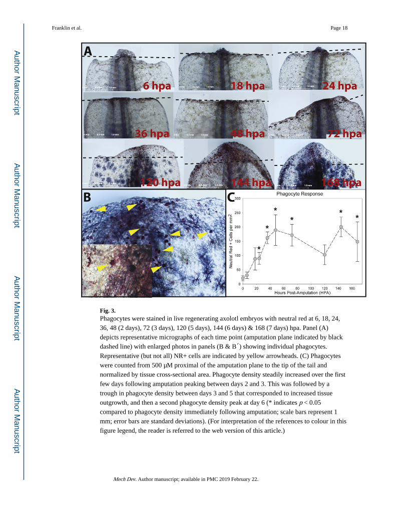
Fig. 3. Phagocytes were stained in live regenerating axolotl embryos with neutral red at 6, 18, 24,
36, 48 (2 days), 72 (3 days), 120 (5 days), 144 (6 days) & 168 (7 days) hpa. Panel (A)
depicts representative micrographs of each time point (amputation plane indicated by black
dashed line) with enlarged photos in panels (B & B′) showing individual phagocytes.
Representative (but not all) NR+ cells are indicated by yellow arrowheads. (C) Phagocytes
were counted from 500 μM proximal of the amputation plane to the tip of the tail and
normalized by tissue cross-sectional area. Phagocyte density steadily increased over the first
few days following amputation peaking between days 2 and 3. This was followed by a
trough in phagocyte density between days 3 and 5 that corresponded to increased tissue
outgrowth, and then a second phagocyte density peak at day 6 (* indicates p < 0.05
compared to phagocyte density immediately following amputation; scale bars represent 1
mm; error bars are standard deviations). (For interpretation of the references to colour in this
figure legend, the reader is referred to the web version of this article.)
Franklin et al. Page 18
Mech Dev. Author manuscript; available in PMC 2019 February 22.
Author M
anuscriptA
uthor Manuscript
Author M
anuscriptA
uthor Manuscript

Fig. 4. Phagocytes were stained in live animals with neutral red at 3 and 6 dpa. Panel (A) depicts
representative micrographs of all time points and treatments (amputation plane indicated by
black dashed line). (B) 4AP inhibits phagocyte activation and recruitment to the wound site
at concentrations of either 5 or 25 μM. This response is concentration dependent at 6dpa but
not 3dpa (* indicates p < 0.05 compared to control and ^ indicates p < 0.05 compared
animals treated with 5 μM 4AP; scale bars represent 1 mm; error bars are standard
deviations).
Franklin et al. Page 19
Mech Dev. Author manuscript; available in PMC 2019 February 22.
Author M
anuscriptA
uthor Manuscript
Author M
anuscriptA
uthor Manuscript

Fig. 5. Phagocytes were labeled by IP injection of Fluorosome®-DiI 24 h prior to amputation and
co-injected with either Encapsome® as a vehicle control (B-B″; n = 10), Clodrosome® for
macrophage depletion (C-C″, n = 15) and a final group co-injected with Encapsome® and
then incubated with 25 μM 4-AP following amputation for KV channel blockade (D-D”; n =
14). (* indicates p < 0.05 compared to control; error bars are standard deviations) Both
macrophage depletion prior to amputation (C-C″) and KV channel blockade following
amputation (D-D″) reduced the number of DiI+ cells at the wound site.
Franklin et al. Page 20
Mech Dev. Author manuscript; available in PMC 2019 February 22.
Author M
anuscriptA
uthor Manuscript
Author M
anuscriptA
uthor Manuscript

Fig. 6. Compared to controls (n = 4) (A-A″), the proliferative response to amputation was reduced
by broad scale chloride channel blockade with either DIDS (n = 4) or DNDS (n = 5) (C) as
well as anoctamin-1 channel blockade with T16a(inh)-A01 (n = 5) (B-B″)) but not when
animals were incubated in chloride free medium (n = 5) (C) (amputation plane indicated by
white dashed line). Taken together these data suggest that chloride channel signaling is a
critical step in the mechanism(s) driving proliferation in response to amputation. (* indicates
p < 0.05 compared to control; error bars are standard deviations) (D&E) Neither DIDS nor
T16a(inh)-A01 had an effect on proliferation in spinal cord (SC) or epidermal (ED) tissues.
In mesenchymal tissues (MM) directly underlying the wound epidermis and within 500 μm
of the amputation plane, proliferation was reduced from 46.6 ± 0.7% in control animals to
either 20.0 ± 3.5% (DIDS) or 23.0 ± 2.9% (T16a(inh)-A01) in treated animals (* indicates p < 0.05 compared to control of same tissue type; error bars are standard deviations).
Franklin et al. Page 21
Mech Dev. Author manuscript; available in PMC 2019 February 22.
Author M
anuscriptA
uthor Manuscript
Author M
anuscriptA
uthor Manuscript

Fig. 7. Apoptosis was assessed following amputation in 3 dpa animals and compared to apoptotic
rates in tissue from un-amputated axolotl tails. Tissues were cut into transverse sections and
then divided into groups consisting of sections of either from the tail tip to 100 μM distal of
the amputation plane and a second group consisting of tissue from 150 to 250 μM distal of
the amputation plane. No differenced were observed in the latter group but the former
exhibited similar increases in apoptotic cells in both the control 3 dpa animals and 3 dpa
animals treated with the Ano1 inhibitor (* indicates p < 0.05 compared to un-amputated tail
tissue).
Franklin et al. Page 22
Mech Dev. Author manuscript; available in PMC 2019 February 22.
Author M
anuscriptA
uthor Manuscript
Author M
anuscriptA
uthor Manuscript
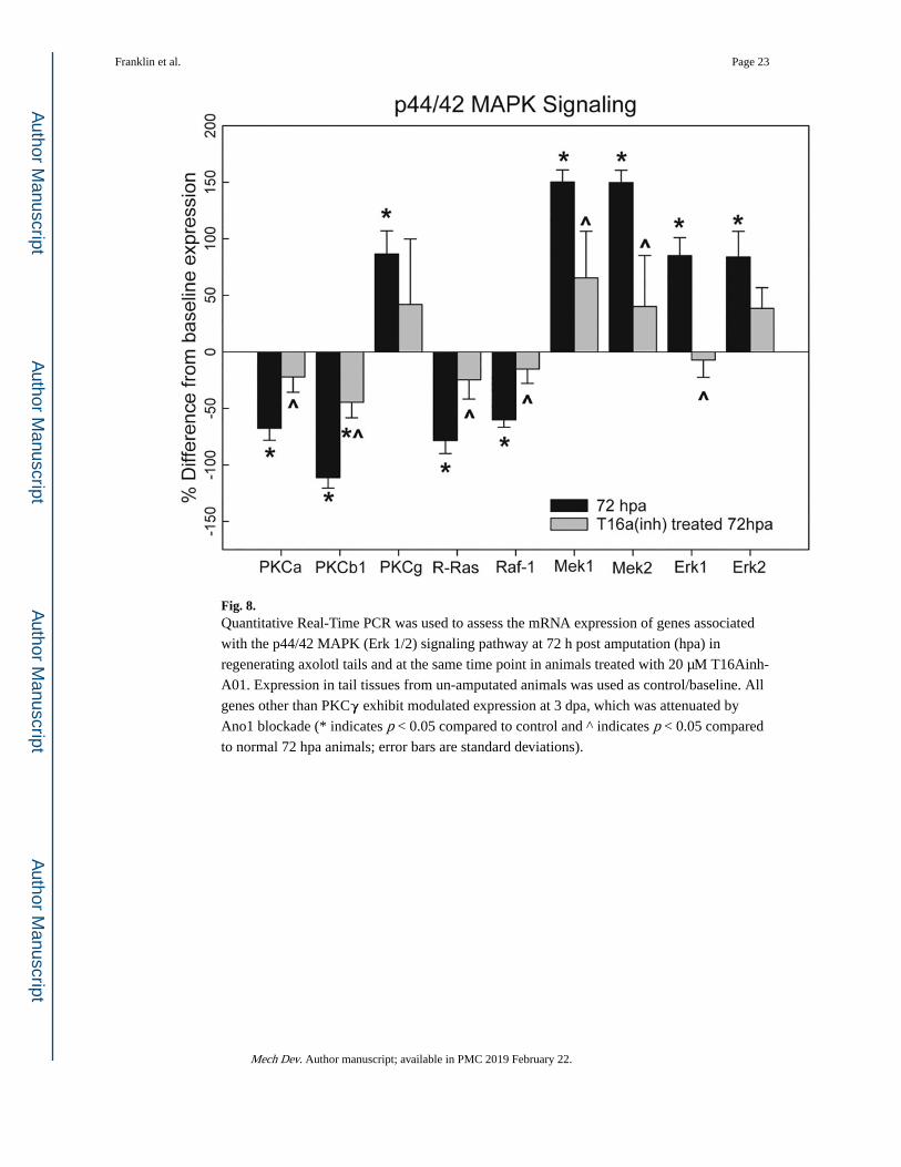
Fig. 8. Quantitative Real-Time PCR was used to assess the mRNA expression of genes associated
with the p44/42 MAPK (Erk 1/2) signaling pathway at 72 h post amputation (hpa) in
regenerating axolotl tails and at the same time point in animals treated with 20 μM T16Ainh-
A01. Expression in tail tissues from un-amputated animals was used as control/baseline. All
genes other than PKCγ exhibit modulated expression at 3 dpa, which was attenuated by
Ano1 blockade (* indicates p < 0.05 compared to control and ^ indicates p < 0.05 compared
to normal 72 hpa animals; error bars are standard deviations).
Franklin et al. Page 23
Mech Dev. Author manuscript; available in PMC 2019 February 22.
Author M
anuscriptA
uthor Manuscript
Author M
anuscriptA
uthor Manuscript

Fig. 9. A comprehensive screen of ion channel blockers was used to identify specific channels
involved in critical cellular processes of growth and regeneration such as proliferation,
migration and differentiation. Drugs that did not affect regeneration are depicted in green,
drugs that resulted in partial regeneration are depicted in yellow, drugs that completely
inhibited regeneration (for any of the concentrations tested) are depicted in red and drugs
that were lethal at all concentrations or caused systemic toxicity (identified by general
atrophy, lethargy and/or tissue degeneration) are depicted in black. (For interpretation of the
references to colour in this figure legend, the reader is referred to the web version of this
article.)
Franklin et al. Page 24
Mech Dev. Author manuscript; available in PMC 2019 February 22.
Author M
anuscriptA
uthor Manuscript
Author M
anuscriptA
uthor Manuscript
Author M
anuscriptA
uthor Manuscript
Author M
anuscriptA
uthor Manuscript
Franklin et al. Page 25
Tab
le 1
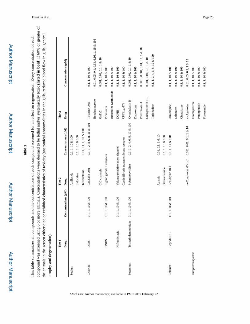
Thi
s ta
ble
sum
mar
izes
all
com
poun
ds a
nd th
e co
ncen
trat
ions
of
each
com
poun
d sc
reen
ed f
or a
n ef
fect
on
rege
nera
tion.
Eve
ry c
once
ntra
tion
of e
ach
com
poun
d w
as s
cree
ned
usin
g 6
or m
ore
anim
als.
Con
cent
ratio
ns w
ere
deem
ed to
be
leth
al a
nd/o
r sy
stem
ical
ly to
xic
(lis
ted
in b
old)
if 6
0% o
r gr
eate
r of
the
anim
als
in th
e sc
reen
eith
er d
ied
or e
xhib
ited
char
acte
rist
ics
of to
xici
ty (
anat
omic
al a
bnor
mal
ities
in th
e gi
lls, r
educ
ed b
lood
flo
w in
gill
s, g
ener
al
atro
phy
and
dege
nera
tion)
.
Tie
r 1
Tie
r 2
Tie
r 3
Dru
gC
once
ntra
tion
s (μ
M)
Dru
gC
once
ntra
tion
s (μ
M)
Dru
gC
once
ntra
tion
s (μ
M)
Sodi
umA
milo
ride
0.1,
1, 1
0 &
100
Lid
ocai
ne0.
1, 1
, 10
& 1
00
Tetr
odot
oxin
0.01
, 0.1
, 1, 1
0 &
100
Chl
orid
eD
IDS
0.1,
1, 1
0 &
100
CaC
Cin
h-A
010.
1, 1
, 2, 4
, 6, 8
, 10
& 1
00T
16A
inh-
A01
0.1,
1, 1
0 &
100
Ben
zbro
mar
one
0.01
, 0.0
5, 0
.1, 0
.33,
0.6
6, 1
, 10
& 1
00
ClC
cha
nnel
sG
aTx2
0.00
1, 0
.01,
0.1
, 1 &
10
DN
DS
0.1,
1, 1
0 &
100
Lig
and
gate
d C
l cha
nnel
sPi
crot
oxin
0.1,
1, 1
0 &
100
Bic
ucul
ine
Met
hiod
ide
0.1,
1, 1
0 &
100
Nif
lum
ic a
cid
0.1,
1, 1
0 &
100
Vol
ume-
sens
itive
ani
on c
hann
elD
CPI
B0.
1, 1
, 10
& 1
00
Cys
tic f
ibro
sis
tran
smem
bran
e re
cept
orC
FTR
inh-
172
0.1,
1, 1
0 &
100
Pota
ssiu
mTe
trae
thyl
amm
oniu
m0.
1, 1
, 10
& 1
004-
Am
inop
yrid
ine
0.1,
1, 2
, 4, 6
, 8, 1
0 &
100
Cyt
ocha
lasi
n B
0.00
1, 0
.01,
0.1
, 1 &
10
Dap
oxet
ine
0.1,
1, 1
0 &
100
Phri
xoto
xin-
10.
0001
, 0.0
01, 0
.01,
0.1
, 1 &
10
Gua
ngxi
toxi
n-1E
0.00
1, 0
.01,
0.1
, 1 &
10
Terf
enad
ine
0.1,
1, 2
, 4, 6
, 8, 1
0 &
100
Apa
min
0.01
, 0.1
, 1 &
10
Glib
encl
amid
e0.
1, 1
, 10
& 1
00
Cal
cium
Bep
ridi
l HC
l0.
1, 1
, 10
& 1
00B
enid
ipin
e H
Cl
0.1,
1, 1
0 &
100
Am
lodi
pine
0.1,
1, 1
0 &
100
Dilt
iaze
m0.
1, 1
, 10
& 1
00
Cin
nari
zine
0.1,
1, 1
0 &
100
ω-C
onot
oxin
MV
IIC
0.00
1, 0
.01,
0.1
, 1 &
10
ω-A
gato
xin
0.01
, 0.0
5, 0
.1, 1
& 1
0
Pum
ps/tr
ansp
orte
rsE
som
epra
zole
0.1,
1, 1
0 &
100
Pant
opra
zole
0.1,
1, 1
0 &
100
Furo
sem
ide
0.1,
1, 1
0 &
100
Mech Dev. Author manuscript; available in PMC 2019 February 22.

Author M
anuscriptA
uthor Manuscript
Author M
anuscriptA
uthor Manuscript
Franklin et al. Page 26
Tie
r 1
Tie
r 2
Tie
r 3
Dru
gC
once
ntra
tion
s (μ
M)
Dru
gC
once
ntra
tion
s (μ
M)
Dru
gC
once
ntra
tion
s (μ
M)
Oua
bain
0.1,
1, 1
0, 5
0 &
100
tBuB
HQ
0.1,
0.5
, 1, 2
, 4, 6
, 8, 1
0 &
100
Mech Dev. Author manuscript; available in PMC 2019 February 22.

Author M
anuscriptA
uthor Manuscript
Author M
anuscriptA
uthor Manuscript
Franklin et al. Page 27
Table 2
This table summarizes the primers used to measure gene transcription by quantitative real-time PCR.
Gene Forward primer Reverse primer
GAPDH ACGTCTCTGTGGTTGACTTG TTCCCTTCATTGGTCCATCAG
PKC-γ GGCAGTCGTGAGATGAGTTT ACCGATACAAGCTGAGTGAAG
PKC-α CGTAGAATGCACGATGGTAGAA TCCTCAGTTTGGAAGCAAGAA
PKC-β CGCATGAAGCTATCCGACTT CATAGAGCTCGTCAGTACCTTTC
RRAS GTCCACATTAAGCCGGATCT CCTACATAGACAGGTGCCAAA
RAF1 AGGAGACCAAGTTTCAGATGTT GTCCCTGTGGATGATGTTCTT
MAPKK1 AGCTCCTGTGAAGCGTATTC CCTAGATCTGCCCTGCATTT
MAPKK2 GAGGAAGGGAAACCGAACATAA CTTAGCTCGTCTACAGCCAATC
MAPK1/ERK2 CGGGCACCAGAGATAATGTT GGAAGATGGGTCTGTTAGATAGC
MAPK3/ERK1 CGCATTGGATCTGCTGGATAA GGTCGTAGTACTGTTCCAGGTA
Mech Dev. Author manuscript; available in PMC 2019 February 22.