iop institute of physics newsletter medical physics group ... · 9th international conference on...
TRANSCRIPT

1
The Medical Physics Group Postgraduate Dissertation PrizeThe Medical Physics Group (MPG) of the Institute of Physics is pleased to announce a new student prize, to be awarded annually for the best master’s level dissertation, on a topic relevant to medical physics and completed by a student member of the MPG during the previous calendar year. Any dissertation submitted as a part of an MSc programme of study at a university in the UK and Ireland is eligible for consideration. The award, which has a monetary value of £250, will be made to the dissertation that, in the opinion of the MPG Committee, has made the greatest contribution to advancement of medical physics.
The closing date for receipt of applications is January 31st of each year for dissertations successfully examined in the previous calendar year (irrespective of submission date). For this year’s inaugural prize, however, the application deadline will be 31st October 2013.
Nominations are invited from supervisors or similar persons (e.g. head of department, programme director) in the form of a 2-page (typed) summary of the dissertation and a letter of support from the proposer detailing the merits of the dissertation. Please send nominations by e-mail to the secretary of the MPG committee ([email protected]) who will be happy to provide any further information required.
Institute of Physics Medical Physics Group
IOP NEWSLETTER Summer 2013
ContentsThe Medical Physics Group Postgraduate Dissertation Prize 1
Meeting reports 1– 20th Annual Meeting & Exhibition of the International Society for
Magnetic Resonance in Medicine– 9th International Conference on the Scientific and Clinical Applications of Magnetic Carriers– Mayneord-Philips Summer School 2012 - Cardiac Imaging and Modelling: Principles, Methods
and Clinical Relevance– Medical Physics and Engineering Conference– Physics in Healthcare– International Society for Therapeutic Ultrasound
Educational resource 10
Journal spotlight 11
Upcoming meeting 12
Meeting reports20th Annual Meeting & Exhibition of the International Society for Magnetic Resonance in Medicine Melbourne, Australia, 5-11th May 2012
In May last year I attended the 20th Annual Meeting & Exhibition of the International Society for Magnetic Resonance in Medicine, organised in Melbourne, Australia. This is one of the biggest international conferences regarding MRI, gathering together clinicians, physicists, engineers, biochemists and technologists to discuss current research topics, exchange knowledge and build the connection between scientific achievements and clinical applications.
The conference started with the Educational Weekend filled with the lectures given by the experts on various MRI topics, designed to provide insight into the theoretical basis. This was very useful for people new to the field or looking for an introduction to the particular subjects in order to extend their research interests.
The main part of the conference was five days long and contained hundreds of oral presentations and traditional and electronic posters, grouped into separate sessions according to their subjects. Hearing about the most current achievements related to my own research was extremely important as it let me look at my work from a different perspective and make me think about new ways of studying the problems I am working on. Presentations regarding more distant subjects allowed me to consider new possible directions of development and also to learn various methodological approaches which might be useful in my research.
Presenting the results of my recent research as two talks and two e-posters gave me a great opportunity to discuss my work with other researchers from the field and take suggestions on how I can improve it. It was also a chance to make the connections which can lead to benefits in the future.
The “outback party” with Australian animals such as wombats, koalas, kangaroos and snakes was an amazing way to finish the conference and an unforgettable experience. I would definitely go back to Australia and especially to Melbourne.
Anna BlazejewskaSir Peter Mansfield Magnetic Resonance Centre, University of Nottingham, UK

2
IOP y Medical Physics Group
20th Annual Meeting & Exhibition of the International Society for Magnetic Resonance in Medicine Melbourne, Australia, 5-11th May 2012
The 2012 International Society of Magnetic Resonance in Medicine (ISMRM) annual meeting held in Melbourne Australia was very well attended by international delegates, and the presentations and discussions were of a very high standard. The field of magnetic resonance imaging (MRI) research is extremely multi-disciplinary, involving physicists, engineers, chemists, biologists and clinicians, and this diversity was well represented at the conference. This conference provides a unique opportunity to see the diversity of research that is occurring in the field of MRI development, and provide a broader context for one’s research relative to the vast array of research presented.
One highlight from the conference was the key-note lecture, the “Lauterbur Lecture”, given on the topic of “MRI: from science to society” by Vivian Lee. This lecture discussed the importance of MRI research, particularly in the cardiovascular field, to the understanding and treatment of major diseases. During the scientific meeting, I was most interested in new developments in cardiovascular imaging methodology, and there were a number of sessions dedicated to this topic. Of particular relevance to my PhD topic were the oral and poster presentations in the “Myocardial Perfusion” sessions. These sessions presented many novel technical developments for the measurement of myocardial perfusion, as well as novel applications of the techniques to different cardiovascular diseases. As a physicist, it is interesting to see how such techniques are translated from
a research interest to a clinically applicable diagnostic method. Another session of great interest was the “Flow” oral presentation session which presented research in the popular area of 3- and 4-dimensional phase contrast MRI. This technique uses the properties of moving protons in the MRI signal to generate stunning movies allowing the visualisation of bulk blood flow through the great vessels and valves. I was particularly impressed in the “MR-guided Intravascular & Percutaneous Interventions” oral presentation session with how the field of MR-guided interventions has grown over the past years to develop the real time imaging techniques and the MR-visible devices necessary to successfully perform MR-guided procedures. These techniques have the potential to replace X-Ray guidance to reduce ionising radiation exposure, particularly in children.
The above are a few specific examples of the many interesting sessions covering several areas of application. As well, the displays in the exhibition hall and the corporate-sponsored sessions were also of value as they provided the opportunity to learn about recent developments made by hardware and software manufacturers. In addition to the scientific meeting, there were two days of educational training sessions which provided an excellent overview of current MRI techniques and ongoing research.
The meeting was useful for me academically, both as a learning experience and as an opportunity to disseminate my thesis research results. I presented three posters at the meeting, all receiving positive feedback and interesting questions. In addition, since I was approaching the end of my PhD, the conference provided an opportunity to look at the broad range of potential research opportunities in the field of MRI physics and methods developments which I may want to explore in my career. It also provided the opportunity to speak to a number of researchers to investigate career options. I am grateful to have had the chance to meet, and learn from, leading researchers in my field.
Overall, I found the ISMRM annual meeting a very valuable experience and I am grateful for the funding that the Institute of Physics provided to assist my attendance.
Adrienne Campbell-WashburnCentre for Advanced Biomedical Imaging, University College London, UK
Meeting reports

3
IOP y Medical Physics Group
9th International Conference on the Scientific and Clinical Applications of Magnetic Carriers Minneapolis, Minnesota, 22-26th May 2012
The magnetic carrier’s series of meetings gives research scientists and industrial suppliers the chance to showcase their latest developments in the field of magnetic carriers. Advances in all fields of magnetic carriers were presented, including diagnostics, therapy, analytics, separation chemistry, labelling, actuation and synthesis.
Urs Hafeli (the conference organiser) opened the meeting with an overview of the highlights. In the field of magnetic nanoparticles (MNPs) and their application, a further 4000 journal citations and 600 patents have been added since the last meeting two years ago. New directions in research were presented including microwave (MW) chemistry, which has become a popular medium of heating with many research teams beginning to study MW effects in syntheses. Research into magnetic catalysts and surfactants (for clearing oil spillages) is proving useful, whilst toxicological studies are allowing the behaviour of MNPs in the body to be better understood. Specifically in biomedicine, MNPs have been successfully shown as agents to improve neovascularisation and reduce atherosclerosis.
In an invited talk, Kannan Krishnan described the fundamentals behind magnetic particle imaging (MPI) and the requirements for good MPI agents. A good imaging agent will exhibit a good signal with high magnetic saturation at low applied magnetic field; the frequency required for a signal is dependent on the coercivity of the particles. Brownian motion should be minimised so that the applied field can be aligned with the anisotropy of the MNPs. To conclude, emphasis was placed upon the requirement for better phase transfer methods, improved control of particle anisotropy and increased size monodispersity.
A particularly active discussion of the fundamentals behind magnetic hyperthermia took place and there appears to still be some confusion around this field, indicating that there is still work to be done in elucidating
the mechanism of heating. It also became clear that the practical method (orientation of particles within field) exhibits high variability and there is a need for standardisation in order for results between teams to be comparable.
In the field of diagnostics, sub pM detection of PSA (for detection of prostate cancers), voice recognition and magnetic tweezers are all showing promise; Tim St Pierre demonstrated the use of MRI in the quantitative analysis of Fe content in patients with sickle cell disease and Steven Saville showed that the relaxometry time of MNP solutions could be adjusted by forming chains.
In other areas, MNPs are finding application as magnetic bar codes; a mini NMR system shows potential with high moment MNPs, a new analytical technique FFF-SAXS allows measurement of core size as opposed to hydrodynamic size (often presented as measurement from photoelectron spectroscopy). Magnetic separation is a growing field with several demonstrations and discussions of systems for
fractionation of magnetic microspheres, cells, proteins and MNPs. In the field of flow chemistry, measurement of flow rates were demonstrated through the use of giant magneto resistance – magnetic cilia disrupt a magnetic field to give an electronic signal.
Personally I found the conference useful on several levels. Lectures showcasing research into the synthesis of MNPs gave me new insights and ideas for my own research into MNP synthesis on a very immediate timescale. One aspect of my research investigates the phase transfer of MNPs from organic to aqueous phase and I was inspired by a novel method (Mingli Peng) involving the chemical change of a stabiliser to make it stable in solution as opposed to via chemical addition or stabiliser exchange. Before attending the conference I attended a careers consultation at the RSC, which made me realise how passionate I am about my research and the potential abroad to widen my capacity. At the conference I really
Meeting reports
...continued on p4

4
IOP y Medical Physics Group
9th International Conference on the Scientific and Clinical Applications of Magnetic Carriers Minneapolis, Minnesota, 22-26th May 2012
The 9th International Conference on the Scientific and Clinical Applications of Magnetic Carriers, held in Minneapolis from 21–26th May 2012, attracted participants from across the scientific community working on the use of magnetic materials for biological applications for a week of talks, discussions and debates. Professor Jian Ping Wang, the host of the conference, and his lab, organised a great program on the University of Minnesota campus.
The conference was focused on the latest findings on the synthesis and use of magnetic materials for a variety of applications ranging from immunoassays to water purification or magnetic resonance imaging (MRI). Speakers such as Professor Tim St. Piere, a major figure in MRI and Professor Kannan M. Krishan, one of the pioneers working on magnetic particle imaging (MPI) were present. Key points such as the characteristics needed from the contrast agents or the new diagnostic techniques on the field were discussed, which was of great value for any research group working on the development of new contrast agents. Moreover, for the first time in this conference series, a debate was held with two seasoned experts:
Professor Robert Ivkov and Professor Carlos Rinaldi, in magnetic hyperthermia. This event permitted not only to directly compare their views but more importantly, it offered the opportunity to the audience to bring their ideas to the open public for discussion, which was a very positive experience for the community. It was possible to point out weak points that need to be addressed and it has given an awareness of the real needs and the direction that the field needs now to move towards.
This conference has been greatly beneficial for my scientific development and career as I was able to interact with other scientists within my research field and find out about the latest advances. Not only has this helped to widen my general scientific knowledge but it has given me the tools to assess some of the research
frontiers that I have been pursuing and it has given me a new and more up-to-date focus to it.
Finally I would like to thank the Medical Physics Group from the Institute of Physics for their financial contribution, which made my attendance to the conference possible.
Cristina Blanco-AndujarThe Davy Faraday Research Laboratories, The Royal Institution of Great Britain, UK
Department of Physics and Astronomy, University College London, UK
took the opportunity to network extensively and make useful contacts with researchers in academia and industry in Australia, Germany and the USA. I hope to continue correspondence with such contacts both on a professional and a personal level.
In my opinion this was a hugely successful conference which inspired and excited me whilst providing useful opportunities for further learning and career progression.
Luke A.W. Green The Davy Faraday Research Laboratories, The Royal Institution of Great Britain, UK
Department of Physics and Astronomy, University College London, UK
...continued from p3
Meeting reports
5
IOP y Medical Physics Group
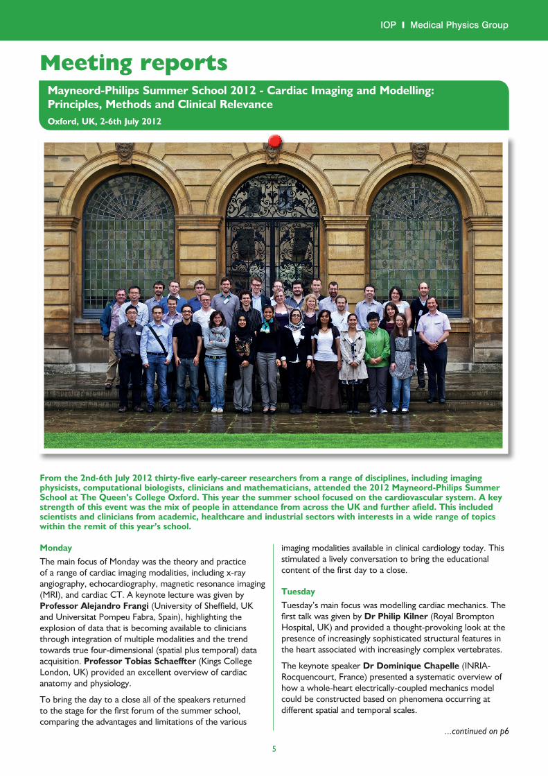
Meeting reportsMayneord-Philips Summer School 2012 - Cardiac Imaging and Modelling: Principles, Methods and Clinical RelevanceOxford, UK, 2-6th July 2012
From the 2nd-6th July 2012 thirty-five early-career researchers from a range of disciplines, including imaging physicists, computational biologists, clinicians and mathematicians, attended the 2012 Mayneord-Philips Summer School at The Queen’s College Oxford. This year the summer school focused on the cardiovascular system. A key strength of this event was the mix of people in attendance from across the UK and further afield. This included scientists and clinicians from academic, healthcare and industrial sectors with interests in a wide range of topics within the remit of this year’s school.
MondayThe main focus of Monday was the theory and practice of a range of cardiac imaging modalities, including x-ray angiography, echocardiography, magnetic resonance imaging (MRI), and cardiac CT. A keynote lecture was given by Professor Alejandro Frangi (University of Sheffield, UK and Universitat Pompeu Fabra, Spain), highlighting the explosion of data that is becoming available to clinicians through integration of multiple modalities and the trend towards true four-dimensional (spatial plus temporal) data acquisition. Professor Tobias Schaeffter (Kings College London, UK) provided an excellent overview of cardiac anatomy and physiology.
To bring the day to a close all of the speakers returned to the stage for the first forum of the summer school, comparing the advantages and limitations of the various
imaging modalities available in clinical cardiology today. This stimulated a lively conversation to bring the educational content of the first day to a close.
TuesdayTuesday’s main focus was modelling cardiac mechanics. The first talk was given by Dr Philip Kilner (Royal Brompton Hospital, UK) and provided a thought-provoking look at the presence of increasingly sophisticated structural features in the heart associated with increasingly complex vertebrates.
The keynote speaker Dr Dominique Chapelle (INRIA-Rocquencourt, France) presented a systematic overview of how a whole-heart electrically-coupled mechanics model could be constructed based on phenomena occurring at different spatial and temporal scales.
...continued on p6
6
IOP y Medical Physics Group
Professor Schaeffter provided an overview of how current imaging techniques can obtain information about the mechanics of the heart. Dr Phillippe Moireau (INRIA-Rocquencourt, France) then talked about estimation techniques for fitting model parameters based on minimising discrepancies between imaging data and cardiac model simulations. A take-home message from this talk was “balancing model complexity with predictivity”.
The cardiac electrophysiology (EP) workshop led by Dr Richard Clayton (University of Sheffield, UK), provided a great introduction and overview of electrical activity in the heart and was a helpful precursor to lectures to follow on Wednesday.
WednesdayThe focus of Wednesday was cardiac electrophysiology. Dr T Alex Quinn (Imperial College London, UK) gave the keynote lecture on different strategies to model the electrophysiology of the heart, providing a history of the development of the models.
He was followed by Dr Sabine Ernst (Royal Brompton Hospital, UK) who explained the clinical protocol of EP studies. Dr Ernst concluded by detailing a wish list of what could further improve EP studies to the audience, hopefully inspiring and motivating future project ideas.
Dr Richard Clayton (University of Sheffield, UK) discussed several different software packages which can be used for EP modelling, providing some tricks and tips for those who are wishing to use these methods in their own work. The last talk of the day was given by Mikael Wallman (University of Oxford, UK) on patient specific EP models. This talk identified the differences between the two sides of modelling - predictive modelling to estimate observable outcomes from a given set of parameters, and the reverse problem of using measured data to estimate potentially unobservable parameters.
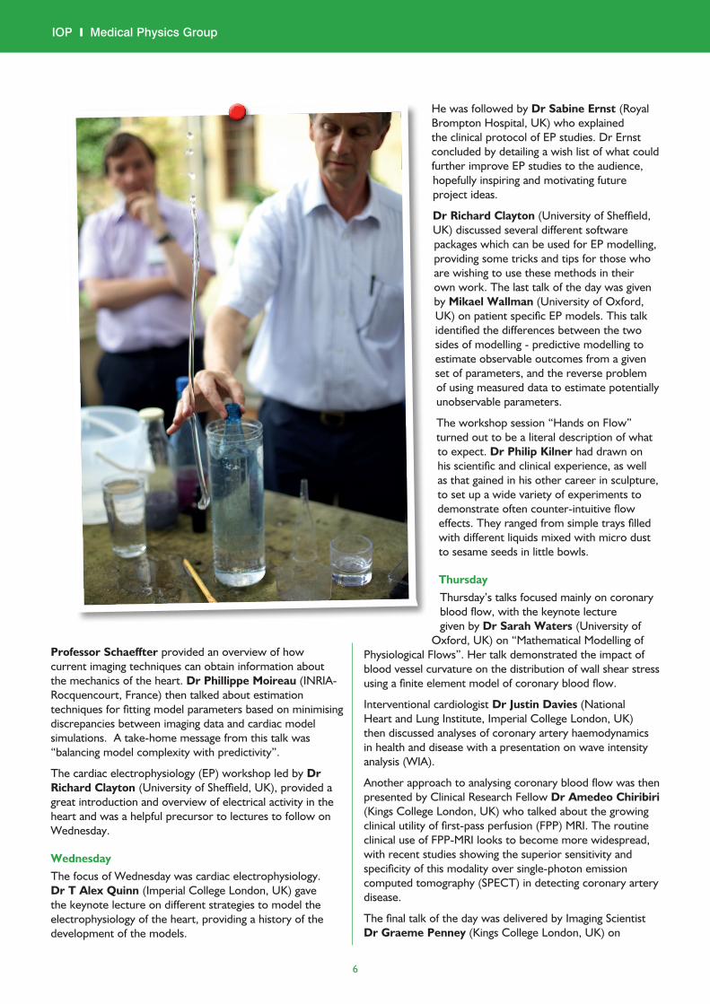
The workshop session “Hands on Flow” turned out to be a literal description of what to expect. Dr Philip Kilner had drawn on his scientific and clinical experience, as well as that gained in his other career in sculpture, to set up a wide variety of experiments to demonstrate often counter-intuitive flow effects. They ranged from simple trays filled with different liquids mixed with micro dust to sesame seeds in little bowls.
ThursdayThursday’s talks focused mainly on coronary blood flow, with the keynote lecture given by Dr Sarah Waters (University of
Oxford, UK) on “Mathematical Modelling of Physiological Flows”. Her talk demonstrated the impact of blood vessel curvature on the distribution of wall shear stress using a finite element model of coronary blood flow.
Interventional cardiologist Dr Justin Davies (National Heart and Lung Institute, Imperial College London, UK) then discussed analyses of coronary artery haemodynamics in health and disease with a presentation on wave intensity analysis (WIA).
Another approach to analysing coronary blood flow was then presented by Clinical Research Fellow Dr Amedeo Chiribiri (Kings College London, UK) who talked about the growing clinical utility of first-pass perfusion (FPP) MRI. The routine clinical use of FPP-MRI looks to become more widespread, with recent studies showing the superior sensitivity and specificity of this modality over single-photon emission computed tomography (SPECT) in detecting coronary artery disease.
The final talk of the day was delivered by Imaging Scientist Dr Graeme Penney (Kings College London, UK) on

7
IOP y Medical Physics Group
methods of image registration to guide clinical interventions and to merge structural and functional information from different imaging modalities. Applications of image registration presented were speckle tracking in cardiac ultrasound to assess cardiac wall motion, registration of anatomical MRI images to real-time invasive angiography for image-guided interventions, and 3D flow quantification from Doppler ultrasound.
On Thursday afternoon a visit to the Oxford Centre for Clinical Magnetic Resonance at the John Radcliffe Hospital took place. An overview of different cardiac pathologies which are routinely diagnosed and assessed with MRI at this hospital was followed by visits to a 3T and newly acquired 7T MRI system.
FridayFriday’s talks addressed the current challenges and unmet needs facing the field of cardiac modelling, with the keynote lecture given by Professor Peter Kohl (University of Oxford, UK and Imperial College London, UK) introducing some concepts of the philosophy behind multi-scale modelling. A vision of how computational modelling and systems biology has the capacity to integrate information spanning from gene expression to proteins to cells to tissues to organs and to an organism was given, with reference made to the Virtual Physiological Human Project. Dr Kilner then provided a complimentary perspective on modelling with a talk about the utility of physical models in understanding blood flow.
Dr Ricardo Petraco (National Heart and Lung Institute, Imperial College London, UK) then presented findings that demonstrated the limitations of using luminal narrowing quantified from angiography as a surrogate metric for fractional flow reserve (FFR) in determining the physiological severity of a coronary stenosis.
The final talk of the summer school was given by Professor Nic Smith (Kings College London) on the limitations and applications of computational cardiac models. A major limitation highlighted was the use of data collected on cell models from a range of species to parameterise a model of a single species. This is something that needs to be addressed especially with many research centres now moving cardiac models into a clinical context with the aim of creating personalised patient models.
Friday’s lectures were followed by the final discussion forum of the Summer School, with the speakers on the day forming the panel. One of the major themes to come out of the discussion was the benefit of sharing information and tools between and across research groups and disciplines. Such actions will make for better understanding and clearer communication of the needs and limitations of both modellers and clinicians, helping to facilitate the translational work that we are aiming to produce together.
Student presentationsEach student prepared a poster and a short talk to be presented in sessions throughout the week. These presentations successfully stimulated discussions both within the sessions and less formally throughout the course of the week, and it was this interaction which many students felt to be the most valuable aspect of the school.
A clinician’s perspectiveCardiology trainees were able to gain insights on the technical foundations of cardiac modelling from different perspectives, such as mathematics, physics and engineering. The discussions were particularly useful, as they permitted opinions and personal views to be presented and challenged in a very friendly atmosphere.
Social activitiesThe evenings were spent exploring some of Oxfords many fine pubs and enjoying meals (including the four-course gala dinner on the Thursday), in the grandeur of The Queen’s College dining hall. Sadly the British summer weather scuppered the plans of taking in the sights of Oxford from the water as no-one fancied punting in the pouring rain. There was time to enjoy some of Oxfords indoor attractions though (such as the Museum of Natural History and the Museum of the History of Science) on the Friday afternoon following the morning sessions that brought the summer school to a close.
Closing remarksThe standard of teaching was excellent but more importantly the school brought together a wide range of students, post-docs and senior figures from a range of modalities together and gave them space and time to discuss ideas freely with each other. To any PhD students, trainees or post-docs reading this; if the next Mayneord-Philips Summer School is relevant to you then we can’t recommend it highly enough.
Following this meeting the idea arose to set up an email list to maintain the professional and personal contacts made in Oxford. Dr Richard Walton (Hôpital Xavier-Arnozan, France) has established a database of attendees and their research interests and we hope this will enable us to continue the dialogue we started during the summer school.
David BroadbentTrainee Clinical Scientist, Leeds Teaching Hospitals, UK
Cristoph Kolbistsch, Matthew Sinclair Imaging Sciences and Biomedical Engineering, Kings College London, UK
Ricardo Petraco The International Centre for Circulatory Health and National Heart and Lung Institute, Imperial College London, UK

8
IOP y Medical Physics Group
Medical Physics and Engineering ConferenceOxford, UK, 10-12th September 2012
The inspirational city of Oxford played host to the 2012 Medical Physics and Engineering Conference (MPEC). The Examination Schools provided a stunning venue for the conference, and delegates were afforded the true ‘Oxford Experience’ by staying in one of the three available college accommodations. Not only did this mean that delegates were able to explore the spectacular surroundings of the colleges, but also that the walk home from the pub in the evening was a short one.
Arranged in partnership with the IPEM Biennial Radiotherapy Physics meeting, the BNMS autumn meeting, EFOMP, and the IoP, the conference provided a great opportunity to meet with colleagues, many of whom had travelled from far and wide. A full and varied programme offered talking points for discussions throughout the three days.
The meeting was opened by Dame Fiona Caldicott. The first invited speaker was Professor Sir Muir Gray, CBE, who gave a thought-provoking talk on “The Third Healthcare Revolution”. This was followed by the Woolmer Lecture, in which Professor Lionel Tarassenko drew from his previous experience in Rolls Royce to show that concepts developed for safety-critical systems such as jet engines could be used to improve healthcare. Preventative maintenance and physiology-driven analysis of vital signs, combined with novel monitoring technology, were shown to have a positive effect on patient care. The HPA Lecture, delivered by Professor Sir Roger Penrose, included colourful cats and questions on artificial intelligence and consciousness, leaving
many of us with more than our lunch to digest. The younger members of the audience left for the break with many questions in their heads: What is life all about? How does wave-particle duality link to Schrödinger’s cat? And what is an overhead projector?
A whole range of scientific sessions followed, with sessions in Radiotherapy, Imaging and Engineering running concurrently. Posters were on display throughout the meeting, providing an interesting read between presentation sessions. The conference also provided a perfect opportunity for trainees to find their feet on the presentation stage in a friendly informal environment, with several dedicated trainee sessions throughout the meeting. The organisers further showed their dedication to the development of trainees by including a number of teaching sessions in the programme – although this meant an early start! If the opportunity to finally get your head round the minefield of statistics wasn’t reason enough to get out of bed in the morning, then the option of learning about Image Analysis from the brains behind IQWorks was an alternative option.
There was one session organised by the IoP, under the heading of “Computational Modelling and Analysis”. Work covering a wide range of topics was presented, demonstrating the importance of computational and programming skills in Medical Physics and Engineering. Carpanen et al. showed how their group used MRI datasets to produce a 3D model of the knee to investigate the relationship between partial meniscectomy size and location with corresponding knee joint contact stress during dynamic loading.
Cocker et al. described the scatter model incorporated in the Standard Attenuation Rate post-processing algorithm used in mammography to calculate a normalised image of tissue density, and compared it to experimental results. A verification of the use of RIS data for the purposes of patient dose audits was carried out by IRS Limited, and the results showed a correlation of above 0.85 for RIS/DICOM data. Gregory et al. demonstrated the use of a VB macro to perform a verification check of imported treatment plans between two software systems. Another use in Radiotherapy for bespoke software was presented by Horsfield et al; their group developed a Matlab program to collect and process Dynalogs, which include large amounts of data including planned and actual MLC positions, beam state, and geometrical information. Finally, Budgell et al. described the use of in-house database software employed in their daily linac QC programme, including the benefits and obstacles encountered.
The conference was, of course, rounded off by a well-organised social programme, including a reception in the University Museum of Natural History, and a conference dinner held in the magnificent New College dining room. No scientific meeting would be complete without a trip to the pub; in this case delegates had the added challenge of trying to find it, but it was worth it in the end.
Patrice BurkeDiagnostic Imaging Medical Physicist, Nottingham University Hospitals NHS Trust, UK
Meeting reports

9
IOP y Medical Physics Group
Physics in HealthcareIOP, London, 31st October 2012
An exciting one-day meeting was organised by the Applied Physics and Technology Division of the Institute of Physics, including the Medical Physics Group. Around forty participants, mainly from UK universities but also including representatives from industry and government bodies, gathered to hear talks on a wide range of topics.
A. C. Fisher (University of Liverpool), on behalf of the Academy for Healthcare Science, began the proceedings with a presentation entitled “A new generation of medical physicists: Education, training and career aspirations” in which the far-reaching implications of the Modernising Scientific Careers initiative were discussed. This was followed by a survey of the current status and future prospects of radiation oncology (N. Burnet, University of Cambridge) from a clinician’s perspective. With over 90,000 patients undergoing radiotherapy each year in the UK, the importance and cost-effectiveness of this type of treatment was pointed out. The crucial role of physicists in providing solutions to the problems identified in the clinic was discussed in the context of current developments such as multi-modal imaging and image-guided radiotherapy. The need for a multi-disciplinary approach was also emphasised.
The clinical theme of the morning session continued with further talks on recent technology developments in medical imaging originating in industry (D. Lewis, CERN), the challenge for physics posed by cancer (K. Peach, University of Oxford) and MeV ion beam therapy (K. Kirkby, University of Surrey). The latter work reported the development of a nanobeam capable of irradiating individual cells in culture with precisely counted numbers of ions to sub-micron precision. Such a system allows the study of cellular response to radiation and the way in which DNA is damaged and repaired. Combined response of radiation with chemo-therapeutic agents can also be studied in this way, as can the targeting of individual organelles in order to observe, in real time, the DNA damage as it occurs.
The afternoon session was mainly concerned with physics at cellular and sub-cellular level. G. Reilly (University of Sheffield) discussed the relevance of mechanobiology to regenerative medicine, examining the mechanisms by which cells orientate their matrix under the influence of intrinsic and external mechanical forces. These considerations form the basis for creating mechanically stable tissues for applications in regenerative medicine. F. Ring (University of Glamorgan) then reviewed the clinical applications
of thermal imaging including the use of this technique in peripheral vascular disease, rheumatology and sports medicine. Current developments, e.g. interest in fever detection especially in airport screening during a pandemic threat, were also discussed.
G. Battaglia (University of Sheffield), in a presentation on the physics of drug delivery – which he reassured the audience, had nothing to do with narcotic trafficking! - gave a fascinating account of the revolution taking place in designing target-specific carriers for the active agent. The presentation included an introduction to “molecular bionics”, a combination of chemistry and soft matter physics used to overcome the biological barriers to effective drug delivery. New tools emerging from chemistry, physics, and biology are being used to better engineer new carriers which can be integrated at much earlier stages of drug development and in some cases even guide the drug discovery process itself.
From “good guys” straight to “bad guys”. P. Stoodley (University of Southampton) gave a frightening account of bacterial biofilms infections. Millions of years of adaptation have provided microbes the ability to colonise manufactured surfaces, as well as biological materials such as skin, teeth, and muscle and bone. Biofilm formation protects bacteria from predation and environmental stresses, and in more recent times antibiotics, biocides and the immune system. Although biofilms are often only a few tens of microns thick they present tremendous problems in industry, shipping and human health. Biofilms can cause material degradation (corrosion), reduced heat transfer, increased pressure drop, performance loss and contamination of process streams. Biofilms in water distribution and cooling tower systems can harbour pathogens with important consequences for public health. In medicine, biofilms growing on artificial implants can result in chronic infections which can be highly tolerant to antibiotics and difficult to diagnose through imaging or
Meeting reports
“lively discussions and exchanges of ideas took place at every opportunity during the meeting and most of us left much better informed and highly inspired”
...continued on p10

10
IOP y Medical Physics Group
Educational resourceSINAPSE (The Scottish Imaging Network: a Platform for Scientific Excellence, http://www.sinapse.ac.uk/) is a network drawing on the expertise of six of Scotland Universities (Aberdeen, Dundee, Edinburgh, Glasgow, St. Andrews and Stirling) along with the NHS associations of each. It supports and funds research on development in all medical imaging specialities and adds strength by pooling the combined resources of the separate establishments.
The group have recently developed an on line program of interactive tutorials for the different modalities (https://www.sinapsecpd.org/). These modules are aimed at the non specialist medical worker or public including the apprehensive patient.
The modules are reasonably and individually priced at £9.95 + VAT for 12 months access. They cover: MRI, PET and SPECT, CT, ultrasound and electrophysiology. At the end of each module is an assessment and on successful completion a certificate can be issued for CPD records.
Geo A CornerNinewells Hospital, Dundee
International Society for Therapeutic UltrasoundShanghai, China, 12-15th May 2013
High Intensity Focused Ultrasound (HIFU) or ultrasounds hyperthermia as it was once called has been of interest for very many years. Therapy using ultrasound predates imaging although issues particularly with dosimetry have limited its uptake. The main country of development has been China with significant interest from the USA. Apart from a few centres there has been very little application in the UK.
Advantages are its non-ionising nature and the fact it can be combined additively with other therapies but there remain many challenges. The fact that China is such a centre of activity in this field made ISTU 2013 organised by the International Society for Therapy Ultrasound in Shanghai particularly interesting. As I was already going to Shanghai to teach at the East China University of Science and Technology I was able to attend.
ISTU is a fairly small conference with a tight knit community. As a developing modality there is much exciting work to do and most of the founders of the society are still active and attend. The delegates were mostly from China and USA but with representation from all over the world. Papers were spread over 3 days with parallel sessions running for most of the time. The
venue was the Regal International East Asia Hotel, one of Shanghai’s leading hotels and there was organisational support from Shanghai Jiao Tong University.
There were 12 invited talks, including Gail ter Haar delivering the Fry Lecture. There was a small poster session (~ 50) and the meeting was surprisingly well supported by manufacturers of components as well as complete systems. Many recurring issues continue to be developed such as dosimetry, bone sparing, and organ movement. Others are seeing accelerating development, particularly non thermal effects often combined with bubbles using cavitation or targeted drugs. The clinical studies session concentrated on liver, uterine fibroids and immune response. Some novel sources especially for dedicated sites were shown.
The proceedings of the conference will be produced later in the year. ISTU continues to be a friendly and vibrate conference for those working in this still developing field. In 2014 the conference will be held in Las Vegas 2–5th April.
Geo A CornerNinewells Hospital, Dundee
Meeting reports
conventional clinical culturing. The speaker examined the challenges associated with preventing, detecting, and treating biofilm infections with illustrative clinical case studies.
The final talk of the day was a whistle-stop tour of the history of medical imaging “from Bertha’s hand to CT and MRI combinations” presented by B. Edwards (The Gordon Museum of Pathology, King’s College London). It reminded us all what a long way medical physics has come in 118 years since Roentgen noticed that mysterious glow in his laboratory.
In addition to the oral presentations, ten posters on diverse topics from mathematical modelling and numerical analysis to technological advances in radiation detectors and optics were on display during the meeting. Lively discussions and exchanges of ideas took place at every opportunity during the meeting and most of us left much better informed and highly inspired. A very successful meeting indeed!
Dareyoush RassiCollege of Human and Health Sciences, Swansea Univeristy
...continued from p9

11
IOP y Medical Physics Group
Journal spotlightPhysica Medica
Background Physica Medica: European Journal of Medical Physics (www.physicamedica.com) is the official journal of the Associazione Italiana di Fisica Medica (AIFM), the European Federation of Organisations for Medical Physics (EFOMP), the Irish Association of Physicists in Medicine (IAMP), and the Société Française de Physique Médicale (SFPM). The Journal has its roots in Europe but attracts submissions and readers from around the world. The continuing increase in high quality submissions over the last few years has allowed the Journal to increase its publication frequency from 4 to 6 issues per year in 2013. Each issue includes review and research articles, book reviews, technical notes and correspondence. The Journal also publishes EFOMP guidelines and policy statements. Papers in the first edition of each volume are made freely available for the course of the year.
ScopeThe Journal provides an international forum for research and reviews on the following main topics:
• Medical Imaging• Radiation Therapy• Radiation Protection• Measuring Systems and Signal
Processing• Education and training in Medical
Physics• Professional issues in Medical Physics.
Contributions on other topics related to applications of Physics to Biology and Medicine and in particular related to new emerging fields such as Molecular Imaging, Hadron therapy, System biology, Nanoparticles and Nanotechnologies are encouraged.
Recent highlights The top 5 most cited articles from the last two years are:
Accuracy of real-time MR temperature mapping in the brain: A comparison of fast sequences, Kickhefel et al. (2010) Phys Med, 26 (4), 192-201. A comparison was made of three fast imaging sequences for real-time MR thermography: gradient echo, segmented echo planar imaging (EPI) and single shot EPI, each mapping the temperature using the proton resonant frequency technique. The single shot EPI approach was found to produce the best high temperature stability, with high temporal and spatial resolution, and was found to be fast, robust and accurate enough for monitoring temperature during thermal therapy.
CT iterative reconstruction in image space: A phantom study, Ghetti et al. (2012) Phys Med, 28 (2), 161-165. This paper addresses the use of iterative reconstruction methods for clinical X-ray CT imaging, widely used for SPECT/PET but not so for CT. This phantom study focused on evaluating image quality metrics such as image noise, low contrast resolution, CT number linearity and accuracy, transverse and z-axis spatial resolution. The iterative reconstruction process was found to preserve spatial resolution, CT number accuracy and linearity, and to decrease image noise. Its potential use in dose reduction while maintaining image quality was highlighted.
Iterative reconstruction methods in X-ray CT, Beister et al. (2012) Phys Med, 28 (2), 94-108. This very interesting review article provides an excellent overview of the field of iterative reconstruction (IR) applied to X-ray CT imaging. Increases in computing power and a drive towards lower ionising radiation doses is motivating progress in this direction. The article provides an overview of the main IR algorithmic concepts in use today, with some
worked-through practical examples, and concludes with thoughts on advantages and disadvantages of IR methods.
Radiochromic film dosimetry: Past, present, and future, Devic (2011) Phys Med, 27 (3), 122-134.This comprehensive review article summarises developments in GAFCHROMICTM film models for use in dosimetric assessment in the field of radiation therapy. Film dosimetry is used for treatment verification and quality assurance – this review summarises developments of these models through improvements in their sensitivity and uniformity as 2-dimensional detectors. Their most important characteristics as well as their limitations are discussed, while new application areas are also covered.
Analytical theory for the fluence, planar fluence, energy fluence, planar energy fluence and absorbed dose of primary particles and their fragments in broad therapeutic light ion beams, Kempe and Brahme (2010) Phys Med, 26 (1), 6-16. In this analytical paper, an investigation into the main mechanisms behind the build-up of absorbed dose and fluence of primary particles and fragments in 7Li and 12C particle beams was carried out, with good agreement between the analytical results obtained and Monte Carlo simulations and experimental data. The analytic expressions derived take into account nuclear interactions, energy losses, range straggling and multiple scattering, and may have application for fast analytical calculations of quantities like mean energy, fluence, energy fluence, absorbed dose, and LET.
Galileo Galilei Award This award is given by the Editors of the Journal every second year to the best paper published in the previous two years. The winner of the award for 2011–2012 was:
CT iterative reconstruction in image space: A phantom study, Ghetti et al. (2012) Phys Med, 28 (2), 161-165.The Journal is indexed by all the major indexing services including Medline / PubMed, Science Citation Index and Scopus.
...continued on p12

12
IOP y Medical Physics Group
Committee
Dimitra Darambara (chair) London [email protected]
George Corner Dundee
David Eaton (secretary and co-newsletter) London [email protected]
Seán Cournane Dublin
Andrew Fagan Dublin
Karen Hampson (co-newsletter) Bradford
Rollo Moore London
Dareyoush Rassi (treasurer) Swansea
Gemma Roberts Edinburgh
Martin Robinson York
Nick Stone* Exeter
*representing the Institute of Physics and Engineering in Medicine (www.ipem.ac.uk)
This newsletter is also available on the web and in larger print sizes
The contents of this newsletter do not necessarily represent the views or policies of the Institute of Physics, except where explicitly stated.
The Institute of Physics, 76 Portland Place, W1B 1NT, UK.
Tel: 020 7470 4800 Fax: 020 7470 4848
Upcoming meetingWhat is on the horizon? – New techniques at the International Conference on Medical Physics
Brighton International Centre, UKWednesday 4th September 20139am-12pm
http://www.icmp2013.org/
A multidisciplinary session organised by the Medical Physics Group at the annual medical physics meeting for the UK. This year the conference sees physicists from around the world gather for the 50th anniversary of the foundation of the International Organisation for Medical Physics (IOMP), and the UK conference joins with the triennial ICMP, so expect a big crowd.
Speakers in this session include:• MikePartridge(Oxford)–Futureofimagingforradiotherapy• KarenHampson(Bradford)–Adaptiveopticsinvisionresearch• ChrisDunsby(London)–Fluorescencespectroscopy• DimitraDarambara(London)–Multi-spectralx-rayimaging• FengWu(Oxford)–Therapeuticultrasound
Notable figures for 2012/13
Impact Factor (Journal Citation Reports® published by Thomson Reuters, 2012) 1.068
Publication times (averages YTD 2013)
Receipt to first decision 7.4 weeks
Accept to first published online DOI citable 3.4 weeks
Acceptance rate (YTD 2013) 35%
Proportion of articles from UK authors (2012) 4.6%
Proportion of articles from EU authors (2012) 65%
Articles published per year (2012) 50
Articles published per year est. 2013 66
Articles downloaded per year (2012) 35,000+
First issue 1984
Andrew J FaganCentre for Advanced Medical Imaging (CAMI), SJH/TCD, Dublin
...continued from p11