iron metabolism in trypanosomatids, and its crucial role
TRANSCRIPT
REVIEW ARTICLE
Iron metabolism in trypanosomatids, and its crucial role
in infection
M. C. TAYLOR* and J. M. KELLY
Pathogen Molecular Biology Unit, Department of Infectious and Tropical Diseases, London School of Hygiene andTropical Medicine, Keppel Street, London WC1E 7HT, UK
(Received 1 October 2009; revised 17 October 2009; accepted 18 November 2009; first published online 15 February 2010)
SUMMARY
Iron is almost ubiquitous in living organisms due to the utility of its redox chemistry. It is also dangerous as it can catalyse
the formation of reactive free radicals – a classical double-edged sword. In this review, we examine the uptake and usage of
iron by trypanosomatids and discuss how modulation of host iron metabolism plays an important role in the protective
response. Trypanosomatids require iron for crucial processes including DNA replication, antioxidant defence, mito-
chondrial respiration, synthesis of the modified base J and, in African trypanosomes, the alternative oxidase. The source
of iron varies between species. Bloodstream-form African trypanosomes acquire iron from their host by uptake of trans-
ferrin, and Leishmania amazonensis expresses a ZIP family cation transporter in the plasma membrane. In other trypa-
nosomatids, iron uptake has been poorly characterized. Iron-withholding responses by the host can be a major
determinant of disease outcome. Their role in trypanosomatid infections is becoming apparent. For example, the cytosolic
sequestration properties of NRAMP1, confer resistance against leishmaniasis. Conversely, cytoplasmic sequestration of
iron may be favourable rather than detrimental to Trypanosoma cruzi. The central role of iron in both parasite metabolism
and the host response is attracting interest as a possible point of therapeutic intervention.
Key words: Trypanosoma, Leishmania, iron transport, transferrin, anaemia, superoxide dismutase.
INTRODUCTION: THE IMPORTANCE OF IRON
Iron is the fourth most common element in the
Earth’s crust. It is present in the vast majority of
living organisms and is the most abundant transition
metal in the human body (Halliwell and Gutteridge,
2007). The biological utility of iron stems from its
redox chemistry, which allows it to catalyse multiple
types of electron transfer reactions. Iron generally
exists in 2 oxidation states, Fe (II) and Fe (III), but
may also be found as Fe (IV), Fe (V) or Fe (VI). The
very ability of iron readily to undergo oxidation/
reduction cycles also leads to its inherent toxicity as
a catalyst for the production of reactive chemical
species, such as the hydroxyl radical (OH.) via the
Fenton reaction:
Fe2++H2O2 ! Fe3++OH�+OHx
This dangerous property requires that organisms
which utilize iron maintain very tight control over its
transport, metabolism and storage.
The balance between the need for iron and its
toxicity is especially apparent during an infection
(Schaible and Kaufmann, 2004; Radtke and
O’Riordan, 2006). Here, the host must fulfil its
own iron requirements, whilst blocking the needs
of the pathogen. In addition, the host may utilize
iron-mediated radical production as part of its own
immune armament, but must minimize harm to
itself. Mammals have evolved an elaborate control
system to sequester iron away from invading
pathogens, whilst pathogens in turn have evolved
multiple and varied methods to obtain it (Schaible
and Kaufmann, 2004). The parasitic trypanosoma-
tids of mammals face different challenges in their
quest for iron, as they inhabit different niches
within the host. In the following sections, we review
current knowledge of the ways that Trypanosoma
brucei, Trypanosoma cruzi and Leishmania obtain
iron from their mammalian host, and describe the
multiple roles that iron plays within these parasites.
We discuss the effect of infection on host iron
metabolism and the role of iron homeostasis in
pathogenesis. Finally, we assess the therapeutic
possibilities of interfering with parasite iron metab-
olism.* Corresponding author: Tel: 0044 207 927 2615. Fax:0044 207 636 8739. E-mail : [email protected]
899
Parasitology (2010), 137, 899–917. f Cambridge University Press 2010
doi:10.1017/S0031182009991880

IRON UPTAKE AND TRANSPORT
Iron uptake and transport in the host
In mammals, most iron is found in erythrocytes
bound to haemoglobin. The majority of this is effec-
tively recycled by macrophages during erythro-
phagocytosis. However, there is inevitably some loss
and iron must be replenished from the diet, either as
free iron or as haem. Dietary iron is usually in the
form of Fe3+ and this must be reduced prior to
transport across the duodenal epithelium. In the
bloodstream, iron is transported as Fe3+ bound to the
carrier glycoprotein transferrin.Mammalian cells ex-
press transferrin receptors (TfR1 and TfR2), which
are internalised on binding of holotransferrin. The
receptor-transferrin complex is trafficked into the
endosomal system. As the sorting endosomematures,
it becomes acidified by the vacuolar ATPase, and
Fe3+ is released from transferrin as the pH drops.
The transferrin is then recycled to the cell surface
alongwith its receptor, where it is released to re-enter
the plasma. Fe3+ is essentially insoluble at physio-
logical temperature and pH, so transport across the
endosomal membrane and into the cytoplasm re-
quires that it be reduced to Fe2+ by a ferric reductase.
Fe2+ is then transported by one of a number of di-
valent cation transporters (e.g.DMT1, also known as
NRAMP2/Slc11a2) into the cytoplasm.
When intracellular iron levels are high, cytoplas-
mic Fe3+ is sequestered in the storage protein ferri-
tin and transferrin uptake is reduced. Ferritin and
transferrin receptor levels are inversely regulated
by the iron regulatory proteins IRP1 (cytoplasmic
aconitase) and IRP2 to ensure that cellular iron
overload or starvation is avoided (Wallander et al.
2006). IRP1 acts as a sensor of iron levels through
its iron-sulphur cluster, which is required for its
aconitase activity. When intracellular iron levels
decrease, the FeS cluster disassembles, causing a
conformational change and exposing the RNA
binding domain (Dupuy et al. 2006; Walden et al.
2006). This allows IRP1 to increase expression of
the transferrin receptor by stabilization of the cor-
responding mRNA. Simultaneously, it represses
translation from ferritin mRNA ensuring that iron
taken in by the cell is available for use. IRP2 is
regulated by iron-dependent ubiquitination and
proteasomal degradation (Salahudeen et al. 2009;
Vashisht et al. 2009). In the bloodstream, iron levels
are controlled principally by macrophages. Macro-
phages recycle iron during phagocytosis and break-
down of senescent erythrocytes. The plasma
membrane of macrophages (and duodenal en-
terocytes) contains the protein ferroportin. Ferro-
portin exports Fe2+ from the macrophage cytoplasm
into the plasma. Ceruloplasmin then oxidizes the
Fe2+ to Fe3+, thus ensuring that the concentration
gradient for Fe2+ is maintained in the plasma di-
rection, and that recycled iron can be taken up by
circulating transferrin. Systemic iron metabolism in
mammals is controlled by the peptide hormone
hepcidin (see below).
Iron transport in trypanosomatids
Trypanosoma brucei. T. brucei differs from other
human pathogenic trypanosomatids in that its life-
style is exclusively extracellular, living as a trypo-
mastigote in the bloodstream and tissue fluids of
the mammalian host. As such, its major source of
iron is transferrin (Schell et al. 1991a). T. brucei
expresses a unique transferrin receptor which is en-
coded by two of the variant surface glycoprotein
(VSG) expression-site associated genes (ESAGs)
(Fig. 1a). Proteins encoded by ESAG6 and ESAG7
form a heterodimeric receptor that is attached to the
membrane by a glycosylphosphatidylinositol (GPI)
lipid anchor (Schell et al. 1991b ; Ligtenberg et al.
1994; Salmon et al. 1994; Steverding et al. 1994).
These two proteins appear to be adaptations of the
VSG, which have evolved to bind host transferrin
(Salmon et al. 1997). Their subcellular location is
restricted to the flagellar pocket, the only part of the
trypanosome surface that has endocytic activity. The
ESAG6/7 transferrin receptor is expressed exclus-
ively in the mammalian stages of the life cycle. When
it is expressed ectopically in the insect stage, its dis-
tribution changes to include the whole of the plasma
membrane. This implies that another factor is re-
quired to maintain flagellar pocket localization
(Ligtenberg et al. 1994).
Each bloodstream-form expression site (ES) con-
tains copies of ESAG6 and 7, which have a short
hypervariable region. It has been proposed by
P. Borst that this could reflect variability in the af-
finity of the different receptors for transferrins from
different mammalian hosts (Bitter et al. 1998; van
Luenen et al. 2005). In light of this hypothesis, it is
notable that the closely related parasite Trypanosoma
equiperdum, which has a more restricted host range,
exhibits much less diversity in its ESAG6 genes than
T. brucei (Isobe et al. 2003; Witola et al. 2005).
Strong evidence in favour of the host-range hy-
pothesis comes from recent studies showing the ex-
tent to which trypanosomes will go to ensure their
transferrin supply (van Luenen et al. 2005). When
trypanosomes expressing the transferrin receptor
from the 221 ES are removed from bovine serum
and grown in canine serum, they experience a cess-
ation in growth after 2 days as their iron level is de-
pleted. Within a week, growth resumes at the normal
rate. Many of the outgrowing trypanosomes have
switched to a new VSG expression site with different
copies of the ESAGs; however, a number retain
expression from the 221 locus. These cells are re-
ferred to as adaptors and have undergone different
changes, all of which result in alterations in the
expression of ESAG6 and/or 7 (Fig. 1b) In the
M. C. Taylor and J. M. Kelly 900

a
b
Fig. 1 (a) Map of the bloodstream-form variant surface glycoprotein expression site in Trypanosoma brucei. The
expression-site associated genes (ESAGs) are illustrated along with their known functions. The promoter is indicated
by the black flag. The ESAG6 and 7 genes encoding the transferrin receptor are located proximal to the promoter,
whilst the VSG gene is always the most distal. (b) Mechanisms for changing transferrin receptor expression in adapted
T. brucei. The transferrin receptor encoded by the ESAG6/7 pair from the 221 expression site (ES) (grey boxes 7 and 6)
is expressed in medium containing bovine transferrin. Each silent ES contains variant copies (green boxes). The
transcribed ES is indicated by the blue arrow. When trypanosomes are switched to canine transferrin, they can adapt in
several ways. (1) In situ transcriptional switch. The old expression site is silenced (red X) and a new one activated,
resulting in the expression of new ESAG6/7 variants. (2) Post-transcriptional upregulation of the previously expressed
ESAG6/7 to increase the amount of surface transferrin receptor by increasing ESAG6/7 mRNA levels. (3) Deletion
(indicated by brackets) of the expressed ESAG7 gene and transcription of a previously silent ESAG7 (dotted blue
arrow). The new ESAG7 protein forms a heterodimer with the previously expressed ESAG6. (4) Gene conversion of
the existing ESAG7 to replace it with a new ESAG7, again resulting in a different heterodimeric receptor. (5) Deletion
of the expressed ESAG7 coupled with gene conversion of the expressed ESAG6. In this case both ESAGs are different
to the previously expressed pair, but are transcribed from different ESs. These mechanisms are based on those
described by van Luenen et al. (2005). For simplicity only 2 expression sites are illustrated, there may be up to 20 in a
single trypanosome.
Iron metabolism in trypanosomatids 901
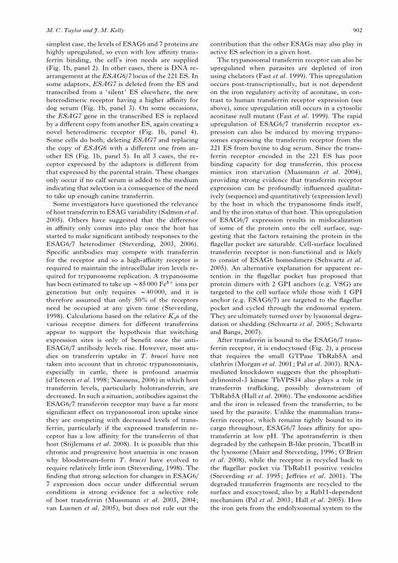
simplest case, the levels of ESAG6 and 7 proteins are
highly upregulated, so even with low affinity trans-
ferrin binding, the cell’s iron needs are supplied
(Fig. 1b, panel 2). In other cases, there is DNA re-
arrangement at the ESAG6/7 locus of the 221 ES. In
some adaptors, ESAG7 is deleted from the ES and
transcribed from a ‘silent ’ ES elsewhere, the new
heterodimeric receptor having a higher affinity for
dog serum (Fig. 1b, panel 3). On some occasions,
the ESAG7 gene in the transcribed ES is replaced
by a different copy from another ES, again creating a
novel heterodimeric receptor (Fig. 1b, panel 4).
Some cells do both, deleting ESAG7 and replacing
the copy of ESAG6 with a different one from an-
other ES (Fig. 1b, panel 5). In all 3 cases, the re-
ceptor expressed by the adaptors is different from
that expressed by the parental strain. These changes
only occur if no calf serum is added to the medium
indicating that selection is a consequence of the need
to take up enough canine transferrin.
Some investigators have questioned the relevance
of host transferrin to ESAG variability (Salmon et al.
2005). Others have suggested that the difference
in affinity only comes into play once the host has
started to make significant antibody responses to the
ESAG6/7 heterodimer (Steverding, 2003, 2006).
Specific antibodies may compete with transferrin
for the receptor and so a high-affinity receptor is
required to maintain the intracellular iron levels re-
quired for trypanosome replication. A trypanosome
has been estimated to take upy85000 Fe3+ ions per
generation but only requires y40000, and it is
therefore assumed that only 50% of the receptors
need be occupied at any given time (Steverding,
1998). Calculations based on the relative Kds of the
various receptor dimers for different transferrins
appear to support the hypothesis that switching
expression sites is only of benefit once the anti-
ESAG6/7 antibody levels rise. However, most stu-
dies on transferrin uptake in T. brucei have not
taken into account that in chronic trypanosomiasis,
especially in cattle, there is profound anaemia
(d’Ieteren et al. 1998; Naessens, 2006) in which host
transferrin levels, particularly holotransferrin, are
decreased. In such a situation, antibodies against the
ESAG6/7 transferrin receptor may have a far more
significant effect on trypanosomal iron uptake since
they are competing with decreased levels of trans-
ferrin, particularly if the expressed transferrin re-
ceptor has a low affinity for the transferrin of that
host (Stijlemans et al. 2008). It is possible that this
chronic and progressive host anaemia is one reason
why bloodstream-form T. brucei have evolved to
require relatively little iron (Steverding, 1998). The
finding that strong selection for changes in ESAG6/
7 expression does occur under differential serum
conditions is strong evidence for a selective role
of host transferrin (Mussmann et al. 2003, 2004;
van Luenen et al. 2005), but does not rule out the
contribution that the other ESAGs may also play in
active ES selection in a given host.
The trypanosomal transferrin receptor can also be
upregulated when parasites are depleted of iron
using chelators (Fast et al. 1999). This upregulation
occurs post-transcriptionally, but is not dependent
on the iron regulatory activity of aconitase, in con-
trast to human transferrin receptor expression (see
above), since upregulation still occurs in a cytosolic
aconitase null mutant (Fast et al. 1999). The rapid
upregulation of ESAG6/7 transferrin receptor ex-
pression can also be induced by moving trypano-
somes expressing the transferrin receptor from the
221 ES from bovine to dog serum. Since the trans-
ferrin receptor encoded in the 221 ES has poor
binding capacity for dog transferrin, this process
mimics iron starvation (Mussmann et al. 2004),
providing strong evidence that transferrin receptor
expression can be profoundly influenced qualitat-
ively (sequence) and quantitatively (expression level)
by the host in which the trypanosome finds itself,
and by the iron status of that host. This upregulation
of ESAG6/7 expression results in mislocalization
of some of the protein onto the cell surface, sug-
gesting that the factors retaining the protein in the
flagellar pocket are saturable. Cell-surface localized
transferrin receptor is non-functional and is likely
to consist of ESAG6 homodimers (Schwartz et al.
2005). An alternative explanation for apparent re-
tention in the flagellar pocket has proposed that
protein dimers with 2 GPI anchors (e.g. VSG) are
targeted to the cell surface while those with 1 GPI
anchor (e.g. ESAG6/7) are targeted to the flagellar
pocket and cycled through the endosomal system.
They are ultimately turned over by lysosomal degra-
dation or shedding (Schwartz et al. 2005; Schwartz
and Bangs, 2007).
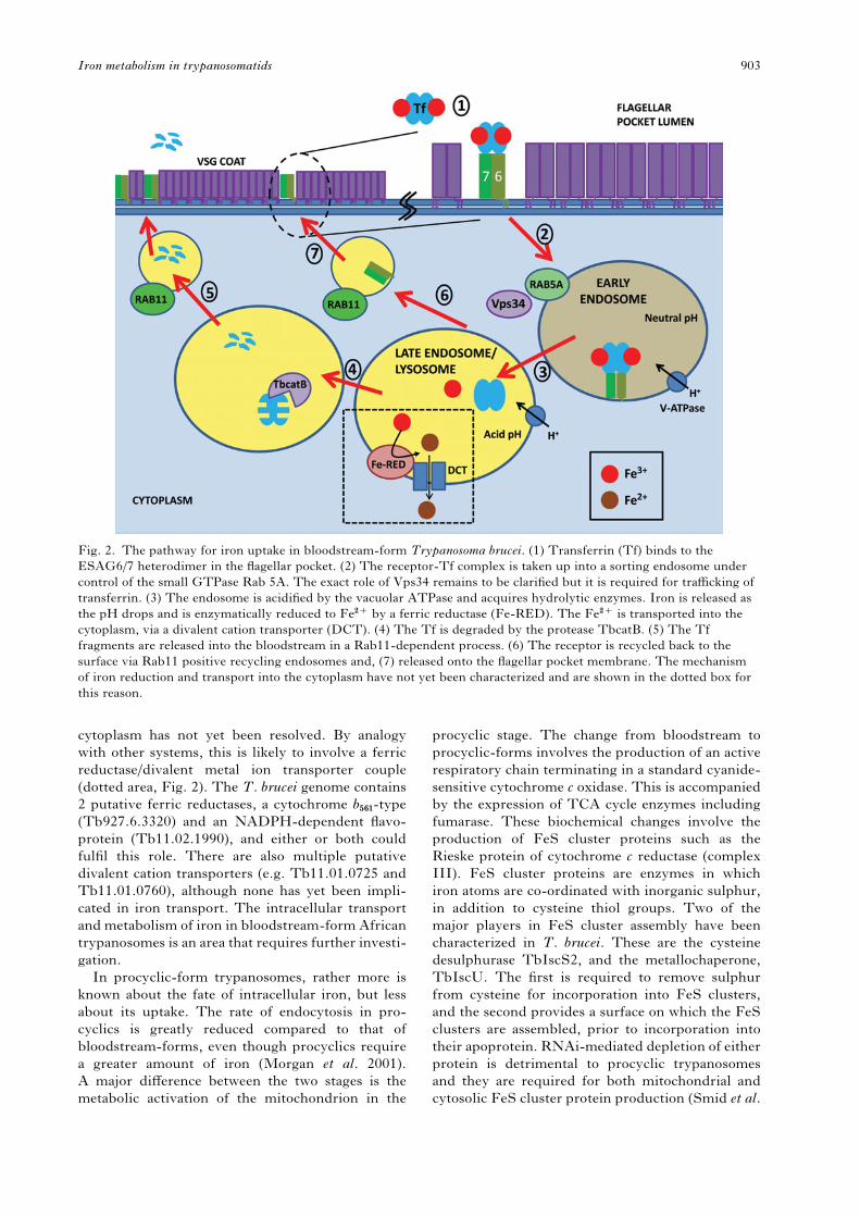
After transferrin is bound to the ESAG6/7 trans-
ferrin receptor, it is endocytosed (Fig. 2), a process
that requires the small GTPase TbRab5A and
clathrin (Morgan et al. 2001; Pal et al. 2003). RNA-
mediated knockdown suggests that the phosphati-
dylinositol-3 kinase TbVPS34 also plays a role in
transferrin trafficking, possibly downstream of
TbRab5A (Hall et al. 2006). The endosome acidifies
and the iron is released from the transferrin, to be
used by the parasite. Unlike the mammalian trans-
ferrin receptor, which remains tightly bound to its
cargo throughout, ESAG6/7 loses affinity for apo-
transferrin at low pH. The apotransferrin is then
degraded by the cathepsin B-like protein, TbcatB in
the lysosome (Maier and Steverding, 1996; O’Brien
et al. 2008), while the receptor is recycled back to
the flagellar pocket via TbRab11 positive vesicles
(Steverding et al. 1995; Jeffries et al. 2001). The
degraded transferrin fragments are recycled to the
surface and exocytosed, also by a Rab11-dependent
mechanism (Pal et al. 2003; Hall et al. 2005). How
the iron gets from the endolysosomal system to the
M. C. Taylor and J. M. Kelly 902
cytoplasm has not yet been resolved. By analogy
with other systems, this is likely to involve a ferric
reductase/divalent metal ion transporter couple
(dotted area, Fig. 2). The T. brucei genome contains
2 putative ferric reductases, a cytochrome b561-type
(Tb927.6.3320) and an NADPH-dependent flavo-
protein (Tb11.02.1990), and either or both could
fulfil this role. There are also multiple putative
divalent cation transporters (e.g. Tb11.01.0725 and
Tb11.01.0760), although none has yet been impli-
cated in iron transport. The intracellular transport
and metabolism of iron in bloodstream-form African
trypanosomes is an area that requires further investi-
gation.
In procyclic-form trypanosomes, rather more is
known about the fate of intracellular iron, but less
about its uptake. The rate of endocytosis in pro-
cyclics is greatly reduced compared to that of
bloodstream-forms, even though procyclics require
a greater amount of iron (Morgan et al. 2001).
A major difference between the two stages is the
metabolic activation of the mitochondrion in the
procyclic stage. The change from bloodstream to
procyclic-forms involves the production of an active
respiratory chain terminating in a standard cyanide-
sensitive cytochrome c oxidase. This is accompanied
by the expression of TCA cycle enzymes including
fumarase. These biochemical changes involve the
production of FeS cluster proteins such as the
Rieske protein of cytochrome c reductase (complex
III). FeS cluster proteins are enzymes in which
iron atoms are co-ordinated with inorganic sulphur,
in addition to cysteine thiol groups. Two of the
major players in FeS cluster assembly have been
characterized in T. brucei. These are the cysteine
desulphurase TbIscS2, and the metallochaperone,
TbIscU. The first is required to remove sulphur
from cysteine for incorporation into FeS clusters,
and the second provides a surface on which the FeS
clusters are assembled, prior to incorporation into
their apoprotein. RNAi-mediated depletion of either
protein is detrimental to procyclic trypanosomes
and they are required for both mitochondrial and
cytosolic FeS cluster protein production (Smid et al.
Fig. 2. The pathway for iron uptake in bloodstream-form Trypanosoma brucei. (1) Transferrin (Tf) binds to the
ESAG6/7 heterodimer in the flagellar pocket. (2) The receptor-Tf complex is taken up into a sorting endosome under
control of the small GTPase Rab 5A. The exact role of Vps34 remains to be clarified but it is required for trafficking of
transferrin. (3) The endosome is acidified by the vacuolar ATPase and acquires hydrolytic enzymes. Iron is released as
the pH drops and is enzymatically reduced to Fe2+ by a ferric reductase (Fe-RED). The Fe2+ is transported into the
cytoplasm, via a divalent cation transporter (DCT). (4) The Tf is degraded by the protease TbcatB. (5) The Tf
fragments are released into the bloodstream in a Rab11-dependent process. (6) The receptor is recycled back to the
surface via Rab11 positive recycling endosomes and, (7) released onto the flagellar pocket membrane. The mechanism
of iron reduction and transport into the cytoplasm have not yet been characterized and are shown in the dotted box for
this reason.
Iron metabolism in trypanosomatids 903
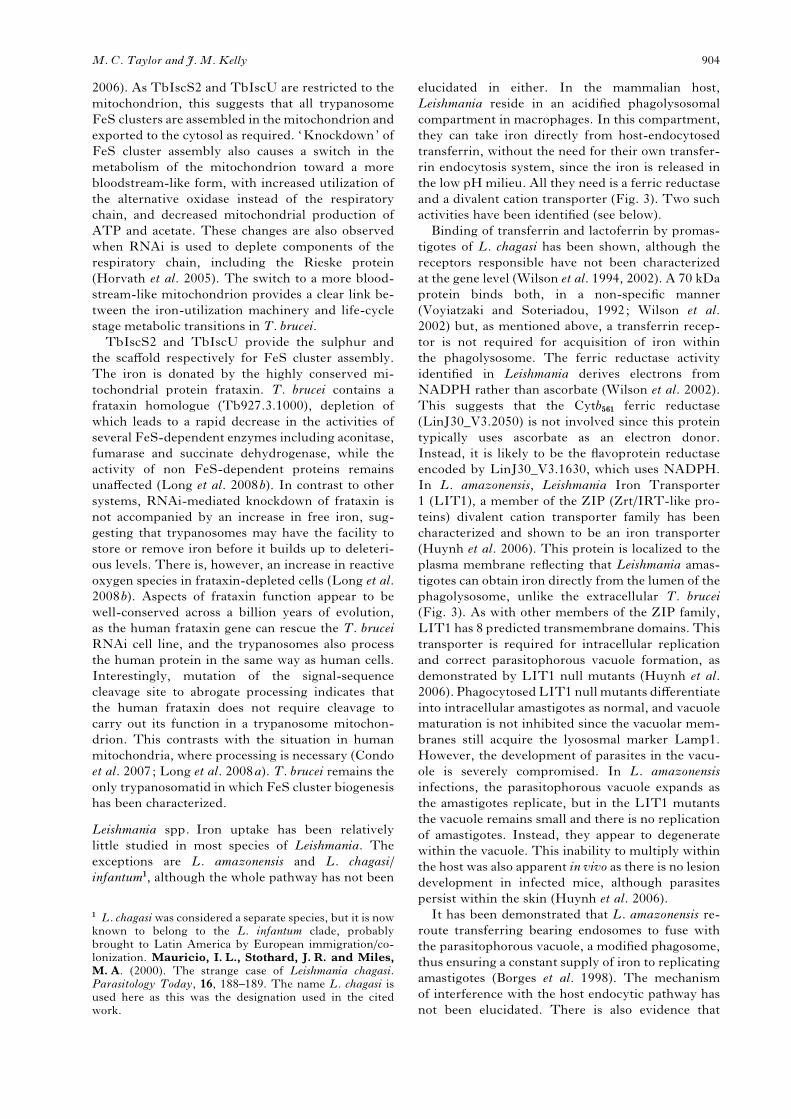
2006). As TbIscS2 and TbIscU are restricted to the
mitochondrion, this suggests that all trypanosome
FeS clusters are assembled in the mitochondrion and
exported to the cytosol as required. ‘Knockdown’ of
FeS cluster assembly also causes a switch in the
metabolism of the mitochondrion toward a more
bloodstream-like form, with increased utilization of
the alternative oxidase instead of the respiratory
chain, and decreased mitochondrial production of
ATP and acetate. These changes are also observed
when RNAi is used to deplete components of the
respiratory chain, including the Rieske protein
(Horvath et al. 2005). The switch to a more blood-
stream-like mitochondrion provides a clear link be-
tween the iron-utilization machinery and life-cycle
stage metabolic transitions in T. brucei.
TbIscS2 and TbIscU provide the sulphur and
the scaffold respectively for FeS cluster assembly.
The iron is donated by the highly conserved mi-
tochondrial protein frataxin. T. brucei contains a
frataxin homologue (Tb927.3.1000), depletion of
which leads to a rapid decrease in the activities of
several FeS-dependent enzymes including aconitase,
fumarase and succinate dehydrogenase, while the
activity of non FeS-dependent proteins remains
unaffected (Long et al. 2008b). In contrast to other
systems, RNAi-mediated knockdown of frataxin is
not accompanied by an increase in free iron, sug-
gesting that trypanosomes may have the facility to
store or remove iron before it builds up to deleteri-
ous levels. There is, however, an increase in reactive
oxygen species in frataxin-depleted cells (Long et al.
2008b). Aspects of frataxin function appear to be
well-conserved across a billion years of evolution,
as the human frataxin gene can rescue the T. brucei
RNAi cell line, and the trypanosomes also process
the human protein in the same way as human cells.
Interestingly, mutation of the signal-sequence
cleavage site to abrogate processing indicates that
the human frataxin does not require cleavage to
carry out its function in a trypanosome mitochon-
drion. This contrasts with the situation in human
mitochondria, where processing is necessary (Condo
et al. 2007; Long et al. 2008a). T. brucei remains the
only trypanosomatid in which FeS cluster biogenesis
has been characterized.
Leishmania spp. Iron uptake has been relatively
little studied in most species of Leishmania. The
exceptions are L. amazonensis and L. chagasi/
infantum1, although the whole pathway has not been
elucidated in either. In the mammalian host,
Leishmania reside in an acidified phagolysosomal
compartment in macrophages. In this compartment,
they can take iron directly from host-endocytosed
transferrin, without the need for their own transfer-
rin endocytosis system, since the iron is released in
the low pH milieu. All they need is a ferric reductase
and a divalent cation transporter (Fig. 3). Two such
activities have been identified (see below).
Binding of transferrin and lactoferrin by promas-
tigotes of L. chagasi has been shown, although the
receptors responsible have not been characterized
at the gene level (Wilson et al. 1994, 2002). A 70 kDa
protein binds both, in a non-specific manner
(Voyiatzaki and Soteriadou, 1992; Wilson et al.
2002) but, as mentioned above, a transferrin recep-
tor is not required for acquisition of iron within
the phagolysosome. The ferric reductase activity
identified in Leishmania derives electrons from
NADPH rather than ascorbate (Wilson et al. 2002).
This suggests that the Cytb561 ferric reductase
(LinJ30_V3.2050) is not involved since this protein
typically uses ascorbate as an electron donor.
Instead, it is likely to be the flavoprotein reductase
encoded by LinJ30_V3.1630, which uses NADPH.
In L. amazonensis, Leishmania Iron Transporter
1 (LIT1), a member of the ZIP (Zrt/IRT-like pro-
teins) divalent cation transporter family has been
characterized and shown to be an iron transporter
(Huynh et al. 2006). This protein is localized to the
plasma membrane reflecting that Leishmania amas-
tigotes can obtain iron directly from the lumen of the
phagolysosome, unlike the extracellular T. brucei
(Fig. 3). As with other members of the ZIP family,
LIT1 has 8 predicted transmembrane domains. This
transporter is required for intracellular replication
and correct parasitophorous vacuole formation, as
demonstrated by LIT1 null mutants (Huynh et al.
2006). PhagocytosedLIT1 null mutants differentiate
into intracellular amastigotes as normal, and vacuole
maturation is not inhibited since the vacuolar mem-
branes still acquire the lyososmal marker Lamp1.
However, the development of parasites in the vacu-
ole is severely compromised. In L. amazonensis
infections, the parasitophorous vacuole expands as
the amastigotes replicate, but in the LIT1 mutants
the vacuole remains small and there is no replication
of amastigotes. Instead, they appear to degenerate
within the vacuole. This inability to multiply within
the host was also apparent in vivo as there is no lesion
development in infected mice, although parasites
persist within the skin (Huynh et al. 2006).
It has been demonstrated that L. amazonensis re-
route transferring bearing endosomes to fuse with
the parasitophorous vacuole, a modified phagosome,
thus ensuring a constant supply of iron to replicating
amastigotes (Borges et al. 1998). The mechanism
of interference with the host endocytic pathway has
not been elucidated. There is also evidence that
1 L. chagasiwas considered a separate species, but it is nowknown to belong to the L. infantum clade, probablybrought to Latin America by European immigration/co-lonization. Mauricio, I. L., Stothard, J. R. and Miles,M. A. (2000). The strange case of Leishmania chagasi.Parasitology Today, 16, 188–189. The name L. chagasi isused here as this was the designation used in the citedwork.
M. C. Taylor and J. M. Kelly 904

Leishmania may act to increase the uptake of holo-
transferrin by the host cell. Infection with L. dono-
vani was shown to stabilize transferrin receptor
mRNA and to promote uptake of radio-labelled
iron from holotransferrin. L. donovani depletes the
intracellular labile iron pool leading to the activation
of iron regulatory proteins, which then decrease
ferritin expression and upregulate transferrin re-
ceptor 1 (Das et al. 2009).
Leishmania also require a source of porphyrins
since they lack the biosynthetic capacity for their
production. Recent work has identified haemoglobin
as a source of iron for intracellular amastigotes
(Carvalho et al. 2009). L. infantum axenic amasti-
gotes were unable to grow on iron-depleted medium,
but growth could be restored by addition of haemo-
globin. A 46 kDa flagellar pocket protein of L. do-
novani had previously been identified as the
haemoglobin receptor (Krishnamurthy et al. 2005).
Carvalho et al. (2009) showed that antibodies against
this receptor could block the utilization of haemo-
globin. The exploitation of haem as an iron source
by amastigotes may explain why the LIT1 ferrous
iron transporter null mutants were able to persist
in infected mice (Huynh et al. 2006). In this respect,
it is notable that the major replication sites of vis-
ceralising Leishmania species are also sites of macro-
phage erythrocyte recycling. Erythrocyte haemoglobin
could therefore provide intracellular amastigotes
with a ready source of haem iron. Leishmania, unlike
the trypanosomes, possess an orthologue of ferro-
chelatase (LmjF17.1460), which is functional, since
protoporphyrin IX can replace haemin in culture
medium for L. amazonensis (Chang and Chang,
1985).
It is clear that Leishmania are capable of obtaining
iron from a number of host sources. The presence
of the iron transport machinery on the surface of
the Leishmania amastigote, together with the dem-
onstration that genetic disruption of iron uptake
completely abrogates infection, implicates this ma-
chinery as an Achilles’ heel of the parasite suitable
for targeting with chemotherapy.
Trypanosoma cruzi. Almost nothing is known about
iron uptake or iron sources in T. cruzi. Given the
wide range of hosts and host cells infected, it is
possible that T. cruzi utilizes several different iron
sources, such as myoglobin in cardiac muscle
cells – a source of both iron and porphyrins. The
Fig. 3. Iron uptake in Leishmania amastigotes. Transferrin is taken up by the infected mammalian cell via its
transferrin receptor (TfR). The Tf:TfR complex is endocytosed and the endosome fuses with the phagosome. Fe3+ is
liberated from the transferrin due to the low pH. A parasite cell surface NADPH-dependent ferric reductase reduces
Fe3+ to Fe2+. The Fe2+ is then transported into the parasite by the divalent ion transporter LIT1. The amastigote may
also acquire iron from the cytoplasmic labile iron pool by an undefined mechanism. Simultaneously, the infected cell
tries to deplete iron from the phagosome through the concerted action of the Dcytb ferric reductase and the NRAMP1
divalent cation transporter. K: kinetoplast.
Iron metabolism in trypanosomatids 905

cytoplasmic habitat of T. cruzi also gives it access to
the host’s iron storage protein, ferritin. This could
be particularly relevant to amastigotes in hepcidin-
responsive macrophages (see below). Insect-stage
epimastigotes have a requirement for both haem
and non-haem iron in vitro (Lalonde and Holbein,
1984).
Transferrin binds to and is taken up by T. cruzi
amastigotes in vitro (Lima and Villalta, 1990).
However, the physiological relevance of this inter-
action is unclear since amastigotes replicate in the
cytoplasm of host cells, a niche in which transferrin
is conspicuously absent. Consistent with this, very
little intracellular staining has been detected in
amastigotes using gold-labelled transferrin (Soares
and de Souza, 1991). This does not exclude the
possibility that trypomastigotes could pick up
transferrin in the bloodstream, or that epimastigotes
can acquire iron from transferrin in the bloodmeal.
Both life-cycle stages have been shown to bind
transferrin, and in epimastigotes it accumulates in
the reservosome, a late endosome/lysosome–like
organelle (Soares and de Souza, 1991; Soares et al.
1992). Despite these preliminary studies, the uptake
and usage of iron by T. cruzi, particularly the intra-
cellular amastigote, is poorly understood. Given the
large number of people infected, or at danger from
this parasite, and the dearth of drugs available, a
deeper understanding of parasite iron requirements
could be fundamental to new chemotherapeutic
strategies.
How do trypanosomatids utilize scavenged haem:
questions to be answered
One problem that arises from the use of haem by
all the pathogenic trypanosomatids is how the haem
ring is broken to release iron (Chang and Chang,
1985; Lara et al. 2007). There is no readily identifi-
able orthologue of haem oxygenase in the genome
sequences of any of the trypanosomatids, although a
putative activity has been measured in promastigotes
of L. donovani (Srivastava et al. 1997). Additionally,
trypanosomes also lack ferrochelatase (present in
Leishmania), and so, theoretically, cannot insert iron
into scavenged porphyrins. This raises the possi-
bility that trypanosomatids accomplish breakage of
the porphyrin ring and insertion of iron into proto-
porphyrin by a different mechanism from other or-
ganisms. However, the simplest explanation for the
use of haem, given the apparent lack of both haem
oxygenase and ferrochelatase, is that trypanosomes
simply incorporate scavenged haem directly into
apoproteins without going through intermediate
steps. This would then require that the iron used by
non-haem proteins be obtained independently from
mammalian or insect sources. It is implicit from this
that one source of iron would not be able to comp-
lement the loss of the other.
THE ROLE OF IRON IN TRYPANOSOMATIDS
Iron has multiple roles in most organisms and try-
panosomatids are no exception, with many activities
common to both parasite and host. These include
the reduction of ribonucleotides for DNA synthe-
sis and cytochrome-based oxidative respiration.
Ribonucleotide reduction utilizes a classical eu-
karyotic enzyme (Dormeyer et al. 1997; Hofer et al.
1997), with the major difference in trypanosomatids
being that the reducing power is provided
by the kinetoplastid-specific thiol trypanothione in
a reaction catalysed by tryparedoxin (Dormeyer
et al. 2001). Below, we discuss several examples
of iron-dependent processes in trypanosomatids,
which are distinct from the mammalian host. These
could have potential as targets for therapeutic inter-
vention.
Antioxidant defences
Fe-dependent superoxide dismutases (Fe-
SODs). Unlike their mammalian hosts, which
contain Cu/Zn and Mn-dependent SODs, trypano-
somatids express 4 different Fe-dependent super-
oxide dismutases (Dufernez et al. 2006; Wilkinson et
al. 2006). Fe-dependent SODs are restricted to
bacteria and some protozoa. These enzymes help to
protect the cell from oxidative stress by catalysing
the dismutation of superoxide radicals into H2O2 and
O2, the H2O2 then being metabolized by peroxidases.
Iron removes an electron to oxidize 1 molecule of
superoxide (step 1), and is then reoxidized, by reac-
tion with a second superoxide molecule in the pres-
ence of protons (step 2).
1. Fe(III)-SOD+O2.xpFe2+-SOD+O2
2. Fe2+-SOD+O2.x+2H+pFe(III)-SOD+H2O2
The net result of these two reactions is regeneration
of Fe(III)-SOD and the production of 1 molecule
each of O2 and H2O2 from 2 superoxide radicals
(Halliwell and Gutteridge, 2007).
The 4 enzymes present in trypanosomatids are
differentially compartmentalized. In T. brucei, SOD
A and SOD C are found in the mitochondrion whilst
SOD B1 and B2 are glycosomal, with some SOD B1
present in the cytoplasm. Phylogenetic analyses have
indicated that the trypanosomatid Fe-SODs belong
to 2 distinct clades, with SOD A and SOD C in one
group and the SOD Bs in the other. Both sets of
SODs appear to have been acquired by lateral gene
transfer from prokaryotes (Dufernez et al. 2006).
RNAi-mediated knockdown of the Fe-SODs in
T. brucei demonstrates that SOD B1/B2 expression
is essential in bloodstream forms. However, the
RNAi construct could not discriminate between the
two isoforms due to their extreme nucleotide se-
quence conservation (Wilkinson et al. 2006). The
ability to generate SOD B1 and SOD B2 individual
M. C. Taylor and J. M. Kelly 906
null mutants with no specific defects in growth or
virulence indicates that the two isoforms can comp-
lement each other, but that at least one must be
expressed for survival (Prathalingham et al. 2007).
However, there was a difference between the two
null mutants in susceptibility to trypanocidal agents.
SOD B1 mutants were more susceptible to benzni-
dazole and nifurtimox than the wild type, whereas
SOD B2 mutants had comparable sensitivity
(Prathalingham et al. 2007). This argues that one-
electron reduction of the drugs takes place in
the cytoplasm where SOD B1 is at a higher level,
rather than the glycosome. The two mitochondrial
isoforms SOD A and SOD C are dispensible for
the bloodstream form; however, RNAi-mediated
knockdown of SODA renders cells more susceptible
to the superoxide generator paraquat (Wilkinson
et al. 2006). SOD A is expressed in the less meta-
bolically active mitochondrion of the bloodstream
form, probably to protect the kinetoplast DNA, and
by implication, turnover of paraquat occurs in this
organelle. In contrast, knockdown of SOD C pro-
duces no discernible effects, either on growth or
susceptibility to oxidizing agents and the role of
SOD C remains to be resolved.
In T. cruzi, SOD A has been implicated in the
control of programmed cell death. Inducible over-
expression of SOD A protects the parasite from
serum-mediated death (Piacenza et al. 2007), dem-
onstrating a direct role for superoxide radicals in the
cell death signalling programme. TcSOD A appears
to be concentrated around the kinetoplast suggesting
a role in protecting mitochondrial DNA from oxi-
dative damage (Taylor and Kelly, 2006). A similar
location has been noted for the mitochondrial per-
oxiredoxin (Wilkinson et al. 2000). It is tempting to
speculate that both enzymes, together with others,
constitute an antioxidant complex guarding kDNA
from oxidative damage.
Increased expression of the T. cruzi SOD B1 iso-
form results in greater susceptibility to the super-
oxide generator gentian violet (Temperton et al.
1998). This seemingly paradoxical outcome has also
been observed in bacteria and mammalian cells,
where SOD activity has been upregulated. A poss-
ible explanation is that higher SOD activity results
in increased H2O2 levels. In situations where the
ability to metabolize H2O2 is limited, this can
result in the production of OH. radicals by the
Fenton reaction. Of more interest, the SOD B1
overexpressers were also more susceptible to the
trypanocidal drug benznidazole, but not to the ni-
trofuran drugs, in contrast to T. brucei (Temperton
et al. 1998). This suggests that at least part of the
trypanocidal mechanism of benznidazole is mediated
through increased oxidative stress in the cytoplasm
or glycosome. The glycosomal SODs B1 and B2 are
developmentally regulated in Leishmania, with SOD
B1 being more highly expressed in amastigotes
and SOD B2 in promastigotes (Plewes et al. 2003).
A single allele deletion of SOD B1 was found to
confer a lower survival rate in macrophages or when
exposed to paraquat, suggesting that glycosomal
SOD activity is important in Leishmania amastigotes
(Plewes et al. 2003).
It is clear that the Fe-dependent SODs, particu-
larly the glycosomal isoforms, play important roles
in pathogenic trypanosomatids and that they or their
iron co-factor are suitable targets for drug develop-
ment. Inhibitors of SOD could then be used in
combination with other trypanocidal drugs, such as
benznidazole, to potentiate their effects, thus mini-
mising the effective dose, and decreasing toxic side
effects.
Ascorbate-dependent peroxidase
Trypanosomatids lack the classical catalase and sel-
enium-dependent glutathione peroxidases which can
rapidly metabolize H2O2. Instead, they express a
battery of different peroxidases including trypar-
edoxin-dependent peroxiredoxins, and non-sel-
enium dependent glutathione peroxidases (Flohe
et al. 1999; Irigoin et al. 2008; Wilkinson and Kelly,
2003). In addition, T. cruzi and Leishmania possess
an ascorbate-dependent peroxidase (APX), which
can metabolize hydrogen peroxide but not organic
peroxides (Wilkinson et al. 2002a ; Adak and Datta,
2005). Ascorbate peroxidases are more usually plant
enzymes and different isoforms are specific to dif-
ferent compartments of the plant cell, including both
stromal and thylakoid compartments of the chloro-
plast. APX is a haemoprotein belonging to the cata-
lase-peroxidase superfamily, and is absent from
African trypanosomes.
The catalytic mechanism of APX includes 3 steps.
The first involves oxidation of the haem iron to
the ferryl (Fe(IV)) state by the H2O2 molecule to
form compound I (underlined below), coupled with
the formation of a cationic radical on the porphyrin
ring. Compound I therefore has an oxidation state
of +5.
1. Fe3+-porphyrin+H2O2pFe4+===O
porphyrin.++H2O
Compound I then reacts sequentially with 2 mol-
ecules of ascorbate (Asc) to produce the ascorbyl free
radical (AFR, monodehydroascorbate) and regener-
ate the resting enzyme (oxidation state +3).
2. Fe4+===O porphyrin.++AscpFe4+===O+AFR.
3. Fe4+===O+AscpFe3+-porphyrin +AFR.
The net reaction is : H2O2+2 AscpH2O+2 AFR.
Ascorbate is only required to return the enzyme
to its resting state. In the absence of ascorbate,
Iron metabolism in trypanosomatids 907
the porphyrin p-cation radical transfers to cysteine
or tryptophan residues in the protein chain and
the enzyme can become irreversibly oxidized
(Hiner et al. 2001; Kitajima et al. 2008). The AFR is
reduced back to Asc by the action of a number of
small molecule reductants (e.g. trypanothione,
(Krauth-Siegel and Ludemann, 1996)) or redox ac-
tive enzymes. It is not clear whether any other re-
ductant can fulfil the role of ascorbate in the above
reaction.
Although Leishmania and T. cruzi proteins are
clearly derived from the same ancestral gene (62%
amino acid identity between L. major and T. cruzi
APXs), they now appear to have different biological
roles. The Leishmania enzyme is mitochondrial and
has a classical mitochondrial targeting sequence,
whereas the T. cruzi protein has a much longer
N-terminal extension, which seems to be related
to plastid transit peptides, and is resident in the
endoplasmic reticulum (Wilkinson et al. 2002a).
Why the proteins should be differentially targeted
in 2 related organisms is not obvious. Leishmania
APX appears to have a role in protection of mi-
tochondrial membrane lipids from oxidative stress
and its expression is induced on exposure to H2O2
(Dolai et al. 2008). In the ER, H2O2 is a constitutive
by-product of the Ero1/protein disulphide iso-
merase redox cycle and it is likely that the T. cruzi
APX plays an analogous role to Leishmania APX,
protecting membrane lipids in the ER membrane
from peroxidation. T. cruzi also expresses another
peroxidase in the ER, glutathione-dependent per-
oxidase II (GPX II), which cannot metabolize
H2O2, but does metabolize phospholipid hydroper-
oxides (Wilkinson et al. 2002b). Thus, it is likely that
these two proteins act in concert to protect the ER
membrane from the consequences of oxidative
stress.
Trypanosome alternative oxidase (TAO)
Bloodstream-form African trypanosomes are de-
pendent on glycolysis since the normal mitochon-
drial respiratory chain is absent. Instead, oxidation
of ubiquinol is carried out by the so-called alterna-
tive oxidase (TAO), resulting in the net transfer of
electrons from glycerol-3-phosphate to molecular
oxygen to produce water. Unlike the conventional
respiratory chain, the transfer of electrons to TAO
does not result in the generation of ATP and it does
not create a proton gradient (Clarkson et al. 1989).
TAO is not present in the mammalian host (or in
T. cruzi and Leishmania, although there is a second
TAO-like sequence in all trypanosomatids examined
(Chaudhuri et al. 2006)). In contrast to the other
respiratory oxidases, TAO is composed of a single
protein of 37.5 kDa and has 2 iron-binding sites
constituted by conserved glutamate and histidine
residues on helices 1,3,4 and 5 (Chaudhuri et al.
2006). The current model for the alternative oxidase
active site suggests that TAO is a member of the
diiron carboxylate protein family (Andersson and
Nordlund, 1999). These proteins are characterized
by the motif E-Xn-E-X-X-H-Xn-E-Xn-E-X-X-H,
where E is glutamate and H is histidine, with X
being any amino acid; n signifies a number of re-
sidues. The 4 carboxylates of the glutamates and
imidazole nitrogens of the histidine co-ordinate 2
iron atoms in the catalytic centre, hence the name
diiron carboxylate protein (Berthold and Stenmark,
2003). Although the T. brucei TAO, like the other
alternative oxidases, has 2 strongly hydrophobic
segments, it is thought to be positioned along the
matrix face of the inner mitochondrial membrane
rather than spanning the membrane (Andersson and
Nordlund, 1999; Chaudhuri et al. 2006). Site-
directed mutagenesis of the putative iron-binding
ligands H165, E214, E266 and H269 in TAO has
demonstrated their importance to activity. Any one
of these mutations abolishes the ability of the TAO
gene to complement an E. coli haem-deficient mu-
tant (Ajayi et al. 2002). Iron-dependence of TAO
has also been established by iron chelation with
o-phenanthroline, resulting in strong inhibition
which was reversed by addition of iron, but not other
metals such as copper (Ajayi et al. 2002).
In T. brucei, TAO is developmentally regulated
with the mRNA level dropping rapidly on differ-
entiation from bloodstream to procyclic-forms
(Chaudhuri et al. 2002). This differential mRNA
accumulation appears to be due to a change in the
half-life of the mRNA. This is mediated by a labile
protein factor since the transcript is stabilized by
cycloheximide treatment. During the transition to
metacyclic trypomastigotes, which are pre-adapted
for infection of the mammalian host, TAO activity is
again upregulated and the standard cyanide-sensitive
respiratory chain downregulated (Bienen et al. 1991).
Although the level of TAO expressed in procyclics
is lower than in bloodstream-forms, it also plays an
important role in this stage, as inhibition of the cya-
nide sensitive respiratory chain is not lethal unless
the TAO is inhibited simultaneously (Coustou et al.
2003).
Due to the reliance of bloodstream-form African
trypanosomes on glycolysis, coupled to the glycerol-
3-phosphate oxidation required to regenerate
NAD+, TAO has been proposed to be a candidate
drug target (Yabu et al. 2003; Chaudhuri et al. 2006).
However, computer modelling of metabolic flux and
RNAi-mediated depletion of TAO suggests, in vitro,
that the TAO protein would have to be inhibited
by more than 95% for there to be a significant effect
on trypanosome growth (Helfert et al. 2001). In
T. brucei, TAO inhibition is only lethal when
coupled with administration of glycerol to block
glycerol-phosphate dehydrogenase or with repeated
administration of the inhibitor (Yabu et al. 1998,
M. C. Taylor and J. M. Kelly 908

2003). Nevertheless, the demonstration that in-
hibitor treatment can cure mice suggests that the
TAO may be a viable drug target. In T. vivax-
infected mice, a single dose of ascofuranone
(50 mg kgx1) has been shown to elicit cure without
glycerol (Yabu et al. 2006).
J-base biosynthesis
A unique feature of trypanosomatids is the pres-
ence of a hypermodified base within their DNA.
This base, b-D-hydroxymethyldeoxyuridine, more
usually known as J, is a derivative of thymidine and
is synthesized within the DNA strand in a two-step
modification process. The first step in J-base syn-
thesis involves the hydroxylation of thymidine re-
sidues in DNA to provide a hydroxyl group to which
the glucose moiety is attached (Fig. 4). The hydro-
xylase which carries this out, is a member of the
Fe2+- and 2-oxoglutarate-dependent dioxygenases,
although the similarity to other members of this
family is not immediately obvious (Yu et al. 2007).
These enzymes use molecular oxygen to donate the
oxygen atom required for the hydroxyl group.
During this process, the iron is probably oxidized to
an oxyferryl (Fe(IV)===O) state as in the APX reac-
tion, except that in this case, the oxygen atom is de-
rived from molecular oxygen rather than H2O2
(Schofield and Zhang, 1999).
There are 2 thymidine hydroxylases in trypano-
somatids, referred to as J-binding protein 1 (JBP1)
and J-binding protein 2 (JBP2). Both contain hom-
ologous thymidine hydroxylase domains and mu-
tation of the putative iron-binding residues in
Leishmania JBP1 (H189, D191 and H239) renders
the protein unable to rescue J-base biosynthesis
in JBP1 null mutant T. brucei, as does mutation
of the conserved arginine residue required for 2-
oxoglutarate binding (R255) (Yu et al. 2007). These
mutations do not affect the DNA binding of the en-
zyme and so the inference is that they affect the
catalytic capability, as would be expected if they
were required for iron incorporation. Similar muta-
genesis studies on iron and 2-oxoglutarate binding
residues, also confirm that JBP2 is a member of this
family (Cliffe et al. 2009; Vainio et al. 2009). In
neither case has the hydroxylase activity been re-
constituted in vitro. Whilst J-base appears to be
dispensible for T. brucei (and is absent in the pro-
cyclic stage), JBP1 is essential in Leishmania (Genest
et al. 2005). The situation in T. cruzi is unclear.
Iron and anti-kinetoplastid drugs
There have been few studies directly addressing the
role of iron in drug-mediated killing of kinetoplastid
parasites. Indeed, for most clinically relevant anti-
kinetoplastid drugs, the exact mechanism of action
remains unknown and many of their effects may be
pleiotropic. However, a recent study has implicated
iron in drug activity (particularly metalloids) in
Leishmania (Mehta and Shaha, 2006). Using defer-
oxamine (DFO), Mehta and Shaha showed that iron
depletion inhibits changes in the mitochondrial
membrane potential and ATPase activity produced
by antimonial or arsenical drugs. This inhibition can
be reversed if the DFO is saturated with iron prior to
incubation with the parasites, showing that the effect
is dependent on iron chelation. In addition, it has
been demonstrated that DFO can reduce the level of
cell death induced by treatment with SbIII or AsIII,
whereas addition of iron causes a slight exacerbation
of cell death. The changes in mitochondrial mem-
brane potential and activity prior to drug-induced
Fig. 4. Hydroxylation of thymidine residues in DNA. Thymidine hydroxylases JBP1 and JBP2 use oxidative
decarboxylation of 2-oxoglutarate in the presence of molecular oxygen to add a hydroxyl group to the methyl carbon of
the thymidine base. The reaction is catalysed by Fe2+. R1 and R2 represent the 5k and 3kends of this strand of the DNA
duplex.
Iron metabolism in trypanosomatids 909
death mirrors the mechanism shown in T. cruzi for
complement-induced programmed cell death of
epimastigotes, which is mediated by mitochondrial
O2.x(Piacenza et al. 2007). Thus, the drug-induced
death of Leishmania may involve activation of their
cell death programme at the mitochondrial level.
Exacerbation of this process by iron is likely to re-
flect increased oxidative stress resulting from OH.
generated by the superoxide-driven Fenton reaction.
Further studies of the role of iron in the mechan-
ism of drug action/resistance in trypanosomatids are
clearly warranted. This is particularly the case with
drugs that may trigger oxidative stress, such as
Ornidyl (difluoromethylornithine). Ornidyl blocks
polyamine biosynthesis and therefore prevents the
production of trypanothione (N1,N8-bisglutathio-
nylspermidine) (Fairlamb et al. 1987). Almost all
the antioxidant defences in trypanosomatids derive
their reducing power ultimately from trypanothione.
Therefore, prolonged inhibition of spermidine syn-
thesis will result in increased oxidative stress by de-
pletion of trypanothione, hence the level of iron
available to the Fenton reaction may be key to the
trypanocidal activity. As iron has also been im-
plicated in the action of benznidazole (via SOD B1),
it would seem important to dissect this interaction
for future drug design (Temperton et al. 1998;
Prathalingham et al. 2007; Francisco et al. 2008).
IRON IN PATHOGENESIS AND IMMUNITY
The peptide hormone hepcidin is released from the
liver during infection. One of its roles is to prevent
iron release from macrophages into the bloodstream.
This is achieved by destabilization of the ferroportin
molecule which is internalized and degraded, lead-
ing to a block in iron export and an increase in fer-
ritin-bound iron in macrophages (Nemeth et al.
2004, 2006). In addition, hepcidin promotes degra-
dation of ferroportin on duodenal enterocytes,
thereby reducing dietary iron intake. It has also been
demonstrated that macrophages can produce hepci-
din themselves under the control of Toll-like recep-
tor 4 signalling, creating an autocrine feedback loop
resulting in iron sequestration (Peyssonnaux et al.
2006). In this way, plasma iron levels are decreased,
and bloodstream pathogens prevented from acces-
sing iron. An unfortunate effect of this innate im-
mune strategy is to create a situation of functional
anaemia in the host. This condition, known as
‘anaemia of chronic infection’ illustrates the ca-
pacity of iron to be both beneficial and dangerous,
often simultaneously.
Trypanosoma brucei. For a bloodstream parasite
such as T. brucei, this anaemia can create problems
as it is dependent on its host for iron. In animal
models, anaemia during T. brucei infection is type
I cytokine driven and appears to be specifically
targeted at removal of iron from the plasma during
the chronic phase, thus reducing the pool of iron
available to the circulating trypomastigotes
(Stijlemans et al. 2008). Using C57Bl/6 mice, it is
apparent that anaemia in experimental trypanoso-
miasis occurs in 2 distinct waves, corresponding to
the acute and chronic phases of the infection. During
the acute phase, there is a rapid drop in the number
of erythrocytes, which then recovers, although not to
uninfected levels. This recovery is followed by a
continuous decrease as the chronic phase takes over.
Transferrin mRNA levels increase rapidly during
the acute phase, possibly in response to the removal
and degradation of transferrin by the multiplying
trypanosomes, but then drop back and fall below the
baseline level during the chronic phase. Ferroportin
mRNA levels also decrease during the chronic
phase. At the same time, ferritin mRNA levels
increase and remain high, as does the mRNA for
divalent metal transporter 1. These findings show
that the infected host responds by increasing the
sequestration of iron within the macrophage cyto-
plasm and decreasing export into the plasma. This
sequestration leads to decreased erythropoiesis as
iron is unavailable for incorporation into haemo-
globin. It may be this cytokine driven anaemia dur-
ing chronic infection that promotes the selection of
different transferrin receptors on the trypanosome.
A higher affinity receptor may be required when
serum holotransferrin levels drop, particularly as the
trypanosome receptor cannot distinguish holo from
apotransferrin.
Leishmania – the role of NRAMP1
For many years it has been established that ‘natu-
ral resistance associated macrophage protein 1’
(NRAMP1, now known as Slc11a12) is involved in
resistance to several intracellular pathogens, includ-
ing Salmonella typhimurium, Mycobacterium bovis
and Leishmania (Vidal et al. 1995). Mice strains
carrying a single mutation in transmembrane helix 4
(G169D) are more susceptible to these pathogens
than are strains carrying wild typeNRAMP1 alleles.
Resistance can be restored by reintroducing a wild
type copy of the gene, whilst NRAMP1 null mutant
mice are susceptible to these pathogens (Govoni
et al. 1996). NRAMP1 is a late endosomal/lysosomal
protein expressed in macrophages and granulocytes.
It functions as a divalent cation/proton symporter
which transports metal ions down a proton gradient
across the phagosomal membrane (Biggs et al.
2001) and is related to the major divalent metal ion
2 We use the name NRAMP1, as this term is used in mostpapers referring to its role in infection. Slc11a1 is thesystematic name: Solute carrier family 11 member 1. TheSlc 11 family are proton-coupled divalent metal iontransporters.
M. C. Taylor and J. M. Kelly 910
transporter DMT1 (NRAMP2/Scl11a2). The net
effect of NRAMP1 activity in acidified phagosomes
is to deplete iron (and some other transition metals)
from the lumen of the phagosome, thus denying
pathogens that reside within this compartment ac-
cess to the metal. Iron chelators can mimic the effect
of NRAMP1 in null mutant macrophages infected
with Salmonella. This suggests that NRAMP1
mediates its antimicrobial effect through its iron
transport properties, although the mechanism by
which this controls such diverse pathogens has yet to
be established (Jabado et al. 2003). In Leishmania,
the LIT1 iron transporter gene is upregulated more
rapidly when parasites infect wild type cells than
NRAMP1 null cells, suggesting that NRAMP1+
phagosomes are depleted in iron (Huynh et al. 2006).
In this context, it is interesting that cells infected
with L. amazonensis mutants defective in the LIT1
iron transporter do not develop the expected para-
sitophorous vacuole. Since the parasites cannot
transport iron from the phagosome, but still express
a ferric reductase, it is probable that they enhance
the natural activity of NRAMP1 by providing re-
duced iron at a greater rate than in cells infected with
wild type Leishmania (Huynh et al. 2006). The me-
chanisms identified in mice may also be present in
humans (Bucheton et al. 2003; Mohamed et al.
2004; Blackwell et al. 2009). Genetic polymorphism
studies in human populations have shown a linkage
between mutations in the NRAMP1 promoter re-
gion and susceptibility to visceral leishmaniasis. In
the new era of genomics and deep sequencing, there
is scope for much wider studies looking at the role of
iron metabolism in susceptibility to VL.
Trypanosoma cruzi. Acute T. cruzi infection in
mouse models involves anaemia which can be re-
versed or blocked by administration of the trypano-
cidal drug nifurtimox (Marcondes et al. 2000). In
addition, anaemia is an indicator of the reactivation
of Chagas disease in heart-transplant recipients
(Theodoropoulos et al. 2009). In the mouse model,
the mechanism for the anaemia was postulated to be
a decrease in the lifespan of erythrocytes. However,
recent studies have shown that resistance to T. cruzi
infection involves a key inducible regulator of hae-
matopoeisis, LRG-47, a member of the p47-
GTPase family of interferon-induced proteins.
These GTPases are thought to play a major role in
defence against intracellular pathogens, particularly
phagosomal pathogens such as Salmonella and
Mycobacteria (MacMicking, 2005). They are also
involved in the regulation of autophagy as an innate
defence mechanism against intracellular pathogens
(Gutierrez et al. 2004).
LRG47 null mutant mice develop severe anaemia
when infected with T. cruzi, coupled with a general
failure of haematopoiesis. These mice are suscep-
tible to trypanosome infection, dying within 19 days,
whereas wild-type mice survive for more than 30
days. In wild-type mice, expression of LRG-47 is
induced on infection, in response to IFN-c. The
infected null mutants develop a profound anaemia
by day 15, accompanied by alterations in splenic
architecture, thrombocytopenia, lymphopenia and a
dramatic depletion of bone marrow cellularity
(Santiago et al. 2005). As well as these haemato-
poietic defects, macrophages derived from the null
mutants also have a decreased ability to kill intra-
cellular amastigotes (Santiago et al. 2005). The pro-
found anaemia, and atrophy of spleen and bone
marrow seen during infection of these null mice,
suggest that LRG-47 plays a critical role in the
control of haematopoiesis in infected animals and
LRG-47 has recently been shown to be required for
the correct response of haematopoietic stem cells to
chemical or pathogenic insult (Feng et al. 2008).
This suggests that the anaemia seen in some models
of T. cruzi infection may be due to interference with
the induction of LRG-47, leading to subsequent
profound haematopietic defects.
The role of the macrophage iron-withholding
response in either acute or chronic T. cruzi infection
has not been studied. Given that T. cruzi replicates
in the cytoplasm of macrophages it could be ex-
pected that such a response would benefit the para-
site by allowing access to iron, unlike the situation
with T. brucei and Leishmania.
IRON METABOLISM AS A DRUG TARGET
Iron is vital for all trypanosomatid parasites and
plays a significant role in pathogenesis and immune
control of these organisms. Depletion of this essen-
tial nutrient rapidly decreases the rate of DNA syn-
thesis, increases oxidative stress levels via loss of
SOD and APX activity, blocks J-base synthesis and
stops electron transfer to the alternative oxidase,
leading inexorably to death of the parasite. Iron
chelation has been tested against all 3 groups
of human pathogenic trypanosomatids. While iron
chelation often has significant effects on parasites
in vitro, during infection it is difficult to separate
direct effects on the parasite from indirect effects
mediated through the immune response. This may
be particularly true for the intracellular trypanoso-
matids, Leishmania and T. cruzi.
In T. brucei, which one would expect to be
especially sensitive as a bloodstream parasite, the
iron chelator deferoxamine (DFO) has been shown,
in vitro, to decrease growth rate, DNA synthesis and
oxygen consumption, and has an IC50 of approxi-
mately 3.3 mM (Breidbach et al. 2002; Merschjohann
and Steverding, 2006). These chelators act by
iron sequestration, preventing the incorporation of
iron into newly synthesized enzymes, rather than
stripping iron from already active proteins. De-
creases in growth rate and oxygen consumption are
Iron metabolism in trypanosomatids 911
almost entirely accounted for by inhibition of
the alternative oxidase, rather than by a cumulative
effect on all iron-dependent proteins. Fe-SOD ac-
tivity is not affected by iron chelation, possibly due
to the protein having a longer half-life. Surprisingly,
there are no published studies of the effects of iron
chelation on T. brucei infection in animal models.
DFO has been tested in animal models of L. major
and T. cruzi infection. Using a cutaneous infection
model (L. major in BALB/c mice), intraperitoneal
injections of DFO have only a modest effect on le-
sion development. However, this may simply reflect
the short half-life of DFO in plasma (5–10 min),
with insufficient concentrations being achieved in
the footpad. Surprisingly, when mice are given iron
supplementation, in the form of intraperitoneal
injections of iron-dextran, lesion development is
significantly retarded. Mice injected with 8 mg iron
dextran show no lesions up to 18 weeks after chal-
lenge (Bisti et al. 2000). The effect of iron sup-
plementation appears to be due to its effect on
the immune response of the host, rather than to any
direct effect on the parasite. (Bisti et al. 2000; Bisti
and Soteriadou, 2006). In this model, iron over-
loading leads to an increased and sustained infil-
tration of neutrophils and a strong oxidative burst
during the initial phase of infection. In the later
stages, there is activation of NF-kB and an increased
number of IFN-c positive CD4+ T-cells are re-
cruited to the draining lymph node. Thus, in this
system iron probably mediates its effect via reactive
oxygen species signalling through NF-kB, leading toa sustained TH1 response against the parasites (Bisti
et al. 2006; Bisti and Soteriadou, 2006).
Experiments on iron depletion or overload in
T. cruzi-infected mice have been carried out using
DFO. In the case of the former the treated mice were
also maintained on an iron-deficient diet to ensure
their iron depleted status (Lalonde and Holbein,
1984). In these experiments, it was clear thatT. cruzi
infection is more severe and produces higher mor-
tality in iron-overloaded mice than in iron-deficient
mice. In a particularly susceptible mouse strain,
C3H, lethality is reduced from 100% to 45% byDFO
treatment. The time to death is also extended from
36.5 to 43.7 days for those that succumbed. A second
study in which Swiss male mice were injected with
DFO daily, also showed reduced parasite growth
and mortality during the acute phase (Arantes et al.
2007). When the trypanocidal drug benznidazole
is coupled with DFO, in the Swiss male mouse/
T. cruzi Y strain model, the result is more effective
than benznidazole alone, but only when the DFO
treatment is started prior to infection. Since these
animals are not maintained on an iron-deficient
diet, pre-treatment was probably necessary to de-
plete iron levels sufficiently to make a difference to
the parasite (Francisco et al. 2008). Nevertheless,
as suggested by the interaction between SOD
overexpression and benznidazole susceptibility
(Temperton et al. 1998), modification of iron
metabolism may potentiate the effects of other drugs
against these parasites.
CONCLUDING REMARKS
Iron is essential for trypanosomatids, and the
mechanism of acquisition and use are potential tar-
gets for new therapeutic approaches. These can be
aimed at stimulating the host to kill the parasite
or directly at the parasite itself. The last 30 years
have seen a rapid increase in our knowledge of the
biology of these parasites, but there are still many
gaps in our understanding of the role of iron in the
life cycle and pathogenesis of these organisms. This
is especially the case with the American trypano-
some T. cruzi, particularly the intracellular amasti-
gote. We hope this review will go some way to
stimulating further research into iron and trypano-
somatids.
ACKNOWLEDGEMENTS
We thank Shane R. Wilkinson and Belinda S. Hall (QueenMary, University of London) for critical reading of themanuscript, and Anne Koerber for help with the figures.We acknowledge the financial support of The WellcomeTrust (Grant No. 084175/Z/07/Z).
REFERENCES
Adak, S. and Datta, A. K. (2005). Leishmania major
encodes an unusual peroxidase that is a close homologue
of plant ascorbate peroxidase: a novel role of the
transmembrane domain. The Biochemical Journal 390,
465–474.
Ajayi, W. U., Chaudhuri, M. and Hill, G. C. (2002).
Site-directed mutagenesis reveals the essentiality of the
conserved residues in the putative diiron active site of
the trypanosome alternative oxidase. Journal of
Biological Chemistry 277, 8187–8193.
Andersson, M. E. and Nordlund, P. (1999). A revised
model of the active site of alternative oxidase. FEBS
Letters 449, 17–22.
Arantes, J. M., Pedrosa, M. L., Martins, H. R.,
Veloso, V. M., de Lana, M., Bahia, M. T.,
Tafuri, W. L. and Carneiro, C. M. (2007).
Trypanosoma cruzi : treatment with the iron chelator
desferrioxamine reduces parasitemia and mortality in
experimentally infected mice. Experimental Parasitology
117, 43–50.
Berthold, D. A. and Stenmark, P. (2003). Membrane-
bound diiron carboxylate proteins. Annu Rev Plant Biol
54, 497–517.
Bienen, E. J., Webster, P. and Fish, W. R. (1991).
Trypanosoma (Nannomonas) congolense : changes in
respiratory metabolism during the life cycle.
Experimental Parasitology 73, 403–412.
Biggs, T. E., Baker, S. T., Botham, M. S., Dhital, A.,
Barton, C. H. and Perry, V. H. (2001). Nramp1
modulates iron homoeostasis in vivo and in vitro :
evidence for a role in cellular iron release involving
M. C. Taylor and J. M. Kelly 912

de-acidification of intracellular vesicles. European
Journal of Immunology 31, 2060–2070.
Bisti, S., Konidou, G., Boelaert, J., Lebastard, M.
and Soteriadou, K. (2006). The prevention of the
growth of Leishmania major progeny in BALB/c
iron-loaded mice: a process coupled to increased
oxidative burst, the amplitude and duration of which
depend on initial parasite developmental stage and dose.
Microbes and Infection 8, 1464–1472.
Bisti, S., Konidou, G., Papageorgiou, F., Milon, G.,
Boelaert, J. R. and Soteriadou, K. (2000). The
outcome of Leishmania major experimental infection
in BALB/c mice can be modulated by exogenously
delivered iron. European Journal of Immunology 30,
3732–3740.
Bisti, S. and Soteriadou, K. (2006). Is the reactive
oxygen species-dependent-NF-kappaB activation
observed in iron-loaded BALB/c mice a key process
preventing growth of Leishmania major progeny and
tissue-damage? Microbes and Infection 8, 1473–1482.
Bitter, W., Gerrits, H., Kieft, R. and Borst, P. (1998).
The role of transferrin-receptor variation in the host
range of Trypanosoma brucei. Nature, London 391,
499–502.
Blackwell, J. M., Fakiola, M., Ibrahim, M. E.,
Jamieson, S. E., Jeronimo, S. B., Miller, E. N.,
Mishra, A., Mohamed, H. S., Peacock, C. S.,
Raju, M., Sundar, S. and Wilson, M. E. (2009).
Genetics and visceral leishmaniasis : of mice and man.
Parasite Immunology 31, 254–266.
Borges, V. M., Vannier-Santos, M. A. and
de Souza, W. (1998). Subverted transferrin trafficking
in Leishmania-infected macrophages. Parasitology
Research 84, 811–822.
Breidbach, T., Scory, S., Krauth-Siegel, R. L. and
Steverding, D. (2002). Growth inhibition of
bloodstream forms of Trypanosoma brucei by the iron
chelator deferoxamine. International Journal for
Parasitology 32, 473–479.
Bucheton, B., Abel, L., Kheir, M. M., Mirgani, A.,
El-Safi, S. H., Chevillard, C. and Dessein, A.
(2003). Genetic control of visceral leishmaniasis in a
Sudanese population: candidate gene testing indicates
a linkage to the NRAMP1 region. Genes Immun 4,
104–109.
Carvalho, S., Cruz, T., Santarem, N., Castro, H.,
Costa, V. and Tomas, A. M. (2009). Heme as a
source of iron to Leishmania infantum amastigotes.
Acta Tropica 109, 131–135.
Chang, C. S. and Chang, K. P. (1985). Heme
requirement and acquisition by extracellular and
intracellular stages of Leishmania mexicana
amazonensis. Molecular and Biochemical Parasitology
16, 267–276.
Chaudhuri, M., Ott, R. D. and Hill, G. C. (2006).
Trypanosome alternative oxidase: from molecule to
function. Trends in Parasitology 22, 484–491.
Chaudhuri, M., Sharan, R. and Hill, G. C. (2002).
Trypanosome alternative oxidase is regulated post-
transcriptionally at the level of RNA stability. Journal
of Eukaryotic Microbiology 49, 263–269.
Clarkson, A. B., Jr., Bienen, E. J., Pollakis, G. and
Grady, R. W. (1989). Respiration of bloodstream forms
of the parasite Trypanosoma brucei brucei is dependent
on a plant-like alternative oxidase. Journal of Biological
Chemistry 264, 17770–17776.
Cliffe, L. J., Kieft, R., Southern, T., Birkeland, S. R.,
Marshall, M., Sweeney, K. and Sabatini, R. (2009).
JBP1 and JBP2 are two distinct thymidine hydroxylases
involved in J biosynthesis in genomic DNA of
African trypanosomes. Nucleic Acids Research 37,
1452–1462.
Condo, I., Ventura, N., Malisan, F., Rufini, A.,
Tomassini, B. and Testi, R. (2007). In vivo
maturation of human frataxin. Hum Mol Genet 16,
1534–1540.
Coustou, V., Besteiro, S., Biran, M., Diolez, P.,
Bouchaud, V., Voisin, P.,Michels, P. A., Canioni, P.,
Baltz, T. and Bringaud, F. (2003). ATP generation
in the Trypanosoma brucei procyclic form: cytosolic
substrate level is essential, but not oxidative
phosphorylation. Journal of Biological Chemistry 278,
49625–49635.
d’Ieteren, G. D., Authie, E., Wissocq, N. and
Murray, M. (1998). Trypanotolerance, an option for
sustainable livestock production in areas at risk from
trypanosomosis. Rev Sci Tech 17, 154–175.
Das, N. K., Biswas, S., Solanki, S. and
Mukhopadhyay, C. K. (2009). Leishmania donovani
depletes labile iron pool to exploit iron uptake capacity
of macrophage for its intracellular growth. Cell
Microbiol 11, 83–94.
Dolai, S., Yadav, R. K., Pal, S. and Adak, S. (2008).
Leishmania major ascorbate peroxidase overexpression
protects cells against reactive oxygen species-mediated
cardiolipin oxidation. Free Radic Biol Med 45,
1520–1529.
Dormeyer, M., Reckenfelderbaumer, N.,
Ludemann, H. and Krauth-Siegel, R. L. (2001).
Trypanothione-dependent synthesis of
deoxyribonucleotides by Trypanosoma brucei
ribonucleotide reductase. Journal of Biological
Chemistry 276, 10602–10606.
Dormeyer, M., Schoneck, R., Dittmar, G. A.
and Krauth-Siegel, R. L. (1997). Cloning,
sequencing and expression of ribonucleotide reductase
R2 from Trypanosoma brucei. FEBS Letters 414,
449–453.
Dufernez, F., Yernaux, C., Gerbod, D., Noel, C.,
Chauvenet, M., Wintjens, R., Edgcomb, V. P.,
Capron, M., Opperdoes, F. R. and Viscogliosi, E.
(2006). The presence of four iron-containing superoxide
dismutase isozymes in trypanosomatidae:
characterization, subcellular localization, and
phylogenetic origin in Trypanosoma brucei. Free Radic
Biol Med 40, 210–225.
Dupuy, J., Volbeda, A., Carpentier, P., Darnault, C.,
Moulis, J. M. and Fontecilla-Camps, J. C. (2006).
Crystal structure of human iron regulatory protein 1 as
cytosolic aconitase. Structure 14, 129–139.
Fairlamb, A. H., Henderson, G. B., Bacchi, C. J. and
Cerami, A. (1987). In vivo effects of
difluoromethylornithine on trypanothione and
polyamine levels in bloodstream forms of Trypanosoma
brucei. Molecular and Biochemical Parasitology 24,
185–191.
Fast, B., Kremp, K., Boshart, M. and Steverding, D.
(1999). Iron-dependent regulation of transferrin
Iron metabolism in trypanosomatids 913

receptor expression in Trypanosoma brucei. The
Biochemical Journal 342, 691–696.
Feng, C. G., Weksberg, D. C., Taylor, G. A., Sher, A.
and Goodell, M. A. (2008). The p47 GTPase Lrg-47
(Irgm1) links host defense and hematopoietic stem cell
proliferation. Cell Stem Cell 2, 83–89.
Flohe, L., Hecht, H. J. and Steinert, P. (1999).
Glutathione and trypanothione in parasitic
hydroperoxide metabolism. Free Radic Biol Med 27,
966–984.
Francisco, A. F., de Abreu Vieira, P. M.,
Arantes, J. M., Pedrosa, M. L., Martins, H. R.,
Silva, M., Veloso, V. M., de Lana, M., Bahia, M. T.,
Tafuri, W. L. and Carneiro, C. M. (2008).
Trypanosoma cruzi : effect of benznidazole therapy
combined with the iron chelator desferrioxamine in
infected mice. Experimental Parasitology 120 314–319.
Genest, P. A., ter Riet, B., Dumas, C.,
Papadopoulou, B., van Luenen, H. G. and Borst, P.
(2005). Formation of linear inverted repeat amplicons
following targeting of an essential gene in Leishmania.
Nucleic Acids Research 33, 1699–1709.
Govoni, G., Vidal, S., Gauthier, S., Skamene, E.,
Malo, D. and Gros, P. (1996). The Bcg/Ity/Lsh locus:
genetic transfer of resistance to infections in C57BL/6J
mice transgenic for the Nramp1 Gly169 allele. Infection
and Immunity 64, 2923–2929.
Gutierrez, M. G., Master, S. S., Singh, S. B.,
Taylor, G. A., Colombo, M. I. and Deretic, V.
(2004). Autophagy is a defense mechanism inhibiting
BCG andMycobacterium tuberculosis survival in infected
macrophages. Cell 119, 753–766.
Hall, B. S., Gabernet-Castello, C., Voak, A.,
Goulding, D., Natesan, S. K. and Field, M. C.
(2006). TbVps34, the trypanosome orthologue of
Vps34, is required for Golgi complex segregation.
Journal of Biological Chemistry 281, 27600–27612.
Hall, B. S., Smith, E., Langer, W., Jacobs, L. A.,
Goulding, D. and Field, M. C. (2005). Developmental
variation in Rab11-dependent trafficking in
Trypanosoma brucei. Eukaryotic Cell 4, 971–980.
Halliwell, B. and Gutteridge, J. M. C. (2007). Free
Radicals in Biology and Medicine, 4th Edn. Oxford
University Press, Oxford, UK.
Helfert, S., Estevez, A. M., Bakker, B., Michels, P.
and Clayton, C. (2001). Roles of triosephosphate
isomerase and aerobic metabolism in Trypanosoma
brucei. The Biochemical Journal 357, 117–125.
Hiner, A. N.,Martinez, J. I., Arnao,M. B., Acosta,M.,
Turner, D. D., Lloyd Raven, E. and
Rodriguez-Lopez, J. N. (2001). Detection of a
tryptophan radical in the reaction of ascorbate
peroxidase with hydrogen peroxide. European Journal
of Biochemistry 268, 3091–3098.
Hofer, A., Schmidt, P. P., Graslund, A. and
Thelander, L. (1997). Cloning and characterization of
the R1 and R2 subunits of ribonucleotide reductase
from Trypanosoma brucei. Proceedings of the National
Academy of Sciences, USA 94, 6959–6964.
Horvath, A., Horakova, E., Dunajcikova, P.,
Verner, Z., Pravdova, E., Slapetova, I.,
Cuninkova, L. and Lukes, J. (2005). Downregulation
of the nuclear-encoded subunits of the complexes III
and IV disrupts their respective complexes but not
complex I in procyclic Trypanosoma brucei. Molecular
Microbiology 58, 116–130.
Huynh, C., Sacks, D. L. and Andrews, N. W. (2006).
A Leishmania amazonensis ZIP family iron transporter
is essential for parasite replication within macrophage
phagolysosomes. Journal of Experimental Medicine 203,
2363–2375.
Irigoin, F., Cibils, L., Comini, M. A., Wilkinson, S. R.,
Flohe, L. and Radi, R. (2008). Insights into the
redox biology of Trypanosoma cruzi : Trypanothione
metabolism and oxidant detoxification. Free Radic Biol
Med 45, 733–742.
Isobe, T., Holmes, E. C. and Rudenko, G. (2003). The
transferrin receptor genes of Trypanosoma equiperdum
are less diverse in their transferrin binding site than
those of the broad-host range Trypanosoma brucei.
Journal of Molecular Evolution 56, 377–386.
Jabado, N., Cuellar-Mata, P., Grinstein, S. and
Gros, P. (2003). Iron chelators modulate the fusogenic
properties of Salmonella-containing phagosomes.
Proceedings of the National Academy of Sciences, USA
100, 6127–6132.
Jeffries, T. R., Morgan, G. W. and Field, M. C. (2001).
A developmentally regulated rab11 homologue in
Trypanosoma brucei is involved in recycling processes.
Journal of Cell Science 114, 2617–2626.
Kitajima, S., Kurioka, M., Yoshimoto, T., Shindo, M.,
Kanaori, K., Tajima, K. and Oda, K. (2008). A
cysteine residue near the propionate side chain of heme
is the radical site in ascorbate peroxidase. FEBS J 275,
470–480.
Krauth-Siegel, R. L. and Ludemann, H. (1996).
Reduction of dehydroascorbate by trypanothione.
Mololecular and Biochemical Parasitology 80, 203–208.
Krishnamurthy, G., Vikram, R., Singh, S. B., Patel,
N., Agarwal, S., Mukhopadhyay, G., Basu, S. K.
and Mukhopadhyay, A. (2005). Hemoglobin receptor
in Leishmania is a hexokinase located in the flagellar
pocket. Journal of Biological Chemistry 280, 5884–5891.
Lalonde, R. G. andHolbein, B. E. (1984). Role of iron in
Trypanosoma cruzi infection of mice. Journal of Clinical
Investigation 73, 470–476.
Lara, F. A., Sant’anna, C., Lemos, D., Laranja, G. A.,
Coelho, M. G., Reis Salles, I., Michel, A.,
Oliveira, P. L., Cunha, E. S. N., Salmon, D. and
Paes, M. C. (2007). Heme requirement and
intracellular trafficking in Trypanosoma cruzi
epimastigotes. Biochemical and Biophysical Research
Communications 355, 16–22.
Ligtenberg, M. J., Bitter, W., Kieft, R., Steverding, D.,
Janssen, H., Calafat, J. and Borst, P. (1994).
Reconstitution of a surface transferrin binding complex
in insect form Trypanosoma brucei. EMBO Journal 13,
2565–2573.
Lima, M. F. and Villalta, F. (1990). Trypanosoma cruzi
receptors for human transferrin and their role.
Molecular and Biochemical Parasitology 38, 245–252.
Long, S., Jirku, M., Ayala, F. J. and Lukes, J. (2008a).
Mitochondrial localization of human frataxin is
necessary but processing is not for rescuing frataxin
deficiency in Trypanosoma brucei. Proceedings of the
National Academy of Sciences, USA 105, 13468–13473.
Long, S., Jirku, M., Mach, J., Ginger, M. L., Sutak, R.,
Richardson, D., Tachezy, J. and Lukes, J. (2008b).
M. C. Taylor and J. M. Kelly 914
Ancestral roles of eukaryotic frataxin: mitochondrial
frataxin function and heterologous expression of
hydrogenosomal Trichomonas homologues in
trypanosomes. Molecular Microbiology 69, 94–109.
MacMicking, J. D. (2005). Immune control of
phagosomal bacteria by p47 GTPases. Current Opinion
in Microbiology 8, 74–82.
Maier, A. and Steverding, D. (1996). Low affinity of
Trypanosoma brucei transferrin receptor to
apotransferrin at pH 5 explains the fate of the ligand
during endocytosis. FEBS Letters 396, 87–89.
Marcondes, M. C., Borelli, P., Yoshida, N. and
Russo, M. (2000). Acute Trypanosoma cruzi infection
is associated with anemia, thrombocytopenia,
leukopenia, and bone marrow hypoplasia : reversal by
nifurtimox treatment. Microbes and Infection 2,
347–352.
Mauricio, I. L., Stothard, J. R. and Miles, M. A.
(2000). The strange case of Leishmania chagasi.
Parasitology Today 16, 188–189.
Mehta, A. and Shaha, C. (2006). Mechanism of
metalloid-induced death in Leishmania spp.: role of
iron, reactive oxygen species, Ca2+, and glutathione.
Free Radic Biol Med 40, 1857–1868.
Merschjohann, K. and Steverding, D. (2006). In vitro
growth inhibition of bloodstream forms of Trypanosoma
brucei and Trypanosoma congolense by iron chelators.
Kinetoplastid Biol Dis 5, 3.
Mohamed, H. S., Ibrahim, M. E., Miller, E. N.,
White, J. K., Cordell, H. J., Howson, J. M.,
Peacock, C. S., Khalil, E. A., El Hassan, A. M. and
Blackwell, J. M. (2004). SLC11A1 (formerly
NRAMP1) and susceptibility to visceral leishmaniasis
in The Sudan. European Journal of Human Genetics 12,
66–74.
Morgan, G. W., Allen, C. L., Jeffries, T. R.,
Hollinshead, M. and Field, M. C. (2001).
Developmental and morphological regulation of
clathrin-mediated endocytosis in Trypanosoma brucei.
Journal of Cell Science 114, 2605–2615.
Mussmann, R., Engstler, M., Gerrits, H., Kieft, R.,
Toaldo, C. B., Onderwater, J., Koerten, H.,
van Luenen, H. G. and Borst, P. (2004). Factors
affecting the level and localization of the transferrin
receptor in Trypanosoma brucei. Journal of Biological
Chemistry 279, 40690–40698.
Mussmann, R., Janssen, H., Calafat, J., Engstler, M.,
Ansorge, I., Clayton, C. and Borst, P. (2003). The
expression level determines the surface distribution of
the transferrin receptor in Trypanosoma brucei.
Molecular Microbiology 47, 23–35.
Naessens, J. (2006). Bovine trypanotolerance: A natural
ability to prevent severe anaemia and haemophagocytic
syndrome? International Journal for Parasitology 36,
521–528.
Nemeth, E., Preza, G. C., Jung, C. L., Kaplan, J.,
Waring, A. J. and Ganz, T. (2006). The N-terminus of
hepcidin is essential for its interaction with ferroportin:
structure-function study. Blood 107, 328–333.
Nemeth, E., Tuttle, M. S., Powelson, J.,
Vaughn, M. B., Donovan, A., Ward, D. M., Ganz, T.
and Kaplan, J. (2004). Hepcidin regulates cellular
iron efflux by binding to ferroportin and inducing its
internalization. Science 306, 2090–2093.
O’Brien, T. C., Mackey, Z. B., Fetter, R. D., Choe, Y.,
O’Donoghue, A. J., Zhou, M., Craik, C. S.,
Caffrey, C. R. and McKerrow, J. H. (2008).
A parasite cysteine protease is key to host protein
degradation and iron acquisition. Journal of Biological
Chemistry 283, 28934–28943.
Pal, A., Hall, B. S., Jeffries, T. R. and Field, M. C.
(2003). Rab5 and Rab11 mediate transferrin and
anti-variant surface glycoprotein antibody recycling in
Trypanosoma brucei. The Biochemical Journal 374,
443–451.
Peyssonnaux, C., Zinkernagel, A. S., Datta, V.,
Lauth, X., Johnson, R. S. and Nizet, V. (2006).
TLR4-dependent hepcidin expression by myeloid cells
in response to bacterial pathogens. Blood 107,
3727–3732.
Piacenza, L., Irigoin, F., Alvarez, M. N., Peluffo, G.,
Taylor, M. C., Kelly, J. M., Wilkinson, S. R. and
Radi, R. (2007). Mitochondrial superoxide radicals
mediate programmed cell death in Trypanosoma cruzi :
cytoprotective action of mitochondrial iron superoxide
dismutase overexpression. The Biochemical Journal 403,
323–334.
Plewes, K. A., Barr, S. D. and Gedamu, L. (2003). Iron
superoxide dismutases targeted to the glycosomes of
Leishmania chagasi are important for survival. Infection
and Immunity 71, 5910–5920.
Prathalingham, S. R., Wilkinson, S. R., Horn, D.
and Kelly, J. M. (2007). Deletion of the
Trypanosoma brucei superoxide dismutase gene
sodb1 increases sensitivity to nifurtimox and
benznidazole. Antimicrobial Agents and Chemotherapy
51, 755–758.
Radtke, A. L. and O’Riordan, M. X. (2006).
Intracellular innate resistance to bacterial pathogens.
Cell Microbiol 8, 1720–1729.
Salahudeen, A. A., Thompson, J. W., Ruiz, J. C.,
Ma, H. W., Kinch, L. N., Li, Q., Grishin, N. V. and
Bruick, R. K. (2009). An E3 ligase possessing an iron
responsive hemerythrin domain is a regulator of iron
homeostasis. Science 326, 722–726.
Salmon, D., Geuskens, M., Hanocq, F., Hanocq-
Quertier, J., Nolan, D., Ruben, L. and Pays, E.
(1994). A novel heterodimeric transferrin receptor
encoded by a pair of VSG expression site-associated
genes in T. brucei. Cell 78, 75–86.
Salmon, D., Hanocq-Quertier, J., Paturiaux-Hanocq,
F., Pays, A., Tebabi, P., Nolan, D. P., Michel, A.
and Pays, E. (1997). Characterization of the ligand-
binding site of the transferrin receptor in Trypanosoma
brucei demonstrates a structural relationship with the
N-terminal domain of the variant surface glycoprotein.
EMBO Journal 16, 7272–7278.
Salmon, D., Paturiaux-Hanocq, F., Poelvoorde, P.,
Vanhamme, L. and Pays, E. (2005). Trypanosoma
brucei : growth differences in different mammalian sera
are not due to the species-specificity of transferrin.
Experimental Parasitology 109, 188–194.
Santiago, H. C., Feng, C. G., Bafica, A., Roffe, E.,
Arantes, R. M., Cheever, A., Taylor, G.,
Vieira, L. Q., Aliberti, J., Gazzinelli, R. T. and
Sher, A. (2005). Mice deficient in LRG-47 display
enhanced susceptibility to Trypanosoma cruzi infection
associated with defective hemopoiesis and intracellular
Iron metabolism in trypanosomatids 915
control of parasite growth. Journal of Immunology 175,
8165–8172.
Schaible, U. E. and Kaufmann, S. H. (2004). Iron and
microbial infection. Nat Rev Microbiol 2, 946–953.
Schell, D., Borowy, N. K. and Overath, P. (1991a).
Transferrin is a growth factor for the bloodstream form
of Trypanosoma brucei. Parasitology Research 77,
558–560.
Schell, D., Evers, R., Preis, D., Ziegelbauer, K.,
Kiefer, H., Lottspeich, F., Cornelissen, A. W. and
Overath, P. (1991b). A transferrin-binding protein of
Trypanosoma brucei is encoded by one of the genes in
the variant surface glycoprotein gene expression site.
EMBO Journal 10, 1061–1066.
Schofield, C. J. and Zhang, Z. (1999). Structural and
mechanistic studies on 2-oxoglutarate-dependent
oxygenases and related enzymes. Curr Opin Struct Biol
9, 722–731.
Schwartz, K. J. and Bangs, J. D. (2007). Regulation of
protein trafficking by glycosylphosphatidylinositol
valence in African trypanosomes. Journal of Eukaryotic
Microbiology 54, 22–24.
Schwartz, K. J., Peck, R. F., Tazeh, N. N. and
Bangs, J. D. (2005). GPI valence and the fate of
secretory membrane proteins in African trypanosomes.
Journal of Cell Science 118, 5499–5511.
Smid, O., Horakova, E., Vilimova, V., Hrdy, I.,
Cammack, R., Horvath, A., Lukes, J. and
Tachezy, J. (2006). Knock-downs of iron-sulfur
cluster assembly proteins IscS and IscU down-regulate
the active mitochondrion of procyclic Trypanosoma
brucei. Journal of Biological Chemistry 281,
28679–28686.
Soares, M. J. and de Souza, W. (1991). Endocytosis
of gold-labeled proteins and LDL by Trypanosoma
cruzi. Parasitology Research 77, 461–468.
Soares, M. J., Souto-Padron, T. and De Souza, W.
(1992). Identification of a large pre-lysosomal
compartment in the pathogenic protozoon Trypanosoma
cruzi. Journal of Cell Science 102, 157–167.
Srivastava, P., Sharma, G. D., Kamboj, K. K.,
Rastogi, A. K. and Pandey, V. C. (1997). Heme
metabolism in promastigotes of Leishmania donovani.
Molecular and Cellular Biochemistry 171, 65–68.
Steverding, D. (1998). Bloodstream forms of
Trypanosoma brucei require only small amounts of iron
for growth. Parasitol Research 84, 59–62.
Steverding, D. (2003). The significance of transferrin
receptor variation in Trypanosoma brucei. Trends in
Parasitology 19, 125–127.
Steverding, D. (2006). On the significance of host
antibody response to the Trypanosoma brucei transferrin
receptor during chronic infection. Microbes and
Infection 8, 2777–2782.
Steverding, D., Stierhof, Y. D., Chaudhri, M.,
Ligtenberg, M., Schell, D., Beck-Sickinger, A. G.
and Overath, P. (1994). ESAG 6 and 7 products of
Trypanosoma brucei form a transferrin binding protein
complex. European Journal of Cell Biology 64, 78–87.
Steverding, D., Stierhof, Y. D., Fuchs, H., Tauber, R.
and Overath, P. (1995). Transferrin-binding protein
complex is the receptor for transferrin uptake in
Trypanosoma brucei. Journal of Cell Biology 131,
1173–1182.
Stijlemans, B., Vankrunkelsven, A., Brys, L.,
Magez, S. and De Baetselier, P. (2008). Role of iron
homeostasis in trypanosomiasis-associated anemia.
Immunobiology 213, 823–835.
Taylor, M. C. and Kelly, J. M. (2006). pTcINDEX: a
stable tetracycline-regulated expression vector for
Trypanosoma cruzi. BMC Biotechnol 6, 32.
Temperton, N. J., Wilkinson, S. R., Meyer, D. J. and
Kelly, J. M. (1998). Overexpression of superoxide
dismutase in Trypanosoma cruzi results in increased
sensitivity to the trypanocidal agents gentian violet and
benznidazole. Molecular and Biochemical Parasitology
96, 167–176.
Theodoropoulos, T. A., Silva, A. G. and Bestetti, R. B.
(2009). Eosinophil blood count and anemia are
associated with Trypanosoma cruzi infection reactivation
in Chagas’ heart transplant recipients. Int J Cardiol
(in the Press).
Vainio, S., Genest, P. A., ter Riet, B., van Luenen, H.
and Borst, P. (2009). Evidence that J-binding protein 2
is a thymidine hydroxylase catalyzing the first step in the
biosynthesis of DNA base J. Molecular and Biochemical
Parasitology 164, 157–161.
van Luenen, H. G., Kieft, R., Mussmann, R.,
Engstler, M., ter Riet, B. and Borst, P. (2005).
Trypanosomes change their transferrin receptor
expression to allow effective uptake of host transferrin.
Molecular Microbiology 58, 151–165.
Vashisht, A. A., Zumbrennen, K. B., Huang, X.,
Powers, D. N., Durazo, A., Sun, D., Bhaskaran, N.,
Persson, A., Uhlen, M., Sangfelt, O., Spruck, C.,
Leibold, E. A. and Wohlschlegel, J. A. (2009).
Control of iron homeostasis by an iron-regulated
ubiquitin ligase. Science 326, 718–721.
Vidal, S., Tremblay, M. L., Govoni, G., Gauthier, S.,
Sebastiani, G., Malo, D., Skamene, E., Olivier, M.,
Jothy, S. and Gros, P. (1995). The Ity/Lsh/Bcg locus:
natural resistance to infection with intracellular
parasites is abrogated by disruption of the Nramp1
gene. Journal of Experimental Medicine 182, 655–666.
Voyiatzaki, C. S. and Soteriadou, K. P. (1992).
Identification and isolation of the Leishmania transferrin
receptor. Journal of Biological Chemistry 267,
9112–9117.
Walden, W. E., Selezneva, A. I., Dupuy, J.,
Volbeda, A., Fontecilla-Camps, J. C., Theil, E. C.
and Volz, K. (2006). Structure of dual function iron
regulatory protein 1 complexed with ferritin IRE-RNA.
Science 314, 1903–1908.
Wallander, M. L., Leibold, E. A. and Eisenstein, R. S.
(2006). Molecular control of vertebrate iron homeostasis
by iron regulatory proteins. Biochimica Biophysica Acta
1763, 668–689.
Wilkinson, S. R. and Kelly, J. M. (2003). The role of
glutathione peroxidases in trypanosomatids. Biological
Chemistry 384, 517–525.
Wilkinson, S. R., Obado, S. O., Mauricio, I. L. and
Kelly, J. M. (2002a). Trypanosoma cruzi expresses a
plant-like ascorbate-dependent hemoperoxidase
localized to the endoplasmic reticulum. Proceedings of
the National Academy of Sciences, USA 99,
13453–13458.
Wilkinson, S. R., Prathalingam, S. R., Taylor, M. C.,
Ahmed, A., Horn, D. and Kelly, J. M. (2006).
M. C. Taylor and J. M. Kelly 916

Functional characterisation of the iron superoxide
dismutase gene repertoire in Trypanosoma brucei. Free
Radic Biol Med 40, 198–209.
Wilkinson, S. R., Taylor, M. C., Touitha, S.,
Mauricio, I. L., Meyer, D. J. and Kelly, J. M.
(2002b). TcGPXII, a glutathione-dependent
Trypanosoma cruzi peroxidase with substrate specificity
restricted to fatty acid and phospholipid
hydroperoxides, is localized to the endoplasmic
reticulum. The Biochemical Journal 364, 787–794.
Wilkinson, S. R., Temperton, N. J., Mondragon, A.
and Kelly, J. M. (2000). Distinct mitochondrial and
cytosolic enzymes mediate trypanothione-dependent
peroxide metabolism in Trypanosoma cruzi. Journal of
Biological Chemistry 275, 8220–8225.
Wilson, M. E., Lewis, T. S., Miller, M. A.,
McCormick, M. L. and Britigan, B. E. (2002).
Leishmania chagasi : uptake of iron bound to lactoferrin
or transferrin requires an iron reductase. Experimental
Parasitology 100, 196–207.
Wilson, M. E., Vorhies, R. W., Andersen, K. A. and
Britigan, B. E. (1994). Acquisition of iron from
transferrin and lactoferrin by the protozoan Leishmania
chagasi. Infection and Immunity 62, 3262–3269.
Witola, W. H., Sarataphan, N., Inoue, N., Ohashi, K.
and Onuma, M. (2005). Genetic variability in ESAG6
genes among Trypanosoma evansi isolates and in
comparison to other Trypanozoon members. Acta
Tropica 93, 63–73.
Yabu, Y., Minagawa, N., Kita, K., Nagai, K.,
Honma, M., Sakajo, S., Koide, T., Ohta, N. and
Yoshimoto, A. (1998). Oral and intraperitoneal
treatment of Trypanosoma brucei brucei with a
combination of ascofuranone and glycerol in mice.
Parasitology International 47, 131–137.
Yabu, Y., Suzuki, T., Nihei, C., Minagawa, N.,
Hosokawa, T., Nagai, K., Kita, K. and Ohta, N.
(2006). Chemotherapeutic efficacy of ascofuranone in
Trypanosoma vivax-infected mice without glycerol.
Parasitology International 55, 39–43.
Yabu, Y., Yoshida, A., Suzuki, T., Nihei, C., Kawai, K.,
Minagawa, N., Hosokawa, T., Nagai, K., Kita, K.
and Ohta, N. (2003). The efficacy of ascofuranone in a
consecutive treatment on Trypanosoma brucei brucei in
mice. Parasitology International 52, 155–164.
Yu, Z., Genest, P. A., ter Riet, B., Sweeney, K.,
DiPaolo, C., Kieft, R., Christodoulou, E.,
Perrakis, A., Simmons, J. M., Hausinger, R. P.,
van Luenen, H. G., Rigden, D. J., Sabatini, R. and
Borst, P. (2007). The protein that binds to DNA base
J in trypanosomatids has features of a thymidine
hydroxylase. Nucleic Acids Research 35, 2107–2115.
Iron metabolism in trypanosomatids 917