is treatment with vaginal pessaries an option in patients
TRANSCRIPT

122 Arabin et al,Treatment with vaginal pessaries
1 Introduction
It is recognized that a short cervix detected bytransvaginal ultrasound (TVS) before 28 weeks’gestation is a strong predictor of spontaneouspreterm birth (SPB) for both singleton [2, 3, 13, 15,18, 19, 34] and twin pregnancies [11, 14, 20, 33, 37,38]. However, the potential prediction has not yetbeen sufficiently translated into effective preven-tive measures. Performing longitudinal TVS inrisk patients and diagnosing a short cervix oropening of the internal os is therefore still com-bined with a dilemma for the obstetrician.
Vaginal pessaries have been used for pelvic organprolapse. Meanwhile, most models are made offlexible silicone so that they can be folded and eas-ily inserted. Specifically designed pessaries havebeen proposed to support the cervix in pregnantpatients with complaints of prolapse (painful pres-sure downwards predominantly during standingand walking) or in patients who are exposed tophysical strain or increased intrauterine pressureor who present with ultrasound signs of an incom-petent cervix. However, prevention of SPB hasnot yet been convincingly proven in prospectivetrials. In the few series known from the literature,
the indication was based on the history or clinicalfindings during digital examination [7, 9, 10, 21, 26,28, 36].
The objective of this pilot study was to determinewhether the placement of a specifically designedvaginal pessary might reduce the rate of SPB inpregnant women with a short cervix, defined as aresult below the 10th centile according to own nor-mal values for singleton [5] and twin pregnancies[6].
2 Patients and methods
Between January 1997 and July 2001, ultrasono-graphic examinations were undertaken with a8.5 MHz transvaginal probe of ATL 5000 HDItechnologies at the high risk outpatient clinic runby one examiner from our center. Examinationswere performed in all twin pregnancies as well asin singleton pregnancies with a risk of SPB, deter-mined by a history of SPB only before 36 gesta-tional weeks or early symptoms of preterm birthsuch as feelings of pressure or contractions. Pa-tients were examined in both a supine and uprightposition. Measurements of the cervical length(CL) and the width of the internal os (funneling)were obtained as described previously [5].
Data from the obstetric and gynecologic history,clinical and laboratory findings, pregnancy out-come and the results obtained by longitudinalTVS were prospectively collected by a SPSS database. All data were obtained by review of the database.
© 2003 by Walter de Gruyter GmbH & Co. KG Berlin · New York
Is treatment with vaginal pessaries an option in patients with asonographically detected short cervix?*
Birgit Arabin1, 2, Johan R. Halbesma1, 2, Fred Vork3, Michael Hübener2, and Jimvan Eyck1, 2
1Department of Perinatology, Isala Hospital Zwolle, The Netherlands, 2ClaraAngela Foundation, Institute of Research & Development Witten, Germany, and3Diakonessen Ziekenhuis Leiden, The Netherlands
J. Perinat. Med.31 (2003) 122–133
* The first author has a direct ownership interest in thecompany that manufactures pessaries including thoseused in the study. The company is privately held andthe profit used to support the Clara Angela Founda-tion for Research and Development.

Arabin et al,Treatment with vaginal pessaries 123
J. Perinat. Med. 31 (2003)
Since patients with an ultrasonographic CL ≤15 mm have nearly a 50% risk of early sponta-neous preterm delivery [15] we decided to use asilicone pessary in patients with the most criticalprognosis, e.g. with a CL ≤ 15 mm between 22 and24 weeks from 1998 onwards. In these patients, thecervix appears to be straight without curvature[35]. The patients were informed about the in-tended therapeutic effect, the possible side effectsand about the fact that there are no prospectiverandomized trials based on TVS. The pessary wasonly used when consent was achieved. Evaluationfor bacterial vaginosis and other infection and thepresence of fetal fibronectin was performed ineach patient before pessary placement.
A flexible ring-like silicone pessary was usedwhereby the outer and inner diameter vary be-tween 65 mm and 70 mm and between 32 and35 cm respectively, the height of the curvature mayvary between 21 and 30 mm. The curvature of thepessary is upwards so that the larger diameter issupported by the pelvic floor. The smaller innerdiameter is supposed to encompass the cervix, af-ter application the pessary changes the inclinationof the cervical canal, directing it more posteriorly(figure 1). Thus the weight of the pregnancy ismore on the anterior lower segment, as can be ob-served by TVS in selected cases (figure 2a and b).The insertion of the pessary can be facilitated byspreading a gliding compound, preferably antibi-otic creams that do not destroy the natural flora.
Since the results of the first 11 patients appeared
promising (table I),patients were already informedabout the treatment possibilities of a vaginal pes-sary when the CL was < the 10th centile according toour own reference values [5, 6]. Iams et al. [19] hadpreviously shown that in patients with a CL < the10th centile, the risk for SPB is increased.
Retrospectively, a matched-pair analysis was per-formed in all patients who underwent TVS at 18 to28 weeks’ gestation and a CL < the 10th centile. Forthe matched control analysis 12 pairs with single-ton pregnancies and 23 pairs with twin pregnan-cies were compared.
To make cases and controls comparable wematched patients with pessary treatment versuspatients without pessary treatment for the gesta-tional week at placement and the absolute CL in asupine position, separately for singleton and twinpregnancies. Thus each case was individuallypaired with a control subject where the cervicallength did not differ by more than 2 mm at thesame gestational week. Singleton pregnancieswere only matched with singleton, twin pregnan-cies only matched with twin pregnancies. Patientswith severe regular contractions,blood loss or pre-mature rupture of membranes were not regardedas candidates for pessary treatment and thus ex-cluded. Further variables which might influencethe outcome (previous preterm birth, prematurecontractions, bacterial vaginosis, fibronectin) werecompared in each subgroup. Patients with iatro-genic preterm birth were excluded from the study.For 3 patients with early cervical shortening no
Figure 1. Sagittal view of a cervix with cerclage pessary demonstrating the movement of the pessary in situ (posteri-or part to the posterior fornix, anterior part towards the symphysis).

124 Arabin et al,Treatment with vaginal pessaries
J. Perinat. Med. 31 (2003)
Arabin et al,Treatment with vaginal pessaries 125
J. Perinat. Med. 31 (2003)
control cases could be found, in all these patientspregnancy was prolonged for more than 12 weeksso that the exclusion could not provoke bias in fa-vor of pessary treatment.
Questionnaire evaluation within the treatmentgroup was performed on a case by case basis, scor-ing complaints of descensus, discharge, pain at in-sertion or removal and whether patients wouldchose this treatment again. Patients were also en-couraged to give open comments.
The clinical characteristics and outcome of pa-tients who underwent pessary treatment werecompared to the group with matched controls.Statistical evaluation was performed by SPSS ver-sion 10.0 and included the student-t test for con-tinuous variables, the χ2-test for categorical vari-ables and the Mann Whitney test for continuousvariables that were not normally distributed. Lifetable analysis was applied to permit comparisonbetween the two subject groups and to exhibit thepattern of gestational age at delivery of individu-als with or without pessary treatment.
3 Results
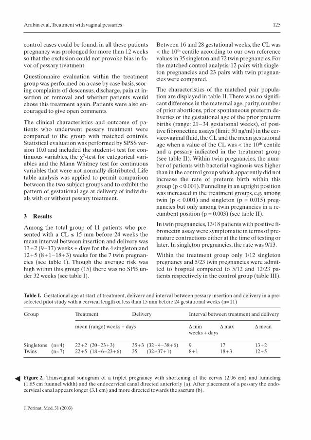
Among the total group of 11 patients who pre-sented with a CL ≤ 15 mm before 24 weeks themean interval between insertion and delivery was13+2 (9–17) weeks + days for the 4 singleton and12+5 (8+1–18+3) weeks for the 7 twin pregnan-cies (see table I). Though the average risk washigh within this group (15) there was no SPB un-der 32 weeks (see table I).
Between 16 and 28 gestational weeks, the CL was< the 10th centile according to our own referencevalues in 35 singleton and 72 twin pregnancies. Forthe matched control analysis, 12 pairs with single-ton pregnancies and 23 pairs with twin pregnan-cies were compared.
The characteristics of the matched pair popula-tion are displayed in table II. There was no signifi-cant difference in the maternal age, parity, numberof prior abortions, prior spontaneous preterm de-liveries or the gestational age of the prior pretermbirths (range: 21–34 gestational weeks), of posi-tive fibronectine assays (limit: 50 ng/ml) in the cer-vicovaginal fluid, the CL and the mean gestationalage when a value of the CL was < the 10th centileand a pessary indicated in the treatment group(see table II). Within twin pregnancies, the num-ber of patients with bacterial vaginosis was higherthan in the control group which apparently did notincrease the rate of preterm birth within thisgroup (p < 0.001). Funneling in an upright positionwas increased in the treatment groups, e.g. amongtwin (p < 0.001) and singleton (p = 0.015) preg-nancies but only among twin pregnancies in a re-cumbent position (p = 0.003) (see table II).
In twin pregnancies,13/18 patients with positive fi-bronectin assay were symptomatic in terms of pre-mature contractions either at the time of testing orlater. In singleton pregnancies, the rate was 9/13.
Within the treatment group only 1/12 singletonpregnancy and 5/23 twin pregnancies were admit-ted to hospital compared to 5/12 and 12/23 pa-tients respectively in the control group (table III).
Table I. Gestational age at start of treatment, delivery and interval between pessary insertion and delivery in a pre-selected pilot study with a cervical length of less than 15 mm before 24 gestational weeks (n=11)
Group Treatment Delivery Interval between treatment and delivery
mean (range) weeks + days Δ min Δ max Δ meanweeks + days
Singletons (n=4) 22+2 (20–23+3) 35+3 (32+4–38+6) 9 17 13+2Twins (n=7) 22+5 (18+6–23+6) 35 (32–37+1) 8+1 18+3 12+5
Figure 2. Transvaginal sonogram of a triplet pregnancy with shortening of the cervix (2.06 cm) and funneling(1.65 cm fuunnel width) and the endocervical canal directed anteriorly (a). After placement of a pessary the endo-cervical canal appears longer (3.1 cm) and more directed towards the sacrum (b).

126 Arabin et al,Treatment with vaginal pessaries
J. Perinat. Med. 31 (2003)
The mean duration of hospital stay among thoseadmitted for the treatment and the control groupsrespectively was 7 and 19 (7–28) days for single-ton and 36 (1–63) and 18 (1–63) days for twinpregnancies (see table III).
Intravenous betamimetics (ritodrine) were admin-istered in only 1/12 singleton pregnancy and5/23 twin pregnancies within the treatment group
compared to 5/12 and 10/23 patients respectively inthe control group (see table III). The mean dura-tion of intravenous tocolytic treatment among thepatients who received tocolysis was 3 and 10 (7–16)days for singleton and 13 (2–28) and 5 (1–11) daysfor twin pregnancies,for the treatment and the con-trol groups respectively (see table III).
The mean interval between TVS (< the 10th cen-
Table II. Patient demographics and risk factors for preterm birth in each subgroup of the case-control study with acervical length of below the 10th centile determined at the gestational week of pessary insertion, respectively the weekof matched controls
Singleton pregnancies (n=24) Twin pregnancies (n=46)
Pessary No pessary Significance* Pessary No pessary Significance*(n=12) (n=12) (n=23) (n=23)
Maternal age (mean/ range) 32 (26–43) 32 (25–38) ns 32 (27–40) 32 (24–40) nsNulliparous (n,%) 1 (8%) 3 (25%) ns 15 (65%) 16 (70%) nsPrior abortions (n,%) 7 (58%) 6 (50%) ns 3 (13%) 1 (4%) nsPrior SPB (n,%) 6 (50%) 7 (53%) ns 3 (13%) 0 nsGestational age of prior SPB 28 (23–34) 28 (21–34) ns 29 (20–34) – ns(mean/ range)Gestational age at TVS 24 (20–27) 24 (20–27) ns 23 (20–27) 24 (21–27) ns(mean/range)Fibronectin+ (n,%)* 4 (33%) 7 (53%) ns 18 (78%) 10 (43%) nsBacterial vaginosis (n,%) 3 (25%) 1 (8%) ns 6 (26%) 1 (4%) p<0.001Funneling supine position (n,%) 1 (8%) 1 (8%) ns 12 (52%) 5 (22%) p=0.003Funneling upright position (n,%) 10 (83%) 5 (42%) p= 0.015 23 (100%) 10 (43%) p<0.001CL supine position 29 (20–35) 28 (10–34) ns 25 (4–33) 27 (7–33) ns(mm,mean/range)CL upright position 24 (11–30) 26 (10–30) ns 17 (0–25) 24 (7–33) ns(mm,mean/range)
*Test: Mann-Whitney*Fibronectin was determined > 24 and < 28 gestational weeks
Table III. Days of admission and intravenous therapy with ß-mimetics for prevention of preterm birth in each sub-group of the case-control study
Singleton pregnancies (n=24) Twin pregnancies (n=46)
Pessary No pessary Pessary No pessary(n=12) (n=12) (n=23) (n=23)
Admission (n patients, total days) n=1, 7 days n=5, 95 days n=6, 216 days n=12, 215 daysAdmission (mean/range per patient admitted) 7 (7) days 19 (7–28) days 36 (3–68) days 18 (1–63) days
Tocolytic treatment (n patients, total days) n=1, 3 days n=3, 30 days n=5, 64 days n=10, 52 daysTocolytic treatment (mean/ range per patient 3 (3) days 10 (7–16) days 13 (2–28) days 5 (1–11) dayswith tocolysis)

Arabin et al,Treatment with vaginal pessaries 127
J. Perinat. Med. 31 (2003)
tile) and delivery was 99 (70–134) days in thetreatment and 67 (2–130) days in the controlgroup (p = 0.0184).For twin pregnancies, the meaninterval was 85 (43–129) days in the treatment
and 67 (21–100) days in the control group(p = 0.001) (table IV). The mean gestational age atdelivery was 38 (36+6–41) weeks for singletonpregnancies in the treatment group and 33+4
Figure 3. Gestational age at delivery expressed as logrank survival with and without pessary treatment started between20 and 28 weeks of gestation:(a) in 24 singleton pregnancies,matched pairs; (b) in 46 twin pregnancies,matched pairs.

128 Arabin et al,Treatment with vaginal pessaries
J. Perinat. Med. 31 (2003)
(26–38) weeks in the control group (p = 0.02), fortwin pregnancies it was 35+6 (33–37+4) and33+2 (24+4–37+2) respectively (p = 0.02) (seetable IV).
Among the 12 singleton pregnancies with pessary,there was no preterm delivery < 36 weeks com-pared to 6 cases in the control group (p < 0.001),whereby 3 cases were even < 32 weeks. Out of the23 twin pregnancies with pessary, there were 8 cas-es of preterm birth < 36 weeks whereby no casewas < 32 weeks, compared to 12 cases of a delivery< 36 weeks and 7 cases of preterm birth < 32 weeksin the control group (p < 0.001) (see table IV).
Life table analysis demonstrated that in patientswith a short cervix treatment with a vaginal sili-cone pessary could prolong pregnancy up to spon-taneous birth compared to a group without thattreatment in both singletons and twin pregnancies(see figure IIIa and b).
Questionnaire evaluation within the treatmentgroup on a case by cases basis indicated that thenumber of patients with complaints of descensusdeclined from 9 before therapy to 3 patients aftertherapy but complaints of discharge increasedfrom 8 to 17 patients after the application of a pes-sary (table V). Patients experienced the removal
as more painful than the application but had nopainful experiences during therapy.
In general, all patients who were treated and de-livered at our center had a positive opinion ofthe treatment; one patient was indifferent but allothers would undergo the same treatment in a fur-ther pregnancy or recommend it to a friend (seetable V).
Nevertheless, though it is generally believed thatvaginal silicone pessaries at least do no harm, wewere confronted with a patient with unexpectedsequelae who had been admitted to our centerdue to threatening preterm birth at 27 weeks.Since the contractions had stopped and the cervixwas shortened with funnelling throughout thecervix we used a vaginal pessary. At 32 gestationalweeks, the patient was discharged and further con-trolled in a peripheral hospital.Though she had in-creasing complaints of pressure, blood loss and fi-nally even premature rupture of membranes, thepessary was only removed during an advancedstage of labor. Soon after forceps delivery due tofetal distress she spontaneously lost a small ring-shaped part of her cervix. Pathologic examinationrevealed cervical tissue with a thrombosis of a cer-vical vein, which was probably due to the in-creased pressure, and edema of the cervix. Sixweeks after delivery, the cervix had recovered a
Table IV. Pregnancy outcome for each subgroup of the case-control study
Preterm birth Singleton pregnancies (n=24) Twin pregnancies (n=46)
Pessary No pessary Significance* Pessary No pessary Significance*(n=12) (n=12) (n=23) (n=23)
< 28 weeks (n,%) 0 2 (17%) ns 0 1 (4%) ns< 32 weeks (n,%) 0 3 (25%) ns 0 7 (30%) p<0.001< 36 weeks (n,%) 0 6 (50%) p<0.001 8 (35%) 12 (52%) ns
Interval (days,mean/ 99 67 p=0.0184 85 67 p=0.001range) between TVS (70–134) (2–130) (43–129) (21–100)before treatment orcontrols and delivery
Gestational age 38 33+4 p=0.02 35+6 33+2 p=0.02(weeks+days) at (36+6–41) (26–38) (33–37+4) (24+4–37+2)delivery (mean/range)
*Test: Mann-Whitney

Arabin et al,Treatment with vaginal pessaries 129
J. Perinat. Med. 31 (2003)
normal shape but was shortened to a length of2.5 cm.
4 Discussion
Transvaginal ultrasonographic examination of thecervix can be regarded as the best imaging modal-ity for the detection or exclusion of patients at riskfor SPB [12]. In contrast to digital examination,TVS allows the examiner to detect that the open-ing of the internal os is combined with a shorten-ing of the endocervical canal length. In a pilotstudy with multiple pregnancies, we demonstratedthat this is even more evident when the mother isin an upright position [4]. Ultrasonographic cervi-cal assessment has suggested that there is a widespectrum of the disease and may detect or excludea risk for preterm delivery irrespective of whethercervical shortening is a primary or secondaryevent.
Cervical incompetence is traditionally consideredas a cause of recurrent mid trimester abortion.Premature cervical ripening may be the result of acongenital disorder of the connective tissue, expo-sure to diethylstilbestrol in utero, traumatic dam-age to the structural integrity, uterine overdisten-
sion, repetitive bleeding or vascular lesions in theplacenta leading to membrane destabilization andlocal or intrauterine infection inducing an in-crease of cytokines and prostaglandines [29].Therefore preterm parturition is regarded as asyndrome with multiple etiologies which may leadto a condition in which there is activation of allcomponents of the common terminal pathwaywith uterine contractility, membrane activationand cervical incompetence [30].
Since the pathophysiology of premature ripeningof the cervix and premature labor varies betweensingleton and multiple pregnancy, we have collect-ed and interpreted our data separately for single-ton and twin pregnancies. An association betweena short cervix and/ or opening of the internal os(funneling) and SPB has been demonstrated inseveral studies both for singleton [2, 3, 13, 15, 18,19, 34] and twin pregnancies [11, 14, 20, 33, 37, 38].Cervical incompetence is not an all-or-nothingphenomenon, but more often a continuum. Wehave therefore performed longitudinal examina-tions in both study groups. While the detection ofa short cervix may alert physicians at an earlystage of pregnancy the optimal management ofpatients has not been determined.
Table V. Subjective experience of pessary treatment within the therapy group of the case-control study
Response (n) Singleton pregnancies Twin pregnancies Total(n=11/12) (n=18/23) (n=29/35)
n (%) Score* n (%) Score* n (%) Score*(x,range) (x, range) (x, range)
Claims of descensus 5 6,6 (4–9) 4 7,1 (7–8) 9 6,9 (4–9)before therapyClaims of descensus 2 6,5 (4–8) 1 7 3 6,7 (5–8)after therapy
Claims of discharge 5 4,2 (2–7) 3 4 (2–6) 8 4,1 (2–7)before therapyClaims of discharge 7 6 (2–9) 10 5 (3–7) 17 5,4 (2–9)after therapy
Pain during insertion 3 5 (3–6) 10 5,5 (1–9) 13 5,3 (1–9)Pain during removal 4 6 (2–9) 11 6 (2–9) 15 6 (2–9)
Chose again? 11 17 (1× ”don’t know) 28 (1× ”don’t know)Recommend to others? 11 17 (1× ”don’t know) 28 (1× ”don’t know)
*Score: the patients were able to rank their answers between 0 (no complaints) and 10 (severe complaints)

130 Arabin et al,Treatment with vaginal pessaries
J. Perinat. Med. 31 (2003)
Hospitalization for bed rest in multiple pregnan-cies was introduced into clinical practice withoutadequate controlled evaluation of its efficacy. Thepolicy has been subjected to limited well-con-trolled evaluation to clarify the beneficial or ad-verse effects. Women’s views of hospitalizationand costs have not yet been assessed. Currently nosound evidence exists to support a policy of rou-tine hospitalization for bed rest in multiple preg-nancies. For women with uncomplicated twinpregnancy, results suggest that such a policy mayeven be harmful since the risk of very pretermbirth seems to be increased [8].
Nevertheless, based on the studies of Papiernik[27] we believe that a reduction of physical stressfor women with multiple pregnancies in an outpa-tient setting is advisable.
Lash introduced the term “incompetent cervix” in1950 describing a first attempt at operative closureof the cervix [22]. Mc Donald [24] and Shirodkar[32] reported on a vaginal approach of a cervicalcerclage during pregnancy to prevent secondtrimester abortion and preterm delivery prefer-ably for patients with a history compatible with anincompetent cervix. With the introduction of TVS,the question arose as to whether cervical cerclagewould be an option to prevent SPB in patientswith a short cervix. Heath et al.[17] reported a re-duction in the rate of SPB in patients who under-went cervical cerclage and had a cervical length≤ 15 mm at around 23 gestational weeks. The rateof spontaneous onset of labor < 32 weeks was low-er (1 versus 11), in the study than in the controlgroup. All infants survived except for 1 child in thecontrol group who died in the neonatal period. Incontrast, within a randomized trial and a larger se-ries of the same group it was found that cerclagewas ineffective at reducing the rate of SPB (Nico-laides, unpublished]. Hassan et al. [16] performeda retrospective study in patients with a shortcervix < 15 mm whereby 25/77 patients underwentcerclage placement. The risk for spontaneous on-set of preterm delivery did not differ, but patientswith a cerclage had a higher rate of prematurerupture of membranes (PROM) (65% vs. 36%,p < 0.05). It was speculated that cerclage place-ment might predispose patients with a short cervixto a local inflammatory reaction. Accordingly,within a randomized controlled trial, Rust et al.[31] stated that cerclage did not reduce the rate of
SPB in patients with a cervical length < 25 mm orfunneling > 25%. However, Althuisius et al. [1]found that cerclage was effective in preventingSPB < 34 weeks’ gestation in patients with a cervix< 2.5 mm compared to a group with expectantmanagement (1/10 versus 5/8). In conclusion, thedata suggesting that cerclage might have a system-atic place in treating risk patients for SPB and ashort cervix as determined by TVS are not yetconvincing.
Vaginal pessaries have been designed for preg-nant women to direct the cervix more posteriorlyand thus change the inclination of the cervicalcanal so that the weight is more directed to the an-terior lower segment. This might prevent furtheropening of the internal os or even premature rup-ture of membranes based on pressure-relatedproblems. Pessaries have the advantage that theyare operator independent, non-invasive, easily toplace or to remove and not expensive. As early as1959, Cross reported on the use of a pessary forthe treatment of cervical incompetence [7]. Sever-al other authors followed using different models.Most of the published studies were either retro-spective or case controlled (9, 21, 26, 28, 36],Forster et al. were the only ones who conducted aprospective randomized study, comparing cer-clage with pessary [10]. They did not find signifi-cant differences between these groups. Neverthe-less, the study included a high number of low riskpatients with the only indication being a history ofSPB. In a more recent review article on the use ofpessaries for the treatment of cervical incompe-tence and prevention of preterm delivery the au-thor concluded that pessaries may be consideredin women who are not eligible for cerclage or incases of cervical changes detected by TVS [25].
To date, only Ludmir et al. compared 15 patientswith cervical shortening and some dilatation diag-nosed by TVS at around 22 weeks who were eithertreated with bed rest (n = 8) or with a vaginal pes-sary (n = 7) [23]. Pessary treatment resulted in aprolongation of pregnancy of a mean of 9.2 ± 4.6gestational weeks whereas bed rest alone only re-sulted in a prolongation of 5.1 ± 3.6 gestationalweeks (p = 0.03). These results are comparable toour results of patients with pessary treatment anda cervical length at around 22 weeks of < 1.5 cm(see table I).

Arabin et al,Treatment with vaginal pessaries 131
J. Perinat. Med. 31 (2003)
None of the previously published studies reportedon a complication following the use of the pes-saries. However, some basic guidelines should beconsidered. We recommend informing patientsthat there is not yet strong evidence that cervicalpessaries prevent SPB.
Using TVS to detect a short cervix and/or funnel-ing and the application of a pessary does not meanthat other screening methods or interventions(e.g. screening for pH-values, infections and an-tibiotic treatment) should be neglected. On thecontrary, since SPB may be a multifactorialprocess, bacterial vaginosis and other infectionshave to be excluded before application and, if nec-essary, treated. The pessary should be removed incases with PROM, blood loss, increasing contrac-tions or pain.
The obstetrician should check whether the cervixis not too firmly surrounded by the upper ring ofthe pessary, especially when the patient reports onspecific complaints such as blood loss or pain. Thepessary may stay in place until around 37 weeks.
There might be some increase of abacterial dis-charge. However, the spectrum of the vaginal flo-ra will usually not be substantially altered.
We experienced a complication (venous thrombo-sis in cervical tissue) following the use of a pessaryin a patient who was followed up by a referringhospital (see above). This might have been pre-vented if the pessary had been removed after thefirst complaints or at least at an earlier stage of la-bor after PROM.
In conclusion, insertion of a vaginal pessary maybe a cost-effective preventive treatment in pa-tients at risk for SPB not due to poor placental in-vasion and/or infection, but mainly based on TVSresults demonstrating a change of mechanicalproperties of the cervix such as shortening of cer-vical length or funneling. Since the allocationwithin retrospective matched pair studies mightbe biased by hidden confounders, prospectivestudies are needed to corroborate our preliminaryresults.
AbstractObjective: The purpose was to determine the effect ofvaginal pessaries in patients at risk for spontaneouspreterm birth (SPB).Study Design: Transvaginal sonography (TVS) was lon-gitudinally performed to measure cervical length (CL)in 258 singleton at risk for SPB and 282 twin pregnancies.Pairs with or without treatment were matched for gesta-tional age and the CL at examination.Results: In 4 singleton and 7 twin pregnancies the CLwas < 15 mm before 24 weeks, the mean interval be-tween pessary insertion and delivery was 13+2 and12+5 weeks respectively. For the matched control analy-sis, 12 pairs with singleton and 23 pairs with twin preg-nancies were compared. For singleton pregnancies, themean interval between TVS and delivery was 99(70–134) days in the treatment and 67 (2–130) days in
the control group (p = 0.0184), the mean gestational ageat delivery was 38 (36+6–41) and 33+4 (26–38) weeksrespectively (p = 0.02). For twin pregnancies, the intervalwas 85 (43–129) days in the treatment and 67 (21–100)days in the control group (p = 0.001), gestational age atdelivery was 35+6 (33–37+4) and 33+2 (24+4–37+2)respectively (p = 0.02). Within singleton pregnancieswith pessary, there was no SPB < 36 weeks compared to6/12 cases in the control group (p < 0.001). Within twinpregnancies, the rates were 8/23 cases with SPB< 36 weeks but none < 32 weeks, compared to 12/23 cas-es with SPB < 36 weeks and 7/23 cases < 32 weeks in thecontrol group (p < 0.001).Conclusions: Insertion of a vaginal pessary may be acost-effective preventive treatment in patients at risk forSPB. Prospective controlled trials are needed.
Keywords: Cervical insufficiency, spontaneous preterm birth, transvaginal ultrasound, vaginal pessaries.
References[1] Althusius SM, GA Dekker, HP van Geijn, BJ
Bekedam, P Hummel: Cervical incompetence pre-vention randomized cerclage trial: study design andpreliminary results. Am J Obstet Gynecol 183(2000) 823
[2] Andersen HF, CE Nugent, SD Wanty, RH Hayashi:Prediction of risk for preterm delivery by ultrasono-graphic measurement of cervical length. Am J Ob-stet Gynecol 163 (1990) 859
[3] Andrews WW, R Copper, JC Hauth, RL Golden-
berg, C Neely, M Dubard: Second-trimester cervi-cal ultrasound: associations with increased risk forrecurrent early spontaneous delivery. Obstet Gy-necol 95 (2000) 222
[4] Arabin B, R Aardenburg, J van Eyck: Maternal po-sition and ultrasonic cervical assess-ment in multi-ple pregnancy. Preliminary observations. J ReprodMed 42 (1997) 719
[5] Arabin B, J van Eyck: Sonographic diagnosis ofcervical incompetence for prevention and manage-ment. Ultrasound Review 1 (2001) 195
[6] Arabin B, M Hübener, J Halbesma, J van Eyck:Sonographic diagnosis of cervical incompetence intwin pregnancies. Ultrasound Review 1 (2001) 340
[7] Cross RG: Treatment of habitual abortion due tocervical incompetence. Lancet 2 (1959) 127
[8] Crowther CA: Hospitalisation and bed rest formultiple pregnancy (Cochrane Review). In: TheCochrane Library, Issue 1. Oxford 2002 (UpdateSoftware)
[9] Dahl J, MS Barz: Prevention of premature labor bymeans of supporting pessaries (1st experiences). ZArztl Fortb 73 (1979) 1010
[10] Forster F, R During, G Schwarzlus: Therapy ofcervix insufficiency-cerclage or support pessary?Zentralbl Gynaekol 108 (1986) 230
[11] Goldenberg RL, JD Iams, M Miodovnik, JP VanDorsten, G Thurnau, S Bottoms, et al: The pretermprediction study: risk factors in twin gestations. AmJ Obstet Gynecol 175 (1996) 1047
[12] Gomez R, M Galasso, R Romero, M Mazor, YSorokin, L Goncalves, et al: Ultrasonographic ex-amination of the uterine cervix is better than cervi-cal digital examination as a predictor of the likeli-hood of premature delivery in patients withpreterm labor and intact membranes. Am J ObstetGynecol 171(1994) 956
[13] Guzman ER, C Mellon, AM Vintzileos, CVAnanth, C Walters, K Gipson: Longitudinal assess-ment of endocervical canal length between 15 and24 weeks’ gestation in women at risk for pregnancyloss or preterm birth. Obstet Gynecol 92 (1998) 31
[14] Guzman ER, C Walters, C O’Reilly-Green, WLKinzler, R Waldron, J Nigam, et al.: Use of cervicalultrasonography in prediction of spontaneouspreterm birth in twin gestations. Am J Obstet Gy-necol 183 (2000) 1103
[15] Hassan SS, R Romero, SM Berry, K Dang, SCBlackwell, MC Treadwell, et al. Patients with an ul-trasonographic cervical length < or = 15 mm havenearly a 50% risk of early spontaneous pretermdelivery. Am J Obstet Gynecol 182 (2000) 1458
[16] Hassan S, R Romero, E Maymon, SM Berry, SCBlackwell, MC Treadwell, et al. Does cervical cer-clage prevent preterm delivery in patients with ashort cervix? Am J Obstet Gynecol 184 (2001)1325
[17] Heath VC, AP Souka, I Erasmus, DM Gibb, KHNicolaides: Cervical length at 23 weeks of gesta-tion: the value of Shirodkar suture for the shortcervix. Ultrasound Obstet Gynecol 12 (1998) 318
[18] Hibbard JU, M Tart, AH Moawad: Cervical lengthat 16–22 weeks’ gestation and risk for preterm de-livery. Obstet Gynecol 96 (2000) 972
[19] Iams JD, J Paraskos, MB Landon, JN Teteris, FFJohnson: Cervical sonography in preterm labor.Obstet Gynecol 84 (1994) 40
[20] Imseis HM, TA Albert, JD Iams: Identifying twingestations at low risk for preterm birth with a trans-vaginal ultrasonographic cervical measurement at24 to 26 weeks’ gestation. Am J Obstet Gynecol177 (1997) 1149
[21] Jorde A, B Hamann, KH Belling: Modification ofthe rate of low birth weight infant. Z Arztl Fortb 76(1982) 553
[22] Lash AF, SR Lash: Habitual abortion: the incom-petent internal os of the cervix. Am J Obstet Gy-necol 59 (1950) 68
[23] Ludmir J, JR Mantione, RH Debbs, HM Sehdev: Ispessary a valid treatment for cervical change dur-ing the late midtrimester. J Soc Gynecol Investig 9(Supplement) 2002) 11
[24] Mc Donald IA: Suture of the cervix for inevitablemiscarriage.J Obstet Gynecol Br Emp 64 (1957) 346
[25] Newcomer J: Pessaries for the treatment of incom-petent cervix and premature delivery. Obstet Gy-necol Survey 55 (2000) 443
[26] Oster S, CT Javert: Treatment of the incompetentcervix with the Hodge pessary. Obstet Gynecol 28(1966) 206
[27] Papiernik E, J Bouyer, D Collin: Prevention ofpreterm births: a perinatal study in Haguenau,France. Pediatrics 76 (1985) 154
[28] Quaas L, HG Hillemans, A du Bois, H Schillinger:The Arabin Cerclage Pessary – An alternative tosurgery. Geburtshilfe Frauenheilk 50 (1990) 429
[29] Romero R, R Gonzales, W Sepulveda, F Brandt, MRamirez, Y Sorokin, et al.: Infection and laborVIII. Microbial invasion of the amniotic cavity inpatients with suspected cervical incompetence:prevalence and clinical significance. Am J ObstetGynecol 167 (1992) 1086
[30] Romero R, M Mazor, H Munoz, R Gomez, MGalasso, DM Sherer: The preterm labor syndrome.Ann N Y Acad Sci 734 (1994) 414
[31] Rust OA, RO Atlas, KJ Jones, BN Benham, J Bal-ducci: A randomized trial with cerclage versus nocerclage among patients with ultrasonographicallydetected second-trimester preterm dilatation ofthe internal os. Am J Obstet Gynecol 183 (2000)830
[32] Shirodkar JN: A new method of operation for ha-bitual abortions in second trimester of pregnancy.Antiseptic 52 (1955) 290
132 Arabin et al,Treatment with vaginal pessaries
J. Perinat. Med. 31 (2003)

[33] Souka AP, V Heath, S Flint, I Sevastopoulou, KHNicolaides: Cervical length at 23 weeks in twins inpredicting spontaneous preterm delivery. ObstetGynecol 94 (1999) 450
[34] Timor-Tritsch IE, F Boozarjomehri, Y Masakows-ki, A Monteagudo, CR Chao: Can a “snapshot”sagittal view of the cervix by transvaginal ultra-sonography predict active preterm labor? Am JObstet Gynecol174 (1996) 990
[35] To MS, C Skentou, S Cicero, KH Nicolaides: Cervi-cal assessment at the routine 23´weeks scan: prob-lems with transabdominal sonography. UltrasoundObstet Gynecol 15 (2000) 288
[36] Vitsky M: The incompetent cervical os and the pes-sary. Am J Obstet Gynecol 87 (1963) 144
[37] Wennerholm UB, B Holm, I Mattsby-Baltzer, TNielsen, J Platz-Christensen, G Sundell, et al.: Fetalfibronectin, endotoxin, bacterial vaginosis and cer-
vical length as predictors of preterm birth andneonatal morbidity in twin pregnancies.Br J ObstetGynaecol 104 (1997) 1398
[38] Yang JH, K Kuhlman, S Daly, V Berghella: Predic-tion of preterm birth by second trimester cervicalsonography in twin pregnancies. Ultrasound Ob-stet Gynecol 15 (2000) 288
Received July 3, 2002. Revised October 13, 2002.Accepted October 23, 2002.
Birgit Arabin, MD, PhDIsala Clinics (Sophia)8025 AB ZwolleNetherlandsTel.: + (31) 38 424 5000Fax: + (31) 38 424 7676e-mail: [email protected]
Arabin et al,Treatment with vaginal pessaries 133
J. Perinat. Med. 31 (2003)