isbn 978-1-904842-39-2 9 781904842392 - scion medical · 7 disorders of red cell survival 114 7.1...
TRANSCRIPT

HAEMATOLOGY 2nd EdITIOn
C.J. PALLISTER AND M.S. WATSON
www.scionpublishing.com
This fully revised and updated edition of Haematology provides an up-to-date review of the scientific principles that underpin our understanding of the blood and blood-forming tissues in health and disease. It describes how advances in biochemistry, physiology, genetics and cell biology have contributed to the study of blood and its disorders. The book is written in an approachable style that focuses on improving understanding and encourages students to think around the subject. The numerous illustrations and defined learning objectives are designed to aid effective learning, while the many ‘fast facts’ boxes broaden the interest and perspective of the material. Further reading lists and website information guide students to more detailed coverage if required.
This book should prove useful for undergraduate and postgraduate students of haematology, as well as medical students and those embarking on higher professional qualifications.
RELATED BIOMEDICAL SCIENCE TITLES:
Cellular Pathology 2e 9781904842309Clinical Biochemistry 2e 9781904842415Human Genetics 2e 9781904842736Immunology 9781904842569Medical Microbiology 9781904842613Transfusion Science 2e 9781904842408
I S BN 978-1-904842-39-2
9 7 8 1 9 0 4 8 4 2 3 9 2
HAEMATOLOGY2nd EdITIOn
C.J. PALLISTER AND M.S. WATSON
HAEMATOLOGY
2nd EdITIOn
PALLISTER & WATSON

HAEMATOLOGY2nd Edition
00-Haematology-Prelims-ppp.indd 1 01/10/2010 13:23

00-Haematology-Prelims-ppp.indd 2 01/10/2010 13:23

HAEMATOLOGY2nd Edition
Chris PallisterManaging Director, SciMed Consulting Ltd
formerly, Reader in Haematology, University of the West of England
and
Malcolm Watsonformerly, Senior Lecturer in Haematology,
University of the West of England
00-Haematology-Prelims-ppp.indd 3 01/10/2010 13:23

Second edition © Scion Publishing Ltd, 2011
ISBN 978 1904842 39 2
First edition published in 1999 by Arnold (ISBN 0 750 62457 4)
All rights reserved. No part of this book may be reproduced or transmitted, in anyform or by any means, without permission.
A CIP catalogue record for this book is available from the British Library.
Scion Publishing Limited�e Old Haylo�, Vantage Business Park, Bloxham Road, Banbury, OX16 9UXwww.scionpublishing.com
Important Note from the Publisher
�e information contained within this book was obtained by Scion PublishingLimited from sources believed by us to be reliable. However, while every e�ort hasbeen made to ensure its accuracy, no responsibility for loss or injury whatsoeveroccasioned to any person acting or refraining from action as a result of informationcontained herein can be accepted by the authors or publishers.
Typeset by Phoenix Photosetting, Chatham, Kent, UKPrinted by �omson Litho, East Kilbride, UK
00-Haematology-Prelims-ppp.indd 4 01/10/2010 13:23

Contents
Preface xiiiAbbreviations xv
1 Content of the blood 1 1.1 Content of the blood 2 Plasma 2 Red cells 3 Platelets 7 White cells 8 Suggested further reading 15 Self-assessment questions 15
2 Haemopoiesis 17 2.1 Ontogeny of haemopoiesis 18 Embryonic haemopoiesis 19 Fetal haemopoiesis 19 Haemopoiesis in the developing child and adult 20 2.2 Sequence of di�erentiation of blood cells 21 2.3 Control of haemopoiesis 22 2.4 The haemopoietic micro-environment 24 2.5 Erythropoiesis 27 2.6 Granulopoiesis and monopoiesis 29 2.7 Thrombopoiesis 29 2.8 Lymphopoiesis 30 B lymphopoiesis 30 T cell di�erentiation 33 NK cell di�erentiation 34 Suggested further reading 35 Self-assessment questions 35
3 Anaemia 36 3.1 De�ning anaemia 37 [Hb] and acute blood loss 37 [Hb] and pregnancy 38 [Hb] and severe burns 38 [Hb] and dehydration 38 [Hb] and hypoxia 38 [Hb] and thalassaemia 38 [Hb] and structural haemoglobinopathy 38 [Hb] and posture 39 [Hb] and the reference range 39 Anaemia as a clinical disorder 39
00-Haematology-Prelims-ppp.indd 5 01/10/2010 13:23

vi Contents
3.2 Mechanisms of anaemia 41 3.3 Describing red cells 42 Anaemia and red cell morphology 44 3.4 Approach to anaemia diagnosis 45 Suggested further reading 46 Self-assessment questions 46
4 Disorders of iron metabolism 48 4.1 Iron 49 The role of iron within the body 49 Body iron distribution 49 Daily iron requirements 49 Iron absorption 50 Iron transport 52 Iron storage 54 4.2 Iron de�ciency 55 Causes of iron de�ciency 56 Pathophysiology 57 4.3 Anaemia of chronic disorders 60 4.4 Sideroblastic anaemias 61 Hereditary sideroblastic anaemia 61 Acquired clonal sideroblastic anaemia 62 Secondary (reversible) sideroblastic anaemia 62 4.5 Iron overload 63 Hereditary haemochromatosis 63 Transfusion-associated haemosiderosis 65 Dietary causes 65 Suggested further reading 66 Self-assessment questions 66
5 Megaloblastic anaemias 67 5.1 Vitamin B12 68 The role of vitamin B12 within the body 68 Daily vitamin B12 requirements and body stores 68 Vitamin B12 absorption 69 Vitamin B12 transport 70 5.2 Folates 70 The role of folate within the body 70 Daily folate requirements and body stores 71 Folate absorption and transport 71 Role of vitamin B12 and folate in DNA and RNA synthesis 71 5.3 Megaloblastic anaemias 73 Vitamin B12 de�ciency 73 Folic acid de�ciency 76 5.4 Causes of megaloblastic anaemia other than haematinic de�ciency 77 Biochemical basis of megaloblastic change 78 Pathophysiology 79 Diagnosis of megaloblastic anaemia 80 Suggested further reading 81 Self-assessment questions 82
00-Haematology-Prelims-ppp.indd 6 01/10/2010 13:23

Contents vii
6 Inherited disorders of haemoglobin 83 6.1 Haemoglobin synthesis 84 Haem synthesis 84 Globin synthesis 85 6.2 Haemoglobin structure 88 Primary structure of globin 88 Secondary structure of globin 88 Tertiary structure of globin 88 Quaternary structure of haemoglobin 89 6.3 Haemoglobin function 90 Haemoglobin oxygen binding 90 Oxygen delivery 92 Carbon dioxide transport 93 Haemoglobin oxygen a�nity 94 6.4 Inherited haemoglobin disorders 95 The thalassaemias 95 The structural haemoglobinopathies 105 Investigation of suspected haemoglobinopathy 111 Suggested further reading 112 Self-assessment questions 113
7 Disorders of red cell survival 114 7.1 Inherited intrinsic haemolytic disorders 116 Composition of the red cell membrane 116 Primary membrane disorders 122 7.2 Acquired (extrinsic) haemolytic disorders 126 Haemolysis secondary to immune mechanisms 126 Autoimmune haemolysis 127 Alloimmune haemolysis 129 7.3 Pathophysiology of haemolytic disorders 132 Suggested further reading 134 Self-assessment questions 134
8 Disorders of red cell metabolism 135 8.1 The Embden–Meyerhof pathway 137 The reaction steps of the Embden–Meyerhof pathway 137 The Rappaport–Luebering shunt 139 8.2 The hexose monophosphate pathway 139 The reaction steps of the hexose monophosphate pathway 140 8.3 The purine salvage pathway 141 8.4 Disorders of red cell metabolism 141 Disorders of the Embden–Meyerhof pathway 142 Disorders of the hexose monophosphate pathway 144 8.5 Laboratory investigation of a suspected enzymopathy 148 Suggested further reading 149 Self-assessment questions 149
9 Non-malignant leucocyte disorders 150 9.1 Disorders of neutrophils 151 Quantitative neutrophil disorders 151 Neutropenia 152
00-Haematology-Prelims-ppp.indd 7 01/10/2010 13:23

viii Contents
Qualitative defects of neutrophils 153 Morphological abnormalities of neutrophils 155 9.2 Disorders of eosinophils and basophils 157 Eosinophilia 157 Basophilia 159 9.3 Disorders of mononuclear phagocytes 159 Quantitative monocyte disorders 159 Langerhans cell histiocytosis 159 Lysosomal storage diseases 162 9.4 Disorders of lymphocytes 162 Lymphocytosis and lymphopenia 162 Suggested further reading 163 Self-assessment questions 163
10 Haematological malignancies 165 10.1 Classi�cation of the haematological malignancies 165 Myeloid neoplasms 168 Lymphoid neoplasms 168 Neoplasms with myeloid and lymphoid lineage 168 10.2 Aetiology of the haematological malignancies 169 Aetiological associations 169 Cytogenetics and haematological malignancy 173 10.3 Biology of malignancy 175 Oncogenes and malignancy 175 Tumour suppressor genes and malignancy 175 DNA repair genes and malignancy 177 The cell cycle 181 Growth factors 185 Programmed cell death and its regulation 185 Epigenetic control of gene transcription 187 10.4 Principles of treatment for haematological malignancy 188 Cytotoxic chemotherapy 188 Haemopoietic stem cell transplantation 193 Radiotherapy 195 Supportive care 195 Other supportive care needs 197 Suggested further reading 197 Self-assessment questions 197
11 Acute leukaemias 199 11.1 Classi�cation of the acute leukaemias 199 History of acute leukaemia classi�cation 199 The WHO classi�cation of acute leukaemia 201 11.2 Acute myeloid leukaemia 204 Epidemiology of AML 205 Aetiological factors 205 Molecular basis of AML 206 Clinical presentation of AML 211 Diagnosis of AML 213 Prognosis of AML 214 Clinical management of AML 216
00-Haematology-Prelims-ppp.indd 8 01/10/2010 13:23

Contents ix
Treatment of relapsed or refractory AML 218 Maintenance therapy 218 11.3 Acute lymphoblastic leukaemia / lymphoma in children 219 Clinical presentation of childhood ALL 219 Diagnosis of childhood ALL 220 Classi�cation of childhood ALL 220 Molecular basis of childhood ALL 223 Prognosis of childhood ALL 224 Clinical management of childhood ALL 225 11.4 Acute lymphoblastic leukaemia / lymphoma in adults 227 Prognosis of adult ALL 228 Molecular basis of adult ALL 228 Clinical management of adult ALL 228 Suggested further reading 229 Self-assessment questions 229
12 Chronic lymphoid leukaemias 231 12.1 Chronic lymphocytic leukaemia 232 Pathophysiology of CLL 233 Cytogenetics in CLL 233 Molecular abnormalities in CLL 234 Clinical presentation of CLL 234 Diagnosis of CLL 234 Clinical staging and prognosis of CLL 235 Transformation of CLL 237 Clinical management of CLL 237 12.2 Hairy cell leukaemia 238 Clinical presentation of HCL 238 Diagnosis of HCL 239 Variant HCL 239 Clinical management of HCL 239 12.3 B-prolymphocytic leukaemia 240 12.4 T-prolymphocytic leukaemia 241 Suggested further reading 241 Self-assessment questions 241
13 The myelodysplastic syndromes 243 13.1 What are myelodysplastic syndromes? 243 13.2 Epidemiology 244 13.3 Classi�cation of MDS 245 The FAB classi�cation system 245 The WHO classi�cation system 248 Risk levels and prognosis 250 13.4 The diagnosis of MDS 252 13.5 Treatment and management of MDS 254 Supportive care 254 Low-intensity therapy 254 High-intensity therapy 255 Suggested further reading 255 Self-assessment questions 255
00-Haematology-Prelims-ppp.indd 9 01/10/2010 13:23

x Contents
14 The chronic myeloproliferative neoplasms 256 14.1 Polycythaemia 257 Polycythaemia vera 258 Primary familial polycythaemia 261 Relative (apparent) polycythaemia 261 Secondary polycythaemias 261 14.2 Essential thrombocythaemia 262 14.3 Primary myelo�brosis 263 14.4 Chronic myeloid leukaemia 263 Chronic phase CML 264 The Philadelphia chromosome 265 Accelerated phase CML 266 Blast phase CML 266 Treatment of CML 267 14.5 Philadelphia chromosome-negative chronic myeloid disorders 268 Chronic neutrophilic leukaemia 268 Atypical CML 269 Chronic myelomonocytic leukaemia 269 Chronic eosinophilic leukaemia 270 Juvenile myelomonocytic leukaemia 271 Suggested further reading 272 Self-assessment questions 272
15 The lymphomas 273 15.1 Hodgkin disease 274 Epidemiology of HD 275 Aetiology of HD 276 Presentation and clinical features of HD 276 Investigation of HD 276 Staging and prognosis of HD 277 Treatment of HD 279 15.2 Non-Hodgkin lymphomas (NHL) 281 Epidemiology of the NHLs 281 Aetiological factors 282 Classi�cation of NHL 284 Clinical features of NHL at presentation 286 Clinical evaluation of the NHL patient 286 Staging and prognosis of NHL 288 Some important subtypes of NHL 289 Clinical management of NHL 292 Suggested further reading 294 Self-assessment questions 295
16 Myeloma and related disorders 296 16.1 Multiple myeloma 296 Epidemiology and aetiology 297 Diagnosis 298 Staging and prognosis 299 Pathophysiology of MM 300 Treatment strategies 307 16.2 Variants of myeloma 308
00-Haematology-Prelims-ppp.indd 10 01/10/2010 13:23

Contents xi
Indolent myeloma 308 Smouldering myeloma 308 Osteosclerotic myeloma (POEMS syndrome) 308 Plasma cell leukaemia 309 Non-secretory myeloma 309 16.3 Monoclonal gammopathy of undetermined signi�cance 309 16.4 Solitary plasmacytoma of bone 310 16.5 Extramedullary plasmacytoma 310 16.6 Waldenström macroglobulinaemia 310 16.7 Heavy chain diseases 311 16.8 Immunoglobulin deposition diseases 311 Systemic light chain disease 311 Primary amyloidosis 312 Suggested further reading 314 Self-assessment questions 314
17 Bone marrow failure 315 17.1 Classi�cation of the bone marrow failures 316 17.2 The inherited bone marrow failures 316 Fanconi anaemia 316 Pearson syndrome 317 Diamond–Blackfan syndrome 317 The congenital dyserythropoietic anaemias 318 Inherited neutropenias 318 Congenital amegakaryocytic thrombocytopenia 320 Familial platelet disorder 320 17.3 Acquired bone marrow failure 320 Aplastic anaemia and chemical or physical agents 321 Immune-mediated aplastic anaemia 322 Classi�cation of aplastic anaemia 323 Transient erythroblastopenia of childhood 324 Acquired pure red cell aplasia 324 Paroxysmal nocturnal haemoglobinuria 325 Suggested further reading 327 Self-assessment questions 328
18 Overview of haemostasis 329 18.1 The vascular system 331 Contribution of the vascular system to haemostasis 331 18.2 Blood platelets 332 Platelet function 333 18.3 The blood coagulation system 336 The role of vitamin K in blood coagulation 337 Blood coagulation in vivo 337 Blood coagulation in vitro 339 18.4 Inhibitors of blood coagulation 341 Tissue factor pathway inhibitor 342 Antithrombin III 342 Heparin cofactor II 342 The protein C pathway 342 18.5 The �brinolytic system 343
00-Haematology-Prelims-ppp.indd 11 01/10/2010 13:23

xii Contents
Plasminogen activators 344 Plasminogen and plasmin 344 Inhibitors of �brinolysis 344 18.6 Tests of haemostasis 344 Tests of vascular function 345 Tests of platelet number and function 345 Tests of coagulation 345 Suggested further reading 346 Self-assessment questions 347
19 Bleeding disorders 348 19.1 Inherited bleeding disorders 349 Inherited structural defects of the vascular system 349 Inherited defects of platelets 351 Inherited thrombocytopenias 351 Inherited thrombocytopathies 352 Inherited coagulopathies 353 19.2 Acquired disorders of haemostasis 362 Disseminated intravascular coagulation 362 Haemostatic disorders associated with malignancy 366 Haemostatic disorders associated with liver disease 366 Haemostatic disorders associated with pregnancy and delivery 367 Haemostatic disorders associated with the neonatal period 367 19.3 Acquired purpuras 368 Vascular purpuras 369 Thrombocytopenic purpuras 369 19.4 Acquired inhibitors of coagulation factors 370 Suggested further reading 371 Self-assessment questions 371
20 Thrombotic disorders 373 20.1 Risk factors for arterial thrombosis 374 20.2 Risk factors for venous thrombosis 374 20.3 Inherited thrombotic disorders 375 Antithrombin de�ciency 375 Protein C 377 Protein S 377 Factor V Leiden 378 Prothrombin allele G20210A 379 Hyperhomocysteinaemia 379 Inherited de�ciency of �brinolysis 379 20.4 Acquired thrombophilia 379 The anti-phospholipid syndrome 379 20.5 Laboratory investigation of thrombophilia 380 20.6 Prophylaxis and treatment of thrombosis 380 Anticoagulants 380 Thrombolytic drugs 383 Suggested further reading 384 Self-assessment questions 384
Answers to self-assessment questions 385Index 393
00-Haematology-Prelims-ppp.indd 12 01/10/2010 13:23

CHAPTER Inherited disorders of haemoglobin6
Learning objectivesAfter studying this chapter you should con�dently be able to:� Outline the structure and biosynthesis of haemoglobin
Although haem and globin syntheses occur separately within developing red cell precursors, their rates of synthesis are carefully coordinated to ensure optimal e�ciency of haemoglobin assembly. Haem is synthesized to some extent in virtually all human tissues but the most important sites are the liver (for incorporation into cytochromes), muscle (for incorporation into myoglobin) and red cell precursors. Globins are all single chain polypeptides and their synthesis is under genetic control. Humans normally synthesize six di�erent types of globin chains (a, b, g, d, z and e) at di�erent stages of life.
� Discuss the relationship between structures and function of haemoglobinThe structure of haemoglobin is intimately related to its function as a transporter of oxygen and carbon dioxide. Each haemoglobin molecule is capable of carrying four oxygen molecules, one for each haem group. Among the most important facets of the structure:function relationship are the haem:haem interaction, the maintenance of iron in the Fe2+ state, the impact of pH, and the binding of 2,3-DPG to b-globin chains.
� Di�erentiate clearly between the thalassaemias and structural haemoglobinopathiesThe thalassaemias are inherited disorders of the rate of globin synthesis whereas structural haemoglobinopathies are disorders of globin structure. In practice, there is overlap between these because some structurally abnormal globins are synthesized at a reduced rate.
� Outline the pathophysiology of a and b thalassaemiaThe thalassaemias are among the most common single gene disorders in the world and are most common in malarial areas, demonstrating balanced polymorphism. They are characterized by reduced, or absent, synthesis of one or more globin chain type. The resultant imbalance in globin chain synthesis leads to ine�ective erythropoiesis and a shortened red cell lifespan. The spectrum of clinical severity of the thalassaemias is very wide.
� Outline the classi�cation of the structural haemoglobinopathiesThe alteration in molecular function induced by structural abnormality is dependent upon the position of the mutation and on the properties of the amino acids involved. Alterations to the amino acid sequence within areas important for normal function typically lead to predictable alterations in molecular behaviour. In general terms, the structural haemoglobinopathies can be classi�ed according to the impact of their altered function as, for example, unstable or thalassaemia-like.
� Describe the pathophysiology of sickle cell diseaseThe clinical consequences of haemoglobin S stem from its tendency to polymerize, forming long, rigid structures called ‘tactoids’, which cause contortion of the red cell into elongated and poorly deformable sickle shapes. Typically, heterozygotes are clinically normal but homozygotes can experience vaso-occlusive sickling crises, aplastic crises, acute splenic sequestration and are susceptible to bacterial infection. Co-inheritance of other haemoglobinopathies can in�uence the clinical course of this disease.
Chapter 6 Inherited disorders of haemoglobin
01-Haematology-Chs1-8-ccp.indd 83 01/10/2010 12:52

84 Chapter 6 Inherited disorders of haemoglobin
�e haemoglobins are red globular proteins which have a molecular weight of about 64 500 and comprise almost one-third of the weight of a red cell. �eir primary function is the carriage of oxygen from the lungs to the tissues. A vital secondary role is the facilitation of the reverse transportation of carbon dioxide. �ey also play a role in blood bu�ering. Over 400 di�erent variants of haemoglobin have been described but all share the same basic structure of four globin polypeptide chains, each with a single prosthetic haem group.
6.1 HAEMOGLOBIN SYNTHESIS
Although haem and globin syntheses occur separately within developing red cell precursors, their rates of synthesis are carefully coordinated to ensure optimal e�ciency of haemoglobin assembly.
Haem synthesis
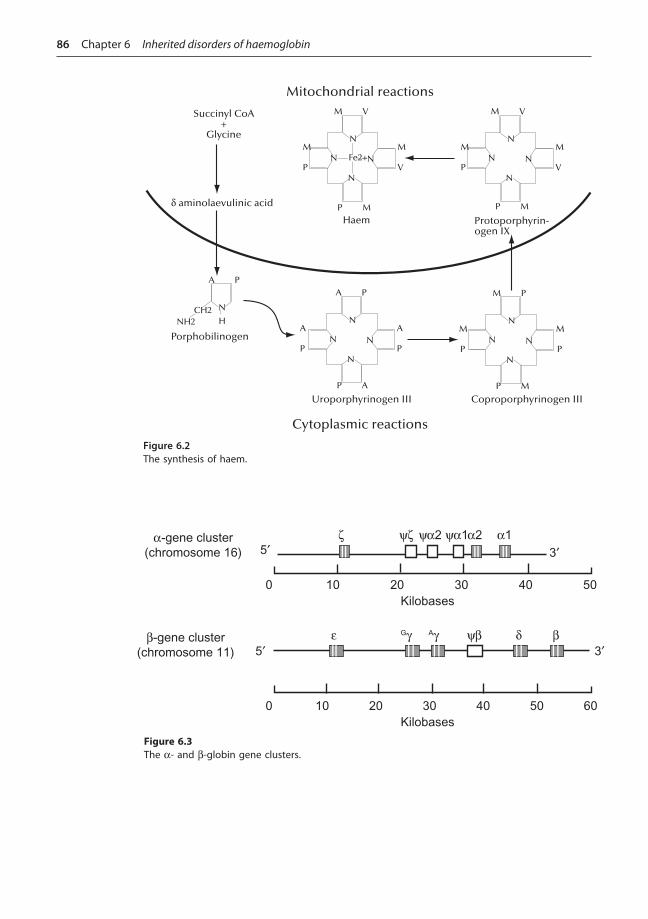
Haem belongs to the class of pigments known as porphyrins. It is composed of four pyrroles linked by methene bridges, with each pyrrole being bound to a central ferrous ion (Fe2+) as shown in Figure 6.1 (see also Box 6.1). Haem is synthesized to some extent in virtually all human tissues but the most important sites are the liver (for incorporation into cytochromes), muscle (for incorporation into myoglobin) and red cell precursors.
�e �rst step of haem synthesis is the rate-limiting reaction for the whole process and involves the combination of glycine and the succinic acid derivative succinyl Co-A to produce d-aminolaevulinic acid (d-ALA). �e reaction is energy-dependent and occurs within the mitochondria. �e catalyst for d-ALA synthesis is the enzyme d-ALA synthetase. �e presence of free globin chains stimulates d-ALA synthesis while the presence of free haem groups is inhibitory, thus providing a control mechanism for the rate of haem synthesis and its coordination with globin synthesis. Several co-factors are required for d-ALA synthesis, including the vitamin B6 derivative pyridoxal phosphate and the presence of free ferrous and copper ions. Synthesis of the enzyme d-ALA synthetase is also inhibited by the presence of free haem, providing a further feedback inhibition mechanism.
Two molecules of d-ALA condense asymmetrically to form a pyrrole called porphobilinogen (PBG) under the in�uence of the enzyme d-ALA dehydrogenase and glutathione. �is and subsequent reactions occur in the cytoplasm of the cell (see also Box 6.2).
�e next step requires the synthesis of the porphyrin ring. �e reactions involved in this process are extremely complex but can be summarized as the condensation of four PBG molecules to form the asymmetric cyclic tetrapyrrole uroporphyrinogen III (UPG III). Synthesis of UPG III requires the presence of two enzymes (uroporphyrinogen I synthetase and uroporphyrinogen III co-synthetase) and involves the formation of several short-lived intermediates.
UPG III is converted to co-proporphyrinogen III (CPG III) by decarboxylation of the acetate side chains under the in�uence of the enzyme uroporphyrinogen decarboxylase. CPG III
Box 6.1 Importance of cyclic tetrapyrroles
Cyclic tetrapyrroles are extremely important structures in the maintenance of life in all of its myriad forms. For example, haem is an essential component of the oxygen transporting proteins of animals (haemoglobin and myoglobin); chlorophyll is central to photosynthesis, which maintains plant life and acts as an important source of atmospheric oxygen; and vitamin B12 is essential for DNA synthesis.
01-Haematology-Chs1-8-ccp.indd 84 01/10/2010 12:52

6.1 Haemoglobin synthesis 85
enters mitochondria where it is converted to protoporphyrinogen IX (PPG IX) by an unknown mechanism. �is reaction is catalysed by the enzyme co-proporphyrinogen oxidase. PPG IX is further converted within the mitochondria to protoporphyrin IX. It only remains for the central ferrous ion to be inserted to complete the synthesis of haem. �is reaction is catalysed by the enzyme ferrochelatase and requires the presence of reducing agents. �e synthesis of haem is depicted schematically in Figure 6.2.
Globin synthesis
�e various globins that combine with haem to form haemoglobins are all single chain polypeptides and, in common with all other proteins, their synthesis is under genetic control. Humans normally carry eight functional globin genes, arranged in two duplicated gene clusters: the b-like cluster (b-, Gg-, Ag-, d- and e-globin genes) on the short arm of chromosome 11 and the a-like cluster (a1-, a2- and z-globin genes) on the short arm of chromosome 16 (see Figure 6.3). �ese genes code for seven di�erent types of globin chains: a-, b-, Gg-, Ag-, d-, z- and e-globin. �e genes in both clusters are arranged in the order of their expression during development. Functional haemoglobins all contain two a-like and two b-like globin chains.
Figure 6.1The structure of haem. M is a methyl group, V is a vinyl group, and P is a propionyl group.
Box 6.2 Porphyrias
The porphyrias are a group of inherited abnormalities of haem metabolism and are characterized by accumulation of intermediates of haem synthesis. One of the rarest forms, congenital erythropoietic porphyria, is associated with severe photosensitivity (exposure to sunlight must be avoided), scarring, excessive hair growth, deformity of the �ngers and �ngernails, reddish discoloration of the teeth and chronic haemolysis, and is associated with psychiatric disorder. The teeth and urine �uoresce under UV light. Some workers believe that the physical appearance and nocturnal habits of these unfortunate people could explain the popular legend of the werewolf, particularly since one of the best studied families, although from South Africa, can trace their ancestry to Transylvania! Alan Bennett’s renowned play ‘The Madness of George III’, subsequently made into a memorable �lm, is based on the widely held belief that King George III of England was a su�erer. There have also been suggestions that porphyria may have been the cause of some of the aberrations of King James V of Scotland, and of his relative Mary, Queen of Scots.
01-Haematology-Chs1-8-ccp.indd 85 01/10/2010 12:52
86 Chapter 6 Inherited disorders of haemoglobin
N
N
N
N Fe2+
M V
M
V
P M
M
P
N
N
N
N
M V
M
V
P M
M
P
Mitochondrial reactions
Cytoplasmic reactions
Haem Protoporphyrin-ogen IX
Succinyl CoA+
Glycine
d aminolaevulinic acid
N
N
N
N
A P
A
P
P A
A
P
N
N
N
N
M P
M
P
P M
M
P
Uroporphyrinogen III Coproporphyrinogen III
N
H
A P
CH2NH2
Porphobilinogen
Figure 6.2The synthesis of haem.
0 10 20 30 40 50 60Kilobases
z yz ya2 ya1a2 a1
bdybAgGge
5¢
5¢ 3¢
3¢
0 10 20 30 40 50Kilobases
a-gene cluster(chromosome 16)
b-gene cluster(chromosome 11)
Figure 6.3The a- and b-globin gene clusters.
01-Haematology-Chs1-8-ccp.indd 86 01/10/2010 12:52

6.1 Haemoglobin synthesis 87
Ontogeny of globin synthesis Globin synthesis is �rst detectable in the primitive erythroid precursor of the yolk sac at about three weeks gestation. At this stage of development, the embryonic globin genes z and e are synthesized, resulting in the formation of haemoglobin Gower I (z2e2). Activation of the a and g genes occurs at about �ve weeks gestation when haemoglobin Portland (z2g2) and haemoglobin Gower II (a2e2) are synthesized. �ese three embryonic haemoglobins are undetectable by routine methods a�er about 10 weeks gestation. �is is coincident with the end of the yolk sac phase of erythropoiesis.
As the rate of synthesis of z- and e-globins decreases, that of a- and g-globins increases sharply. �us, the predominant haemoglobin for the remainder of fetal life is haemoglobin F (a2g2). �e predominant form of g-globin during fetal life is Gg. At birth, approximately 50–80% of the haemoglobin content is haemoglobin F, although the rate of synthesis of g-globin is by this time markedly reduced. Maximal synthesis of g-globin coincides with the hepatic phase of fetal erythropoiesis.
Synthesis of b-globin begins at about the same time as a- and g-globin but it remains a minor component until well into the third trimester of pregnancy. �e sharp increase in synthesis of b-globin coincides with the establishment of the bone marrow as the main site of erythropoiesis. A�er birth, b-globin synthesis rapidly replaces g-globin synthesis and by around six months 97% of the haemoglobin present is haemoglobin A (a2b2) and haemoglobin F accounts for less than 1%. �e predominant form of g-globin during adult life is normally Ag. �ere is no known physiological reason for the change from Gg to Ag. �e remaining 2–3% of haemoglobin consists of haemoglobin A2 (a2d2). d-globin synthesis begins at about 30 weeks gestation but remains a minor component throughout life. �e mechanism of switching from embryonic to fetal to adult globin chain synthesis is extremely complex and appears to involve a region known as the locus control region (LCR) and as yet incompletely understood epigenetic mechanisms. �e relative rates of synthesis of the di�erent globins throughout life are depicted in Figure 6.4.
ζ
δ
γ
γ
�
b
αα50
40
30
20
10
Tota
l hae
mog
lobi
n (%
)
BirthGestational age(in weeks)
Postnatal age(in weeks)
6 12 18 24 30 36 1 6 12 18 24 30 36 42 48
Figure 6.4Changes in globin concentrations at di�erent stages of life.
01-Haematology-Chs1-8-ccp.indd 87 01/10/2010 12:52

88 Chapter 6 Inherited disorders of haemoglobin
6.2 HAEMOGLOBIN STRUCTURE
Primary structure of globin
�e primary structure of globin refers to the amino acid sequence of the various chain types. �e position of individual amino acids is identi�ed by numbering from the N-terminal end. �us, the sixth amino acid from the N-terminal end of the b-globin chain is designated b6. However, certain amino acids perform the same essential role in all normal globin chains. �e identity and position of these amino acids cannot be changed without causing gross impairment to molecular function. Numbering according to primary sequence does not reveal the positional similarities of these so-called ‘invariant amino acids’, however.
Secondary structure of globin
�e secondary structure of all globin chain types comprises nine non-helical sections joined by eight helical sections as shown in Figure 6.5. �e helical sections are identi�ed by the letters A–H while the non-helical sections are identi�ed by a pair of letters corresponding to the adjacent helices, e.g. NA (N-terminal end to the start of A helix), AB (joins the A helix to the B helix), etc.
One complete turn of the helix requires between three and four amino acid residues. �e amino acid side chains point outwards from the axis of the helix. �is means that the side chains of amino acids which are in closest apposition, and therefore most likely to interact with each other, are not those of sequentially adjacent amino acids but of amino acids which are one turn of the helix apart. Individual amino acids can be identi�ed according to their position in the secondary structure. �is is a useful notation because invariant amino acids o�en appear in the same positions in all types of globin chain. For example, the a58 and b63 amino acids are both histidine residues, but their position in the primary structure provides no clue that their function may be related. However, both residues occupy the position E7 in the secondary structure of their respective globin chains. A histidine residue must always occupy this position because it is one of the two sites to which the haem group is bound (see also Box 6.3).
Tertiary structure of globin
�e tertiary folding of each globin chain forms an approximate sphere. �e intra-molecular bonds that give rise to the helical parts of the chain impart considerable structural rigidity, causing chain folding to occur in the non-helical parts. Tertiary folding gives rise to at least three functionally important characteristics of the haemoglobin molecule, as follows:
� Polar or charged side chains tend to be directed to the outside surface of the subunit and, conversely, non-polar structures tend to be directed inwards. �e e�ect of this is to make the surface of the molecule hydrophilic and the interior hydrophobic.
N C NA AB BC CD FG GH HC
A B C G H
Figure 6.5The secondary structure of globin.
01-Haematology-Chs1-8-ccp.indd 88 01/10/2010 12:52

6.2 Haemoglobin structure 89
� An open-topped cle� in the surface of the subunit known as the haem pocket is created. Each globin subunit has one haem pocket in which a single haem group is bound. Within this hydrophobic cle�, the ferrous ion of the haem group is protected from the oxidative e�ects of water, which would destroy its oxygen-binding capability.
� �e amino acids that form the inter-subunit bonds responsible for maintaining the quaternary structure, and thus the function, of the haemoglobin molecule are brought into the correct spatial orientation to permit these bonds to form.
Quaternary structure of haemoglobin
�e quaternary structure of haemoglobin has four subunits arranged tetrahedrally as shown in Figure 6.6. �e structure of haemoglobin is o�en written as ‘a2b2’, but this is misleading because the structure is that of a double dimer, so the more correct formula should be ‘(ab)2’. Each dimer is held together very �rmly and in�exibly by strong inter-subunit bonds involving over 30 amino acid residues, which impart stability. �e area of contact is known as the a1b1 or a2b2 junction. ab dimers have a limited ability to exist separately and a small proportion of all normal haemoglobin exists in this dissociated form.
�e a1b2 and a2b1 contact areas that hold the tetramer together are much less tight than the a1b1 and a2b2 contact areas. �e a1b2 and a2b1 contact areas involve less than 20 amino acid residues each, and it is across these junctions that the sliding, rotational molecular movements that accompany oxygen uptake and release occur.
�ere are two other areas of contact between globin subunits in the haemoglobin tetramer, i.e. the a1a2 and the b1b2 contact areas. Bonding at these contact areas is, of necessity,
Box 6.3 Key dates
1660 Air recognized as absolute requirement for life by Boyle1777 Oxygen identi�ed as vital component of air by Lavoisier1848 Guinea pig haemoglobin crystallized1864 Name haemoglobin coined and oxygen binding capacity demonstrated by Hoppe–Seyler1894 Hüfner demonstrated haemoglobin oxygen capacity of 1.34 ml O2/g Hb1913 Suggestion that haem is a cyclic tetrapyrrole1929 Synthesis of protoporphyrin by Fischer1946–60 Elucidation of haem biosynthetic pathway1960 Elucidation of structure of haemoglobin by Perutz using X-ray crystallography1978 β-globin gene cloned1978–present Rapid advances in understanding of molecular biology of globin synthesis and abnormal haemoglobins
1
α2α1
2Figure 6.6The quaternary structure of haemoglobin.
01-Haematology-Chs1-8-ccp.indd 89 01/10/2010 12:52

90 Chapter 6 Inherited disorders of haemoglobin
considerably weaker than at the other contact areas. In deoxyhaemoglobin, the two b chains are too far apart for bonding to occur, and a functionally signi�cant area, the b cle�, is created. It is in this cle� that 2,3-DPG binds. On oxygenation, however, the whole molecule contracts and the b cle� disappears as the two chains move very close together, and 2,3-DPG is ejected. Weak interaction between the b chains is possible in this state. Strong bonding would interfere with oxygen release because deoxygenation involves the two b chains separating again. Conversely, the two a chains are some distance apart in the oxygenated state and move closer together on deoxygenation when weak interaction is possible; strong bonding between a chains would inhibit oxygenation.
6.3 HAEMOGLOBIN FUNCTION
Haemoglobin oxygen binding
Each haemoglobin molecule is capable of carrying four oxygen molecules, one for each haem group. �e ferrous (Fe2+) ions at the centre of each haem molecule are capable of forming six covalent bonds using electrons in their outer shell (see Box 6.4). Four of these bonds bind to the nitrogen atoms of the four pyrrole groups and the remaining two bind to invariant histidine residues in the attached globin chain. �e distal histidine (E7) residue binds closely, but the distance between the proximal histidine (F8) and the ferrous ion is large enough to permit the insertion of a single oxygen molecule. It is important that the iron in the haem is maintained in the ferrous state. If it is oxidized to the ferric (Fe3+) form, the ion can only form �ve bonds and oxygen cannot be bound. �is form is known as methaemoglobin.
Oxygenation of haemoglobin is associated with considerable movement within the molecule. In the deoxygenated state, the central ferrous ions of the four haem groups are too large to �t into the plane of their porphyrin rings without causing severe distortion of the optimal ring structure. Oxygenation of the �rst haem group causes distortion of the electron cloud of the ferrous ion and facilitates the assumption of a truly planar con�guration. Movement of the ferrous ion into the plane of the porphyrin ring pulls the attached a-globin chain inwards, thereby reducing the width of the haem pocket and allowing the haem group to tilt from its upright, deoxygenated position.
Movement of the �rst a-globin chain pulls on the other globin chains and causes a conformational change in the whole haemoglobin molecule, which results in increased oxygen a�nity. As each haem group is oxygenated, reduction in the size of its haem pocket induces further conformational change, which further increases the oxygen a�nity of the molecule. �is process, whereby the sequential oxygenation of haem groups has an e�ect on the subsequent oxygenation of the others is known as haem–haem interaction, and is an important feature of normal haemoglobin function.
Box 6.4 Iron chemistry
Iron is a transition metal with atomic number 26. The electrons in an atom of iron are arranged as 1s2 2s2 2p6 3s2 3p6 3d6 4s2. The ferrous ion (Fe2+) is formed by the loss of the two 4s electrons. This leaves six electrons in the outer shell that can form covalent bonds. Thus the ferrous ion in haem can bond as described in the text and is capable of binding oxygen. Ferric ions (Fe3+) are formed by loss of one of the 3d electrons, leaving only �ve electrons available for covalent bonding. Thus the ferric ions in methaemoglobin bond with the four pyrrole nitrogen atoms and the distal histidine residue at E7. It cannot bind to the proximal histidine at F8 or to oxygen.
01-Haematology-Chs1-8-ccp.indd 90 01/10/2010 12:52

6.3 Haemoglobin function 91
In the process of complete oxygenation, the diameter of the haemoglobin molecule is reduced by about 10%. Molecular compaction makes the binding of an oxygen molecule to the fourth haem group di�cult despite the high oxygen a�nity of this binding site. �e ‘e�ort’ required to oxygenate the four haem groups of a typical haemoglobin molecule is depicted graphically in Figure 6.7a. Oxygenation of the �rst haem group requires the greatest e�ort but haem–haem interaction ensures that oxygenation of the second and third haem groups is made progressively easier. Steric hindrance caused by molecular compaction makes oxygenation of the last haem group more di�cult than expected.
�e derivation of Figure 6.7a is clearly hypothetical and simplistic. Oxygenation is an integrated process, involving all four subunits acting in concert. It is the whole molecule that oxygenates or releases oxygen, not the individual subunits. For this reason, each haemoglobin molecule exists only in the fully oxygenated or fully deoxygenated states. Based on the free energy required or generated as the transition between them occurs, the fully deoxygenated and fully oxygenated conformations are known as ‘tense’ (T) and ‘relaxed’ (R) forms of haemoglobin respectively.
Exposure of a haemoglobin solution to increasing partial pressures of oxygen results in an increasing proportion of haemoglobin molecules that exist in the fully oxygenated (R) form. �e relationship between partial pressure of oxygen and the degree of oxygen saturation it causes is shown graphically in Figure 6.7b. �is curve is known as the haemoglobin:oxygen dissociation curve and characteristically is sigmoidal in shape (see also Box 6.5). �e shape of the curve for a range of saturation from around 5% to 95% can be represented mathematically by a complex equation known as the Hill equation, but at either extremity the curve can only be de�ned empirically.
(a)
100
75
50
25
0‘Effort’ required
Oxy
gen
satu
ratio
n (%
)
(b)
10080
60
4020
10
00
pO2 (mmHg)
Oxy
gen
satu
ratio
n (%
)20 40 60 80 100 120 140
Figure 6.7(a) ‘E�ort’ required to sequentially oxygenate the four haem groups of a single molecule of haemoglobin. (b) The haemoglobin:oxygen dissociation curve.
Box 6.5 Myoglobin
The oxygen-carrying protein of muscle, myoglobin, consists of a single polypeptide chain with a single prosthetic haem group. Because it has no haem–haem interaction, the oxygen dissociation curve of myoglobin is hyperbolic rather than sigmoidal.
01-Haematology-Chs1-8-ccp.indd 91 01/10/2010 12:52

92 Chapter 6 Inherited disorders of haemoglobin
Oxygen delivery
�e haemoglobin:oxygen dissociation curve shown in Figure 6.7b is of limited value as drawn because it is di�cult to interpret in terms of oxygen carriage and delivery. �e y-axis can be recalibrated to show the volume of oxygen carried per 100 ml of blood. Under physiological conditions, 100 ml of normal adult blood is capable of carrying a theoretical maximum of about 20 ml of oxygen (see Box 6.6).
Although the mean alveolar pO2 of 100 mmHg is su�cient to saturate the oxygen-binding capacity of haemoglobin, for various physiological reasons, blood that enters the aorta typically carries only about 97% of the theoretical maximum quantity of oxygen, i.e. about 19.5 ml of oxygen per 100 ml of blood. When this oxygenated blood reaches the systemic capillaries where the pO2 is typically about 40 mmHg, it is no longer in equilibrium with its environment and so releases about 4.5 ml of oxygen per 100 ml of blood to the tissues. �is reduces the oxygen saturation of the haemoglobin to 75%. �e partially deoxygenated blood is then transported back to the lungs where it will, again, encounter a mean pO2 of 100 mmHg and start the cycle all over again with 19.5 ml of oxygen per 100 ml of blood. �is chain of events is shown in Figure 6.8a.
�e extra oxygen, which is apparently needlessly carried, acts as an instantly available reserve when oxygen demand increases. For example, when a muscle contracts oxygen utilization increases, causing a localized fall in pO2. When blood arrives at the capillaries supplying that muscle, it encounters a pO2 of, for example, 30 mmHg and is forced to give up more oxygen as it equilibrates. Because of the sigmoidal shape of the haemoglobin:oxygen dissociation curve, a small drop in partial pressure of oxygen causes a disproportionately large increase in oxygen donation. �is chain of events is shown in Figure 6.8b.
2
2
2 mm g2 2
ml
2 dl
blo
od
2
2
2 mm g2 2
ml
2 dl
blo
od
a b
Figure 6.8(a) Delivery of oxygen under normal physiological conditions. (b) Increased delivery of oxygen in response to decreased tissue pO2.
Box 6.6 Oxygen binding capacity
The theoretical maximum oxygen binding capacity of ‘normal blood’ assumes a haemoglobin concentration of 14.6 g/dl. Since 1 g of haemoglobin can bind up to 1.34 ml of oxygen, it follows that 100 ml of normal blood can bind up to (1.34 x 14.6) ml of oxygen, i.e. about 20 ml. Obviously, if the haemoglobin concentration is higher or lower, the maximum oxygen binding capacity alters proportionately.
01-Haematology-Chs1-8-ccp.indd 92 01/10/2010 12:52

6.3 Haemoglobin function 93
As shown in Figure 6.8, a reduced alveolar pO2 of 70 mmHg would still result in about 90% haemoglobin saturation (around 18 ml O2/dl blood). It is therefore extremely unlikely that, under normal atmospheric conditions, a su�ciently low alveolar pO2 could ever arise which would reduce signi�cantly the oxygen saturation of blood leaving the lungs. Indeed, breathing could cease altogether for a considerable time before an alveolar pO2 of 70 mmHg was reached and, even then, the oxygen saturation of the arterial blood would be su�cient to meet the demands of the tissues for several minutes. Even when oxygen saturation reduces markedly, the maximum hypoxic e�ect on ventilation is less than double (see also Box 6.7). �is means that oxygen saturation cannot be the driver for ventilation. �e true principal driver is the pH of cerebrospinal �uid (CSF), which sensitively re�ects systemic pH. �us it could be said that the function of ventilation is the maintenance of brain pH; take care of that and, at sea level at least, oxygen will look a�er itself!
At rest, the typical cardiac output is about 5 litres of blood per minute. �is is the minimum blood �ow that meets the resting oxygen demand of the body. Since each 100 ml of blood delivers about 4.5 ml of oxygen, the resting, or basal, oxygen requirement is about 225 ml of oxygen per minute. Oxygen demand increases rapidly when activity exceeds the basal level. Failure to meet this increased demand by an appropriate increase in oxygen delivery severely limits performance. �e necessary increase in oxygen delivery can be achieved by increased release of oxygen per 100 ml of blood, by increasing cardiac output, or both. In normal individuals these and other mechanisms ensure that oxygen delivery to the tissues is adequate over a wide range of activity levels.
When severe anaemia is present, increases in oxygen demand may be di�cult to meet. �e value of 20 ml of oxygen carried per 100 ml of blood assumes a normal haemoglobin concentration of 14.6 g/dl. If the haemoglobin concentration falls to, for example, 7 g/dl, maximal oxygen carriage falls to less than 10 ml per 100 ml of blood. Greater proportional donation of oxygen by the mechanism described above can still meet basal oxygen demands in this case, but a greatly increased cardiac output and respiration rate is required when activity levels rise. �is is manifest as a normal heart rate at rest, but an exaggerated cardiorespiratory response to exercise: the degree of exaggeration is directly related to the severity of the anaemia. �is gives rise to the classical signs of anaemia: shortness of breath on exertion and palpitations.
Carbon dioxide transport
Carbon dioxide is excreted via the lungs in exhaled air. It is transported from the tissues to the lungs in the blood in three forms, as follows.
� Approximately 78% is transported in the form of bicarbonate ions (HCO3–), formed by the
ionization of carbonic acid (H2CO3). Carbonic acid is formed by the reaction of carbon dioxide and water, a reaction catalysed by the red cell enzyme carbonic anhydrase. Haemoglobin, acting as a bu�er, is an important acceptor of the H+ ions that are produced along with the HCO3
– ions.
Box 6.7 Hypoxia and sport
Long distance runners often train at high altitude before an important race. Running in a reduced partial pressure of oxygen stimulates erythropoiesis which increases their oxygen carrying capacity and triggers a rise in red cell 2,3-DPG concentration which improves oxygen delivery to the tissues. These changes favour the prolonged e�ort required of distance runners, for a short time after they return to lower altitudes.
01-Haematology-Chs1-8-ccp.indd 93 01/10/2010 12:52

94 Chapter 6 Inherited disorders of haemoglobin
� Approximately 13% is transported bound to proteins as carbaminoproteins. About half of the total is bound to plasma proteins, principally as carbaminoalbumin, and the other half is bound intracellularly to globin chains as carbaminoglobins. �e mechanism of carbon dioxide binding to globin is completely di�erent to that of oxygen binding to haem. Carbon dioxide is acidic and therefore binds to the basic groups that are present on all proteins at physiological pH.
� Approximately 9% is transported in solution in plasma and cell water.
Haemoglobin oxygen a�nity
�e haemoglobin molecule must achieve two apparently contradictory functions, i.e. it must have a high enough a�nity for oxygen to load up fully in the lungs, but have a low enough a�nity to deoxygenate in the tissues (see also Box 6.8). A number of substances help to solve this dilemma by modulating the oxygen a�nity of haemoglobin, e.g. carbon dioxide, bicarbonate ions (HCO3
–), H+ and 2,3-DPG. �e oxygen a�nity of haemoglobin is a�ected by the presence of carbon dioxide in two
ways:
� H+ ions formed by the ionization of carbonic acid within the red cell bind preferentially to deoxyhaemoglobin, thereby favouring the deoxygenated form of haemoglobin
� carbon dioxide binds to basic amino groups at the N-terminal ends of deoxygenated a-and b-globin chains, again favouring the deoxygenated form; the e�ect of pH on haemoglobin oxygen a�nity is much more important and is known as the Bohr e�ect.
�is is a physiologically appropriate response because the pCO2 and H+ concentrations are relatively high in the tissues where pO2 is relatively low, thereby maximizing oxygen donation to the tissues. In the lungs, pO2 is high and pCO2 and H+ concentration are relatively low, thereby promoting expulsion of CO2 from the haemoglobin and uptake of oxygen as shown in Figure 6.9.
One of the products of red cell glycolysis is 2,3-diphosphoglyceric acid (2,3-DPG), which binds to the b-globin chains of deoxygenated haemoglobin A ((ab)2). One molecule of 2,3-DPG occupies the b cle�, and must be ejected before oxygenation can occur. �us, the relatively high concentration of 2,3-DPG found in red cells decreases the oxygen a�nity of haemoglobin A and improves oxygen delivery to the tissues. It is important to realize that, as far as 2,3-DPG is
Box 6.8 Adaptation
Animals that live only at high altitude such as Andean llamas typically have haemoglobins that have a high a�nity for oxygen. This adaptation ensures maximum uptake of oxygen in the lungs, even at the low partial pressures of oxygen found at altitude. Llamas do not develop the raised red cell count that typi�es the physiological response of humans at altitude.
Humans can meet the challenge of high-altitude life in three di�erent ways as exempli�ed by indigenous Andean, Tibetan and Ethiopian high-altitude populations. Andean highlanders respond to the chronic hypobaric hypoxia at altitude by increasing the red cell count and [Hb]. This represents the normal physiological response to hypoxia. Ethiopian highlanders and Tibetans who live below 4000 m above sea level do not respond to hypoxia with an erythrocytosis. Tibetans typically have profoundly low oxygen saturation, and an arterial oxygen content about 10% lower than sea-level reference values. There is evidence for a genetically determined physiological adaptation that means Tibetans experience less physiological hypoxic stress under these conditions. Ethiopian highlanders typically have [Hb], oxygen saturation, and arterial oxygen content comparable to those of healthy sea-level populations. The mechanism underlying this observation has not been determined.
01-Haematology-Chs1-8-ccp.indd 94 01/10/2010 12:52

6.4 Inherited haemoglobin disorders 95
concerned, only two states can exist: either a molecule is bound to b-globin or it is not. �us, there are only two physiological oxygenation states in which the haemoglobin A molecule is stable, either 100% saturated (no 2,3-DPG bound) or 0% saturated (2,3-DPG bound), all three intermediate states being unstable and, physiologically, extremely transient.
2,3-DPG binds avidly to b-globin, but not to other non-a-globins, since only b-globin chains have the required amino acids in the right position to bind 2,3-DPG. �is means that haemoglobins that lack b-globin chains, such as haemoglobin F ((ag)2) do not have to eject 2,3-DPG before oxygenating and thus have a higher oxygen a�nity than haemoglobin A in the presence of 2,3-DPG. �is increased oxygen a�nity of haemoglobin F confers its ability to extract oxygen from maternal haemoglobin A at the placental barrier. However, the high oxygen a�nity also means oxygen is released to the tissues somewhat less readily than in the adult. �is does not present a problem because fetal activity levels are necessarily restricted and the excess oxygen demand that accompanies strenuous physical exercise does not arise.
6.4 INHERITED HAEMOGLOBIN DISORDERS
Haemoglobin disorders, or haemoglobinopathies, fall into two main types:
� the thalassaemias are disorders of the rate of globin synthesis; globin structure is normal in thalassaemia
� the structural haemoglobinopathies where globin structure is abnormal
Classi�cation is not always straightforward, however, since many structurally abnormal globins are synthesized at a reduced rate and so do not �t clearly into either category.
The thalassaemias
�e thalassaemias are characterized by reduced, or absent synthesis of one or more globin chain type (see also Box 6.9). �e resultant imbalance of globin chain synthesis leads to formation of A or B tetramers rather than exclusively ab dimers which causes ine�ective erythropoiesis and a shortened red cell lifespan.
Figure 6.9How physiological conditions in the lungs and tissues promote oxygen and carbon dioxide transport.
n sp pC
i h a init
iss esp pC
o a init
Arterialcirc lation
eno scirc lation
01-Haematology-Chs1-8-ccp.indd 95 01/10/2010 12:52

96 Chapter 6 Inherited disorders of haemoglobin
Classi�cation�e thalassaemias are classi�ed according to three criteria:
� the a�ected globin gene(s) e.g. a, b, db, etc.� whether the reduction in the rate of synthesis of the a�ected globin is partial (designated
by a + superscript a�er the a�ected gene, e.g. b+) or absolute (designated by a 0 superscript a�er the a�ected gene e.g. b0)
� the genotype, i.e. homozygous, heterozygous or compound heterozygous, e.g. homozygous b0
Incidence and distribution�e thalassaemias are among the most common single gene disorders in the world. �ey are most common in parts of the world where malaria is endemic because the heterozygous state a�ords some protection against malaria (see Figure 6.10). �is phenomenon, where an apparently severely detrimental genetic abnormality persists because it confers a survival advantage is known as a ‘balanced polymorphism’. �e incidence of thalassaemia is also high in immigrant populations that originate in these parts of the world. �e distribution of the di�erent forms of thalassaemia is not uniform.
� b thalassaemia is most common in people from the Mediterranean, Africa, India, SE Asia and Indonesia. �e incidence of b thalassaemia mutations is almost 10% in some parts of Greece.
Box 6.9 Naming thalassaemia
The eminent haematologist George Whipple coined the name thalassaemia in 1932 as an alternative to the eponymous ‘Cooley’s anaemia’. He wanted a name that would convey the sense of an anaemia that is prevalent in the region of the Mediterranean Sea, since most of the early cases originated there. Thalassaemia is derived by contraction of thalassic anaemia (from the Greek thalassa - sea, an - none and haima - blood).
Figure 6.10The worldwide distribution of the thalassaemias. The shaded area represents areas where the disease is most common, but it is found in most populations of the world.
01-Haematology-Chs1-8-ccp.indd 96 01/10/2010 12:52

6.4 Inherited haemoglobin disorders 97
� a+ thalassaemia is most common in African-Americans and in people from Indonesia, SE Asia, the Middle East, India, the Mediterranean and the S. Paci�c islands. 30% of African-Americans are ‘silent’ carriers of a+ thalassaemia, while about 3% are homozygous. Homozygotes express minimal symptoms of disease.
� a0 thalassaemia is most common in people from the Philippines, SE Asia and southern China. �e population incidence of deletions that lead to this form of a thalassaemia reaches 25% in some parts of �ailand.
a thalassaemia. Normal individuals carry four α-globin genes, two tandem pairs on the short arm of each chromosome 16. More than 95% of α thalassaemias result from the deletion of one or both of the tandem α-globin genes. �is gives rise to six possible genotypes (see Figure 6.11):
Type GenotypeNormal aa/aaa+ heterozygote (silent carrier) a-/aaa+ homozygote (a thalassaemia trait) a-/a-a0 heterozygote (a thalassaemia trait) aa/--a0 homozygote (Barts hydrops fetalis) --/--a0a+ double heterozygote (haemoglobin H disease) --/a-
�e frequencies of the di�erent deletions that give rise to a thalassaemia vary widely in di�erent races. Deletion of both a-globin genes on one chromosome 16 is relatively common in SE Asia and the Philippines, leading to a high incidence of haemoglobin H disease and haemoglobin Barts hydrops fetalis (see also Box 6.10). Conversely, the most common deletion in African-Americans is of only one a-globin gene: haemoglobin H disease is rare in this population and haemoglobin Barts hydrops fetalis exceedingly so.
b thalassaemia. Normal individuals carry two b-globin genes, one on the short arm of each chromosome 11. Most b thalassaemias result from a point mutation within or close to the
2
2
2
I
II
III
a thalassaemia gene
ormal a globin gene
Figure 6.11Pedigree chart of a typical family with a thalassaemia. The investigation was prompted by the stillbirth of baby III4.
01-Haematology-Chs1-8-ccp.indd 97 01/10/2010 12:52

98 Chapter 6 Inherited disorders of haemoglobin
b-globin gene complex. Each mutation can result in a reduction or abolition of b-globin gene function and so to b+ or b0 thalassaemia. �erefore, the classi�cation of b thalassaemia is similar to that for a thalassaemia:
Type GenotypeNormal b /bb+ heterozygote b+/bb+ homozygote b+/b+
b0 heterozygote b0/bb0 homozygote b0/b0
Molecular basis�e classi�cation scheme outlined above, although useful, greatly underestimates the complex nature of the relationship between genotype and phenotype in the thalassaemia syndromes. More than 100 di�erent gene defects which cause thalassaemia have been described.
Gene deletion form – a thalassaemia. Most a thalassaemias result from gross deletions within the a-globin gene complex. At least nine deletions that result in complete abolition of a-globin synthesis have been described. Each is most closely associated with a particular population. For example, the most common deletions found in SE Asia are designated --SEA, --FIL (Philippines) and --THAI (�ailand). Two deletions are common in the Mediterranean region and these are designated --MED and --20.5 (see Figure 6.12). All of these deletions remove both a-globin genes on one chromosome and so result in an a0 haplotype. Haemoglobin Barts hydrops fetalis is most common in those parts of the world where such deletions are prevalent. Deletions of only one a-globin gene have been described in many populations and are the most common deletions in a thalassaemia. Such deletions are denoted according to the size of the deleted region as shown in Figure 6.13. �ese deletions arise from unequal crossing-over of two chromosome 16s within the homologous a-globin gene complexes as shown in Figure 6.14 (see also Box 6.11).
Box 6.10 Classifying α thalassaemia
Because there are two α-globin genes on each chromosome 16, classi�cation of the α thalassaemias relies on applying the synthesis rules to each pair of genes rather than each individual gene, e.g. deletion of one gene reduces but does not abolish α-globin synthesis by that pair of genes so α+ thalassaemia results. The picture is complicated, however, by the fact that the two genes in each pair are not expressed equally. Normally, the α2 gene encodes most of the α-globin present. This means that identical mutations in the α2 and α1 genes would have di�ering impacts on total α-globin synthesis and so di�erent clinical impact.
Box 6.11 Crossing-over
Crossing-over describes the reciprocal exchange of chromosomal material between homologous chromosome pairs during prophase I of meiosis. The exchange occurs when two chromatids align, break at corresponding locations, swap sides and rejoin. Unequal crossing-over occurs when the material swapped is of di�erent length, and results in gene deletion on one chromosome and the acquisition of an extra gene on the other. As will be seen later in this chapter, the same mechanism is an important cause of some structurally abnormal haemoglobins.
Chromosomes with three α genes are relatively common in the populations in which these deletions are found. These represent the reciprocal αααanti3.7 and the αααanti4.2 formed during the process of unequal crossing-over. The extra α-globin gene in these cases is usually functional.
01-Haematology-Chs1-8-ccp.indd 98 01/10/2010 12:52

6.4 Inherited haemoglobin disorders 99
Gene deletion form – b thalassaemia. Several deletions that a�ect only the b-globin gene and result in b thalassaemia have been described, but all except one are extremely rare. About one-third of b thalassaemias from the Indian subcontinent result from deletion of a large part of the 3¢ end of the b-globin gene. �is mutation is most common in the Gujarati and Sind peoples.
ζ2 ψζ1 ψα2 ψα1 α2 α1
T I
2
- b b b 2 b b
FIL
S
Figure 6.12Gene deletions that result in a0 thalassaemia. The solid bars correspond to the extent of the deleted area for each type of deletion.
X X
Ψα1 α2 α1
−α4.2
−α3.7
Figure 6.13Gene deletions that result in a+ thalassaemia. The solid bars represent the extent of the deleted area for each type of deletion. The regions labelled X, Y and Z denote areas of homology that are hot spots for recombination events.
01-Haematology-Chs1-8-ccp.indd 99 01/10/2010 12:52

100 Chapter 6 Inherited disorders of haemoglobin
Non-deletion forms. In contrast to a thalassaemia, most cases of b thalassaemia result from point mutations within or close to the b-globin gene. More than 200 di�erent such mutations have been described. As well as mutations within the exons that code directly for b-globin, mutations of the promoter region, mRNA cap site, the 5¢ untranslated region, the intron splice sites, the polyadenylation region, as well as within the introns themselves, have all been described as causing b thalassaemia. However, the distribution of mutations is non-random, with certain mutations having distinct geographic associations.
Mutations that do not lie within the b-globin introns can cause a thalassaemic phenotype in several ways, for example:
� they may impair gene transcription, resulting in reduced b-globin synthesis.� mutations within the mRNA cap site or the polyadenylation region can lead to defective
stabilization of transcribed b-globin mRNA, with consequent failure of b-globin synthesis.
X X
Ψα1 α2 α1
X X
Ψα1 α2 α1
isalignment during meiosis
X X
Ψα1 α2 α1
X X
Ψα1 α2 α1
change of homologous material
α1Ψα1−α
2 deletion
Ψα1 α2 α2 α1
Figure 6.14The process of unequal crossing-over that results in -a4.2 deletion.
01-Haematology-Chs1-8-ccp.indd 100 01/10/2010 12:52

6.4 Inherited haemoglobin disorders 101
� defective splicing of the mRNA transcript due to mutations at the splice sites or within the intron means that functional b-globin cannot be synthesized.
Point mutations that result in an a thalassaemia phenotype are rare. Examples include:
� a point mutation within the stop codon of the a2-globin gene (a2 codon 142 TAAÆCAA) that leads to synthesis of a long chain unstable a-globin variant called Haemoglobin Constant Spring. �is is one of a small family of similar chain extension abnormalities, all of which have an identical extra 31 amino acids, but which di�er in the point mutation at 142.
� mutation of a2 codon 125 (CTGÆCCG) results in proline instead of leucine at this point in the a-globin and results in an extremely unstable variant called Haemoglobin Quong Sze.
Two rare forms of a thalassaemia exist that are associated with severe mental retardation and other characteristic clinical �ndings. �ese are caused by mutations of ATR genes on chromosome 16 or chromosome X. ATR-16 encodes the SOX8 transcription factor, while ATR-X encodes a protein that is involved in chromatin remodelling (see also Box 6.12).
Pathophysiology�e thalassaemia syndromes encompass a wide spectrum of clinical severity. �e heterogeneity is further increased by the frequency of coincident inheritance of a structural haemoglobinopathy such as haemoglobin S (see also Box 6.13).
�e myriad manifestations of this complex group of disorders can all be traced to a single cause, the imbalance of the synthesis of a-like and non-a-like globin chains. All normal mammalian haemoglobins are composed of two a-like and two non-a-like globin chains. Under normal circumstances, the rate of a-globin synthesis is matched by the total synthesis of b-, d- and g-globin chains. Impaired synthesis of a-globin results in the accumulation of unpaired non-a-globins within the developing erythroblast and vice versa. Unpaired globin chains are short-lived; they form aggregates and precipitate within the cell, causing membrane damage and selective removal of the damaged cell by the spleen.
Red cells possess mechanisms that block a moderate imbalance in globin chain synthesis through the action of the a haemoglobin stabilizing protein (AHSP), an abundant erythroid-speci�c protein that is induced by the transcription factor GATA-1 and forms a stable complex with free b-globin chains (see also Box 6.14). �is mechanism of protection of erythroid cells,
Box 6.12 Key dates
1925 First clinical description by Cooley and Lee1932 Name thalassaemia �rst coined by Whipple and Bradford; �rst published suggestion that
thalassaemia is an inherited disease1946 Demonstration of raised HbF level in β thalassaemia1955 Demonstration of raised HbA2 level in β thalassaemia; �rst description of HbH disease by Rigas1958 First description of Hb Barts hydrops fetalis1964 First use of α:β globin synthetic ratio by Weatherall1970 First description of Hb Constant Spring1978 β-globin gene cloned1982–present Rapid advances in knowledge of molecular genetics1980 First antenatal diagnosis using linked RFLP1982 Concept of haplotype analysis introduced1987 First use of polymerase chain reaction in diagnosis of thalassaemia
01-Haematology-Chs1-8-ccp.indd 101 01/10/2010 12:52

102 Chapter 6 Inherited disorders of haemoglobin
however, is insu�cient to protect the cells when the globin chain synthesis is greatly unbalanced, as occurs in thalassemia.
a thalassaemia. Although a thalassaemia encompasses the complete spectrum of disease severity, a�ected individuals are considered to belong to one of four groups, according to the increasing severity of their symptoms:
� ‘silent’ carriers� a thalassaemia trait� haemoglobin H disease� haemoglobin Barts hydrops fetalis
�e groups correspond approximately to the functional equivalent of the deletion of 1, 2, 3 or 4 a-globin genes respectively. �us, if a point mutation completely prevents expression of an a-globin gene, the result is equivalent to deletion of that gene. Similarly, the presence of two point mutations, which reduce the output of their respective genes to about 50% of normal, approximates to deletion of a single gene.
Silent carriers. Deletion of a single a-globin gene (heterozygous a+ thalassaemia) has no signi�cant e�ect on the well-being of the a�ected individual. In adults, no haematological abnormality can be demonstrated using standard laboratory techniques (i.e. excluding DNA analysis; see also Box 6.15). Blood drawn from the umbilical cord of newborns contains 1% of haemoglobin Barts (g4). Such individuals are usually identi�ed by deduction from a pedigree chart in the course of a family study. �ey can only be de�ned with complete reliability by DNA analysis.
Box 6.13 Malaria and inherited disease
The geographic distribution of the thalassaemias overlaps with that of sickle cell disease and G6PD de�ciency. This is because carriage of these abnormal genes a�ords some protection against malaria. Thus, being heterozygous for one of these conditions o�ers a selective survival advantage and increases the opportunity for these genes to be passed on, another example of balanced polymorphism.
Box 6.14 Unpaired globin chains
Unpaired α-globin chains are extremely insoluble and cause severe damage to developing erythroblasts. Unpaired β-globin chains, on the other hand, form haemoglobin H, which is relatively stable and only precipitates as the red cell ages. Thus moderate impairment of β-globin synthesis is associated with a greater degree of ine�ective erythropoiesis and haemolysis than an equivalent impairment of α-globin synthesis.
Box 6.15 Haemoglobin electrophoresis
Di�erent forms of haemoglobin can often be separated from each other by electrophoresis. This method involves application of a blood lysate to a carrier such as a cellulose acetate strip soaked in bu�er at pH 8.4. When a current is applied across the strip, the haemoglobins present migrate towards the cathode at a rate proportional to the charges present on the haemoglobin molecule, i.e. changes in the amino acid structure a�ect electrophoretic mobility and permit identi�cation of abnormal forms. Not all haemoglobins are separated by this method, however. Supplementary tests are required to con�rm the identity of any abnormal haemoglobins.
01-Haematology-Chs1-8-ccp.indd 102 01/10/2010 12:52

6.4 Inherited haemoglobin disorders 103
a thalassaemia trait. Individuals with the equivalent of two a-globin genes deleted may be a+ homozygotes (a-/a-) or a0 heterozygotes (--/aa). It is important to know to which group a given individual belongs so that accurate genetic counselling may be o�ered, in particular about the risk of bearing a child with HbH disease or Hb Barts hydrops fetalis (see Box 6.16). �e two groups are clinically indistinguishable and present identical laboratory pro�les using standard laboratory techniques.
A�ected individuals show a mild microcytic, hypochromic anaemia but exhibit no signi�cant symptoms of disease. Precipitated haemoglobin H (b4) can be demonstrated by supravital staining in a small minority of red cells. Blood drawn from the umbilical cord can be shown to contain up to 10% of haemoglobin Barts.
Haemoglobin H disease arises from the deletion of three a-globin genes or the equivalent and is seen most commonly in SE Asian populations. �e most common genotype is that of a compound heterozygote for a+ and a0 thalassaemia (a-/--). Haemoglobin H disease is characterized by a moderately severe anaemia and hepatosplenomegaly. Typically, the haemoglobin level is maintained at around 8 g/dl and transfusion support is unnecessary. Characteristic �ndings on the peripheral blood �lm include microcytosis, hypochromasia, fragmented red cells, poikilocytosis, polychromasia and target cells. Multiple haemoglobin H inclusions are demonstrable in most of the red cells and are the main cause of the haemolytic anaemia that characterizes the condition. Adult blood contains between 5 and 35% of haemoglobin H and traces of haemoglobin Barts. Umbilical cord blood contains up to 40% haemoglobin Barts. �e a:b globin chain synthesis ratio is typically reduced to between 0.2 and 0.4. Haemoglobin molecules that are composed of four identical globin chains such as haemoglobin H and haemoglobin Barts do not carry oxygen e�ectively.
Haemoglobin Barts hydrops fetalis. �e most severe form of a thalassaemia results from the deletion of all four a-globin genes and is seen most commonly in SE Asia and the Mediterranean region. Because of the absence of a-globin synthesis, no functionally normal haemoglobins are formed a�er the cessation of z-globin synthesis at about 10 weeks gestation. Instead, functionally useless tetrameric molecules such as haemoglobin Barts (g4) and haemoglobin H (b4) are synthesized (see also Box 6.17). �us, although the haemoglobin concentration at delivery is typically about 6 g/dl, the functional anaemia is much more severe. �e severity of the anaemia causes gross oedema secondary to congestive cardiac failure and massive hepatosplenomegaly. Pregnancy usually terminates in a third trimester stillbirth, o�en a�er a di�cult delivery. �e peripheral blood smear shows marked microcytosis, hypochromasia, poikilocytosis, fragmentation and numerous nucleated red cells. Haemoglobin electrophoresis con�rms the presence of haemoglobin Barts and haemoglobin H.
Box 6.16 Haemoglobin H
Haemoglobin H within red cells is demonstrated by incubation in a redox dye such as 1% Brilliant Cresyl Blue at 37∞C for 1 hour, followed by microscopic examination. Precipitated HbH appears as multiple blue inclusions, giving a ‘golf ball’ appearance. This method can also be used, with a much shorter incubation time, to demonstrate reticulocytes.
Box 6.17 Naming haemoglobin Barts
An abnormal haemoglobin which travelled faster than haemoglobin A on electrophoresis was identi�ed in the blood of a baby with haemoglobin H disease in 1958 by Ager and Lehmann. Since the letters of the alphabet had been exhausted by then, the new variant was called after the hospital where the baby was a patient – St Bartholomew’s Hospital in London. Haemoglobin Barts was subsequently shown to be a γ-globin tetramer.
01-Haematology-Chs1-8-ccp.indd 103 01/10/2010 12:52

104 Chapter 6 Inherited disorders of haemoglobin
b thalassaemia. Because b thalassaemia usually results from point mutations within the b-globin gene cluster, the relationship between genotype and phenotype is less straightforward than for a thalassaemia. It is still convenient, however, to group a�ected individuals according to the severity of their symptoms. �ree groups are recognized:
� b thalassaemia minor (or trait)� b thalassaemia major� b thalassaemia intermedia
b thalassaemia minor. �e mildest form of b thalassaemia arises from the inheritance of a single abnormal b-globin gene. A�ected individuals exhibit no signi�cant signs of disease, and may be unaware of their condition. Laboratory analysis reveals a mild microcytic, hypochromic anaemia, with target cells a prominent feature on the peripheral blood �lm. In most cases, the red cell count appears to be inappropriately high given the degree of anaemia. Most cases can be demonstrated to have raised levels of one or both of the minor haemoglobins A2 and F. It is important to di�erentiate b thalassaemia minor from iron de�ciency anaemia, which produces super�cially similar results. Iron de�ciency causes a greater decrease in the level of Hb A2 than Hb A, which may obscure the diagnosis of b thalassaemia minor when iron de�ciency is also present (see also Box 6.18).
b thalassaemia major results from the inheritance of two b thalassaemia genes. A�ected individuals are thus either homozygous for a particular gene defect or doubly heterozygous for two distinct mutations. In the absence of treatment, the condition is characterized by severe anaemia, gross hepatosplenomegaly, failure to thrive and skeletal deformities such as bossing of the skull and maxillary prominence. �e skeletal deformities are the result of marked erythroid hyperplasia with consequent expansion of the bone marrow volume, which causes outward pressure and marked thinning of the bones.
�e peripheral blood �lm shows marked microcytosis, hypochromasia, numerous target cells and nucleated red cells. �e prominent haemolytic component of the anaemia is manifest as teardrop poikilocytes, fragmented red cells and microspherocytes. Analysis of the haemoglobins present reveals a marked increase in the proportion of haemoglobin F, the precise value of which is dependent on the genetic defect(s) present. In homozygous b0 thalassaemia, for example, Hb F accounts for up to 98% of the total. Marked reduction in the synthesis of b-globin is re�ected in an a:b globin synthetic ratio of greater than 2.5.
Bone marrow examination reveals extreme erythroid hyperplasia with marked ine�ective erythropoiesis. �e presence of precipitated aggregates of excess a-globin chains promotes intramedullary death of developing erythroblasts and a reduced lifespan of circulating red cells.
�e mainstay of current treatment is regular blood transfusion, aimed at maintaining the haemoglobin level above about 10–12 g/dl, thereby e�ectively suppressing erythropoiesis and preventing skeletal changes and extramedullary haemopoiesis. However, such a programme of regular, lifelong transfusion leads to the accumulation of large amounts of iron within the body, which is severely toxic. Treatment of iron overload involves chelation therapy, which binds excess iron so that it can be excreted in urine or faeces. At best, this treatment is currently only partially e�ective.
Box 6.18 Discriminant function
One approach to the discrimination between thalassaemia and iron de�ciency anaemia is the use of a mathematical formula known as the discriminant function: DF=MCV–([5xHb]–RBC–k)where MCV is mean cell volume, [Hb] is haemoglobin concentration, RBC is red cell count, and k is a locally determined constant. Using this formula, positive values of DF indicate iron de�ciency and negative values suggest thalassaemia. A number of other variants of discriminant function exist.
01-Haematology-Chs1-8-ccp.indd 104 01/10/2010 12:52

6.4 Inherited haemoglobin disorders 105
b thalassaemia intermedia. Not all cases of homozygous or doubly heterozygous b thalassaemia have severe disease. Because of the diversity and high frequency of thalassaemic mutations, a complete spectrum of disease severity exists. �alassaemia intermedia encompasses all cases of b thalassaemia with signi�cant symptoms of disease which do not require regular transfusion to maintain their haemoglobin level above about 7 g/dl. Typically, thalassaemia intermedia arises from one of three circumstances:
� inheritance of ‘mild’ b thalassaemia mutation(s);� co-inheritance of a gene which increases the rate of g-globin synthesis; � co-inheritance of a thalassaemia; reduction in a-globin synthesis reduces the imbalance in
the a:non-a globin synthetic ratio.
�e laboratory and clinical features of thalassaemia intermedia mirror those of the more severe phenotype. Paradoxically, despite the absence of regular transfusion, iron overloading remains a major cause of morbidity.
The structural haemoglobinopathies
�e structural haemoglobinopathies are characterized by the synthesis of structurally abnormal globin chains. �ese abnormal globins can exert a wide range of e�ects on the behaviour of the haemoglobin molecule. More than 1000 di�erent structurally abnormal haemoglobins have been described, but most are rare and clinically silent. In some respects, the scienti�c importance of the structural haemoglobinopathies lies in what they tell us about the structure–function relationship of the normal haemoglobin molecule. �e four most common examples are, in order of decreasing incidence, haemoglobins S, C, D and E. Haemoglobin S is by far the most common structural haemoglobinopathy and is most prevalent in Afro-Caribbeans but it also is relatively common in central India, the eastern Arabian peninsula and the southern Mediterranean (see also Box 6.19). About 30% of live births in Nigeria carry the abnormal gene. In common with thalassaemia, the carrier state for haemoglobin S confers selective resistance to the malarial parasite Plasmodium falciparum. Haemoglobin C is most common in West and central Africa, particularly in Ghana. It is frequently co-inherited with haemoglobin S. Haemoglobin D is most common in the Punjab but is seen in a wide range of populations. Haemoglobin E is most common in SE Asia.
Classi�cation�e alteration in molecular function induced by structural abnormality is dependent upon the position of the mutation and on the properties of the amino acids involved. Functionally, the haemoglobin molecule can be considered to have a number of important areas:
� the exterior surface of the molecule, which confers water solubility.� the a1b1 and a2b2 contact areas, which confer stability on the molecule.� the a1b2 and a2b1 contact areas, which confer �exibility during oxygenation and deoxygenation.� the a1a2 and b1b2 contact areas.
Box 6.19 Naming haemoglobins
In 1953 a standard nomenclature for haemoglobins was proposed. It was decided that normal Adult haemoglobin should be designated HbA, Fetal haemoglobin as HbF and Sickle haemoglobin HbS. The two abnormal haemoglobins discovered by Itano in 1950 and 1953 were designated HbC and HbD, respectively. There is no haemoglobin B because some workers had used this term to describe sickle haemoglobin. When the term HbS was adopted HbB was permanently abandoned.
01-Haematology-Chs1-8-ccp.indd 105 01/10/2010 12:52

106 Chapter 6 Inherited disorders of haemoglobin
� the 2,3-DPG binding site, which a�ects the oxygen a�nity of the molecule.� the hydrophobic haem pockets, which protect the haem groups from oxidation.� the C-terminal histidines of the b chains, which contribute to the Bohr e�ect.
Alterations to the amino acid sequence within these areas typically lead to predictable alterations in molecular behaviour. For example, changes in the a1b1 and a2b2 contact areas are likely to a�ect molecular stability, while changes in the a1b2 and a2b1 contact areas are likely to a�ect oxygen a�nity.
In general terms, the phenomena resulting from the presence of an abnormal haemoglobin will fall under one or more of the following headings:
� clinically silent. � thalassaemia-like syndrome.� methaemoglobinaemia.� molecular instability.� altered oxygen a�nity.� miscellaneous atypical e�ects, e.g. haemoglobin S.
Clinically silent haemoglobinopathiesMost structurally abnormal haemoglobins have no apparent e�ect. Over 300 such ‘silent’ abnormal haemoglobins have been described. Almost all are caused by alterations to amino acids on the external surface of the haemoglobin molecule, a position where change is readily tolerated; many involve the so-called ‘variable’ amino acid residues. Most of these silent haemoglobinopathies have been discovered by chance and are mainly of interest to population geneticists.
The thalassaemia-like syndromesAny abnormality of globin that leads to mRNA instability is likely to lead to a thalassaemia-like syndrome. Abnormalities of this type are more common in b-globin than in a-globin, probably because severe abnormalities of a-globin synthesis are more likely to be incompatible with life. Important examples of thalassaemic haemoglobinopathies include haemoglobin E and the chain fusion haemoglobins such as haemoglobin Lepore and haemoglobin anti-Lepore, which are d-b and b-d fusions respectively, and haemoglobin Kenya which is a Ag-b fusion. �ese haemoglobins result from unequal crossing-over during meiosis with the formation of hybrid globin genes.
Haemoglobin Constant Spring arises as a result of a mutation in the a-globin gene stop codon, which results in chain extension (the a-globin chain is 172 residues long instead of the normal 141). �e resulting mRNA is unstable and is partially degraded prior to protein synthesis. Furthermore, the mutant a-globin is itself unstable. �e result is a thalassaemic phenotype.
Haemoglobin Wayne arises as a result of a deletion of the third nucleotide of codon 139 of the a2-globin gene, resulting in a frameshi� mutation that causes the carboxyl-terminal tripeptide sequence, Lys-Tyr-Arg to be replaced by the octapeptide sequence Asx-�r-Val-Lys-Leu-Glu-Pro-Arg. In a�ected patients, two abnormal haemoglobins are present; Hb Wayne I which is as described above, and Hb Wayne II which is formed by a post-translational deamidation of the new asparagine residue. Haemoglobin Wayne is a relatively stable variant.
Haemoglobin Tak results from an insertion of the dinucleotide CA into codon 147 of the b-globin gene, which abolishes the normal stop codon (TAA) at position 147, leading to an abnormal elongation of the beta chain by 11 amino acids – (147)THR Lys-Leu-Ala-Phe-Leu-Leu-Ser-Asn-Phe-(157)Tyr-COOH. �e resulting haemoglobin molecule is unstable and has a high oxygen a�nity. In contrast to the a-globin chain extension variants, which are typically expressed as a thalassaemias, the haematological e�ect of HbTak is a mild polycythaemia caused by the increased oxygen a�nity.
01-Haematology-Chs1-8-ccp.indd 106 01/10/2010 12:52

6.4 Inherited haemoglobin disorders 107
MethaemoglobinaemiaMethaemoglobinaemia is the result of irreversible oxidation of the ferrous (Fe2+) ion in haem to the ferric (Fe3+) state. In all instances where this occurs, molecular instability also results. Similarly, in many cases where an amino acid change causes the formation of an unstable haemoglobin, there is an increase in methaemoglobin formation. Whether the abnormal haemoglobin is classi�ed as unstable or as a methaemoglobinaemia is to some extent arbitrary, but as a general rule it is classi�ed according to which of the results predominates.
�e four members of the haemoglobin M group are the most important of the abnormal haemoglobins that cause methaemoglobinaemia (see also Box 6.20). All result from the substitution of the haem-binding histidine by tyrosine as shown in Table 6.1.
Haemoglobin Milwaukee was originally also classi�ed as a haemoglobin M but the substitution involved is not of a haem-binding histidine, but of b67 (E11) valine by glutamic acid. Being four amino acids away, the highly active glutamic acid is spatially adjacent to the haem binding histidine with which it interacts, resulting in methaemoglobin formation. Similarly, haemoglobin Zürich (b63 (E7) distal His → Arg) was originally classi�ed as a haemoglobin M because of the methaemoglobinaemia which results. However, the predominant e�ect of this substitution is molecular instability and haemoglobin Zürich is now classi�ed as an unstable haemoglobin.
Unstable haemoglobinsMore than 60 haemoglobin variants have been described where the principal defect is instability (see also Box 6.21). Frequently the amino acid substitution involves the invariant amino acids. �e reasons for the instability can be grouped under three main headings:
� weakened haem–globin contact.� weakened tetrameric structure. � disruption of normal or helical structure.
Weakened haem–globin contact. �e abnormalities leading to weak haem–globin contact usually involve amino acids in the haem pocket. �e most common unstable variant, haemoglobin Köln, results from a Val→Met substitution at b98, which destabilizes the haem pocket. In haemoglobin Hammersmith, the invariant b42 (CD1) phenylalanine is substituted by serine. Serine is a much smaller molecule than phenylalanine so the distance between the globin chain and the haem at this point is greater. �e consequent weakening of the haem–b-
Box 6.20 Haemoglobin M
Medical workers in northern Japan had been puzzled for some time about the origin of a familial disease, which they called kuchikuru or ‘black mouth’. The mystery was solved when the abnormality was linked to haemoglobin M. The black discoloration of the mouth was due to the peripheral cyanosis that accompanies methaemoglobinaemia.
Table 6.1 The M haemoglobins
Haemoglobin Mutation
Hb M Saskatoon b63 (E7) distal His � Tyr
Hb M Hyde Park b92 (F8) proximal His � Tyr
Hb M Boston a58 (E7) distal His � Tyr
Hb M Iwate a87 (E7) proximal His � Tyr
01-Haematology-Chs1-8-ccp.indd 107 01/10/2010 12:52
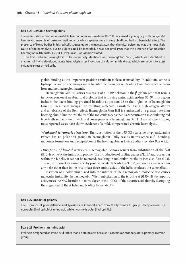
108 Chapter 6 Inherited disorders of haemoglobin
globin binding at this important position results in molecular instability. In addition, serine is hydrophilic and so encourages water to enter the haem pocket, leading to oxidation of the haem iron and methaemoglobinaemia.
Haemoglobin Gun Hill arises as a result of a 15 BP deletion in the b-globin gene that results in the expression of an abnormal b-globin that is missing amino acid residues 93–97. �is region includes the haem-binding proximal histidine at position 92 so the b-globins of haemoglobin Gun Hill lack haem groups. �e resulting molecule is unstable, has a high oxygen a�nity and an absence of the Bohr e�ect. Haemoglobin Gun Hill is synthesized at a greater rate than haemoglobin A but the instability of the molecule means that its concentration in circulating red blood cells remains low. �e clinical consequences of haemoglobin Gun Hill are relatively minor; most reported cases have shown evidence of a mild, compensated chronic haemolysis.
Weakened tetrameric structure. �e substitution of the b35 (C1) tyrosine by phenylalanine (which has no polar OH group) in haemoglobin Philly results in weakened a1b1 bonding, monomer formation and precipitation of the haemoglobin as Heinz bodies (see also Box 6.22).
Disruption of helical structure. Haemoglobin Genova results from substitution of the b28 (B10) leucine by the imino acid proline. �e introduction of proline causes a ‘kink’ and, occurring within the B helix, it cannot be tolerated, resulting in molecular instability (see also Box 6.23). �e substitution of an amino acid by proline inevitably leads to a ‘kink’, and such a change within any helix other than in the �rst or last three amino acids of the helix produces the same e�ect.
Insertion of a polar amino acid into the interior of the haemoglobin molecule also causes molecular instability. In haemoglobin Wien, substitution of the tyrosine at b130 (H8) by aspartic acid causes the NA2 histidine to move closer to the –COO– of the aspartic acid, thereby disrupting the alignment of the A helix and leading to instability.
Box 6.21 Unstable haemoglobins
The earliest description of an unstable haemoglobin was made in 1952. It concerned a young boy with congenital haemolytic anaemia of unknown aetiology for whom splenectomy in early childhood had no bene�cial e�ect. The presence of Heinz bodies in his red cells suggested to the investigators that chemical poisoning was the most likely cause of the haemolysis, but no culprit could be identi�ed. It was not until 1970 that the presence of an unstable haemoglobin, Hb Bristol (β67 (E11) valÆasp), was demonstrated.
The �rst unstable haemoglobin to be de�nitively identi�ed was haemoglobin Zürich, which was identi�ed in a young girl who developed acute haemolysis after ingestion of sulphonamide drugs, which are known to exert oxidative stress on red cells.
Box 6.22 Impact of polarity
The R groups of phenylalanine and tyrosine are identical apart from the tyrosine OH group. Phenylalanine is a non-polar (hydrophobic) amino acid while tyrosine is polar (hydrophilic).
Box 6.23 Proline is an imino acid
Proline is designated an imino acid rather than an amino acid because it contains a secondary, not a primary, α amine group.
01-Haematology-Chs1-8-ccp.indd 108 01/10/2010 12:52

6.4 Inherited haemoglobin disorders 109
Altered oxygen a�nitySeveral mechanisms exist that result in altered oxygen a�nity:
� direct interference with oxygen binding, especially of a haem.� abnormality of the a1b2 (a2b1) contact area.� alteration of a1a2 or b1b2 interaction.� changes in 2,3-DPG binding.� changes in Bohr e�ect H+ binding.
Interference with oxygen binding. �e unstable haemoglobin Hammersmith also has a reduced oxygen a�nity because the CD1 phenylalanine helps to maintain the correct orientation of the haem group and is responsible for the tilt of the haem on oxygenation. In the absence of phenylalanine, the haem group remains in the upright, deoxygenated position, which lowers its oxygen a�nity. In addition, haemoglobins M Boston and Iwate have lowered oxygen a�nity since they involve a chains, whereas in haemoglobins M Saskatoon and Hyde Park the oxygen a�nity is normal because they involve b chains.
Abnormality of the a1b2 (a2b1) contact. Interference with the a1b2 (a2b1) contact is a common cause of altered oxygen a�nity. For example, in haemoglobin Chesapeake the a92 (FG4) arginine is substituted by leucine. �e a92 arginine is normally in Van der Waals’ contact with the arginine at b35 (C2), stabilizing the deoxygenated conformation of haemoglobin. In haemoglobin Chesapeake, this interaction is absent and the deoxygenated conformation is no longer favoured thereby causing increased oxygen a�nity.
Conversely, the oxygenated conformation is normally stabilized by a bond between the b102 (G4) asparagine and a94 (G1) aspartic acid. In haemoglobin Kansas, formation of this stabilizing bond is precluded by the substitution of threonine for the b102 (G4) asparagine. �is mutation therefore reduces the stability of the oxygenated conformation and results in reduced oxygen a�nity.
Alteration of a1a2 and b1b2 interaction. Abnormalities a�ecting a1a2 are very rare. No mutations a�ecting b1b2 interaction have yet been reported. In theory, stabilizing b1b2 interactions should increase oxygen a�nity; conversely stabilizing a1a2 substitutions should reduce oxygen a�nity.
In the deoxygenated form, the N-terminal (a1) valine of each a chain bonds with C-terminal arginine (a141) and aspartic acid (a126) on the opposite a chain. �ese bonds stabilize the deoxygenated conformation, since they must be broken during oxygenation. In haemoglobin Suresnes the a141 arginine is replaced by histidine, which precludes bonding with the a1 valine on the opposite a chain and results in increased oxygen a�nity.
Changes in 2,3-DPG binding. �ere are three amino acid residues on the b-globin chain which are responsible for the binding of 2,3-DPG; the b1 (NA1) valine, the b82 (EF6) lysine and the b143 (H21) histidine. One of the sequence di�erences between b- and g-globin is at (H21) where the histidine is replaced by serine. �is explains the inability of g-globin to bind 2,3-DPG and the resultant elevated oxygen a�nity of haemoglobin F (a2g2).
�e b82 lysine is involved in at least three abnormal haemoglobins, Helsinki (ÆMet), Rahere (Æ�r) and Providence (ÆAsn/Asp). In all three the positive charge is lost, resulting in weakened DPG binding and increased a�nity.
Altered H+ binding. Hydrogen ions (H+) are bound in the process of deoxygenation, but must be released for association of oxygen to occur. �e shi� in the position of the haemoglobin–oxygen dissociation curve that results from changes in pH is called the Bohr e�ect. Half of the
01-Haematology-Chs1-8-ccp.indd 109 01/10/2010 12:52

110 Chapter 6 Inherited disorders of haemoglobin
H+ binding is due to the interaction of the C-terminal histidines of the b chains (b146, HC3), via their side chains, to the aspartic acid at b94 (FG1) of their own chains. Mutations that remove the b146 (HC3) histidine therefore a�ect H+ binding and the oxygen a�nity of the molecule. For example, the increased oxygen a�nity of haemoglobin York is explained by the substitution of the b146 (HC3) histidine by proline.
Miscellaneous e�ectsPolymerization. By far the most clinically signi�cant structural haemoglobinopathy is haemoglobin S. �e clinical consequences of the presence of this abnormal haemoglobin all stem from its tendency to polymerize in hypoxic conditions, forming long, rigid structures called ‘tactoids’, causing contortion of the red cell into elongated and poorly deformable sickle shapes.
Haemoglobin S results from a substitution of the glutamic acid in the external position b6 (A3) by valine. Genetic analysis of HbS mutations has identi�ed �ve di�erent disease haplotypes. �e HbS gene is always the same in these haplotypes, but there is co-inherited variation in the surrounding (non-coding) DNA. �ese haplotypes are each associated with a particular geographic region and are named to re�ect this: HbS (Benin), HbS (Senegal), HbS (Central African Republic), HbS (Cameroon), HbS (Asian). In general, the Asian and Senegal haplotypes tend to be less clinically severe than the Central African Republic haplotype (see also Box 6.24).
Sickle cell anaemia follows a highly variable clinical course; some patients die in infancy from the disabling e�ects of recurrent crises or overwhelming infection; others may live for a normal lifespan. �e precise causes of this variability are incompletely understood but the physical and social environment plays a large part. Symptoms of sickle cell anaemia are seldom manifest before the age of about 6 months when the level of circulating fetal haemoglobin falls to the adult level. Between crises, the condition is characterized by a chronic haemolytic state with jaundice and a relatively constant haemoglobin level of 7–8 g/dl. �e anaemia may be exacerbated by the presence of folate de�ciency. �is quiescent state is punctuated by crises of four main types:
� vaso-occlusive sickling crises are the most common manifestation and occur when poorly deformable sickle cells occlude small blood vessels, e.g. in the spleen, leading to downstream tissue hypoxia and infarction. Repeated sickling crises cause cumulative tissue damage (see also Box 6.25).
� aplastic crises are manifest as a sudden fall in haemoglobin concentration with no compensatory reticulocytosis and are usually secondary to infection, most commonly with parvovirus.
� acute splenic sequestration, where a large proportion of the circulating red cell mass is suddenly sequestered by the spleen, is a major cause of infant mortality in sickle cell anaemia.
Box 6.24 Sickle cell anaemia
James Herrick published the �rst description of a case of sickle cell anaemia in 1910. The patient described was a young West Indian student who demonstrated many of the classical clinical features of this condition recognized today (Archives of Internal Medicine, 6: 517–521).
Box 6.25 Autosplenectomy
Repeated sickling crises within the microcirculation of the spleen cause cumulative damage, which can ultimately lead to the loss of a functional spleen. This condition is relatively common in adults with sickle cell disease and is known as autosplenectomy.
01-Haematology-Chs1-8-ccp.indd 110 01/10/2010 12:52

6.4 Inherited haemoglobin disorders 111
� susceptibility to bacterial infection is a hallmark of sickle cell anaemia, and a common cause of infant mortality. �e most common infections are pneumococcal and staphylococcal.
�ere are several known risk factors that may trigger a sickling crisis:
� hypoxia due to cigarette smoke, high altitude or �ying in an unpressurized aircra�;� exposure to cold air, wind or water;� dehydration with slowing of blood �ow;� infection;� stress;� strenuous exercise.
Heterozygotes for haemoglobin S are said to have sickle cell trait, while homozygotes have sickle cell anaemia. Typically, individuals with sickle cell trait are clinically normal and may be unaware of their condition. Laboratory analysis reveals the presence of about 40% HbS. Sickle cell crises are extremely rare in heterozygotes but a�ected individuals should be warned of the potential dangers of severe hypothermia or hypoxia.
Co-inheritance of other haemoglobinopathies can in�uence the clinical course of sickle cell trait or anaemia. For example, HbS homozygotes that also inherit a thalassaemia trait typically have higher haemoglobin levels and a less severe clinical picture. On the other hand, compound heterozygotes for HbS and b0 thalassaemia have relatively severe disease. One particularly important example of such an interaction is where sickle cell anaemia is co-inherited with the benign condition hereditary persistence of fetal haemoglobin (HPFH) in which levels of haemoglobin F do not fall. �e presence of signi�cant amounts of haemoglobin F has a protective e�ect against sickling and such individuals have relatively mild disease.
Substitution of the b6 glutamic acid (the same amino acid as HbS) by lysine gives rise to haemoglobin C, the second most common of the structural haemoglobinopathies. �is mutation is most closely associated with West Africa. Haemoglobin C is associated with decreased solubility and crystallization and dehydration and poor deformability of red cells. Homozygotes and HbC–b0 thalassaemia compound heterozygotes have a chronic haemolytic anaemia.
Investigation of suspected haemoglobinopathy
�e laboratory diagnosis of a haemoglobinopathy usually requires a combination of di�erent tests, with results being assessed in relation to clinical features, the ethnic origin of the patient, and the blood count and blood �lm. Serum ferritin is also performed in those patients who present with microcytosis, because iron status may a�ect some test results (including HbA2 estimation).
�e basic laboratory screening tests aim to readily detect the most common forms of haemoglobinopathy and include measurement of the red blood cell indices (RBC, MCV, MCH in particular), quanti�cation of HbA2 and HbF levels, detecting the reduced solubility of HbS, and haemoglobin electrophoresis or equivalent technique to detect structural haemoglobinopathy. �e results of these tests, interpreted together with any evidence of family history and ethnic origin, will identify the vast majority of abnormalities. It is important that all of the components of this screen are performed because of the common inheritance of more than one abnormality, e.g. HbS and b thalassaemia. If HPLC is used to determine HbF and HbA2 levels, the most clinically important structural variants will be determined simultaneously (see also Box 6.26).
�e results of this screen can be divided into �ve groups:
� reduced MCH (<27 pg) and raised HbA2 (>3.5%) is typical of heterozygous b thalas-saemia. �e HbF level is also raised (1–3%) in about one-third of cases. Because of the huge
01-Haematology-Chs1-8-ccp.indd 111 01/10/2010 12:52

112 Chapter 6 Inherited disorders of haemoglobin
variety of mutations and the possible co-inheritance of other abnormalities, the range of possible results in this category is wide.
� normal HbA2 (<3.5%) and reduced MCH (<27 pg) is most commonly explained by iron de�ciency. If this has been excluded, the most likely explanations are heterozygous a0 thalassaemia, homozygous a+ thalassaemia, mild b thalassaemia trait, and rarer forms of thalassaemia such as db thalassaemia or egdb thalassaemia. Genetic testing may be required to di�erentiate between these.
� normal HbA2 (<3.5%), reduced MCH (<27 pg) and raised HbF (>1%) raises the suspicion of db thalassaemia trait or HPFH. Genetic testing is required for de�nitive diagnosis here.
� normal HbA2 (<3.5%) and normal MCH (>27 pg) is most likely normal but may be explained by the uncommon silent b thalassaemia trait. Where a family history is positive for thalassaemia intermedia, the only way to exclude silent b thalassaemia trait is by genetic testing.
� abnormal haemoglobin present. �e most common clinically relevant abnormal haemoglobins (HbS, C, D-Punjab, O-Arab and E) can all be identi�ed using haemoglobin electrophoresis or HPLC. Identi�cation of less common abnormalities requires genetic testing or amino acid analysis. Identi�cation of an abnormal band on haemoglobin electrophoresis should trigger a sickle solubility test to identify the presence of HbS. Using alkaline cellulose acetate electrophoresis, HbS co-migrates with HbD and HbC co-migrates with HbA2, HbE and HbOArab. In cases of doubt, acid agar gel electrophoresis permits resolution of these subtypes (see also Box 6.27).
Box 6.26 HPLC
Charge-dependent HPLC has largely replaced standard alkaline cellulose acetate electrophoresis for haemoglobinopathy screening, because it can simultaneously detect the most clinically important structural haemoglobinopathies and quantitate HbA2 and HbF with greater accuracy.
Box 6.27 Isoelectric focusing
Isoelectric focusing (IEF) is an alternative form of haemoglobin electrophoresis. Although it is more technically demanding than cellulose acetate electrophoresis, the resolution of minor haemoglobin fractions is better.
SUGGESTED FURTHER READING
Giardina, B., Messana, I., Scatena, R. and Castagnola, M. (1995) �e multiple functions of hemoglobin. Crit. Rev. Biochem. Mol. Biol. 30: 165–196.
Hazelwood, L. (2003) Can’t Live Without It: the story of hemoglobin in sickness and in health. New York: Nova Biomedical.
Perutz, M.F. (1998) Science Is Not a Quiet Life: Unravelling the Atomic Mechanism of Haemoglobin (Series in 20th Century Biology). Singapore: World Scienti�c Publishing.
Steinberg, M.H., Forget, B.G., Higgs, D.R. and Nagel, R.L. (eds) (2009). Disorders of Haemoglobin: genetics, pathophysiology and clinical management. Cambridge: Cambridge University Press.
01-Haematology-Chs1-8-ccp.indd 112 01/10/2010 12:52

Self-assessment questions 113
SELF-ASSESSMENT QUESTIONS
1. What is the rate-limiting step in haem synthesis?2. Where in the cell does haem synthesis occur?3. Name three embryonic haemoglobins and de�ne their globin content.4. Why is it important that haem does not come into contact with water?5. What is the normal alveolar pO2?6. Why is anaemia associated with shortness of breath on exertion?7. Why is anaemia associated with pallor?8. Do you think that d0 thalassaemia would be a severe disease?9. Why is haemoglobin Barts hydrops fetalis rare in African-Americans?10. What e�ect might coincident inheritance of heterozygous a0 thalassaemia have on the
severity of a moderately severe b thalassaemia?11. Why are the haemoglobins M associated with peripheral cyanosis?12. Why should patients with sickle cell disease avoid �ying in unpressurized aircra�?
01-Haematology-Chs1-8-ccp.indd 113 01/10/2010 12:52