ischemic preconditioning efficacy following anabolic
TRANSCRIPT
Vol.:(0123456789)1 3
Cardiovascular Toxicology https://doi.org/10.1007/s12012-018-9497-4
Ischemic Preconditioning Efficacy Following Anabolic Steroid Usage: A Clear Difference Between Sedentary and Exercise-Trained Rat Hearts
Zahra Akbari1 · Mansour Esmailidehaj2 · Ebrahim Avarand3 · Mehrdad Shariati3 · Khalil Pourkhalili1
© Springer Science+Business Media, LLC, part of Springer Nature 2018
AbstractPrevious studies show that anabolic steroids impair innate cardioprotective mechanisms. Here, we investigated the effect of supraphysiological doses of nandrolone on ischemic preconditioning (IPC) as a potent cardioprotective tool against ischemia reperfusion (IR) injury in rat hearts. Male Wistar rats in two experimental settings of sedentary and exercise-trained (60 min/day swimming, 5 days/week, for 8 weeks) were either pretreated with intramuscular injections of arachis oil (Arach, n = 16) as vehicle or nandrolone decanoate (ND, n = 8), 10 mg/kg/week, for 8 weeks. At the end, the hearts were excised and perfused in a Langendorff system. Then, the vehicle-treated hearts subdivided into the IR (30 min of LAD coronary artery occlusion and 120 min reperfusion, n = 8) and IPC (three cycles of 3-min ischemia and 3-min reperfusion before test ischemia, n = 8) groups and nandrolone-treated hearts served as ND + IPC (nandrolone pretreatment before IR and IPC protocols, n = 8) group. Post-ischemic cardiac function and infarct size were assessed. Reperfusion arrhythmias were analyzed using a standard scor-ing system. In sedentary hearts, ND slightly increased heart-to-body weight ratio and increased baseline cardiac contractile function. In trained hearts, ND markedly increased heart-to-body weight ratio which was also associated with enhanced baseline cardiac function. ND pretreatment enhanced protective effects of IPC in sedentary group; however, abolished these effects in exercise-trained group. The arrhythmia score was not significantly different between nandrolone-treated groups vs. respective preconditioned groups. Our findings show that ND impairs IPC-induced cardioprotection in exercise-trained rat hearts. Cardiac hypertrophy seems to play a crucial role in this response.
Keywords Ischemic preconditioning · Nandrolone · Hypertrophy · Cardioprotection
Introduction
Myocardial ischemia–reperfusion (IR) injury occurs in many cardiovascular interventions such as coronary angioplasty, thrombolysis and coronary by-pass grafting [1]. Prolonged ischemia leads to severe cardiomyocytes injury and cell death, which might be exacerbated by reperfusion after reopening of the involved vessel. Ischemic preconditioning
(IPC) described by Murrry et al. in 1986 [2], markedly pro-tects the heart against IR-induced injuries including cardio-myocytes necrosis and apoptosis [3, 4], contractile dysfunc-tion and arrhythmias [2, 5, 6].
Anabolic androgenic steroids (AASs), synthetic deriva-tives of testosterone, are abused by athletes and non-athletes to improve their exercise performance and physical appear-ance, respectively [7]. AASs are also used in many clinical setting for therapeutic purposes [8]. However, long-term supra-physiological doses of AASs may impair the normal cardiac remodeling process [9] which in turn can increase the susceptibility of the heart to cardiovascular pathologies such as hypertension [10], hypertrophy [11], atherosclerosis [12], infarction and sudden cardiac death [13, 14].
A growing number of studies have shown the adverse effects of AASs on IR-induced injury in the cardiovascular system. Du Toit et al. [15] showed that in long term, nan-drolone lead to left ventricular hypertrophy and increased IR injury in isolated rat hearts. Chaves et al. [16] demonstrated
Handling Editor: James Kang.
* Khalil Pourkhalili [email protected]
1 Department of Physiology, Faculty of Medicine, Bushehr University of Medical Sciences, Bushehr, Iran
2 Department of Physiology, Faculty of Medicine, Shahid Sadoughi University of Medical Sciences, Yazd, Iran
3 Department of Biology, Islamic Azad University, Kazerun, Iran
Cardiovascular Toxicology
1 3
that high doses of nandrolone impaired the exercise-induced cardioprotection against IR injury. Bissoli et al. [17] dem-onstrated that long-term therapy with nandrolone attenuates the sensitivity of the Bezold–Jarisch reflex control of heart rate and blood pressure. Recently, increased susceptibility of the heart to ventricular arrhythmias has also been shown in exercised rats treated with nandrolone decanoate [18, 19].
Regarding these adverse effects of AASs on cardiovascu-lar system, the usefulness of innate cardioprotective mecha-nisms such as IPC against ischemia–reperfusion injury in the presence of high doses of AASs might be controversial. In an animal model of IR injury, Penna et al. showed that nandrolone abolishes the cardioprotective effects of ischemic postconditioning in long term, however, it potentiates post-conditioning-induced cardioprotection against IR injury in short term [20, 21]. This discrepancy was attributed to the occurrence of left ventricular hypertrophy along with nan-drolone decanoate pretreatment in long term. Accordingly, hypertrophy seems to be a determining factor in maintain-ing or eliminating the innate cardioprotective mechanisms against IR injury. Because combination of exercise training with high doses of AASs increases the risk of ventricular hypertrophy and makes the heart more susceptible to dam-age [9, 22], we supposed that response to ischemic precondi-tioning in nandrolone-treated sedentary and exercise-trained rat hearts would be different.
Therefore, we hypothesized that chronic administration of supra-physiological doses of nandrolone would impair protective effects of ischemic preconditioning in exercise-trained rat hearts. To the best of our knowledge, this is the first study that addresses the effect of nandrolone pretreat-ment on ischemic preconditioning phenomena.
Materials and Methods
Chemicals
Nandrolone decanoate and arachis oil (solvent of nan-drolone) were prepared from Iran Hormone CO. (Tehran, Iran). Evans blue and triphenyl tetrazolium chloride (TTC) were purchased from Sigma-Aldrich (USA).
Animals and Group Assignment
Male Wistar rats (12–13 weeks old and 250–300 g weight; n = 48) were purchased from Razi Vaccine and Serum Research Institute, Karaj, Iran. Rats were housed in trans-parent polycarbonate collective cages (eight rats per cage) at 24 ± 2 °C with 12:12 h dark–light cycles in a stress-free environment with ad libitum access to food and water. The experimental protocols used in this study were approved by the Ethics Committee of Bushehr University of Medical
Sciences, Bushehr, Iran. After 1 week of acclimatization with animal house conditions, animals were randomly assigned to sedentary (n = 24) and exercise-trained (n = 24) settings. Then, 16 animals from each setting were treated with arachis oil which were subsequently assigned to control ischemia–reperfusion (IR, n = 8) and ischemic precondition-ing (n = 8) groups, and the remained eight animals of each setting were received nandrolone decanoate, 10 mg/kg/week, for 8 weeks and were assigned for the assessment of anabolic steroid effect on ischemic preconditioning (ND + IPC, n = 8). The dose of nandrolone decanoate used in the present study was selected based on the previous researches on animals [23, 24], which has the potential for inducing cardiac remod-eling and/or ventricular hypertrophy and is comparable with the non-therapeutic doses used by athletes in doping pro-cesses (ranging from 200 to 3200 mg/week) [25–27].
Arachis oil was used as solvent to prepare nandrolone decanoate solution at a final concentration of 25 mg/ml. The arachis oil and/or nandrolone decanoate (0.1–0.15 ml) were injected intramuscularly to the gluteal region of hind limbs, 30 min before the exercise training session. The injection site was alternated weekly between the two hind limbs. The volume of injectable solution was so low that no change in the swimming performance of the animals was observed.
Exercise Training Program
A program of swimming exercise training was adapted from a previously described method [28]. Briefly, training was performed by swimming in a pool (length 100 cm, width 100 cm, depth 50 cm) containing tap water maintained at 32–34 °C. Rats were submitted to exercise training, 5 days/week for 8 weeks. The first week was familiarization to swimming exercise. On the first day of the week 1, the ani-mals were trained in the pool for 10 min. In the subsequent days, the swimming time was extended to 10 min each day till 60 min per day at the end of first week. Then, the animals were exercised 60 min/day for 5 days/week for subsequent 7 weeks. All experimental procedures were performed in the morning between 8:00 and 12:00.
Heart Isolation and Perfusion on the Langendorff Apparatus
After 8 weeks treatment with arachis oil or nandrolone decanoate, animals were anaesthetized with pentobarbital sodium (60 mg/kg, i.p.) and heparinized (500 IU, i.p.). Then, their hearts removed and perfused retrogradely through aorta with carbogenized Krebs–Henseleit buffer (37 °C) under a constant-pressure Langendorff perfusion apparatus. For assessment of heart functional parameters; heart rate, left ventricular developed pressure (LVDP), rate pressure prod-uct (RPP), and the maximum rate of increase and decrease of
Cardiovascular Toxicology
1 3
left ventricular pressure during the time (max dp/dt and min dp/dt) were determined. Coronary flow (CF) was measured by timed collections of the coronary effluent. A water-filled latex balloon connected to a pressure transducer placed in the left ventricle to record pressures by a data acquisition system (MLT 844, AD instruments, Australia).
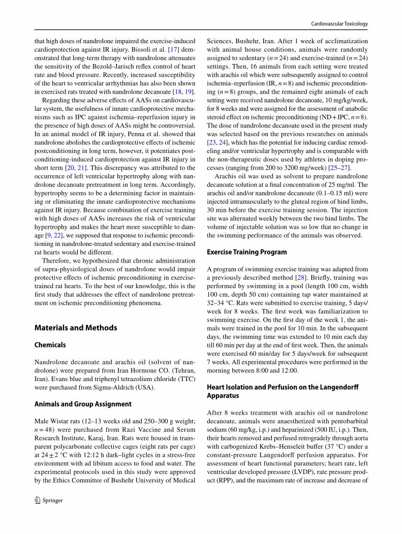
Experimental IR and IPC Protocols
After mounting on Langendorff perfusion system, all hearts experienced a stabilization period according to the experi-mental protocol and baseline cardiac functional parameters were obtained. Thereafter, all hearts subjected to 30 min regional ischemia and 120 min reperfusion, respectively, by tightening and loosening a 5 − 0 silk suture placed under the left anterior descending coronary artery (LAD). Precondi-tioning was achieved by three cycles of 3-min occlusion and 3-min reperfusion of the LAD before main test ischemia (Fig. 1), [29].
Determination of Myocardial Infarct Size
At the end of 120 min reperfusion, the LAD was re-occluded, and the risk zone was delineated by perfusing the hearts with 1 ml of 2% Evans blue (Sigma) solution into the aortic cannula. After freezing at − 20 °C, the hearts were cut into transverse slices of 2 mm thickness from the apex to the base. The slices were stained with 1% triphenyl tetrazolium chloride (TTC, Sigma) at 37 °C for 20 min. To increase the contrast between viable and non-viable (infarcted) areas, the slices were placed in 10% formalin solution for 1 h. Then, the both sides of slices were photographed by a digital cam-era (Canon PowerShot G11). Area at risk and infarct size was determined by computerized planimetry using image analysis software (Image Tool 3.0). The infarct size was cal-culated as the percentage of area at risk.
Quantification of Arrhythmias
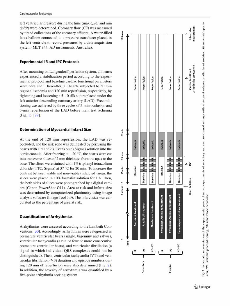
Arrhythmias were assessed according to the Lambeth Con-ventions [30]. Accordingly, arrhythmias were categorized as premature ventricular beats (single, bigeminy and salvos), ventricular tachycardia (a run of four or more consecutive premature ventricular beats), and ventricular fibrillation (a signal in which individual QRS complexes could not be distinguished). Then, ventricular tachycardia (VT) and ven-tricular fibrillation (VF) duration and episode numbers dur-ing 120 min of reperfusion were also determined (Fig. 2). In addition, the severity of arrhythmia was quantified by a five-point arrhythmia scoring system. Fi
g. 1
Sc
hem
atic
repr
esen
tatio
n of
the
expe
rimen
tal p
roto
col i
n tw
o ex
perim
ents
of s
eden
tary
and
exe
rcis
e-tra
ined
setti
ngs w
ith su
bseq
uent
subg
roup
s afte
r hea
rt is
olat
ion.
IR is
chem
ia/re
perf
u-si
on, I
PC is
chem
ic p
reco
nditi
onin
g, N
D n
andr
olon
e de
cano
ate
Cardiovascular Toxicology
1 3
Statistics
All data were analyzed by SPSS program, version 19.0 (SPSS Inc., Chicago, IL, USA) and expressed as means ± S.E.M. Mann–Whitney U test were used to deter-mine the statistical differences between groups. P value of less than 0.05 was considered statistically significant.
Results
Body Weight
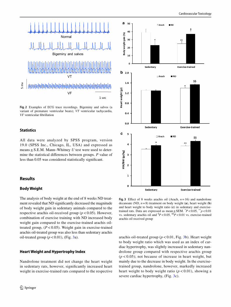
The analysis of body weight at the end of 8 weeks ND treat-ment revealed that ND significantly decreased the magnitude of body weight gain in sedentary animals compared to the respective arachis oil-received group (p < 0.05). However, combination of exercise training with ND increased body weight gain compared to the exercise-trained arachis oil-treated group, (P < 0.05). Weight gain in exercise-trained arachis oil-treated group was also less than sedentary arachis oil-treated group (p < 0.01), (Fig. 3a).
Heart Weight and Hypertrophy Index
Nandrolone treatment did not change the heart weight in sedentary rats, however, significantly increased heart weight in exercise-trained rats compared to the respective
arachis oil-treated group (p < 0.01, Fig. 3b). Heart weight to body weight ratio which was used as an index of car-diac hypertrophy, was slightly increased in sedentary nan-drolone group compared with respective arachis group (p < 0.05); not because of increase in heart weight, but mainly due to the decrease in body weight. In the exercise-trained group, nandrolone, however, markedly increased heart weight to body weight ratio (p < 0.01), showing a severe cardiac hypertrophy, (Fig. 3c).
Fig. 2 Examples of ECG trace recordings. Bigeminy and salvos (a variant of premature ventricular beats); VT ventricular tachycardia, VF ventricular fibrillation
Fig. 3 Effect of 8 weeks arachis oil (Arach, n = 16) and nandrolone decanoate (ND, n = 8) treatment on body weight (a), heart weight (b) and heart weight to body weight ratio (c) in sedentary and exercise-trained rats. Data are expressed as mean ± SEM. *P < 0.05, **p < 0.01 vs. sedentary arachis oil and $P < 0.05, $$P < 0.01 vs. exercise-trained arachis oil received group
Cardiovascular Toxicology
1 3
Baseline Hemodynamic Parameters Following Heart Isolation
Baseline cardiac function parameters were assessed imme-diately before the start of ischemia. Values of LVDP, RPP, dP/dtmax, dP/dtmin and CF were significantly increased in sedentary ND + IPC and exercise-trained ND + IPC groups. Heart rate was increased in exercise-trained ND + IPC group compared to the respective IPC group (Table 1).
Post‑ischemic Cardiac Function Assessment
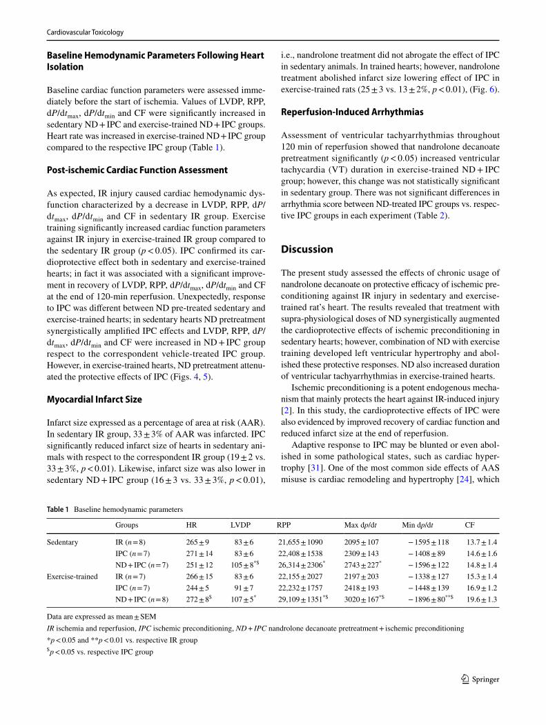
As expected, IR injury caused cardiac hemodynamic dys-function characterized by a decrease in LVDP, RPP, dP/dtmax, dP/dtmin and CF in sedentary IR group. Exercise training significantly increased cardiac function parameters against IR injury in exercise-trained IR group compared to the sedentary IR group (p < 0.05). IPC confirmed its car-dioprotective effect both in sedentary and exercise-trained hearts; in fact it was associated with a significant improve-ment in recovery of LVDP, RPP, dP/dtmax, dP/dtmin and CF at the end of 120-min reperfusion. Unexpectedly, response to IPC was different between ND pre-treated sedentary and exercise-trained hearts; in sedentary hearts ND pretreatment synergistically amplified IPC effects and LVDP, RPP, dP/dtmax, dP/dtmin and CF were increased in ND + IPC group respect to the correspondent vehicle-treated IPC group. However, in exercise-trained hearts, ND pretreatment attenu-ated the protective effects of IPC (Figs. 4, 5).
Myocardial Infarct Size
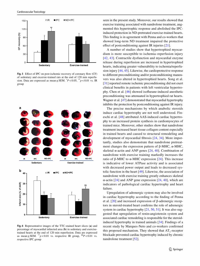
Infarct size expressed as a percentage of area at risk (AAR). In sedentary IR group, 33 ± 3% of AAR was infarcted. IPC significantly reduced infarct size of hearts in sedentary ani-mals with respect to the correspondent IR group (19 ± 2 vs. 33 ± 3%, p < 0.01). Likewise, infarct size was also lower in sedentary ND + IPC group (16 ± 3 vs. 33 ± 3%, p < 0.01),
i.e., nandrolone treatment did not abrogate the effect of IPC in sedentary animals. In trained hearts; however, nandrolone treatment abolished infarct size lowering effect of IPC in exercise-trained rats (25 ± 3 vs. 13 ± 2%, p < 0.01), (Fig. 6).
Reperfusion‑Induced Arrhythmias
Assessment of ventricular tachyarrhythmias throughout 120 min of reperfusion showed that nandrolone decanoate pretreatment significantly (p < 0.05) increased ventricular tachycardia (VT) duration in exercise-trained ND + IPC group; however, this change was not statistically significant in sedentary group. There was not significant differences in arrhythmia score between ND-treated IPC groups vs. respec-tive IPC groups in each experiment (Table 2).
Discussion
The present study assessed the effects of chronic usage of nandrolone decanoate on protective efficacy of ischemic pre-conditioning against IR injury in sedentary and exercise-trained rat’s heart. The results revealed that treatment with supra-physiological doses of ND synergistically augmented the cardioprotective effects of ischemic preconditioning in sedentary hearts; however, combination of ND with exercise training developed left ventricular hypertrophy and abol-ished these protective responses. ND also increased duration of ventricular tachyarrhythmias in exercise-trained hearts.
Ischemic preconditioning is a potent endogenous mecha-nism that mainly protects the heart against IR-induced injury [2]. In this study, the cardioprotective effects of IPC were also evidenced by improved recovery of cardiac function and reduced infarct size at the end of reperfusion.
Adaptive response to IPC may be blunted or even abol-ished in some pathological states, such as cardiac hyper-trophy [31]. One of the most common side effects of AAS misuse is cardiac remodeling and hypertrophy [24], which
Table 1 Baseline hemodynamic parameters
Data are expressed as mean ± SEMIR ischemia and reperfusion, IPC ischemic preconditioning, ND + IPC nandrolone decanoate pretreatment + ischemic preconditioning*p < 0.05 and **p < 0.01 vs. respective IR group$ p < 0.05 vs. respective IPC group
Groups HR LVDP RPP Max dp/dt Min dp/dt CF
Sedentary IR (n = 8) 265 ± 9 83 ± 6 21,655 ± 1090 2095 ± 107 − 1595 ± 118 13.7 ± 1.4IPC (n = 7) 271 ± 14 83 ± 6 22,408 ± 1538 2309 ± 143 − 1408 ± 89 14.6 ± 1.6ND + IPC (n = 7) 251 ± 12 105 ± 8*$ 26,314 ± 2306* 2743 ± 227* − 1596 ± 122 14.8 ± 1.4
Exercise-trained IR (n = 7) 266 ± 15 83 ± 6 22,155 ± 2027 2197 ± 203 − 1338 ± 127 15.3 ± 1.4IPC (n = 7) 244 ± 5 91 ± 7 22,232 ± 1757 2418 ± 193 − 1448 ± 139 16.9 ± 1.2ND + IPC (n = 8) 272 ± 8$ 107 ± 5* 29,109 ± 1351*$ 3020 ± 167*$ − 1896 ± 80**$ 19.6 ± 1.3

Cardiovascular Toxicology
1 3
in turn increases susceptibility of the heart to IR injury [21, 32]. Accordingly, we proposed that because of their anabolic nature; combination of exercise training and AAS administration may predispose the heart more to the ven-tricular hypertrophy and alter responsiveness of the heart to ischemic preconditioning against IR injury.
Despite studies showing the destructive effects of nan-drolone on the cardiovascular system, unexpectedly in the present study, we showed that in sedentary hearts, nan-drolone pretreatment potentiated cardioprotective effects of IPC and this synergism was characterized by greater recov-ery of LVDP, RPP, dP/dt and CF at the end of reperfusion.
In similar setting, Penna et al. demonstrated that short-term nandrolone pretreatment synergistically enhanced sys-tolic heart function induced by postconditioning [20, 21]; however, in long-term, nandrolone increased susceptibility to IR injury and abolished postconditioning-induced cardio-protection [21]. They related this disparity to the absence and presence of cardiac hypertrophy with short-term and long-term nandrolone pretreatment, respectively.
Our results showed that in sedentary rats, nandrolone decanoate induced a slight raise in heart to body weight ratio, and synergistically enhanced protective effects of IPC. This finding was in agreement with the results of the previ-ous studies showing slight increase in heart weight to body weight ratio in anabolic steroid-treated animals [21, 33–36]. Penna et al. [21] showed short-term (2 weeks) nandrolone treatment slightly increased cardiac/body weight ratio and potentiated cardioprotective effect of postconditioning. Per-golizzi et al. [37] related this benefit of nandrolone to the limitation of myocardial redox response through the induc-tion of antioxidant protein expression in the heart which in turn ameliorates postischemic heart performance. However, in contrast to the observations by Pergolizzi et al., Chaves et al. demonstrated that 10 weeks nandrolone decanoate pretreatment impaired exercise-induced cardioprotection by reduction of cardiac antioxidant capacity [38]. This incon-sistency may be due to differences in treatment duration and nandrolone dosage.
In the present study, it was also shown that exercise by itself increased heart weight and heart weight to body weight ratio compared to the control sedentary group. It is clear that exercise is a powerful stimulus for physiological cardiac
hypertrophy [39] without collagen deposition and systolic dysfunction [24, 40]. This adaptation supports the myocar-dium for increased demands of exercise while preserving or even augmenting normal heart function [41] as we have
Fig. 4 Effect of ischemic preconditioning (IPC) on post-ischemic recovery of myocardial mechanical function at the end of 120 min reperfusion in two groups of sedentary and exercised-trained rats. a Left ventricular developed pressure (LVDP); b rate pressure product (RPP); c, d maximum and minimum rate of left ventricular pressure changes (dp/dtmax, and dp/dtmin). IR control ischemia–reperfusion group, IPC ischemic preconditioning group, ND + IPC ischemic pre-conditioned group received nandrolone decanoate. Data are expressed as mean ± SEM. *P < 0.05, **p < 0.01 vs. respective IR group, $P < 0.05, $$P < 0.01 vs. respective IPC group
▸
Cardiovascular Toxicology
1 3
seen in the present study. Moreover, our results showed that exercise training associated with nandrolone treatment, aug-mented this hypertrophic response and abolished the IPC-induced protection in ND-pretreated exercise-trained hearts. This finding is in agreement with Penna and co-workers that showed long-term ND treatment impaired the protective effect of postconditioning against IR injuries [21].
A number of studies show that hypertrophied myocar-dium is more susceptible to ischemia–reperfusion injury [42, 43]. Contractile dysfunction and myocardial enzyme release during reperfusion are increased in hypertrophied hearts, indicating greater vulnerability to ischemia/reperfu-sion injury [44, 45]. Likewise, the cardioprotective response to different preconditioning and/or postconditioning maneu-vers was also altered in hypertrophied hearts. Song et al. [31] reported remote ischemic preconditioning did not exert clinical benefits in patients with left ventricular hypertro-phy. Chen et al. [46] showed isoflurane-induced anesthetic preconditioning was attenuated in hypertrophied rat hearts. Wagner et al. [47] demonstrated that myocardial hypertrophy inhibits the protection by postconditioning against IR injury.
The precise mechanisms by which anabolic steroids induce cardiac hypertrophy are not well understood. Fin-eschi et al. [48] attributed AAS-induced cardiac hypertro-phy to an increased protein synthesis in cardiomyocytes of trained mice. Moreover, other studies show that nandrolone treatment increased heart tissue collagen content especially in trained hearts and caused to structural remodeling and development of myocardial fibrosis [24, 34]. More impor-tantly, studies also demonstrate that nandrolone pretreat-ment changes the expression pattern of β-MHC, α-MHC, skeletal α-actin and ANP genes [24, 40]. Combination of nandrolone with exercise training markedly increases the ratio of β-MHC to α-MHC expression [24]. This increase is indicative of lower ATPase activity and is associated with decreased power output and leads to decreased sys-tolic function in the heart [49]. Likewise, the association of nandrolone with exercise training greatly enhances skeletal α-actin [24] and ANP gene expression [24, 40], which are indicators of pathological cardiac hypertrophy and heart failure.
Upregulation of adrenergic system may also be involved in cardiac hypertrophy according to the finding of Penna et al. [20] and increased expression of β-adrenergic recep-tors in steroid-treated heart confirms the role of adrenergic system in cardiac hypertrophy [21, 50, 51]. It was also sug-gested that upregulation of renin-angiotensin system and associated cardiac remodeling is responsible for the steroid-induced hypertrophy in trained animals [34]. Findings of a recent study by Marques-Neto and co-workers confirmed this proposed mechanism. They showed that AT1-receptor blockade prevented cardiac hypertrophy induced by chronic nandrolone treatment [52].
Fig. 5 Effect of IPC on post-ischemic recovery of coronary flow (CF) of sedentary and excersie-trained rats at the end of 120 min reperfu-sion. Data are expressed as mean ± SEM. *P < 0.05, **p < 0.01 vs. IR group
Fig. 6 Representative images of the TTC-stained heart slices (a) and percentage of myocardial infarcted area (b) in sedentary and exercise-trained hearts at the end of 120 min reperfusion. Data are expressed as mean ± SEM. **p < 0.01 vs. respective IR group, $$P < 0.01 vs. respective IPC group

Cardiovascular Toxicology
1 3
Hypertrophy is associated with various morphological and biochemical changes in cardiomyocytes. Impairment in mitochondrial biogenesis and function, as the cellular power plants for ATP production, has been regarded as one of the main pathogenesis of pathological cardiac hypertro-phy. This impairment reduces cellular oxidative capacity and leads to energy starvation and cardiac dysfunction [53]. In this regard, recent studies have shown that anabolic steroids along with the development of cardiac hypertrophy also cause severe damages to the subcellular structures. Abeer et al. [54] indicated that nandrolone treatment has been associated with histochemical and ultrastructural changes such as nuclear fragmentation, vacuolation, loss of stria-tion, mitochondrial fragmentation and increased collagen fibers in the endomysium of cardiac muscle. Sun et al. [55] showed that while exercise training greatly enhances expres-sion of transcription factors involved in mitochondrial bio-genesis and fusion, combination of nandrolone and exercise training attenuated normal aortic adaptation to exercise by downregulation of mitochondrial biogenesis and intensive autophagy. Mitochondrial destruction along with abnor-mality of other cytoplasmic organelles was also reported by other researchers [56, 57]. These subcellular changes may explain the impaired systolic function of the exercise-trained nandrolone-received hearts at the end of 120 min of reperfu-sion in the current study.
Another concern about using AAS is the occurrence of cardiac arrhythmias. Our results show that ND pre-treatment increased the severity of tachyarrhythmias in exercise-trained hearts which is consistent with the cardiac functional parameters. In line with the present findings, Achar et al. [58] showed that chronic nandrolone admin-istration for 5 years increased the risk of ventricular fibril-lation. Abdollahi et al. [18] showed that administration of AAS increases the severity of VF in exercise-trained rat hearts and this response was attributed to the develop-ment of cardiac hypertrophy. Phillis et al. [59] also dem-onstrated that nandrolone augments the incidence of fatal
arrhythmia during ischemia without any effect on arrhyth-mia score. In the present study, ND pretreatment in seden-tary hearts, however, like its effects on cardiac functional data, did not significantly change VT and VF duration. This result is consistent with the finding by Phillis et al. They showed that chronic administration of 15 mg/kg ND for 17 days had no effect on QTc, QRS and PR intervals of rat hearts [59]. The reason for this inconsistency between sedentary and exercise-trained heart may be due to the cardiac hypertrophy and related cellular changes.
According to the results of the present study and some other mentioned investigations, it seems that presence of cardiac hypertrophy is necessary for the emergence of destructive effects of nandrolone decanoate. But on the other hand, this study contrasts with other studies that show the use of anabolic androgenic steroids along with exercise reduces their destructive effects. Therefore, it is recommended to design future studies in such a way that the efficacy of cardiac preconditioning protocols along with anabolic steroid drugs be examined simultaneously in the presence and absence of cardiac hypertrophy in sed-entary or exercise-trained hearts.
In conclusion, the results of the present study showed that cardioprotective effects of IPC were not lost by nan-drolone treatment in sedentary hearts; however, admin-istration of nandrolone combined with exercise training induced severe cardiac hypertrophy and attenuated ben-eficial effects of IPC on post ischemic recovery of heart function. This may be attributed to the nandrolone-induced fibrotic remodeling in cardiomyocytes, altered expression pattern of fetal genes and/or mitochondrial biogenesis and function. Further studies are needed to better clarify the precise mechanisms involved in different response to IPC in sedentary and exercise-trained hearts exposed to nandrolone.
Acknowledgements The authors appreciate the Vice Chancellor for Research and Technology, Bushehr University of Medical Sciences for financial support.
Table 2 Ventricular tachyarrhythmias episodes and durations
Data are expressed as mean ± SEMVT ventricular tachycardia, VF ventricular fibrillation** p < 0.01 vs. respective IR group, $P < 0.05 vs. respective IPC group
Groups VT episodes VT duration (s) VF episodes VF duration (s) Arrhythmias score
Sedentary IR (n = 8) 4 ± 0.9 13 ± 6 1.25 ± 0.45 26 ± 16 3.88 ± 0.23IPC (n = 7) 2 ± 0.7 12 ± 5 0.33 ± 0.21 8 ± 7 3.43 ± 0.37ND + IPC (n = 7) 5 ± 1.1** 16 ± 8 0.40 ± 0.24 14 ± 11 3.44 ± 0.24
Exercise-trained IR (n = 7) 4 ± 1 10 ± 5 0.40 ± 0.24 43 ± 28 3.86 ± 0.34IPC (n = 7) 2 ± 1 5 ± 2 0.60 ± 0.24 45 ± 24 4.2 ± 0.47ND + IPC (n = 8) 5.5 ± 1.2 30 ± 17$ 2.13 ± 1.58 107 ± 77 3.37 ± 0.32
Cardiovascular Toxicology
1 3
Compliance with Ethical Standards
Conflict of Interest The authors declare that they have no conflict of interest.
References
1. Hausenloy, D. J., & Yellon, D. M. (2013). Myocardial ischemia-reperfusion injury: A neglected therapeutic target. Journal of Clinical Investigation, 123, 92–100.
2. Murry, C. E., Jennings, R. B., & Reimer, K. A. (1986). Precon-ditioning with ischemia: A delay of lethal cell injury in ischemic myocardium. Circulation, 74, 1124–1136.
3. Lai, C. C., Tang, C. Y., Chiang, S. C., Tseng, K. W., & Huang, C. H. (2015). Ischemic preconditioning activates prosurvival kinases and reduces myocardial apoptosis. Journal of the Chinese Medical Association, 78, 460–468.
4. Iliodromitis, E. K., Lazou, A., & Kremastinos, D. T. (2007). Ischemic preconditioning: Protection against myocardial necro-sis and apoptosis. Vascular Health and Risk Management, 3, 629–637.
5. Li, Y. W., Whittaker, P., & Kloner, R. A. (1992). The transient nature of the effect of ischemic preconditioning on myocardial infarct size and ventricular arrhythmia. American Heart Journal, 123, 346–353.
6. Galagudza, M. M., Sonin, D. L., Vlasov, T. D., Kurapeev, D. I., & Shlyakhto, E. V. (2016). Remote vs. local ischaemic precondition-ing in the rat heart: Infarct limitation, suppression of ischaemic arrhythmia and the role of reactive oxygen species. International Journal of Experimental Pathology, 97, 66–74.
7. Kersey, R. D., Elliot, D. L., Goldberg, L., Kanayama, G., Leone, J. E., Pavlovich, M., et al. (2012). National athletic trainers’ associa-tion position statement: Anabolic–androgenic steroids. Journal of Athletic Training, 47, 567–688.
8. Woerdeman, J., & de Ronde, W. (2011). Therapeutic effects of anabolic androgenic steroids on chronic diseases associated with muscle wasting. Expert Opinion on Investigational Drugs, 20, 87–97.
9. Marqueti, R. C., Micocci, K. C., Leite, R. D., & Selistre-de-Araujo, H. S. (2012). Nandrolone inhibits MMP-2 in the left ventricle of rats. International Journal of Sports Medicine, 33, 181–185.
10. Rosca, A. E., Stoian, I., Badiu, C., Gaman, L., Popescu, B. O., Iosif, L. et al. (2016). Impact of chronic administration of ana-bolic androgenic steroids and taurine on blood pressure in rats. Brazilian Journal of Medical and Biological Research. https ://doi.org/10.1590/1414-431X2 01651 16.
11. Pirompol, P., Teekabut, V., Weerachatyanukul, W., Bupha-Intr, T., & Wattanapermpool, J. (2016). Supra-physiological dose of testosterone induces pathological cardiac hypertrophy. Journal of Endocrinology, 229, 13–23.
12. Ammar, E. M., Said, S. A., & Hassan, M. S. (2004). Enhanced vasoconstriction and reduced vasorelaxation induced by testoster-one and nandrolone in hypercholesterolemic rabbits. Pharmaco-logical Research, 50, 253–259.
13. Nascimento, J. H., & Medei, E. (2011). Cardiac effects of ana-bolic steroids: Hypertrophy, ischemia and electrical remodelling as potential triggers of sudden death. Mini Reviews in Medicinal Chemistry, 11, 425–429.
14. Fineschi, V., Riezzo, I., Centini, F., Silingardi, E., Licata, M., Beduschi, G., et al. (2007). Sudden cardiac death during anabolic steroid abuse: Morphologic and toxicologic findings in two fatal
cases of bodybuilders. International Journal of Legal Medicine, 121, 48–53.
15. Du Toit, E. F., Rossouw, E., Van, Rooyen, J., & Lochner, A. (2005). Proposed mechanisms for the anabolic steroid-induced increase in myocardial susceptibility to ischaemia/reperfusion injury. Cardiovascular Journal of South Africa, 16, 21–28.
16. Chaves, E. A., Fortunato, R. S., Carvalho, D. P., Nascimento, J. H. M., & Oliveira, M. F. (2013). Exercise-induced cardioprotection is impaired by anabolic steroid treatment through a redox-dependent mechanism. The Journal of Steroid Biochemistry and Molecular Biology, 138, 267–272.
17. Bissoli, N. S., Medeiros, A. R. S., Santos, M. C. S., Busato, V. C. W., Jarske, R. D., Abreu, G. R., et al. (2009). Long-term treatment with supraphysiological doses of nandrolone decanoate reduces the sensitivity of Bezold–Jarisch reflex control of heart rate and blood pressure. Pharmacological Research, 59, 379–384.
18. Abdollahi, F., Joukar, S., Najafipour, H., Karimi, A., Masumi, Y., & Binayi, F. (2016). The risk of life-threatening ventricular arrhythmias in presence of high-intensity endurance exercise along with chronic administration of nandrolone decanoate. Ster-oids, 105, 106–112.
19. Binayi, F., Joukar, S., Najafipour, H., Karimi, A., Abdollahi, F., & Masumi, Y. (2016). The effects of nandrolone decanoate along with prolonged low-intensity exercise on susceptibility to ven-tricular arrhythmias. Cardiovascular Toxicology, 16, 23–33.
20. Penna, C., Abbadessa, G., Mancardi, D., Tullio, F., Piccione, F., Spaccamiglio, A., et al. (2008). Synergistic effects against post-ischemic cardiac dysfunction by sub-chronic nandrolone pretreat-ment and postconditioning: Role of beta2-adrenoceptor. Journal of Physiology and Pharmacology, 59, 645–659.
21. Penna, C., Tullio, F., Perrelli, M. G., Moro, F., Abbadessa, G., Piccione, F., et al. (2011). Ischemia/reperfusion injury is increased and cardioprotection by a postconditioning protocol is lost as cardiac hypertrophy develops in nandrolone treated rats. Basic Research in Cardiology, 106, 409–420.
22. Do Carmo, E. C., Fernandes, T., Koike, D., Da Silva, N. D., Mat-tos, K. C., Rosa, K. T., et al. (2011). Anabolic steroid associated to physical training induces deleterious cardiac effects. Medicine and Science in Sports and Exercise, 43, 1836–1848.
23. Pereira-Junior, P. P., Chaves, E. A., Costa, E. S. R. H., Masuda, M. O., de Carvalho, A. C., & Nascimento, J. H. (2006). Cardiac autonomic dysfunction in rats chronically treated with anabolic steroid. European Journal of Applied Physiology, 96, 487–494.
24. Tanno, A. P., Neves, V. J. d., Rosa, K. T., Cunha, T. S., Giordano, F. C. L., & Calil, C. M. (2011). Nandrolone and resistance training induce heart remodeling: Role of fetal genes and implications for cardiac pathophysiology. Life Sciences, 89, 631–637.
25. Pope, Jr. H. G., Katz, D. L. (1988). Affective and psychotic symp-toms associated with anabolic steroid use. American Journal of Psychiatry, 145, 487–490.
26. Norton, G. R., Trifunovic, B., Woodiwiss, A. J. (2000). Attenuated beta-adrenoceptor-mediated cardiac contractile responses follow-ing androgenic steroid administration to sedentary rats. European Journal of Applied Physiology, 81, 310–316.
27. Evans, N. A. (1997). Gym and tonic: A profile of 100 male steroid users. British Journal of Sports Medicine, 31, 54–58.
28. Shokri, S., Aitken, R. J., Abdolvahhabi, M., Abolhasani, F., Ghasemi, F. M., Kashani, I., et al. (2009). Exercise and supra-physiological dose of nandrolone deconoate increase apoptosis in spermatogenic cells. Basic and Clinical Pharmacology and Toxicology, 106, 324–330.
29. Schultz, J. E., Hsu, A. K., & Gross, G. J. (1998). Ischemic pre-conditioning in the intact rat heart is mediated by delta1- but not mu- or kappa-opioid receptors. Circulation, 97, 1282–1289.
30. Walker, M. J., Curtis, M. J., Hearse, D. J., Campbell, R. W., Janse, M. J., Yellon, D. M., et al. (1988). The Lambeth Conventions:

Cardiovascular Toxicology
1 3
Guidelines for the study of arrhythmias in ischaemia infarction, and reperfusion. Cardiovascular Research, 22, 447–455.
31. Song, Y., Song, J. W., Lee, S., Jun, J. H., Kwak, Y. L., & Shim, J. K. (2017). Effects of remote ischemic preconditioning in patients with concentric myocardial hypertrophy: A randomized, con-trolled trial with molecular insights. International Journal of Cardiology, 249, 36–41.
32. Seara, F. A. C., Barbosa, R. A. Q., de Oliveira, D. F., Gran da Silva, D. L. S., Carvalho, A. B., Ferreira, A. C. et al. (2017). Administration of anabolic steroid during adolescence induces long-term cardiac hypertrophy and increases susceptibility to ischemia/reperfusion injury in adult Wistar rats. Journal of Ster-oid Biochem Molecular Biology, 171, 34–42.
33. Beutel, A., Bergamaschi, C. T., & Campos, R. R. (2005). Effects of chronic anabolic steroid treatment on tonic and reflex cardio-vascular control in male rats. The Journal of Steroid Biochemistry and Molecular Biology, 93, 43–48.
34. Rocha, F. L., Carmo, E. C., Roque, F. R., Hashimoto, N. Y., Ros-soni, L. V., Frimm, C., et al. (2007). Anabolic steroids induce cardiac renin-angiotensin system and impair the beneficial effects of aerobic training in rats. American Journal of Physiology; Heart Circulation Physiology, 293, H3575–H3583.
35. Lunz, W., Oliveira, E. C., Neves, M. T. D., Fontes, E. P. B., Dias, C. M. G. C., & Natali, A. J. (2006). Anabolic steroid and exercise induced cardiac stress protein (HSP72) in the rat. Brazillian Jour-nal of Medical and Biological Research, 39, 889–893.
36. Woodiwiss, A., Trifunovic, G., Philippides, M., & Norton, G. (2000). Effect of an androgenic steroid on exercise-induced cardiac remodeling in rats. Journal of Applied Physiology, 88, 409–415.
37. Pergolizzi, B., Carriero, V., Abbadessa, G., Penna, C., Berchialla, P., De Francia, S., et al. (2017). Subchronic nandrolone adminis-tration reduces cardiac oxidative markers during restraint stress by modulating protein expression patterns. Molecular Cell Bio-chemistry, 434, 51–60.
38. Chaves, E. A., Pereira-Junior, P. P., Fortunato, R. S., Masuda, M. O., de Carvalho, A. C., de Carvalho, D. P., et al. (2006). Nan-drolone decanoate impairs exercise-induced cardioprotection: Role of antioxidant enzymes. Journals of Steroid Biochemistry Molecular Biology, 99, 223–230.
39. Xiao, J., Xu, T., Li, J., Lv, D., Chen, P., Zhou, Q., et al. (2014). Exercise-induced physiological hypertrophy initiates activation of cardiac progenitor cells. International Journal of Clinical and Experimental Pathology, 7, 663–669.
40. Brasil, G. A., Lima, E. M., Nascimento, A. M., Caliman, I. F., Medeiros, A. R., Silva, M. S., et al. (2015). Nandrolone decanoate induces cardiac and renal remodeling in female rats, without mod-ification in physiological parameters: The role of ANP system. Life Sciences, 137, 65–73.
41. Iemitsu, M., Miyauchi, T., Maeda, S., Sakai, S., Kobayashi, T., Fujii, N., et al. (2001). Physiological and pathological cardiac hypertrophy induce different molecular phenotypes in the rat. American Journal of Physiology-Regulatory, Integrative and Comparative Physiology, 281, R2029–R2036.
42. Molgaard, S., Faricelli, B., Salomonsson, M., Engstrom, T., & Treiman, M. (2016). Increased myocardial vulnerability to ischemia–reperfusion injury in the presence of left ventricular hypertrophy. Journal of Hypertension, 34, 513–523.
43. Yano, T., Miki, T., Tanno, M., Kuno, A., Itoh, T., Takada, A., et al. (2011). Hypertensive hypertrophied myocardium is vulnerable to infarction and refractory to erythropoietin-induced protection. Hypertension, 57, 110–115.
44. Snoeckx, L. H., van der Vusse, G. J., van der Veen, F. H., Cou-mans, W. A., & Reneman, R. S. (1989). Recovery of hypertro-phied rat hearts after global ischemia and reperfusion at different perfusion pressures. Pflugers Archiv, 413, 303–312.
45. Friehs, I., Moran, A. M., Stamm, C., Choi, Y. H., Cowan, D. B., McGowan, F. X., et al. (2004). Promoting angiogenesis protects severely hypertrophied hearts from ischemic injury. The Annals of Thoracic Surgery, 77, 2004–2010.
46. Chen, C. H., Wu, C. W., Shih, C. D., Lien, W. H., Huang, S. L., & Huang, C. C. (2016). Attenuation of isoflurane precondi-tioning-induced acute cardioprotection in hypertensive hypertro-phied hearts. Journal of Cardiothorac Vascular Anesthesia, 30, 1317–1323.
47. Wagner, C., Ebner, B., Tillack, D., Strasser, R. H., & Weinbrenner, C. (2013). Cardioprotection by ischemic postconditioning is abro-gated in hypertrophied myocardium of spontaneously hyperten-sive rats. Journal of Cardiovascular Pharmacolology, 61, 35–41.
48. Fineschi, V., Di Paolo, M., Neri, M., Bello, S., D’Errico, S., Dinucci, D., et al. (2011). Anabolic steroid- and exercise-induced cardio-depressant cytokines and myocardial beta1 receptor expression in CD1 mice. Current Pharmacology Biotechnology, 12, 275–284.
49. Stelzer, J. E., Brickson, S. L., Locher, M. R., & Moss, R. L. (2007). Role of myosin heavy chain composition in the stretch activation response of rat myocardium. Journal of Physiology, 579, 161–173.
50. Penna, C., Abbadessa, G., Mancardi, D., Spaccamiglio, A., Racca, S., & Pagliaro, P. (2007). Nandrolone-pretreatment enhances car-diac [beta]2-adrenoceptor expression and reverses heart contrac-tile down-regulation in the post-stress period of acute-stressed rats. The Journal of Steroid Biochemistry and Molecular Biology, 107, 106–113.
51. das Neves, V. J., Tanno, A. P., Cunha, T. S., Fernandes, T., Guz-zoni, V., da Silva, C. A., et al. (2013). Effects of nandrolone and resistance training on the blood pressure, cardiac electrophysiol-ogy, and expression of atrial beta-adrenergic receptors. Life Sci-ence, 92, 1029–1035.
52. Marques-Neto, S. R., Ferraz, E. B., Rodrigues, D. C., Njaine, B., Rondinelli, E., Campos de Carvalho, A. C., et al. (2014). AT1 and aldosterone receptors blockade prevents the chronic effect of nandrolone on the exercise-induced cardioprotection in perfused rat heart subjected to ischemia and reperfusion. Cardiovascular Drugs Therapy, 28, 125–135.
53. Zhou, L. Y., Liu, J. P., Wang, K., Gao, J., Ding, S. L., Jiao, J. Q., et al. (2018). Mitochondrial function in cardiac hypertrophy. International Journal of Cardiology, 167, 1118–1125.
54. Abeer, A. M., Noura, H. M., & Maha, Z. M. (2018). The Nan-drolone effect on cardiac muscle of adult male albino rat and the possible role of nigella sativa: Light and electron microscopic studies. Journal of Biochemistry and Cell Biology, 1, 109.
55. Sun, M., Shen, W., Zhong, M., Wu, P., Chen, H., & Lu, A. (2013). Nandrolone attenuates aortic adaptation to exercise in rats. Car-diovascular Research, 97, 686–695.
56. Hanan, A. E. E., Adel, A. E. A., Mona, A. E. E. S., & Afaf, T. A. (2018). Effect of anabolic steroids on the cardiac and skeletal muscles of adult male rats. International Journal of Clinical and Developmental Anatomy, 4, 1–14.
57. Hassan, N. A., Salem, M. F., & Sayed, M. A. (2009). Doping and effects of anabolic androgenic steroids on the heart: Histologi-cal, ultrastructural, and echocardiographic assessment in strength athletes. Human Experimental Toxicology, 28, 273–283.
58. Achar, S., Rostamian, A., & Narayan, S. M. (2010). Cardiac and metabolic effects of anabolic-androgenic steroid abuse on lipids, blood pressure, left ventricular dimensions, and rhythm. American Journal of Cardiology, 106, 893–901.
59. Phillis, B. D., Abeywardena, M. Y., Adams, M. J., Kennedy, J. A., & Irvine, R. J. (2007). Nandrolone potentiates arrhythmo-genic effects of cardiac ischemia in the rat. Toxicology Science, 99, 605–611.