isolation and culture of primary adult skin fibroblasts ... · isolation and culture of primary...
TRANSCRIPT
Submitted 27 September 2017Accepted 9 January 2018Published 24 January 2018
Corresponding authorKorakot Nganvongpanit,[email protected]
Academic editorMartin Anger
Additional Information andDeclarations can be found onpage 13
DOI 10.7717/peerj.4302
Copyright2018 Siengdee et al.
Distributed underCreative Commons CC-BY 4.0
OPEN ACCESS
Isolation and culture of primary adultskin fibroblasts from the Asian elephant(Elephas maximus)Puntita Siengdee1, Sarisa Klinhom2, Chatchote Thitaram2 andKorakot Nganvongpanit1,3
1Animal Bone and Joint Research Laboratory, Department of Veterinary Biosciences and Public Health,Faculty of Veterinary Medicine, Chiang Mai University, Chiang Mai, Thailand
2Center of Excellence in Elephant and Wildlife Research, Faculty of Veterinary Medicine,Chiang Mai University, Chiang Mai, Thailand
3 Excellence Center in Veterinary Bioscience, Department of Veterinary Biosciences and Public Health, Facultyof Veterinary Medicine, Chiang Mai University, Chiang Mai, Thailand
ABSTRACTBackground. Primary cultures from Asian elephants (Elephas maximus) allow sci-entists to obtain representative cells that have conserved most of their originalcharacteristics, function, physiology and biochemistry. This technique has thus gainedsignificant importance as a foundation for further cellular, cell biology and molecularresearch. Therefore, the aim of this study was to describe conditions for the successfulestablishment of primary adult fibroblasts from Asian elephant carcasses.Methods. Ear tissue sample collection from Asian elephant carcasses and our recom-mendations are given. We describe here a simple modified protocol for successfulisolation and maintenance of primary adult fibroblasts from elephant ear skin. Earsamples from each individual (five 3 × 3 cm2 pieces) were brought to the laboratorywithin 3 h after collection, kept in transportationmediumat 0–4 ◦C.The ear tissueswereprepared by a combination of 10% collagenase type II digestion procedure togetherwitha simple explant procedure. Primary fibroblasts were cultured at 37 ◦C in Dulbecco’smodified Eagle’s medium (DMEM) with 20% fetal calf serum (FCS) in a humidifiedatmosphere containing 5% CO2. After the third passage, fibroblasts were routinelytrypsinized with 0.25% trypsin/EDTA and cultured in DMEM with 10% FCS at 37 ◦Cand 5% CO2. Traditional cell counting method was used to measure cell viability andgrowth curve. Long-term storage of cells used freezing medium consisting of 40% FCS(v/v).Results. We explored the most suitable conditions during sample collection (post-mortem storage time and sample storage temperature), which is the most importantstep in determining primary outgrowth.Our study successfully established and culturedprimary adult skin fibroblasts obtained from post-mortem E. maximus ear skin tissuesfrom six carcasses, with a success rate of around 83.3%. Outgrowth could be seen4–12 days after explantation, and epithelial-like cells were found after 4–7 days ofculture, while fibroblasts appeared at around day 7–10. The fibroblasts had viabilityand post-freezing recovery rates of around 97.3± 4.3% and 95.5± 7.3%, respectively,and doubling time was about 25 h (passage 6).Discussion. To our knowledge, this report is the first to describe primary cell culturesderived from adult Asian elephant skin. Future studies should benefit from the
How to cite this article Siengdee et al. (2018), Isolation and culture of primary adult skin fibroblasts from the Asian elephant (Elephasmaximus). PeerJ 6:e4302; DOI 10.7717/peerj.4302
information and useful suggestions herein, which may be used as a standard methodfor establishing primary skin fibroblast cultures in future experiments.
Subjects Cell Biology, Veterinary MedicineKeywords Asian elephant, Culture, Fibroblasts, Skin
INTRODUCTIONThe elephant is the largest landmammal and is the only living species of the genus Loxodontaand Elephas.The Asian elephant (Elephas maximus) distributed in Asia, includingmainlandAsia and the islands of Sri Lanka, Borneo and Sumatra (Shoshani & Eisenberg, 1982).Four subspecies have been described: Elephas maximus maximus (Sri Lanka); Elephasmaximus indicus (mainland Asia, including India, Nepal, Bangladesh, Bhutan, Myanmar,Thailand, Malay Peninsula, Laos, China, Cambodia and Vietnam); Elephas maximussumatranus (Sumatra) (Sukumar, 1992); and a new subspecies (based on mitochondrialDNA), Elephas maximus borneensis (Fernando et al., 2003) called the Borneo pygmyelephant, from the island of Borneo (specifically the Malaysian states of Sabah andKalimantan).
Most domesticated elephants today are found working in the tourism industry,particularly throughout Thailand. However, populations of domesticated elephants havedeclined more than 97% over the past century (Magda et al., 2015). Elephants are listed asprotected animals under Thailand’sWild Animal Reservation and Protection Act, B.E. 2535(1992). In recent decades, numerous organizations have been established to provide betterlives for the elephants, including improved healthcare and protection from exploitation bythe tourismand logging industries.However, health issues still exist, with themajor problembeing wounds and abscesses (Angkawanish et al., 2009). Damaged skin consequently causespainful lesions on the elephant’s body. A high prevalence (∼64.4%) of active lesions, mostlocated on the back region, was found to be associated with working conditions (Magda etal., 2015), and causing trauma and death in elephants at a rate of around 2.2% of majorpostmortem pathologic findings reported by survey respondents (Miller et al., 2015).
At the same time, wild elephants are suffering from poaching and severe habitatloss via encroachment and deforestation. Destruction of the forests gives rise to seriousconflicts with people that share the same habitat, usually ending with more elephants beingpoisoned, injured or even killed. Effective management of the environment and goodmedical treatment are required in order to resolve these issues and enhance the animals’well-being. However, there is little published literature on the treatment of elephant diseasesand injuries, and a lack of basic information on Asian elephants from the cellular to theorganismal level.
Cell culture technology has become a widely used method in biology, medical researchand applications. Establishing primary cultures of fibroblasts allows researchers toobtain representative cells that have conserved most of their original characteristicsand functions, which is an important foundation for further cell biology and cell
Siengdee et al. (2018), PeerJ, DOI 10.7717/peerj.4302 2/16

engineering. Cryopreservation of animal cells is an excellent technique for long-termpreservation of animal genetic resources, which is critical to guarantee genomics andgenetic analyses (Groeneveld et al., 2008; Guan et al., 2010; Mestre-Citrinovitz et al., 2016).Only two elephant cell lines are available in the cell bank at the present time: LACF-NaNaI(RIKEN Cell Bank: RCB2319) and LACF-NaNaII (RIKEN Cell Bank: RCB2320), whichwere derived from the gum and ear, respectively, of the African savannah elephant.
To our knowledge, two previous studies briefly mentioned successful methods ofisolating skin fibroblasts from Asian elephant skin, by direct culture of elephant skinexplants (Sathanawongs et al., 2010) and trypsin digestion procedure (Techakumphu et al.,2017). However in these procedures, fibroblasts were limited to those isolated from youngor stillborn donors with thin connective tissues that provided fast-growing fibroblasts andgenerally possessed greater proliferative capacity and good characteristics and function(Brink, Bernstein & Nicoll, 2009; Sriram, Bigliardi & Bigliardi-Qi, 2015). Even though densetissues from older donors are less desirable for fibroblast culture establishment, adultelephant carcasses are more readily available and hence more frequently obtained. Isolatingadult skin fibroblasts from direct culture of elephant skin explants (Aoued & Singh, 2015;Singh & Ma, 2014; Vangipuram et al., 2013) and also with collagenase digestion technique(Seluanov, Vaidya & Gorbunova, 2010) was ineffective in our preliminary study.
Therefore, the present study aimed to establish primary elephant skin fibroblasts takenfrom post-mortem adult Asian elephant ear skin tissues. Moreover, we have exploredpreliminary information about the development of methods of approach, problems andrecommendations, and useful suggestions for culturing primary skin fibroblasts from anadult Asian elephant, to serve as standard methods for regular establishment of primaryskin fibroblast cultures, which is of significant importance for further biological studies,including cell biology and cellular and molecular research.
MATERIALS AND METHODSTissue sample collectionTissue samples were obtained from six Asian elephant carcasses housed at differentfacilities/field settings (Licence number U1006312558). Details of the elephants are givenin Table 1, including age, sex, cause of death, location, and length of time after death untilsamples were brought to the laboratory.
Reagents used in this study are listed in Table 2, including the main components ofreagents and medium formulations and their vendor and concentration.
Ear samples were excised after the mahout had conducted a death ritual or funeralceremony according to Thai belief (Keyes, 1977), which occurred around 2 to 14 hafter death. Ear skins were washed with water and scrubbed with povidone–iodine orchlorhexidine solution, using an impregnated brush to remove any remaining dirt, thenrinsed with phosphate-buffered saline (PBS) or normal saline and dried with a sterile gauzepad. Ear samples were cut with scissors into 3 × 3 cm2 pieces; these were transferred toa sterile 50 mL test tube containing 25 mL of transportation medium with ten times thenormal concentration of a routine antibiotic dose of antibiotic/antimycotic. Note that the
Siengdee et al. (2018), PeerJ, DOI 10.7717/peerj.4302 3/16

Table 1 Details of the elephant carcasses used in this study.
Animal Age(years)
Sex Cause of death Location Hours post-mortem
E1 40 Female The necropsy indicated death fromendotoxic shock due to impaction colic
Hang Chat district,Lampang, Thailand
6 h (including 1.5 h intransportation medium)
E2 33 Female Found dead in enclosure; a necropsy was notperformed
Mae Taeng district, ChiangMai, Thailand
9 h (including 3 h intransportation medium)
E3 68 Male Complications resulting from large bowel ob-struction (food)
Mae Rim district, ChiangMai, Thailand
7 h (including 2.5 h intransportation medium)
E4 ∼35–40 Female The necropsy revealed that death was due tobleeding in the abdomen
Mae Taeng district, ChiangMai, Thailand
8 h (including 2 h intransportation medium)
E5 30 Female Signs of ataxia for a month before deathrelated to suspected nervous system disorder
Mae Taeng district, ChiangMai, Thailand
8 h (including 2 h intransportation medium)
E6 2.9 Female The necropsy indicated death fromendotoxic shock as a result of Clostridiumspecies infection
Mae Wang district, ChiangMai, Thailand
20 h (including 1 h intransportation medium)(No outgrowth)
solution volume should be enough to entirely cover the ear samples. The collection tubewith tissue samples was then transported to the laboratory in a chilled carrier at 0−4 ◦C(on ice or in the presence of ice packs, but not in direct contact with the coolant to avoidsample freezing) within 3 h after collection.
Explant preparationFor the isolation of skin fibroblasts, ear tissues were prepared and cultured under amodifiedprotocol, as described in previous reports (Mestre-Citrinovitz et al., 2016; Reiisi, Esmaeili &Shirazi, 2010; Rittié & Fisher, 2005; Vangipuram et al., 2013). In brief, the ear skin biopsieswere dried on cellulose filter paper (Fig. 1A) (Whatman R©; Sigma-Aldrich, St. Louis MO,USA), soaked once in 70% ethanol for 1 min, allowed to dry, and then washed threetimes with washing PBS containing ten times the routine antibiotic/antimycotic dose plusgentamicin. Tissue fragments were transferred into a 100 mm tissue culture dish using asterile scalpel. A 5 mm perimeter was trimmed from the edges of the excised skin (Fig. 1B);the front and back of excised skin samples were then separated (Fig. 1C). Subcutaneoustissue (loose connective tissue and lobules of fat) was removed and rinsed with washingPBS (Figs. 1D and 1E). Excised samples were first cut with a scalpel into∼1-cm-long strips,then chopped into pieces approximately 1 to 2 mm2 in size (explants) (Figs. 1F and 1G)and placed in washing DMEM containing ten times the routine antibiotic/antimycotic doseplus gentamicin. These skin explants were minced with iris scissors (Fig. 1H). Samples werecentrifuged at 200 g for 10 min to remove the supernatant, then digested in collagenasesolution (DMEM + 10% v/v collagenase type II + 10× antibiotic/antimycotic) in a60-mm-diameter culture dish and incubated at 37 ◦C and 5% CO2 for 21 h (Fig. 1I). Thenext day, the explant samples were further washed twice with PBS. After the first 1 to 3days of culture, explant samples were incubated in a 60-mm-diameter culture dish withexplant medium containing 20% heat-inactivated FCS together with a routine antibioticdose to protect against microbial contamination.
Siengdee et al. (2018), PeerJ, DOI 10.7717/peerj.4302 4/16
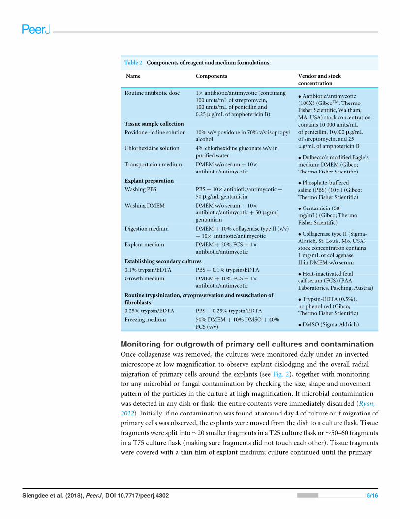
Table 2 Components of reagent andmedium formulations.
Name Components Vendor and stockconcentration
Routine antibiotic dose 1× antibiotic/antimycotic (containing100 units/mL of streptomycin,100 units/mL of penicillin and0.25 µg/mL of amphotericin B)
Tissue sample collectionPovidone–iodine solution 10% w/v povidone in 70% v/v isopropyl
alcoholChlorhexidine solution 4% chlorhexidine gluconate w/v in
purified waterTransportation medium DMEM w/o serum+ 10×
antibiotic/antimycotic
Explant preparationWashing PBS PBS+ 10× antibiotic/antimycotic+
50 µg/mL gentamicinWashing DMEM DMEM w/o serum+ 10×
antibiotic/antimycotic+ 50 µg/mLgentamicin
Digestion medium DMEM+ 10% collagenase type II (v/v)+ 10× antibiotic/antimycotic
Explant medium DMEM+ 20% FCS+ 1×antibiotic/antimycotic
Establishing secondary cultures0.1% trypsin/EDTA PBS+ 0.1% trypsin/EDTAGrowth medium DMEM+ 10% FCS+ 1×
antibiotic/antimycotic
Routine trypsinization, cryopreservation and resuscitation offibroblasts0.25% trypsin/EDTA PBS+ 0.25% trypsin/EDTAFreezing medium 50% DMEM+ 10% DMSO+ 40%
FCS (v/v)
• Antibiotic/antimycotic(100X) (GibcoTM; ThermoFisher Scientific, Waltham,MA, USA) stock concentrationcontains 10,000 units/mLof penicillin, 10,000 µg/mLof streptomycin, and 25µg/mL of amphotericin B
• Dulbecco’s modified Eagle’smedium; DMEM (Gibco;Thermo Fisher Scientific)
• Phosphate-bufferedsaline (PBS) (10×) (Gibco;Thermo Fisher Scientific)
• Gentamicin (50mg/mL) (Gibco; ThermoFisher Scientific)
• Collagenase type II (Sigma-Aldrich, St. Louis, Mo, USA)stock concentration contains1 mg/mL of collagenaseII in DMEM w/o serum
•Heat-inactivated fetalcalf serum (FCS) (PAALaboratories, Pasching, Austria)
• Trypsin-EDTA (0.5%),no phenol red (Gibco;Thermo Fisher Scientific)
• DMSO (Sigma-Aldrich)
Monitoring for outgrowth of primary cell cultures and contaminationOnce collagenase was removed, the cultures were monitored daily under an invertedmicroscope at low magnification to observe explant dislodging and the overall radialmigration of primary cells around the explants (see Fig. 2), together with monitoringfor any microbial or fungal contamination by checking the size, shape and movementpattern of the particles in the culture at high magnification. If microbial contaminationwas detected in any dish or flask, the entire contents were immediately discarded (Ryan,2012). Initially, if no contamination was found at around day 4 of culture or if migration ofprimary cells was observed, the explants were moved from the dish to a culture flask. Tissuefragments were split into∼20 smaller fragments in a T25 culture flask or∼50–60 fragmentsin a T75 culture flask (making sure fragments did not touch each other). Tissue fragmentswere covered with a thin film of explant medium; culture continued until the primary
Siengdee et al. (2018), PeerJ, DOI 10.7717/peerj.4302 5/16

1.5 cm
2.5 cm
1.0 cm
1.5 cm 1.0 cm
B C
D E F
H G I
A
1.5 cm
2.5 cm
1.0 cm 1.0 cm
E4 E4 E4
E4 E5
E5 E1 E1
E1
Figure 1 Ear explant preparation. (A) The ear skin samples were soaked in 70% ethanol, washed threetimes with washing PBS and dried on cellulose filter paper. (B) and (C) edges of the skin were trimmed,separating the front and back, respectively. (D) and (E) loose connective tissue and lobules of fat were re-moved and rinsed with washing PBS. (F) and (G) excised samples were cut with a scalpel into∼1-cm-longstrips, then chopped into pieces approximately 1 to 2 mm2 in size. (H) explants were washed with wash-ing DMEM and then minced with iris scissors. (I) explants were digested in collagenase solution and incu-bated at 37 ◦C and 5% CO2 for 21 h; after that, the explant samples were further washed twice with PBS.
Full-size DOI: 10.7717/peerj.4302/fig-1
outgrowing cells reached confluence around the explants or reached ∼80% confluence inculture flasks (Figs. 3A and 3B).
Establishing secondary culturesOnce reaching confluence (Fig. 4A), fibroblast cultures surrounding the pieces of skin werefurther expanded. The explant medium was poured out of the culture flasks and the cellsurfaces washed with PBS three times. In these second and third passages, keratinocyteswere removed from the cultures by short trypsinization with 0.1% trypsin/EDTA, as
Siengdee et al. (2018), PeerJ, DOI 10.7717/peerj.4302 6/16
I Day 14
E2
Day 4 Day 4 C E2 E1
Day 7 D E1
Day 9 E E2
A Day 4
E1 E5
Day 4 B
Day 9
E5 F
H Day 14
E5 G Day 14
E1
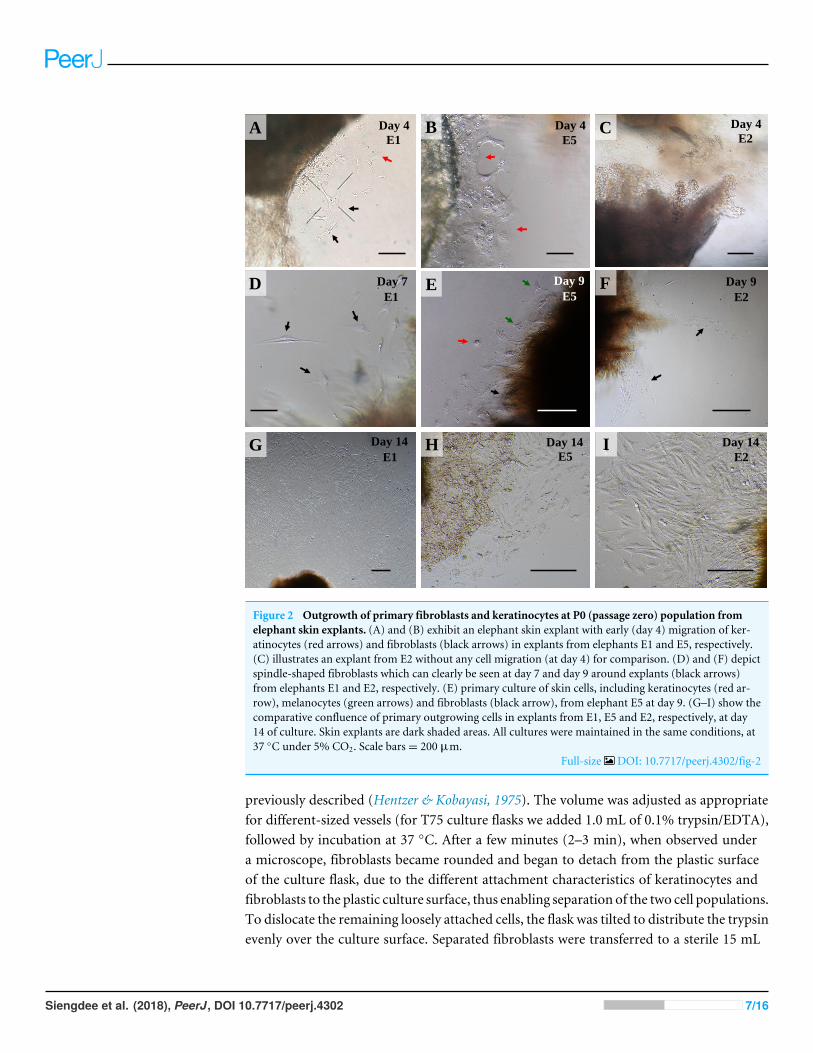
Figure 2 Outgrowth of primary fibroblasts and keratinocytes at P0 (passage zero) population fromelephant skin explants. (A) and (B) exhibit an elephant skin explant with early (day 4) migration of ker-atinocytes (red arrows) and fibroblasts (black arrows) in explants from elephants E1 and E5, respectively.(C) illustrates an explant from E2 without any cell migration (at day 4) for comparison. (D) and (F) depictspindle-shaped fibroblasts which can clearly be seen at day 7 and day 9 around explants (black arrows)from elephants E1 and E2, respectively. (E) primary culture of skin cells, including keratinocytes (red ar-row), melanocytes (green arrows) and fibroblasts (black arrow), from elephant E5 at day 9. (G–I) show thecomparative confluence of primary outgrowing cells in explants from E1, E5 and E2, respectively, at day14 of culture. Skin explants are dark shaded areas. All cultures were maintained in the same conditions, at37 ◦C under 5% CO2. Scale bars= 200 µm.
Full-size DOI: 10.7717/peerj.4302/fig-2
previously described (Hentzer & Kobayasi, 1975). The volume was adjusted as appropriatefor different-sized vessels (for T75 culture flasks we added 1.0 mL of 0.1% trypsin/EDTA),followed by incubation at 37 ◦C. After a few minutes (2–3 min), when observed undera microscope, fibroblasts became rounded and began to detach from the plastic surfaceof the culture flask, due to the different attachment characteristics of keratinocytes andfibroblasts to the plastic culture surface, thus enabling separation of the two cell populations.To dislocate the remaining loosely attached cells, the flask was tilted to distribute the trypsinevenly over the culture surface. Separated fibroblasts were transferred to a sterile 15 mL
Siengdee et al. (2018), PeerJ, DOI 10.7717/peerj.4302 7/16

Skin explant
Fibroblasts
A Skin explant
Keratinocytes
Fibroblasts
B
Figure 3 The labeled example illustrates the outgrowth of keratinocytes and fibroblasts from an ele-phant skin biopsy (E3) after three weeks of culture. (A) an example of fibroblast-like cells growing outof the elephant skin explant. (B) skin explant surrounded by polygonal cells, referred to as epithelial ker-atinocytes (squamous epithelia), that approach confluence until producing stratified multilayer sheets.Scale bars= 200 µm.
Full-size DOI: 10.7717/peerj.4302/fig-3
centrifuge tube, followed by immediately adding at least five volumes of fresh growthmedium containing 10% FBS and re-cultivating to expand cell numbers in a new T75culture flask. Cultures were grown at 37 ◦C in a humidified atmosphere containing 5%CO2. During this time, keratinocytes were still attached to the plastic bottom and preservedtheir membranous shapes, as seen before trypsin treatment. To culture keratinocytes, cellswere incubated at 37 ◦C for ∼10 min longer to disperse the cells; then the skin explantswere removed and fresh growth medium added to re-cultivate keratinocytes. Moreover, toobtain a larger amount of fibroblasts, explants were left in the first flask for a second roundof cell growth and incubated as before, while keratinocytes were separated and moved to anew T75 culture flask instead. The culture medium was changed once every 48 h. The 2ndto 4th passages of cultures of these cells were frozen for long-term storage stock. Fibroblastsat the 4th to 7th passages were used for further experiments.
Routine trypsinization, cryopreservation and resuscitation offibroblastsCryogenic preservation of elephant skin fibroblasts is generally the same as cryopreservationofmost continuous cell lines: by using commondimethyl sulfoxide (DMSO), which permitslong-term storage of cells in liquid nitrogen. After routine trypsinization of cells with 0.25%trypsin/EDTA and incubation for 5 min, cell suspensions were diluted with fresh growthmedium and the sample thoroughly mixed. Cells were centrifuged at 200 g for 10 min.The pellets were re-suspended in freezing medium consisting of 50% DMEM, 10% DMSOand 40% FBS (v/v). Cell suspensions were aliquoted into cryogenic storage vials (atapproximately 1.5–2.0 × 10⁶ cells/vial) and frozen at −80 ◦C in a deep freezer overnight.
Siengdee et al. (2018), PeerJ, DOI 10.7717/peerj.4302 8/16

B
Skin explant
C
Skin explant
E3
E3 E4
E4 A
D
Figure 4 Establishing secondary cultures of fibroblasts. (A) the outgrowths surrounding the pieces ofskin reached confluence and covered 80% of the flask. (B) 2 to 3 min after short trypsinization with 0.1%trypsin/EDTA application, fibroblasts were becoming rounded. (C) 3 min after short trypsinization, fi-broblasts appeared rounded and were detached from the culture flasks, while keratinocytes were still at-tached to the plastic bottom. (D) after 15 min of trypsinization, both fibroblasts and keratinocytes weredetached from the culture flasks. Scale bar= 20 mm.
Full-size DOI: 10.7717/peerj.4302/fig-4
The next day the vials were transferred to a liquid nitrogen tank for long-term storage untilused for further experiments.
To resuscitate fibroblasts or determine post-cryopreservation cell viability, the cellsin the frozen vials were quickly thawed at 37 ◦C and promptly mixed with 6.0 mL ofgrowth medium in a sterile centrifugal tube, then cultured in T75 culture flasks withoutcentrifugation. Viable cells were counted manually using a hemocytometer and expressedas the % of live cells from the total cell count. The next day, attached cells were washedtwice with PBS and new growth medium added.
Measuring cell viability and generating a growth curveCells were counted to assess viability using a traditional cell-counting method, with abright-line hemocytometer and trypan blue dye (Jauregui et al., 1981; Louis & Siegel, 2011).
Siengdee et al. (2018), PeerJ, DOI 10.7717/peerj.4302 9/16

Briefly, cells were first trypsinized with a routine subculture. Cell suspension was mixed 1:1with 0.4% trypan blue solution and let stand for 5 min at room temperature. Then 20 µL ofthe cell suspension was applied between the cover slip and the edge of the hemocytometerchamber and examined immediately under a microscope following the method of Louis& Siegel (2011). For measuring cell growth, seed cells at passage 6 (P6) of approximately5,000 cells per well in 0.5 mL of growth medium in a 24-well plate were cultured undernormal culture conditions. Viable cells in each well were counted over a 12-day periodusing a hemocytometer. The total count was calculated to obtain the growth curve andpopulation-doubling time.
RESULTS AND DISCUSSIONTissue sample collectionTo our knowledge this is the first report on the successful isolation and culture of skinfibroblasts from post-mortem ear tissues of a mature Asian elephant. In addition toproviding convenience of tissue sampling, the ear serves as ideal tissue for fibroblastisolation, with an excellent number and quality of cells (Mestre-Citrinovitz et al., 2016).We collected ear samples after the elephant had died and the traditional requiem ritualhad been performed by the mahout (Keyes, 1977). Including post-mortem examination(necropsy), ear cleaning and transportation time, the ear samples arrived at the lab around10 to 24 h after the elephant’s death. This was still considered good quality remains forprimary cell culture (Seluanov, Vaidya & Gorbunova, 2010). Previous studies have showneffective methods of preserving tissues and protocols to isolate outgrowth of fibroblast-likecells several days after death, taking into account different factors such as the storagetemperature and animal species, e.g., 12 days after animal death from ears stored at 4 ◦C ingoats and sheep (Silvestre, Sánchez & Gómez, 2004), 10 days post-mortem for sheep ear skinwhen skin tissues were exposed to around 25 to 26 ◦C (Singh & Ma, 2014), and recentlypublished data showing a recovery method for fibroblast cells from goat skin up to 160 dayspost-mortem at 4 ◦C storage (Aoued & Singh, 2015). Of these protocols mentioned, theimportant point is that tissues were processed for good preservation within ‘‘an hour’’ ofcollection, which is almost impossible in the case of an elephant carcass in Thailand due toThai cultural beliefs concerning the death ceremony. In this study, we failed to isolate anyoutgrowth of fibroblast cells from post-mortem tissues harvested 20 h after the animal’sdeath (elephant E6), even after culturing the explants for 1 month and finding no presenceof contamination. It is possible that elephant E6 had been left in unsuitable conditions,i.e., at a high environmental temperature (∼33–35 ◦C in June) for too long a time, beforeear samples were collected and transferred to the laboratory. Another important thingthat should not be overlooked is the awareness of zoonoses. Wild animals may containpathogens, such as anthrax tuberculosis and leptospirosis, that can be transmitted fromanimals to humans; and most samples were collected under field conditions, so takingcare to avoid sharp surfaces that can pierce the skin and awareness of sterile techniqueare essential at all steps to minimizes any risks from infectious diseases (Kruse, Kirkemo &Handeland, 2004).
Siengdee et al. (2018), PeerJ, DOI 10.7717/peerj.4302 10/16

Outgrowth of primary cell culturesFor our study we therefore adapted a collagenase digestion technique together withan explant technique to establish elephant fibroblasts (Mestre-Citrinovitz et al., 2016;Pruniéras, Delescluse & Regnier, 1976; Reiisi, Esmaeili & Shirazi, 2010; Rittié & Fisher, 2005;Vangipuram et al., 2013). Because trypsin digestion technique (Orazizadeh et al., 2015) wasnot completed to separate the dermis and epidermis layers in our study, outgrowth ofepidermis (keratinocytes) and dermis (fibroblasts) could therefore be seen (Pruniéras,Delescluse & Regnier, 1976). Explant dislodging and overall radial migration aroundexplants could be observed, as shown in Fig. 2 at low magnification. Both fibroblast-like and epithelial-like cells could be seen migrating from the tissue pieces 4–12 daysafter explanting. The earliest observed outgrowth around explants that dislodged wasat day 4 of culture in E1 and E5 (Figs. 2A and 2B), compared with E2, E3 and E4 onthe same day (Fig. 2C for comparison) in which primary cells were observed aroundday 7–10 (Figs. 2D–2F). We found outgrowths of epithelial keratinocytes around someexplants in our experiments. They grew in squamous epithelia shapes until confluent andproduced stratified multilayer sheets around the explants beginning after 4–7 days, whilefibroblasts started later at around 7–10 days. At the time of fibroblast appearance during thefollowingweeks the fibroblasts overgrew the keratinocytes and the culturesmainly consistedof fibroblasts with only small islands of epidermal cells around the explants (Fig. 3).Cells continued to proliferate and were subcultured when they reached 80% confluence(Figs. 3A and 3B).
Establishing secondary culturesDuring the first few minutes (2–3 min) of observation under a microscope, fibroblastsbecame rounded and began to dislodge from the plastic surface of the culture flasks,while the epidermal cells stuck to the bottom and preserved their membranous shapes(Figs. 4B and 4C). After 15 min of trypsinization, both fibroblasts and keratinocytesbecame detached from the culture flasks (Fig. 4D). After subculture, the fibroblast cellsgrew rapidly, gradually outgrowing.
Measuring cell viability and generating a growth curveThe viabilities of elephant skin fibroblasts before freezing and recovery (percent cell viabilitywas calculated immediately post-thaw) were around 97.3 ± 4.3%, and 95.5 ± 7.3%,respectively, when their post-freezing recovery and survival was improved with freezingmedium consisting of 50%DMEM+ 10%DMSO+ 40% FCS (v/v). This indicates that thecells were grown in good culture conditions and that the freezing medium was appropriate.Note that at 10–20% FBS freezing medium the cells had very low recovery rates and cellmorphology was different from cells observed prior to cryopreservation, which may becaused by apoptosis (Xu et al., 2010). In this study after 12 days continuous count theelephant skin fibroblast growth curve was as shown in Fig. 5. The curve was sigmoidal witha population doubling time (at the 6th passage) of about 25 h.
Siengdee et al. (2018), PeerJ, DOI 10.7717/peerj.4302 11/16

0.00E+00
2.00E+05
4.00E+05
6.00E+05
8.00E+05
1.00E+06
1.20E+06
0 1 2 3 4 5 6 7 8 9 10 11 12
Cel
l n
um
ber
s
Days in culyure
E1
E2
E3
E4
E5
Figure 5 Growth curve of elephant skin fibroblasts at passage 6 (P6) obtained from five elephants.Full-size DOI: 10.7717/peerj.4302/fig-5
CONCLUSIONSThis study has described the simple methods and requirements for isolating and culturinglarge numbers of primary cultures of E. maximus fibroblasts obtained from post-mortemear skin tissues. The protocol presented here is a modified collagenase digestion proceduretogether with a simple explant procedure that can be applied to elephant fibroblasts. Theproblems we encountered and their successful resolution, employing the most efficientapproach, are also demonstrated here. This knowledge should represent a step forwardfor the use of this technology for research in cell aging, in the treatment of age-associatedimpairments in dermal integrity and chronic wound healing, and in further cellularstudies in Asian elephants. This study also suggests that suitable conditions during samplecollection—such as post-mortem storage time, storage temperature, or other possiblefactors—may be associated with cell isolation success; however, a statistical test for this wasnot performed. Moreover, no correlation was observed between the age of an elephant atthe time of death and the outgrowth of fibroblasts or the proliferation rate of cells. Thisshould extend the usefulness of adult Asian elephant fibroblasts for further studies.
ACKNOWLEDGEMENTSWe wish to thank the Thai Elephant Conservation Center, Hang Chat, Lampang, Thailand;Panda Elephant Camp, Mae Taeng, Chiang Mai, Thailand; Mae Sa Elephant Camp, MaeRim, Chiang Mai, Thailand; Water Runner and Bobby’s Elephant Home, Mae Taeng,Chiang Mai, Thailand; Elephant Nature Park, Mae Taeng, Chiang Mai, Thailand; and
Siengdee et al. (2018), PeerJ, DOI 10.7717/peerj.4302 12/16

Elephant Jungle, Mae Wang, Chiang Mai, Thailand, for donating tissue samples used inthis study.
ADDITIONAL INFORMATION AND DECLARATIONS
FundingThis workwas supported by research funding received fromChiangMaiUniversity throughthe Research Administration Office, which provided a budget to the Center of Excellence inElephant Research and Education and Excellence Center in Veterinary Bioscience, Facultyof Veterinary Medicine, Chiang Mai University, Thailand. The authors also received aresearch grant from the Brian Nixon Fund for the protection of elephants in Thailand. Thefunders had no role in study design, data collection and analysis, decision to publish, orpreparation of the manuscript.
Grant DisclosuresThe following grant information was disclosed by the authors:Chiang Mai University.Brian Nixon Fund.
Competing InterestsThe authors declare there are no competing interests.
Author Contributions• Puntita Siengdee conceived and designed the experiments, performed the experiments,analyzed the data, contributed reagents/materials/analysis tools, wrote the paper,prepared figures and/or tables, reviewed drafts of the paper.• Sarisa Klinhom performed the experiments, contributed reagents/materials/analysistools, reviewed drafts of the paper.• Chatchote Thitaram conceived and designed the experiments, reviewed drafts of thepaper.• Korakot Nganvongpanit conceived and designed the experiments, analyzed the data,wrote the paper, prepared figures and/or tables, reviewed drafts of the paper.
Animal EthicsThe following information was supplied relating to ethical approvals (i.e., approving bodyand any reference numbers):
According to the Animals for Scientific Purposes Act, B.E. 2558 (2015), since a part ofthis experiment was performed on an elephant carcass from a private owner, no ethicalapproval was required for this study and confirmed by the Animal Ethics Committee,Faculty of Veterinary Medicine, Chiang Mai University (Licence number U1006312558).However, the owner allowed the research team to take a sample of skin for this study.
Data AvailabilityThe following information was supplied regarding data availability:
The raw data is included in a Supplemental File.
Siengdee et al. (2018), PeerJ, DOI 10.7717/peerj.4302 13/16

Supplemental InformationSupplemental information for this article can be found online at http://dx.doi.org/10.7717/peerj.4302#supplemental-information.
REFERENCESAngkawanish T, Boonprasert K, Homkong P, Sombutputorn P, Mahasawangkul S,
Jansittiwate S, Keratimanochaya T, Clausen B. 2009. Elephant health status inThailand: the role of mobile elephant clinic and elephant hospital. Gajah 31:15–20.
Aoued H, SinghM. 2015. Recovery of fibroblast-like cells after 160 days of postmortemstorage of goat skin tissues in refrigerated media. Journal of Veterinary Science andTechnology 6:Article 1000236 DOI 10.4172/2157-7579.1000236.
Brink H, Bernstein J, Nicoll S. 2009. Fetal dermal fibroblasts exhibit enhanced growthand collagen production in two- and three-dimensional culture in comparison toadult fibroblasts. Journal of Tissue Engineering and Regenerative Medicine 3:623–633DOI 10.1002/term.204.
Fernando P, Vidya T, Payne J, StueweM, Davison G, Alfred R, Andau P, Bosi E,Kilbourn A, Melnick D. 2003. DNA analysis indicates that Asian elephants are nativeto Borneo and are therefore a high priority for conservation. PLOS Biology 1:e6DOI 10.1371/journal.pbio.0000006.
Groeneveld E, Tinh N, KuesW, Vien N. 2008. A protocol for the cryoconserva-tion of breeds by low-cost emergency cell banks—a pilot study. Animal 2:1–8DOI 10.1017/S1751731107000869.
GuanW, Liu C, Li C, Liu D, ZhangW,Ma Y. 2010. Establishment and cryopreservationof a fibroblast cell line derived from Bengal tiger (Panthera tigris tigris). Cryo Letters31:130–138.
Hentzer B, Kobayasi T. 1975. Separation of human epidermal cells from fibrob-lasts in primary skin culture. Archiv für Dermatologische Forschung 252:39–46DOI 10.1007/BF00582429.
Jauregui H, Hayner N, Driscoll J, Williams-Holland R, LipskyM, Galletti P. 1981.Trypan blue dye uptake and lactate dehydrogenase in adult rat hepatocytes–freshly isolated cells, cell suspensions, and primary monolayer cultures. In Vitro17:1100–1110 DOI 10.1007/BF02618612.
Keyes CF. 1977. The golden Peninsula: culture and adaptation in mainland Southeast Asia.Honolulu: University of Hawaii Press.
Kruse H, Kirkemo A, Handeland K. 2004.Wildlife as source of zoonotic infections.Emerging Infectious Diseases 10:2067–2072 DOI 10.3201/eid1012.040707.
Louis K, Siegel A. 2011. Cell viability analysis using trypan blue: manual and automatedmethods.Methods in Molecular Biology 740:7–12 DOI 10.1007/978-1-61779-108-6_2.
Magda S, Spohn O, Angkawanish T, Smith D-A, David L. 2015. Risk factors for saddle-related skin lesions on elephants used in the tourism industry in Thailand. BMCVeterinary Research 11:117 DOI 10.1186/s12917-015-0438-1.
Siengdee et al. (2018), PeerJ, DOI 10.7717/peerj.4302 14/16

Mestre-Citrinovitz A, Sestelo A, Ceballos M, Barañao J, Saragüeta P. 2016. Isolationof primary fibroblast culture from wildlife: the Panthera onca case to preservea South American endangered species. Current Protocols in Molecular Biology116:128.117.111–128.117.114 DOI 10.1002/cpmb.25.
Miller D, Jackson B, Riddle H, Stremme C, Schmitt D, Miller T. 2015. Elephant (Elephasmaximus) health and management in Asia: variations in veterinary perspectives.Veterinary Medicine International 2015:Article 614690 DOI 10.1155/2015/614690.
OrazizadehM, Hashemitabar M, Bahramzadeh S, Dehbashi F, Saremy S. 2015. Com-parison of the enzymatic and explant methods for the culture of keratinocytes iso-lated from human foreskin. Biomedical Reports 3:304–308 DOI 10.3892/br.2015.442.
Pruniéras M, Delescluse C, Regnier M. 1976. The culture of skin. A review of theo-ries and experimental methods. Journal of Investigative Dermatology 67:58–65DOI 10.1111/1523-1747.ep12512483.
Reiisi S, Esmaeili F, Shirazi A. 2010. Isolation, culture and identification of epidermalstem cells from newborn mouse skin. In Vitro Cellular & Developmental Biology-Animal 46:54–59 DOI 10.1007/s11626-009-9245-y.
Rittié L, Fisher GJ. 2005. Isolation and culture of skin fibroblasts.Methods in MolecularMedicine 117:83–98 DOI 10.1385/1-59259-940-0:083.
Ryan J. 2012. Understanding and managing cell culture contamination. Available athttp://www.level.com.tw/html/ ezcatfiles/ vipweb20/ img/ img/20297/ contamination-COR.pdf (accessed on 19 June 2017).
Sathanawongs A, Jarujinda Y, Rojanasthien S, Oranratnachai A. 2010. Production ofcloned Asian elephant embryos using an interspecies somatic cell nuclear transfer(iSCNT) technique. Kasetsart Journal (Natural Science) 44:610–620.
Seluanov A, Vaidya A, Gorbunova V. 2010. Establishing primary adult fibrob-last cultures from rodents. Journal of Visualized Experiments 44:Article 2033DOI 10.3791/2033.
Shoshani J, Eisenberg JF. 1982. Elephas maximus.Mammalian Species 182:1–8.Silvestre M, Sánchez J, Gómez E. 2004. Vitrification of goat, sheep, and cattle skin sam-
ples from whole ear extirpated after death and maintained at different storage timesand temperatures. Cryobiology 49:221–229 DOI 10.1016/j.cryobiol.2004.08.001.
SinghM,Ma X. 2014. In vitro culture of fibroblast-like cells from sheep ear skin stored at25–26 ◦C for 10 days after animal death. International Journal of Biology 6:96–102DOI 10.5539/ijb.v6n4p96.
Sriram G, Bigliardi P, Bigliardi-Qi M. 2015. Fibroblast heterogeneity and its impli-cations for engineering organotypic skin models in vitro. European Journal of CellBiology 94:483–512 DOI 10.1016/j.ejcb.2015.08.001.
Sukumar R. 1992. The Asian elephant: ecology and management. Cambridge: CambridgeUniversity Press.
TechakumphuM, Rungsiwiwut R, Numchaisrika P, Thongphakdee A. 2017. ClonedAsian elephant (Elephas maximus) embryos reconstructed from rabbit recipientoocytes. Thai Veterinary Medicine 40:63–68.
Siengdee et al. (2018), PeerJ, DOI 10.7717/peerj.4302 15/16

VangipuramM, Ting D, Kim S, Diaz R, Schüle B. 2013. Skin punch biopsy explant cul-ture for derivation of primary human fibroblasts. Journal of Visualized Experiments7:e3779 DOI 10.3791/3779.
Xu X, Cowley S, Flaim C, JamesW, Seymour L, Cui Z. 2010. The roles of apoptoticpathways in the low recovery rate after cryopreservation of dissociated humanembryonic stem cells. Biotechnology Progress 26:827–837 DOI 10.1002/btpr.368.
Siengdee et al. (2018), PeerJ, DOI 10.7717/peerj.4302 16/16