isolation and molecular characterization of a hyperglycemic neuropeptide from the sinus gland of the...
TRANSCRIPT
Eur. J. Biochem. 211, 601-607 (1993) 0 FEBS 1993
Isolation and molecular characterization of a hyperglycemic neuropeptide from the sinus gland of the terrestrial isopod Armadillidium vulgare (Crustacea) Gilbert MARTIN', Odile SOROKINE' and Alain VAN DORSSELAER2
Laboratoire de biologie animale URA 1452, UniversitC de Poitiers, France Laboratoire de spectrom6trie de masse bioorganique UA31, FacultC de chimie, CNRS, Strasbourg, France
(Received August 26, 1992) - EJB 92 1224
The major peptide from the sinus gland of the terrestrial isopod Armadillidium vulgare (Crusta- cea) has been extracted and purified by reverse-phase HPLC. This neuropeptide exhibited a high hyperglycemic activity and was therefore named A. vulgare crustacean hyperglycemic hormone (Am-CHH). Its average molecular mass measured by mass spectrometry was 8729.3 Da. Its com- plete amino acid sequence was determined by a combination of Edman degradation and mass spec- trometry. The N-terminal amino acid was found to be unblocked, the C-terminal residue was found amidated and none of the other 72 residues was affected by any post-translational modification. Disulfide bond assignment was made unambigously by mass spectrometry and Edman degradation was performed on peptides produced by enzymatic cleavage. Relationships with other, similar neuro- peptides from decapod sinus glands are discussed.
The X-orgadsinus gland neurosecretory system located in the protocerebrum is the major neuroendocrine center in the isopod Armadillidium vulgare [l]. The sinus gland is a neurohemal organ, in which the neurohormones, synthesised in the neurosecretory cells of the X-organ are stored until their release. The X-organ location differs in isopods and decapods. This difference is related to the fact that the former have sessile eyes leading to a reduction of the optic medulla; as a consequence, the X-organ is shifted to the median part of the brain. However, in both groups the sinus gland neuro- hormones are involved in monitoring almost all known physiological processes [2]. The crustacean hyperglycemic hormone (CHH) has been shown to be the most abundant hormone in the sinus gland of isopods and decapods and controls the blood sugar level [3, 41. Molting and repro- duction are regulated by inhibitory hormones : molt inhibiting hormone (MIH), vitellogenesis inhibiting hormone (VIH), and androgenic inhibiting hormone (AIH) [5]. From the re- sults published in recent years [4, 6, 71 it could not be ex- cluded that CHH also interacts in various physiological pro- cesses. Furthermore, CHH was also reported to act as a stress hormone [2]. From a phylogenetic point of view, a compari- son of such a hormone in marine and terrestrial related forms
Correspondence to A. Van Dorsselaer, Laboratoire de spectro- metrie de masse bioorganique UA31, faculte de chimie, CNRS, 1 rue B. Pascal, F-67008 Strasbourg Cedex, France
Abbreviations. CHH, crustacean hyperglycemic hormone ; A w - CHH, Armadillidiurn vulgare crustacean hyperglycemic hormone ; MIH, molt-inhibiting hormone ; VIH, vitellogenesis-inhibiting hor- mone ; AIH, androgenic-inhibiting hormone ; LSIMS, liquid second- ary-ion mass spectrometry ; ESMS, electrospray mass spectrometry ; MIKES, mass-analysed ion-kinetic-energy spectrum ; EW, reverse- phase.
(Crustacea) should be very fruitful. Until now very little structural work has been done on isopod sinus gland pep- tides. We first reported the CHH amino-acid composition [3] and, later, a partial sequence of two peptides of the CHH family [8]. Recently several papers have been published on the sinus gland peptides of decapods. These peptides exist as different isoforms but share CHH and other biological activi- ties [9].
This paper reports for the first time the complete amino- acid sequence of an isopod CHH. It was determined by a combination of Edman degradation and mass spectrometry [lo-131. The assignment of the position of the disulfide bridges was made unambiguously by a two-step procedure which combined Edman degradation and mass spectrometry.
MATERIALS AND METHODS Experimental animals
Armadillidium vulgare woodlice (Crustacea) or pillbugs belong to the Isopoda and are classified as Oniscoydea terres- trial crustacean. The woodlice were reared in the laboratory. According to the terminology proposed by Raina and Gade [14], our peptide was labeled as Am-CHH.
Collection and extraction of sinus glands Sinus glands were excised from adults. Their localization
and appearance were as published elsewhere [l]. During col- lection, we were selective with regard to the molting stages (animals in premolt or in molt were discarded) and the sex of the woodlice (batches from males and females were kept separate). For each HPLC run batches of about 600 male or

602
female sinus glands were extracted with 0.2 M acetic acid as previously described [3].
Liquid chromatography Reverse-phase high-performance liquid chromatography
(RP-HPLC) was performed using a Waters liquid chromatog- raphy 625 LC system and a Waters 991 photodiode array detector. The separation was performed with an 80-min water/acetonitrile linear gradient of 0 - 60 % acetonitrile with 0.1% trifluoroacetic acid at a flow rate of 0.9 mumin. The eluate was monitored by measuring the absorbance at 214 nm. CHH was first purified on a pBondapack C,, col- umn (Waters Associates, 0.39 X 30 cm) ; then a second run was performed on a Speri 300-5 RP-8 column (Brownlee, 0.46 X 25 cm, 1 mumin) with the same gradient. Cleavage peptides were purified using a Lichrocart C,, column (Merck 0.4 X 2.5 cm) with a water/acetonitrile gradient of 0 % - 80 % acetonitrile (0.1% trifluoroacetic acid) at a rate of l%/min and a flow rate of 1 ml/min.
Enzymatic cleavages Trypsin. Trypsin (Boehringer, sequencing grade) cleaves
at the C-terminal side of Arg and Lys residues. Tryptic diges- tion was performed in 0.1 M Tris, pH 8.5, 0.02 M CaC1, with a peptide/enzyme mass ratio of 1 O : l for 4 h at 38°C. The solution was directly injected onto the RP-HPLC column and the different peaks were collected, oxidized (performic oxi- dation) and then submitted to Edman degradation.
Endoproteinuse Glu-C. The endoproteinase V8 (Boehr- inger, sequencing grade) digestion was performed in 0.1 M potassium phosphate pH 7.5 with a peptide/enzyme mass ra- tio of 10: 1 for 4 h at 37"C, leading to cleavage at the C- terminal side of Asp and Glu residues. The mixture of cleav- age peptides was injected onto the RP-HPLC column and the different peaks were characterized by mass spectrometry.
Endoproteinuse Asp-N. Digestion by endoproteinase Asp-N (Boehringer, sequencing grade) leading to cleavage at the Asp N-terminus was done by incubation for 4 h at 37°C with a peptide/enzyme mass ratio of 10: 1 in 0.1 M Tris pH 8.5. After incubation, the digest was directly injected on the RP-HPLC column.
Performic oxidation Performic acid was prepared by adding hydrogen per-
oxide (30 vol. %) to 100% formic acid (5/95, by vol.). The mixture was allowed to react 4 h before use. Peptide was dissolved in this performic acid preparation and left at -8OC during 2 h. This sample was then dried in a Speed-Vac (Sav- ant) redissolved in water (10% acetic acid) and injected on RP-HPLC.
Amino acid analysis Peptides (1 nmol) were hydrolyzed in constant-boiling
HCl (6 M) at 106°C for 24 h. After hydrolysis, the amino acids were converted to their phenylthiocarbamoyl deriva- tives using phenylisothiocyanate and identified as described in the Waters Picotag manual.
Peptide quantification
evaluated by amino acid quantification (Pico-Tag method). The amount of peptides collected by RP-HPLC was
Mass spectrometry Mass measurement of the CHH was performed using two
techniques : liquid secondary ion mass spectrometry (LSIMS) and electrospray mass spectrometry (ESMS).
LSIMS was done in the positive mode on a VG Analyti- cal ZAB-2SE double-focussing instrument (mass range 15 kDa at 8 keV ion energy) and recorded on a VG 11-250 data system (VG Analytical Ltd, England). Ionization of the sample was performed with approximatively 2 pA of 30- keV-energy cesium ions using the cesium ion gun. Wide scans were performed at a resolution of 1000 by magnetic scanning over the mass range 12-1.5 kDa in 20 s [lo, 12, 151.
ESMS was done on a VG-Bio-Q (Bio-Tech, Manchester, UK) in the positive mode [13]. The protein was dissolved in H,O/MeOH (50/50, by vol.) with 1% acetic acid at a concen- tration of about 5 pmoVp1 (by vol).
Edman degradation
stem model 473 A in the liquid pulse mode. Edman degradation was performed on an Applied Biosy-
Bioassays Assays were based on glucose determination. Animals to
be tested were transferred from the rearing boxes onto moist filter paper in petri dishes at least one day prior to the bioas- says [8]. Test solutions were injected as 0.5-1.0-p1 samples through a small hole in the exoskeleton (tergite 5) carefully pierced with a needle; 2 h after injection, 5 pl hemolymph sample was withdrawn through the membrane between the tergites into a calibrated capillary. The hemolymph sample was mixed with 500p1 glucose oxidase reagent test kit (Boehringer) in a micro plate and the absorbance was meas- ured at 436 nm after 45 min.
RESULTS Purification by RP-HPLC
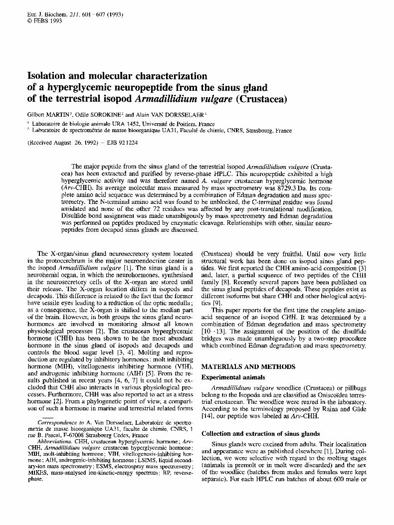
A typical chromatogram obtained by RP-HPLC analysis of the 0.2 M acetic acid extract from 600 female sinus glands is presented in Fig. 1. All peaks were separately collected and tested for different biological activities (MIH, VIH and CHH) [8]. The major peak (labelled CHH on Fig. 1) of the chromatogram displayed a high hyperglycemic activity. Peak labeled VIH on Fig. 1 was shown to be a potent inhibitor of vitellogenin synthesis [8].
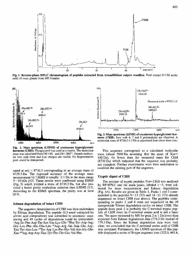
Mass spectrometry A mass spectrometry characterization of CHH was then
undertaken. Mass spectra of this hormone were first obtained by LSIMS scanning over a large mass range (1500- 12000 Da). Two major peaks (Fig. 2) were detected at m/z = 8730.3 and m/z = 4365.3. These values corresponded re-
spectively to a singly charged (M+H)' and a doubly charged ion (M+2H)*+ of an average molecular mass of 8728.9 Da. Peaks clearly visible at m/z = 2910.5 and m/z = 2183.4, cor- responding to three and four charges, confirmed this molecu- lar mass. To improve the accurracy of this result another measurement was performed in the narrow-scan mode [12]. The centroid of the protonated molecular peak was then mea-
603
0.5
0.3
3.1
-
CHH
ni, I I 1 I 1 1 1 0 20 40 60 time (min.)
-+
Fig. 1. Reverse-phase HPLC chromatogram of peptides extracted from Armadillidium vulgare woodlice. Total extract (0.2 M acetic acid) of sinus glands from 600 females.
100 31
Fig. 2. Mass spectrum (LSIMS) of crustacean hyperglycemic hormone (CHH). Thioglycerol was used as a matrix. The molecular mass was calculated from [M+H]+ and [M+2HI2+. Pseudo-molecu- lar ions with three and four charges are visible. No fragmentation peak could be interpreted.
sured at mlz = 8730.3 corresponding to an average mass of 8729.3 Da. The expected accuracy of the average mass measurement by LSIMS is usually 5 1 Da in the mass range 5-10 kDa [12]. These results were confirmed using ESMS (Fig. 3) which yielded a mass of 8730.2 Da, but also pro- vided a better purity evaluation criterion then LSIMS [13]. According to the ESMS spectrum, the purity was at least 90%.
Edman degradation of intact CHH
The sequence determination of CHH was then undertaken by Edman degradation. The peptide (0.5 nmol estimated by amino acid composition) was submitted to automatic sequ- encing and 49 cycles of degradation could be interpreted: 'Arg-Ile-Phe-Asp-Thr-Ser-Xaa-Lys-Gly-"Phe-Tyr-Asp-Arg- Gly-Leu-Phe-Ala-Gln-Leu-20Asp-Arg-Val-Xaa-Glu-Asp- Xaa-Tyr-Asn-Leu-30Tyr-Arg-Lys-Pro-His-Val-Ala-Ala-Glu- Xaa-40Arg-Arg-Asp-Xaa-Tyr-Thr-Thr-Glu-Val-Phe.
100
%? 8 Measured mass = 8730.2 +_ 2 B 2 5
.n 2 .r +
3
1000 1200 1400 mlz
Fig. 3. Mass spectrum (ESMS) of crustacean hyperglycemic hor- mone (CHH). Ions with 6, 7 and 8 protonations are observed. A molecular mass of 8730.2 2 2 Da is calculated from these three ions.
This sequence corresponds to a calculated molecular mass (about 5860Da assuming that the mass of Xaa= 100Da), far lower than the measured mass for CHH (8730 Da) which indicated that the sequence was probably not complete. Further experiments were then undertaken to establish the missing part of the sequence.
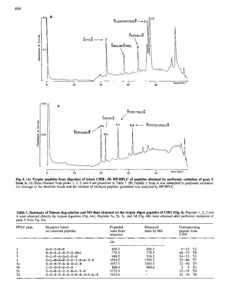
Tryptic digest of CHH The mixture of tryptic peptides from CHH was analysed
by RP-HPLC and the main peaks, labeled 1-5, were col- lected for mass measurement and Edman degradation (Fig. 4A). Results are given in Table 1. Peaks 1 and 3 corre- sponded to the peptides 9-13 (T2) and 14-21 (T3) already sequenced on intact CHH (see above). The peptides corre- sponding to peaks 2 and 4 were not sequenced in the 49 interpretable Edman degradation cycles on intact CHH. The peptide from peak 2 is probably the C-terminal tryptic pep- tide of CHH since its C-terminal amino acid is not a basic one. The mass measured by MS for peak 2 is 1 Da lower than expected from Edman degradation data (778.3 Da instead of 779.3 Da). Since the Edman degradation data were very clear, we concluded that the C-terminal amino acid of CHH was amidated. Furthemore, the LSIMS spectrum of this pep- tide displayed a series of B-type sequence ions (332.0,445.4,
604
I.:
A
9 7 3 GFYDR -
~ L M M H D F I N E Y ~ - 4,
~ L F A Q L D ~
\ .-.-. 1 1 I I 1 0 10 20 30 40 time (min.)
.
0.04
0.02
B
~FDTSCB
f 32 40 / KPHVAAECR
42 54 f - DCYTTEVFESCLK
J vCEDCYNLYil
I
1 I I 1 I 0 10 20 30 40 time (min.)
*
Fig. 4. (A) Tryptic peptides from digestion of intact CHH; (B) RP-HPLC of peptides obtained by performic oxidation of peak 5 from A. (A) Data obtained from peaks 1, 2, 3, and 4 are presented in Table 1. (B) Peptide 5 from A was submitted to performic oxidation for cleavage of the disulfide bonds and the mixture of oxidized peptides generated was analysed by W-HPLC.
Table 1. Summary of Edman degradation and MS data obtained on the tryptic digest peptides of CHH (Fig. 4). Peptides 1, 2, 3 and 4 were obtained directly by trypsin digestion (Fig. 4A). Peptides Sa, 5b, 5c, and 5d (Fig. 4B) were obtained after performic oxidation of peak 5 from Fig. 4A.
HPLC peak Sequence found on collected peptides
Expected Measured Corresponding mass from mass by MS peptide from sequence CHH
1 2 3 4 5a 5b 5c 5d
G-F-Y-D-R E-M-A-L-M-V-S-NH2 G-L-F-A-Q-L-D-R D-L-M-M-H-D-F-I-N-E-Y-K K-P-H-V-A-A-E-C-R I-F-D-T-S-C-K V-C-E-D-C-Y-N-L-Y-R D-C-Y-T-T-E-V-F-E-S-C-L-K
656.3 778.5 918.5
1554.5 1057.5 860.4
1372.5 1632.6
Da
656.3 778.3 918.3
1554.7 1057.5 860.4 -
9-13 T2 64-73 T8 14-21 T3 55-66 T7 32-40 T5 2- 8 T1
22-31 T4 42-54 T6

605
576.2, and 675.3) corresponding to a C-terminal sequence: - Leu-Met-Val-Ser-NH, and locating the 1 Da mass difference on Ser. Finally, a MIKES analysis performed on ion 779.4 produces two intense ions : 675.3 and 576.3 also correspond- ing to a sequence -Val-Ser-NH, (data not shown). These data unambiguously demonstrate that the C-terminal amino acid of peptide from peak 2 is amidated.
Peak 5 from Fig. 4A was submitted to performic oxi- dation in order to separate peptides attached by disulfide bonds (cysteine residues are converted to -SO,H groups). After analysis of the oxidized mixture by RP-HPLC (Fig. 4B), four peaks were collected (5a, 5b, 5c, 5d) for mass measurement and Edman degradation. The data collected are presented in Table 1. Peaks 5a, 5b, and 5c corresponded to peptides 32-40 (T5), 2-8 (Tl) and 22-31 (T4), respectively, already completely sequenced on intact CHH. Peak 5d corre- sponds to peptide 42-54 which was only partially sequenced on the intact CHH (last interpretable Edman degradation cy- cle was no. 49) and is now complete since the last amino acid is Lys.
By adding the molecular masses corresponding to the se- quences established for the eight tryptic peptides described above (Table 1) and the mass of the two arginyl residues which were expected to escape the tryptic digestion analysis (Argl and Arg41), a molecular mass of 8735.0 Da was ex- pected for CHH (after substraction of nine molecules of water). This value is in very good agreement with the meas- ured mass (8729.3 Da), assuming that the six cysteine resi- dues are involved in the formation of three disulfide bridges.
From the above data all of the peptides could be aligned, the measured molecular mass is in very good agreement with the sequence proposed and the difference of 6 Da between the measured mass and the calculated mass may be compat- ible with the six cysteines involveed in three disulfide briges. The C-terminal amino acid is amidated. Most of the peptides were sequenced twice, and their molecular mass measured for correlation with sequence data. Fig. 5 summarizes the se- quence data. The elucidation of the position of the disulfide bridges has been performed as described below.
Disulfide bridges Disulfide bridge attribution was performed using two dif-
ferent enzymatic cleavages : (a) digestion of intact CHH with endoproteinase Glu-C (C-terminal cleavage at Asp and Glu), (b) digestion with endoproteinase Asp-N (N-terminal cleav- age at Asp) of peptide 5 from the tryptic digest of intact CHH (Fig. 4A). After RP-HPLC on each digest mixture, the different peptides were submitted to mass spectrometry and Edman degradation. These data were interpreted taking into account the known sequence of CHH and the expected cleav- age sites of enzymes.
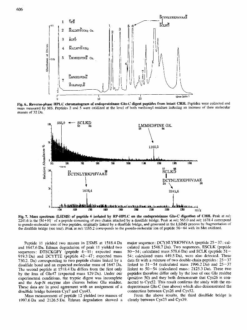
Endoproteinase Glu-C
After RP-HPLC of the enzymatic digest of the intact CHH (Fig. 6), seven purified peptides were submitted to MS measurement. LSIMS on peak 6 (Fig. 7) yielded a mass of 2241.6 Da. This mass spectrum also contains two large peaks at mlz 565.0 and at mlz 1678.4 that we interpreted as frag- ments obtained from a tryptic peptide after rupture of an in- terchain disulfide bond. It has been shown that peptides con- sisting of two chains linked by a disulfide bridge undergo, in LSIMS, an intense fragmentation at the disulfide bridge which generates an [M+H]+ ion for each peptide chain [16].
I
Fig. 5. The complete sequence of CHH and the comparison with the peptide from Keller’s group [17]. On the complete sequence of CHH (top of figure) the fragments marked with a dashed line (----) show all tryptic identified peptides ; the fragments marked with a full line (-) show peptides identified after endoprotein- ase Glu-C cleavage. The positions of disulfide bridges (7-43, 23- 39 and 26-51) are indicated. The bottom of the figure shows the comparison between our CHH sequence and the sequence of the CHH from Keller’s group. Identical residues are boxed; Pgl = 5- oxoproline (‘pyroglutamic acid’).
The mass calculated for peptides at mlz 565.0 and at mlz 1678.4 linked by a disulfide bond would be 2241.4 Da which fits very well with the observed mass (2241.6 Da). From the known sequence of the CHH, one can predict which peptide could be generated by endoproteinase Glu-C digestion and only one solution was possible to explain the spectrum of the peptide from peak 6 [16]. One chain would be SCLKD (peptide 51-55; expected mass 564.2 Da) and the other DCYNLYRKPHVAAE (peptide 25 - 38 ; expected mass 1677.8 Da). Thus it was concluded that a disulfide bridge occurs between Cys26 and Cys52.
Endoproteinase Asp-N on peak 5 nfer tryptic digestion of CHH
After Asp-N digestion of peak 5 from Fig. 4A the mix- ture of cleavage peptides was analysed by RP-HPLC. Two peptides (11 and 12) were sequenced and submitted to mass measurement because of their high absorbance at 254 nm and 280 nm suggesting the presence of Phe and Tyr in the ex- pected cleavage peptides containing Cys.
606
~ ~ N L Y R K P H V ~
0.1
0.05
. I I I time (min ) 0 10 20
Fig, 6.. Reverse-phase HPLC chromatogram of endoproteinase Glu-C digest peptides from intact CHH. Peptides were collected and mass measured by MS. Peptides 2 and 5 were oxidized at the level of both methionyl residues inducing an increase of their molecular masses of 32 Da.
565.0 - SCLY5 51 I LMMHDFINE OX.
64
1678.4 I
I DCYNLYRKPHVAAE 38 25 \ I
1 22456
Fig. 7. Mass spectrum (LSIMS) of peptide 6 isolated by FW-HPLC on the endoproteinase Glu-C digestion of CHH. Peak at rnlz 2241.6 is the [M+H]+ of a peptide consisting of two chains attached by a disulfide bridge. Peak at mlz 565.0 and m l z 1678.4 correspond to pseudo-molecular ions of two peptides, originally linked by a disulfide bridge, and generated in the LSIMS process by fragmentation of the disulfide bridge (see text). Peak at mlz 1165.2 corresponds to the pseudo-molecular ion of peptide 56-64 with its Met oxidized.
Peptide 11 yielded two masses in ESMS at 1518.4Da and 1647.6 Da. Edman degradation of peak 11 yielded two sequences : DTSCKGFY (peptide 4- 11 ; expected mass 919.3 Da) and DCYTTE (peptide 42-47 ; expected mass 730.2. Da) corresponding to two peptide chains linked by a disulfide bond and an expected molecular mass of 1647 Da. The second peptide at 1518.4 Da differs from the first only by the loss of Glu47 (expected mass 129 Da). Under our experimental conditions, the tryptic digest was incomplete and the Asp-N enzyme also cleaves before Glu residue. These data are in good agreement with an assignment of a disulfide bridge between Cys7 and Cys43.
Mass measurement of peptide 12 yielded two masses of 1997.6 Da and 2126.5 Da. Edman degradation showed a
major sequence : DCYNLYRKPHVAA (peptide 25-37 ; cal- culated mass 1548.7 Da). Two sequences, ESCLK (peptide 50-54; calculated mass 578.6 Da) and SCLK (peptide 51- 54; calculated mass 449.5 Da), were also detected. These data fit with a mixture of two double-chain peptides: 25-37 linked to 51-54 (calculated mass 1996.2Da) and 25-37 linked to 50-54 (calculated mass: 2125.3 Da). These two peptides therefore differ only by the loss of one Glu residue (position 50) and they both demonstrate that Cys26 is con- nected to Cys52. This result confirms the study with the en- doproteinase Glu-C (see above) which also demonstrated the connection between Cys26 and Cys52.
From the above results, the third disulfide bridge is clearly between Cys23 and Cys39.
607
DISCUSSION Our results present the first amino acid sequence of a
peptide with hyperglycemic activity in a crustacean species not belonging to the decapods and adapted to terrestrial life. Aw-CHH is a 73-amino-acid peptide with a calculated mol- ecular mass of 8729Da that matches with the measured mass. Therefore we assert that a single post-translational modification affects CHH and occurs on the C-terminus which is amidated. The N-terminus is not blocked. Putative maturation cleavage sites are present in the sequence occur- ring as dibasic amino acid pairs (Arg-Lys in position 31- 32; Arg-Arg in position 40-41).
This peptide is a somewhat larger molecule than expected from earlier SDSPAGE results [3]. These results were prob- ably distorted by the presence of three disulfide bridges. The assignment of these bridges is essential for valid comparisons with neuropeptide peptides from other species, but these bridges have not always been fully reported. Our work estab- lishes the three disulfide bridges in Aw-CHH unambigously. It is noteworthy that the assignment of disulfide bridges in Aw-CHH corroborates perfectly with Keller 's group results on Carcinus maenas [4, 171
Am-CHH displays a sequence similarity with different sinus gland neurohormones such as Carcinus maenas (Cam) CHH [4] and Homarus americanus (Hoa) MIH [6]. Aw- CHH shares 37 amino acid residues with Cam-CHH [4] and 34 with Hoa-MIH [6]. There are two stretches of six and seven residues at position 4 - 9 and 25 - 3 1, respectively, identical with those of Cam-CHH and a long stretch of 15 residues at position 19-33 identical with those of Hoa-MIH. As in Cam-CHH [4, 171 no Trp residue was found. In the light of the position of the Cys residues of the crustacean peptides so far sequenced, it is obvious that they all belong to the same family. In various decapod species [18, 191 CHH isoforms have been characterized as duplicated genes, and it has been suggested [20] that these isoforms might have 'dif- ferent target tissues and act through different metabolic path- ways'. Such a polymorphism is much less likely in A. vul- gare sinus gland which stores a large amount of a major CHH peptide. This peptide is eluted in RP-HPLC as a single peak; however, a minor form eluting just before CHH with a shorter C-terminal (unpublished data) has no hyperglycemic activity but is shown to inhibit vitellogenin synthesis [8]. From a biological viewpoint Aw-CHH has no potent effect on molt duration nor on vitellogenin synthesis. We reckon that small differences in the C-terminal sequence may change the biological activity. Peptides of the CHH family are prob- ably multifunctional. From available sequences, it appears that Cam-CHH[4], Hoa-CHH [7], Hoa-MIH [6] and Aw- CHH should be designed as true CHH; Cam-MIH [6] and Hoa-VIH [21] should be considered as related peptides (Trp residues, molecular mass > 9 kDa, N- and C-termini free).
In conclusion, decapods and isopods are two phyla among crustacea in which the more evolved forms are ob- served, but it seems that the interspecific evolution of the
sinus gland peptides is somewhat different. It would be inter- esting to determine whether the sequenced peptide is released without further processing. Additional studies are in progress in our laboratory to answer this question.
We thank C. Debenest and M. Frelon for their help in collecting sinus glands.
REFERENCES 1. Martin, G. (1988) in Neurohormones in invertebrates (Thorn-
dike, M. C. & Goldworthy, G., e d ~ ] pp. 79-96, University Press, Cambridge.
2. Webster, S. G. & Keller, R. (1988) in Neurohormones in invert- ebrates (Thorndike, M. C. & Goldworthy, G., eds) pp. 173- 196, University Press, Cambridge.
3. Martin, G., Keller, R., Kegel, G., Besse, G. & Jaros, P. P. (1984) Gen. Comp. Endocrinol. 55, 208-216.
4. Kegel, G., Reichwein, B., Weese, S., Peter-Katalinic, J. & Kel- ler, R. (1989) FEBS Lett. 255, 10-14.
5. Juchault, P. & Legrand, J. J. (1978) Gen. Comp. Endocrinol. 36, 175-186.
6. Chang, E. S., Prestwich, G. D. & Bruce, M. J. (1990) Biochem. Biophys. Res. Commun. 171, 81 8 - 826.
7. Tensen, C. P., De Kleijn, D. P. V. & Van Herp, F. (1991) Eur. J. Biochem. 200, 103-106.
8. Martin, G., Souty-Grosset, C., Sorokine, 0. & Van Dorsselaer, A. (1990) in Neurobiology and endocrinology of selected in- vertebrates (Loughton B. G. & Saleuddin A. s. M., eds) pp. 1-9, Captus University Publications.
9. Webster, S. G. (1991) Proc. R. SOC. Lon. B 244, 247-252. 10. Barber, M. & Green, B. (1987) Rapid Commun. Mass Spectrom.
1, 80-83. 11. Boyot, P., Trifilieff, E., Van Dorsselaer, A. & Luu, B. (1988)
Anal. Biochem. 173, 75-85. 12. Van Dorsselaer, A., Lepage, P., Bitsch, F., Whitechurch, O.,
Riehl-Bellon, N., Fraisse, D., Green, B. & Roitsch, C. (1989) Biochemistry 28, 2949-2956.
13. Van Dorsselaer, A., Bitsch, F., Green, B., Jarvis, S., Lepage, P., Bischoff, R., Kolbe, H. V. J. & Roitsch, C. (1990) Biomed. Environ. Mass Spectrom. 19, 692-704.
14. Raina, A. K. & Gade, G. (1988) Insect Biochem. 18, 785-787. 15. Green, B. N. & Bordoli, R. S. (1986) in Mass spectrometry in
biomedical research (Gaskell S. J., ed.) pp. 235-250, John Wiley & Sons Ltd, Chichester.
16. Lepage, P., Bitsch, F., Roecklin, D., Kepi, E., Dimarcq, J. L., Reichhart, J. M., Hoffmann, J. A., Roitsch, C. & Van Dorsse- laer, A. (1991) Eur. J. Biochem. 196, 735-742.
17. Weidemann, W., Gromoll, J. & Keller, R. (1989) FEBS Lett.
18. Tensen, C . P., Janssen, K. P. C., Soyez, D. & Van Herp, F.
19. Huberman, A. & Aguilar, M. B. (1988) Comp. Biochem. Phy-
20. Santos, E. A. & Stefanello, T. M. (1991) Brazil J. Med. Biol.
21. Soyez, D., Le Caer, J. P., Noel, P. Y. & Rossier, J. (1991) Neuro-
257, 31-34.
(1991) Peptides 12, 241-249.
siol. B 91, 345-349.
Res. 24, 267 - 270.
peptides 20, 25-32.