isolation, diversity, and biotechnological potential of
TRANSCRIPT

©FUNPEC-RP www.funpecrp.com.br Genetics and Molecular Research 18 (3): gmr18320
Isolation, diversity, and biotechnological potential of maize (Zea mays) grains bacteria
F.C. dos Santos1, F.F. de Castro1, T.M. Apolonio1, L. Yoshida1, D.B. Martim1, D.J. Tessmann2 and I.P. Barbosa-Tessmann1 1 Departamento de Bioquímica, Universidade Estadual de Maringá, Maringá, PR, Brasil 2 Departamento de Agronomia, Universidade Estadual de Maringá, Maringá, PR, Brasil Corresponding author: I.P. Barbosa-Tessmann E-mail: [email protected] Genet. Mol. Res. 18 (3): gmr18320 Received April 08, 2019 Accepted July 11, 2019 Published July 24, 2019 DOI http://dx.doi.org/10.4238/gmr18320 ABSTRACT. Brazil is the third world largest maize producing country, and Paraná state is the second largest producer state of this essential crop in this country. The bacterial microbiota of cereal grains depends on the environment where they were grown, handled, and processed – and it can influence plant growth and food safety. The industrial enzymes market is rapidly increasing around the globe, and new producer microorganisms are in demand. Hydrolases correspond to 75% of all industrial enzymes. Considering the dearth of information about maize bacterial microbiota in Brazil and that this microbiota might produce hydrolases for degrading maize grains biomolecules, we examined the bacteria of maize grains within a region of Paraná state and looked for hydrolytic enzymes producers. Harvest leftover dried maize ears presenting rotting symptoms were collected from three different farms in two towns of the North Central region of Paraná state. The ears were threshed, and a grain portion of each ear was incubated in peptone water. Aliquots of this suspension were diluted and inoculated in nutrient agar. Individualized and morphologically diverse colonies were transferred to selective media containing starch, microcrystalline cellulose, skimmed milk, or triolein. Halo development around the bacterial colonies was representative of hydrolase production. Isolates (n = 137) presenting hydrolytic activity were stored, and their biochemical profile was analyzed. Fifty-five isolates that had unique biochemical characteristics

©FUNPEC-RP www.funpecrp.com.br Genetics and Molecular Research 18 (3): gmr18320
F.C. Santos et al. 2
were chosen to be molecularly identified by DNA barcoding. A phylogenetic tree showed that most of the bacterial strains belonged to the phylum Proteobacteria, but some also were from the phyla Actinobacteria and Firmicutes. Some of the isolated species had already well-characterized enzymes. However, new producers were also found, including amylase producing isolate of Massilia timonae and a lipase producing isolate of Pantoea dispersa. Key words: Bacteria; Hydrolases; Bioprospection; Maize; Ecology
INTRODUCTION Maize (Zea mays) is originated in Central America, and it is an essential human food
resource and livestock feed worldwide. This cereal is the second-largest crop in Brazil, only behind soybean. In 2018/2019, maize production in Brazil is expected to reach 94.5 million metric tons, which makes Brazil the third largest world maize producer, only behind the United States and China (USDA, 2019). In Brazil, Paraná state alone is expected to produce 16.6 million metric tons of maize in 2018/2019 (SEAB, 2019). In this state, 10 million metric tons will be produced in the North, Northwestern, and West regions (SEAB, 2019).
Plant seed associated microorganisms contribute to germination, performance, and survival, but some are also involved in seed pathology (Nelson, 2018). However, the knowledge of the seed microbiota is far behind that of the root, rhizosphere, and leaves (Nelson, 2018). Although there are reports about bacteria present in seeds, roots, or rhizosphere in the East region of Paraná state, in other states in Brazil, and other parts of the world (Chelius & Triplett, 2001; Johnston-Monje & Raizada, 2011; Ikeda et al., 2013; Niu et al., 2017; Silva et al., 2017), this data is missing in the North, Northwestern, and West regions of Paraná state. Considering the maize production and the unique characteristics of these regions in the Paraná state, such as humid subtropical climate, red latosol soil, and geographic position (latitude and longitude), it was assumed that maize grains in these regions have a particular microbiota.
The maize nutritional importance is characterized by its high content in starch, protein, triacylglycerol, and fibers. Because of this, we have hypothesized that associated maize bacteria could produce hydrolytic enzymes to degrade those components. Microorganism’s bioprospecting for enzymes production is generally performed in substrate rich environments. Previous studies by our group isolated several fungi that were identified as new producers of hydrolytic enzymes from maize grains with rot symptoms (Abe et al., 2015).
The global market for industrial enzymes is expected to reach nearly 6.2 billion dollars by 2020 (Singh et al., 2016). Microbial enzymes are preferred due to their stability, higher catalytic activity, and supply that is not affected by seasonal fluctuations (Gurung et al., 2013). Microorganisms grow abundantly on low-cost substrates and offer accessible culture optimization and genetic manipulation to increase enzyme production (Gurung et al., 2013). However, only about 200 microbial enzymes are used commercially, and only about 20 of them are produced on an industrial scale (Li et al., 2012). Therefore, this significant market is continuously looking for new microorganisms and their proteins.
Almost 75% of all industrial enzymes are hydrolytic enzymes (Li et al., 2012; Gurung et al., 2013). Proteases represent over 60% of the enzymes global market (Adrio & Demain, 2014). Detergent alkaline proteases hold the largest commercial proteases segment, but they are also employed in the food and pharmaceutical industries, leather treatment, and bioremediation (Adrio & Demain, 2014). Amylases embody 25% of the worldwide enzyme commercialization

Genetics and Molecular Research 18 (3): gmr18320 ©FUNPEC-RP www.funpecrp.com.br
Diversity and biotechnological potential of maize grains bacteria 3
(Gurung et al., 2013). They are used in textile, brewery, bakery, chemical, and pharmaceutical industries and bio-ethanol production (Sun et al., 2010; Gurung et al., 2013). After proteases and amylases, lipases are the third enzyme group in terms of sales volume (Gurung et al., 2013). Lipases are used in the detergent, paper, and food industries, and effluent treatment and biodiesel production (Gurung et al., 2013). Finally, cellulases are sold in significant volumes and are applied to the food, fruit juices, paper, brewery, textile, and laundry industries and bio-ethanol production (Gurung et al., 2013).
Considering the lack of information about bacterial microflora in maize grains in the main maize producing regions of Paraná state, we decided to study the bacterial flora associated with dried maize grains with rot symptoms collected as harvest leftover in Maringá and Marialva, two preeminent agricultural cities in the North Central region of the Paraná state, Brazil. We also examined the potential of these bacterial isolates for producing hydrolytic enzymes.
MATERIAL AND METHODS
Maize grain samples Harvest leftovers of dry maize ears containing grains presenting rotten symptoms, such
as cracks, color change, mold infestation, or boring insect holes, were collected from three different locations. Damaged grains can often be more susceptible to microorganism degradation. The first two locations were the Iguatemi Experimental Farm and Irrigation Technology Center, both belonging to the Universidade Estadual de Maringá and located in the city of Maringá, Paraná, Brazil, with 40 and five hectares of maize cultivated area, respectively. The third location was a private farm, with 20 hectares of maize cultivated area, in the Marialva (MVA) municipality, also in Paraná, Brazil. After thrashing the maize ears (samples), the grains were treated with aerosol spray household insecticide (pyrethroids) and stored in paper bags at room temperature for about one month, until analysis. Each ear was called a “sample,” and seven-grain samples were collected: five from the Iguatemi Experimental Farm (FEI1, FEI2, FEI3, FEI4, FEI5), one from the Irrigation Technology Center (CTI), and one from the Marialva municipality farm (MVA).
Bacterial isolates A subsample of grains (2.5 g) from each sample was incubated in 250 mL flasks
containing 47.5 mL of sterile 0.1% (w/v) peptone water for one hour at 37°C, with orbital shaking at 100 rpm. Aliquots (100 L) of this suspension, pure or diluted 2 and 4 times, were spread on the surface of a 10-cm diameter Petri dish (triplicate) containing nutrient agar [3.0 g/L (w/v) yeast extract, 5.0 g/L (w/v) peptone, pH 8.0, 15 g/L (w/v) agar,] and malachite green (2.5 μg/mL of media) to inhibit fast-growing fungi. The inoculated dishes were incubated for 16 h at 37°C.
Detection of enzymatic activity in solid media Individual bacterial colonies from the nutrient agar culture (about 160 isolates from each
grain sample) were transferred with a toothpick to the specific substrate media plates. The dishes were inoculated with some 40 colonies, equidistantly (1 cm) placed from each other.

©FUNPEC-RP www.funpecrp.com.br Genetics and Molecular Research 18 (3): gmr18320
F.C. Santos et al. 4
Protease producers were identified by casein hydrolysis in skimmed milk-agar [300 mL/L of non-fat milk and 20 g/L agar] (Sarath et al., 1989). As the milk agar is opaque, the enzyme activity was revealed by a transparent degradation halo around the colonies after 24 - 48 h of culture at 37°C.
The bacterial isolates ability to degrade starch was used as a criterion to check for amylases production, using nutrient agar containing 2 g/L of soluble starch (Hankin & Anagnostakis, 1975). After cultivating the bacteria for 48 h at 37°C, the dishes were treated with 5 mL of the iodine reagent [2% (w/v) KI; 0.2% (w/v) I2] for the remaining substrate detection. The amylase activity was detected by a transparent yellow halo around the bacterial colony (Hankin & Anagnostakis, 1975).
The lipolytic activity was evaluated in Rhodamine B medium [2.0% (v/v) olive oil; 1.0% (v/v) Tween 80; 2 mg/L Rhodamine B; 0.5% (w/v) yeast extract; 0.3% (w/v) peptone; 0.125% (w/v) tryptone; 4 g/L NaCl; pH 7.0; 15 g/L agar]. A Rhodamine B solution (2 mg/mL) was prepared in sterile water:absolute ethanol (1:1) and added into the medium after sterilization in autoclave and cooling to 60°C in the proportion of 1 L/mL of media (Jaeger & Kouker, 1987). After incubation for 72 h at 37°C, lipase production was detected by a pink fluorescent halo around the colonies visualized under UV light (312 nm) in a transilluminator.
A medium containing microcrystalline cellulose was used to determine cellulase activity production [7.0 g/L KH2PO4; 2.0 g/L K2HPO4; 0.1 g/L MgSO4·7H2O; 1.0 g/L (NH4)2SO4; 0.6 g/L yeast extract; and 10 g/L microcrystalline cellulose (Sigmacell Type 20, Sigma-Aldrich, St Louis, MO, USA); pH 5.0; and 15 g/L agar]. After cultivating the isolates for 48 - 72 h, as described above, the dishes were incubated for 16 h at 50°C, since cellulases optimum temperatures are in this range. To visualize the hydrolytic halo, the dishes were revealed by the addition of 5 mL of the iodine reagent and distaining by several washes with distilled water (Kasana et al., 2008).
Maintenance of the isolates Bacteria isolates that showed the ability to produce at least one hydrolytic activity
and presented the most extensive halos (larger than 1.0 cm) were inoculated in nutrient agar slants and incubated for 24 - 48 h at 37°C. After growth, the cultures were stored at 4°C and room temperature. The same bacteria isolates were also inoculated in Luria Bertani medium (LB) [10 g/L tryptone; 5 g/L NaCl; 5 g/L yeast extract] and, after 14 h of incubation at 37°C, under orbital shaking (100 rpm), the resulting cultures were added to sterile glycerol at a final concentration of 50% (v/ v) and stored at -20°C.
Biochemical characterization of the isolates All stored isolates were stained by Gram and their morphology was evaluated under
optical microscopy. Gram-negative and positive isolates were confirmed by culture at 37°C, for 24 h, in EMB agar (Eosin Methylene Blue) [10 g/L peptone; 10 g/L lactose; 2 g/L K2HPO4; 0.4 g/L eosin Y; 0.065 g/L methylene blue; 15 g/L agar]. EMB culture also indicated lactose fermentation. The isolates capacity to use glucose, lactose, or sucrose as a carbon source and to produce H2S and gas was verified by culture in Triple Sugar Iron slants (TSI) [0.3% (w/v) meat peptone; 0.3% (w/v) yeast extract; 2% (w/v) casein peptone; 0.5% (w/v) NaCl; 1% (w/v) lactose; 1% (w/v) sucrose; 0.1% (w/v) glucose; 0.03% (w/v)
Genetics and Molecular Research 18 (3): gmr18320 ©FUNPEC-RP www.funpecrp.com.br
Diversity and biotechnological potential of maize grains bacteria 5
ammonium iron(III) citrate; 0.03% (w/v) sodium thiosulphate (Na2S2O3); 0.0024% (w/v) phenol red; 1.2% (w/v) agar]. The inoculated medium was incubated at 37°C for 18 to 24 h.
The isolated bacteria were also cultured in Simmons citrate agar [0.02% (w/v) MgSO4∙7H2O; 0.02% (w/v) NH4H2PO4; 0.08% (w/v) sodium ammonium phosphate; 0.2% (w/v) tribasic sodium citrate; 0.5% (w/v) NaCl; 0.008% (w/v) bromothymol blue; 1.5% (w/v) agar] to verify their ability to use citrate as the main carbon and energy source. The inoculated slants tubes were incubated for 24 - 48 h at 37°C.
The Sulfide, Indole, and Motility (SIM) medium [2% (w/v) tryptone; 0.61% (w/v) peptone; 0.02% (w/v) ammonium iron (II) sulfate hexahydrate ((NH4)2Fe(SO4)2·6H2O); 0.02% (w/v) sodium thiosulphate, 0.35% (w/v) agar] was used to detect H2S and indole production, in addition to motility. The tubes were incubated for 24 - 48 h at 37°C. The indole production was evaluated by adding drops of the Kovacs Reagent [0.6% (w/v) p- dimethylaminobenzaldehyde and 3.2% (v/v) hydrochloric acid in ethyl alcohol] to the medium surface.
Isolates of most considerable interest, regarding their enzyme profile, were further characterized to discriminate them better. For the Methyl Red (MR) and Voges-Proskauer (VP) tests, the isolates were inoculated in 2 mL of glucose broth [7 g/L peptone; 5 g/L KH2PO4; 5 g/L glucose; pH 7.0] for 24 h, without agitation, at 37°C. The MR test examined an aliquot of 1 mL of each culture by adding 4 - 5 drops of the methyl red solution [0.1 g methyl red, 800 mL of ethanol 35%]. The MR test determines stable acid production. Another aliquot of 1 mL of the culture was inspected by the VP test by the addition of 600 L of 5% (w/v) alpha-naphthol in absolute ethanol and 200 L of 40% (w/v) KOH. The VP test detects acetyl methyl carbinol production. To test the use of urea by the selected isolates, they were inoculated and cultured for 18 h at 37°C in agar urea [1 g/L glucose; 1 g/L peptone; 2 g/L KH2PO4; 5 g/L NaCl; 0.012 g/L phenol red; 20 g/L agar; pH 6.8; urea was added after autoclaving and cooling to 60°C to a final concentration of 0.4 % (w/v)]. The selected isolates were also subjected to fermentation tests with single sugars. Samples were inoculated in specific sugar broth [5 g/L peptone; 5 g/L NaCl; 0.018 g/L phenol red; 10% (w/v) of one of the following sugars: glucose, lactose, raffinose, sucrose, or mannitol; pH 7.4]. The culture was performed in tubes with 10 mL of the liquid medium containing an inverted Durham tube inside, for 18 - 24 h, at 37°C.
The general and specific biochemical results were analyzed by ABIS online program (http://www.tgw1916.net/bacteria_abis.html) and classification tables (Holt, 1994).
Molecular identification of the selected isolates The isolates were grouped according to the biochemical tests results. One or two
isolates from each group and out-group isolates were chosen for DNA barcoding identification. The strains were selected to meet the most significant number of different species. For the DNA extraction, the isolates were inoculated in 5 mL of LB medium and incubated for 18 h, at 37°C under orbital stirring at 100 rpm. Cells from 1.5 mL of this culture were harvested by centrifugation and washed three times with 500 L TE buffer (10 mM Tris; 1 mM Na2EDTA; pH 8.0). Then, cells were resuspended in 200 L of TE buffer and boiled for 10 min. The obtained lysate was centrifuged for 1 min (10,000×g), and the supernatant obtained was used as the DNA source and stored at -20°C. Bacterial isolates that did not grow in LB liquid medium were inoculated in solid medium (LB or nutrient

©FUNPEC-RP www.funpecrp.com.br Genetics and Molecular Research 18 (3): gmr18320
F.C. Santos et al. 6
agar) and cultured at 30 or 37°C for 24 - 72 h. The obtained colonies were suspended in 1.0 mL of nutrient broth. The cells were collected by centrifugation (9,000×g, 2 min) and treated as described above.
PCR was carried out for the 16S rRNA partial gene amplification, using the universal primers 91E (5´-GGAATTCAAAKGAATTGACGGGGGC) and 13B (5´-CGGGATCCCAGGCCCGGGAACGTATTCAC) (Relman, 1993), which amplify a 440 bp fragment from positions 930 to 1370 of the Escherichia coli 16S rDNA.
The PCR was performed with 1 U of Platinum® Taq DNA Polymerase (Thermo Fisher Scientific, USA), 1 X enzyme buffer, 1.5 mM MgCl2, 0.2 mM of each dNTP, 25 pmol of each primer, and 2 L of the DNA solution, in a total volume of 25 µL. The PCR consisted of an initial incubation of 3 min at 95οC and 25 cycles of 1 min at 94ºC, 1 min at 55ºC, and 2 min at 72ºC. Then, the samples were incubated at 72ºC for 10 min for the complete extension of the fragments. The amplicons were cleaned by the ExoSAP-IT PCR Clean-up Kit (GE Healthcare, USA), following the manufacturers protocol. Alternatively, a double-volume PCR reaction was run in a 1.0% agarose gel and the PCR product was cleaned from the gel by the Wizard® SV Gel and PCR Clean-Up System (Promega, USA), following the manufacturer’s protocol. The obtained amplicons were sequenced in the Centro de Estudos do Genoma Humano (CEGH) at the Universidade de São Paulo (USP), Brazil. Each amplicon was sequenced once using the 13B primer in the sequencing reaction. After trimming at 5′ and 3′ extremities, the resulting sequences were compared with sequences deposited in databanks. All 16S rRNA partial gene obtained sequences were deposited in GenBank, and the accession numbers are listed in Table 1. The identification was considered only at the genus level when the identity was high for several species or lower than 98% for all found species.
To evaluate the identified bacteria phylogenetic distances, the 16S-rDNA obtained sequences (388 to 393 bp) were aligned by ClustalW, and the alignment was used to build a phylogenetic tree with the Neighbor-Joining method by the MEGA 7.0 program (Kumar et al., 2016). Bootstrap analyses were conducted to assess the confidence limits of the branching with 1000 heuristic replicates. Values higher than 70% in the bootstrap test of phylogenetic accuracy have indicated a reliable grouping among bacterial isolates. The pairwise deletion was used to remove gaps because their complete removal could eliminate a large part of phylogenetically meaningful sites. The evolutionary distances were computed by the maximum composite likelihood method and the number of base substitutions per site. The grouping was also performed using other methods, such as maximum parsimony, minimum evolution, and UPGMA with similar results.
RESULTS
Maize samples and bacterial isolates Maize grains samples FEI1 and FEI2 did not yield bacterial colonies. The maize
samples FEI3, FEI4, FEI5, CTI, and MVA rendered many colonies on nutrient agar in all tested dilutions. However, the highest dilutions were the best to obtain isolated colonies. One hundred and thirty-seven isolates that produced the most extensive hydrolytic clear or fluorescent halos in the selective media were stored.
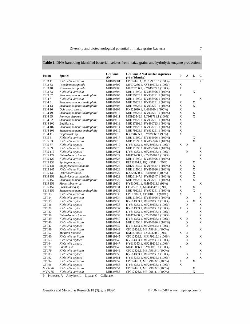
Identification of the isolates The DNA barcoding identified strains are shown in Table 1 and Figure 1.
Genetics and Molecular Research 18 (3): gmr18320 ©FUNPEC-RP www.funpecrp.com.br
Diversity and biotechnological potential of maize grains bacteria 7
Table 1. DNA barcoding identified bacterial isolates from maize grains and hydrolytic enzyme production.
Isolate Species GenBank AN
GenBank AN of similar sequences (% of identity) P A L C
FEI3 11 Klebsiella variicola MH819801 CP012426.1, MF179616.1 (100%) X FEI3 33 Pseudomonas putida MH819802 MF079266.1, KY849572.1 (100%) X FEI3 40 Pseudomonas putida MH819803 MF079266.1, KY849572.1 (100%) X FEI3 53 Klebsiella variicola MH819804 MH111590.1, KY856926.1 (100%) X FEI3 62 Stenotrophomonas maltophilia MH819805 MH179323.1, KY933291.1 (100%) X FEI4 3 Klebsiella variicola MH819806 MH111590.1, KY856926.1 (100%) X FEI4 6 Stenotrophomonas maltophilia MH819807 MH179323.1, KY933291.1 (100%) X X FEI4 13 Stenotrophomonas maltophilia MH819808 MH179323.1, KY933291.1 (100%) X X FEI4 16 Ochrobactrum sp. MH819809 KX822680.1, FJ603030.1 (100%) X FEI4 49 Stenotrophomonas maltophilia MH819810 MH179323.1, KY933291.1 (100%) X X FEI4 65 Pantoea dispersa MH819811 MG923542.1, LT969731.1 (100%) X X FEI4 92 Stenotrophomonas maltophilia MH819812 MH179323.1, KY933291.1 (100%) X FEI4 106 Bacillus sp. MH819813 MH337993.1, KY860723.1 (100%) X FEI4 107 Stenotrophomonas maltophilia MH819814 MH179323.1, KY933291.1 (100%) X FEI4 108 Stenotrophomonas maltophilia MH819815 MH179323.1, KY933291.1 (100%) X FEI4 119 Isoptericola sp. MH819816 KX034605.1, KY939565.1 (98%) X FEI5 8 Klebsiella variicola MH819817 MH111590.1, KY856926.1 (100%) X FEI5 63 Klebsiella variicola MH819818 MH111590.1, KY856926.1 (100%) X FEI5 87 Klebsiella oxytoca MH819819 KY614353.1, MF289236.1 (100%) X X FEI5 89 Klebsiella variicola MH819820 MH111590.1, KY856926.1 (100%) X FEI5 117 Klebsiella oxytoca MH819821 KY614353.1, MF289236.1 (100%) X X FEI5 124 Enterobacter cloacae MH819822 MF471480.1, KY495207.1 (100%) X FEI5 127 Klebsiella variicola MH819823 MH111590.1, KY856926.1 (100%) X FEI5 139 Sphingomonas sp. MH819824 FR750304.1, DQ145741.1 (99%) X X FEI5 141 Staphylococcus hominis MH819825 MH201347.1, KY992547.1 (100%) X X FEI5 145 Klebsiella variicola MH819826 MH111590.1, KY856926.1 (100%) X X FEI5 146 Ochrobactrum sp. MH819827 KX822680.1, FJ603030.1 (100%) X X FEI5 151 Staphylococcus hominis MH819828 MH201347.1, KY992547.1 (100%) X X FEI5 152 Stenotrophomonas maltophilia MH819829 MH179323.1, KY933291.1 (100%) X X FEI5 153 Modestobacter sp. MH819830 KY510681.1, FM995613.1 (98%) X FEI5 157 Burkholderia sp. MH819831 LC385678.1, MF464547.1 (99%) X X FEI5 159 Stenotrophomonas maltophilia MH819832 MH179323.1, KY933291.1 (100%) X X CTI 13 Klebsiella variicola MH819833 CP013985.1, CP001891.1 (100%) X X CTI 14 Klebsiella variicola MH819834 MH111590.1, KY856926.1 (100%) X X CTI 15 Klebsiella oxytoca MH819835 KY614353.1, MF289236.1 (100%) X X X CTI 16 Klebsiella oxytoca MH819836 KY614353.1, MF289236.1 (100%) X X CTI 20 Klebsiella oxytoca MH819837 KY614353.1, MF289236.1 (100%) X X X CTI 37 Klebsiella oxytoca MH819838 KY614353.1, MF289236.1 (100%) X X CTI 38 Enterobacter cloacae MH819839 MF471480.1, KY495207.1 (100%) X CTI 39 Klebsiella oxytoca MH819840 KY614353.1, MF289236.1 (100%) X X X CTI 40 Klebsiella variicola MH819841 MH111590.1, KY856926.1 (100%) X X CTI 47 Klebsiella oxytoca MH819842 KY614353.1, MF289236.1 (100%) X CTI 49 Klebsiella variicola MH819843 CP012426.1, MF179616.1 (100%) X CTI 57 Massilia timonae MH819844 KM187207.1, JX566630.1 (99%) X X CTI 60 Klebsiella variicola MH819845 CP012426.1, MF179616.1 (100%) X X CTI 63 Klebsiella oxytoca MH819846 KY614353.1, MF289236.1 (100%) X CTI 64 Klebsiella oxytoca MH819847 KY614353.1, MF289236.1 (100%) X X CTI 70 Bacillus sp. MH819848 MF418036.1, KY860716.1 (100%) X CTI 79 Klebsiella variicola MH819849 CP012426.1, MF179616.1 (100%) X CTI 83 Klebsiella oxytoca MH819850 KY614353.1, MF289236.1 (100%) X X CTI 92 Klebsiella oxytoca MH819851 KY614353.1, MF289236.1 (100%) X X CTI 94 Klebsiella variicola MH819852 CP012426.1, MF179616.1 (100%) X X CTI 96 Klebsiella oxytoca MH819853 KY614353.1, MF289236.1 (100%) X MVA 26 Klebsiella variicola MH819854 CP012426.1, MF179616.1 (100%) X MVA 35 Klebsiella variicola MH819855 CP012426.1, MF179616.1 (100%) X P – Protease, A – Amylase, L – Lipase, C – Cellulase.

Genetics and Molecular Research 18
Figure length = 0.9439bootstrap test (1000 replicates) are shown next to the branches. The tree is drawn to scale, with branch lengths in number of base substitutions per siteremoved for each sequence pair. There were a total of 398 positions in the final dataset. Evolutionary analyses were made with the MEGA 7.0 program (Kumar et al., 2016).
Grambuilt phylogenetic tree (Figure 2) shows these species separations. All thenegative isolates belonged to the phylum Proteobacteria, with the predominance of the proteobacteria class, followed by positive strains belong to the phyla Firmicutes and Actinobacteria. The phylum bacterial isolates percentages are shown in Figure 3.
Figure agar containing soluble starch. casein. Stenotrophomonas maltophiliaCellulolytic activity. Medium containing microcrystalline cellulose.
netics and Molecular Research 18
Figure 1. The evolutionary history of the 16S rRNA partial gene sequences. The optimal tree with the sum of branch length = 0.94393273 is shown. The percentages of replica trees in which the associated taxa clustered together in the bootstrap test (1000 replicates) are shown next to the branches. The tree is drawn to scale, with branch lengths in number of base substitutions per siteremoved for each sequence pair. There were a total of 398 positions in the final dataset. Evolutionary analyses were made with the MEGA 7.0 program (Kumar et al., 2016).
Among the 55 identified isolates, there were 13 different genera, of which nine were Gram-negative bacilli, one was Grambuilt phylogenetic tree (Figure 2) shows these species separations. All thenegative isolates belonged to the phylum Proteobacteria, with the predominance of the proteobacteria class, followed by positive strains belong to the phyla Firmicutes and Actinobacteria. The phylum bacterial isolates percentages are shown in Figure 3.
Figure 2. Petri dishesagar containing soluble starch. casein. Stenotrophomonas maltophiliaStenotrophomonas maltophiliaCellulolytic activity. Medium containing microcrystalline cellulose.
netics and Molecular Research 18
The evolutionary history of the 16S rRNA partial gene sequences. The optimal tree with the sum of branch 3273 is shown. The percentages of replica trees in which the associated taxa clustered together in the
bootstrap test (1000 replicates) are shown next to the branches. The tree is drawn to scale, with branch lengths in number of base substitutions per site. The analysis involved 55 nucleotide sequences for rDNA. All ambiguous positions were removed for each sequence pair. There were a total of 398 positions in the final dataset. Evolutionary analyses were made with the MEGA 7.0 program (Kumar et al., 2016).
Among the 55 identified isolates, there were 13 different genera, of which nine were negative bacilli, one was Gram
built phylogenetic tree (Figure 2) shows these species separations. All thenegative isolates belonged to the phylum Proteobacteria, with the predominance of the proteobacteria class, followed by positive strains belong to the phyla Firmicutes and Actinobacteria. The phylum bacterial isolates percentages are shown in Figure 3.
Petri dishes with distinct substrate media for the enzyme activities detection. A) Amylolytic activity. Nutrient agar containing soluble starch. Klebsiella oxytoca
Stenotrophomonas maltophiliaStenotrophomonas maltophilia FEI4 6. D) The underside of the dish shown in C) photographed under UV light. E) Cellulolytic activity. Medium containing microcrystalline cellulose.
(3): gmr18320
F.C. Santos
The evolutionary history of the 16S rRNA partial gene sequences. The optimal tree with the sum of branch 3273 is shown. The percentages of replica trees in which the associated taxa clustered together in the
bootstrap test (1000 replicates) are shown next to the branches. The tree is drawn to scale, with branch lengths in number . The analysis involved 55 nucleotide sequences for rDNA. All ambiguous positions were
removed for each sequence pair. There were a total of 398 positions in the final dataset. Evolutionary analyses were made with the MEGA 7.0 program (Kumar et al., 2016).
Among the 55 identified isolates, there were 13 different genera, of which nine were negative bacilli, one was Gram-positive cocci, and three were Gram
built phylogenetic tree (Figure 2) shows these species separations. All thenegative isolates belonged to the phylum Proteobacteria, with the predominance of the proteobacteria class, followed by the -proteobacteria and positive strains belong to the phyla Firmicutes and Actinobacteria. The phylum bacterial isolates percentages are shown in Figure 3.
with distinct substrate media for the enzyme activities detection. A) Amylolytic activity. Nutrient Klebsiella oxytoca CTI 63; B) Proteolytic activity. Medium containing skimmed milk
Stenotrophomonas maltophilia FEI4 49. C) Lipolytic activity. Medium containing triolein and rhodamine B. FEI4 6. D) The underside of the dish shown in C) photographed under UV light. E)
Cellulolytic activity. Medium containing microcrystalline cellulose.
F.C. Santos et al.
The evolutionary history of the 16S rRNA partial gene sequences. The optimal tree with the sum of branch
3273 is shown. The percentages of replica trees in which the associated taxa clustered together in the bootstrap test (1000 replicates) are shown next to the branches. The tree is drawn to scale, with branch lengths in number
. The analysis involved 55 nucleotide sequences for rDNA. All ambiguous positions were removed for each sequence pair. There were a total of 398 positions in the final dataset. Evolutionary analyses were
Among the 55 identified isolates, there were 13 different genera, of which nine were positive cocci, and three were Gram
built phylogenetic tree (Figure 2) shows these species separations. All thenegative isolates belonged to the phylum Proteobacteria, with the predominance of the
proteobacteria and positive strains belong to the phyla Firmicutes and Actinobacteria. The phylum bacterial isolates
with distinct substrate media for the enzyme activities detection. A) Amylolytic activity. Nutrient CTI 63; B) Proteolytic activity. Medium containing skimmed milk
I4 49. C) Lipolytic activity. Medium containing triolein and rhodamine B. FEI4 6. D) The underside of the dish shown in C) photographed under UV light. E)
Cellulolytic activity. Medium containing microcrystalline cellulose. Klebsiella oxytoca
©FUNPEC-
The evolutionary history of the 16S rRNA partial gene sequences. The optimal tree with the sum of branch 3273 is shown. The percentages of replica trees in which the associated taxa clustered together in the
bootstrap test (1000 replicates) are shown next to the branches. The tree is drawn to scale, with branch lengths in number . The analysis involved 55 nucleotide sequences for rDNA. All ambiguous positions were
removed for each sequence pair. There were a total of 398 positions in the final dataset. Evolutionary analyses were
Among the 55 identified isolates, there were 13 different genera, of which nine were positive cocci, and three were Gram
built phylogenetic tree (Figure 2) shows these species separations. All thenegative isolates belonged to the phylum Proteobacteria, with the predominance of the
proteobacteria and -proteobacteria classpositive strains belong to the phyla Firmicutes and Actinobacteria. The phylum bacterial isolates
with distinct substrate media for the enzyme activities detection. A) Amylolytic activity. Nutrient CTI 63; B) Proteolytic activity. Medium containing skimmed milk
I4 49. C) Lipolytic activity. Medium containing triolein and rhodamine B. FEI4 6. D) The underside of the dish shown in C) photographed under UV light. E)
bsiella oxytoca CTI 15.
-RP www.funpecrp.com.br
The evolutionary history of the 16S rRNA partial gene sequences. The optimal tree with the sum of branch 3273 is shown. The percentages of replica trees in which the associated taxa clustered together in the
bootstrap test (1000 replicates) are shown next to the branches. The tree is drawn to scale, with branch lengths in number . The analysis involved 55 nucleotide sequences for rDNA. All ambiguous positions were
removed for each sequence pair. There were a total of 398 positions in the final dataset. Evolutionary analyses were
Among the 55 identified isolates, there were 13 different genera, of which nine were positive cocci, and three were Gram-positive bacilli. The
built phylogenetic tree (Figure 2) shows these species separations. All the identified Gramnegative isolates belonged to the phylum Proteobacteria, with the predominance of the
proteobacteria classes. The Grampositive strains belong to the phyla Firmicutes and Actinobacteria. The phylum bacterial isolates
with distinct substrate media for the enzyme activities detection. A) Amylolytic activity. Nutrient CTI 63; B) Proteolytic activity. Medium containing skimmed milk
I4 49. C) Lipolytic activity. Medium containing triolein and rhodamine B. FEI4 6. D) The underside of the dish shown in C) photographed under UV light. E)
CTI 15.
www.funpecrp.com.br
8
The evolutionary history of the 16S rRNA partial gene sequences. The optimal tree with the sum of branch 3273 is shown. The percentages of replica trees in which the associated taxa clustered together in the
bootstrap test (1000 replicates) are shown next to the branches. The tree is drawn to scale, with branch lengths in number . The analysis involved 55 nucleotide sequences for rDNA. All ambiguous positions were
removed for each sequence pair. There were a total of 398 positions in the final dataset. Evolutionary analyses were
Among the 55 identified isolates, there were 13 different genera, of which nine were positive bacilli. The
identified Gram-negative isolates belonged to the phylum Proteobacteria, with the predominance of the -
. The Gram-positive strains belong to the phyla Firmicutes and Actinobacteria. The phylum bacterial isolates
with distinct substrate media for the enzyme activities detection. A) Amylolytic activity. Nutrient
CTI 63; B) Proteolytic activity. Medium containing skimmed milk I4 49. C) Lipolytic activity. Medium containing triolein and rhodamine B.
FEI4 6. D) The underside of the dish shown in C) photographed under UV light. E)

Ge
Figure 3. rDNA sequence similarity.
Enzyme activities
summarized in Table 1. Examples of the obtained hydrolSome of the stored isolates produced more than one hydrolytic activity (Table 1). Among 55 isolates, 47% presented protease, 33% amylase, 29% lipase, and 45%(Table 1).
DISCUSSIONS
teosinte. Its seeds are reported to have a set of associated bacteria, despite 9,000 years of culture, selection, and geneticMonje & Raizada, 2011)because of the culturthe culture conditions emplosamples to develop. Nisolates can be considered as epiphytic as well as endophytic. All the identified Gramnegative isolates belonproteobacteria class, followed by the 1). For Gramalso obtained in this work. A total of 13 bacterial genera were representwith our results, Johnstonbacteria from the presence of bacteria from the Firmicutes and Actinobacteria phyla in maize grains isolated from Central and North America. Besides, those authors have described the presence of 26 bacterial genera as endophytic bacteria, but they have used an approach of c
Genetics and Molecular Research 18
Diversity and biotechnological potential of maize grain
Figure 3. Percentage composition of different phyla of bacteria isolated from maize grains on the basis of 16S rDNA sequence similarity.
Enzyme activities The DNA barcoding identified isolates, and their hydrolytic enzymes prod
summarized in Table 1. Examples of the obtained hydrolSome of the stored isolates produced more than one hydrolytic activity (Table 1). Among 55 isolates, 47% presented protease, 33% amylase, 29% lipase, and 45%(Table 1).
DISCUSSIONS Maize is originated from southern Mexico, and it is a domestication product of wild
teosinte. Its seeds are reported to have a set of associated bacteria, despite 9,000 years of culture, selection, and geneticMonje & Raizada, 2011)because of the culturthe culture conditions emplosamples to develop. Nisolates can be considered as epiphytic as well as endophytic. All the identified Gramnegative isolates belonproteobacteria class, followed by the
). For Gram-positive bacteria, genera from the phyla Firmicutes and Actinobacteria were also obtained in this work. A total of 13 bacterial genera were representwith our results, Johnstonbacteria from the presence of bacteria from the Firmicutes and Actinobacteria phyla in maize grains isolated from Central and North America. Besides, those authors have described the presence of 26 bacterial genera as endophytic bacteria, but they have used an approach of c
ics and Molecular Research 18
Diversity and biotechnological potential of maize grain
Percentage composition of different phyla of bacteria isolated from maize grains on the basis of 16S rDNA sequence similarity.
Enzyme activities
The DNA barcoding identified isolates, and their hydrolytic enzymes prodsummarized in Table 1. Examples of the obtained hydrolSome of the stored isolates produced more than one hydrolytic activity (Table 1). Among 55 isolates, 47% presented protease, 33% amylase, 29% lipase, and 45%
DISCUSSIONS
Maize is originated from southern Mexico, and it is a domestication product of wild teosinte. Its seeds are reported to have a set of associated bacteria, despite 9,000 years of culture, selection, and geneticMonje & Raizada, 2011). The absence of colonies in samples FEI1 and FEI2 might be because of the culture medium used, the chemical added to control rapid fungal growth, or the culture conditions employed, which were not adequate for the bacterial flora of those samples to develop. No grain disinfection was performed in our methodology, and our isolates can be considered as epiphytic as well as endophytic. All the identified Gramnegative isolates belonged to the phylum Proteobacteria, with a predominance of the proteobacteria class, followed by the
positive bacteria, genera from the phyla Firmicutes and Actinobacteria were also obtained in this work. A total of 13 bacterial genera were representwith our results, Johnston-Monje & Raizada (2011) have described the massivebacteria from the - Proteobacteria class as maize seed endophytes and also observed the presence of bacteria from the Firmicutes and Actinobacteria phyla in maize grains isolated from Central and North America. Besides, those authors have described the presence of 26 bacterial genera as endophytic bacteria, but they have used an approach of c
ics and Molecular Research 18 (3): gmr18320
Diversity and biotechnological potential of maize grain
Percentage composition of different phyla of bacteria isolated from maize grains on the basis of 16S
The DNA barcoding identified isolates, and their hydrolytic enzymes prodsummarized in Table 1. Examples of the obtained hydrolSome of the stored isolates produced more than one hydrolytic activity (Table 1). Among 55 isolates, 47% presented protease, 33% amylase, 29% lipase, and 45%
Maize is originated from southern Mexico, and it is a domestication product of wild teosinte. Its seeds are reported to have a set of associated bacteria, despite 9,000 years of culture, selection, and genetic improvement by primitive and modern breeders (Johnston
. The absence of colonies in samples FEI1 and FEI2 might be e medium used, the chemical added to control rapid fungal growth, or
yed, which were not adequate for the bacterial flora of those o grain disinfection was performed in our methodology, and our
isolates can be considered as epiphytic as well as endophytic. All the identified Gramged to the phylum Proteobacteria, with a predominance of the
proteobacteria class, followed by the -proteobacteria and positive bacteria, genera from the phyla Firmicutes and Actinobacteria were
also obtained in this work. A total of 13 bacterial genera were representMonje & Raizada (2011) have described the massive
Proteobacteria class as maize seed endophytes and also observed the presence of bacteria from the -Proteobacteria and Firmicutes and Actinobacteria phyla in maize grains isolated from Central and North America. Besides, those authors have described the presence of 26 bacterial genera as endophytic bacteria, but they have used an approach of c
Diversity and biotechnological potential of maize grain
Percentage composition of different phyla of bacteria isolated from maize grains on the basis of 16S
The DNA barcoding identified isolates, and their hydrolytic enzymes prodsummarized in Table 1. Examples of the obtained hydrolSome of the stored isolates produced more than one hydrolytic activity (Table 1). Among 55 isolates, 47% presented protease, 33% amylase, 29% lipase, and 45%
Maize is originated from southern Mexico, and it is a domestication product of wild teosinte. Its seeds are reported to have a set of associated bacteria, despite 9,000 years of
improvement by primitive and modern breeders (Johnston. The absence of colonies in samples FEI1 and FEI2 might be
e medium used, the chemical added to control rapid fungal growth, or yed, which were not adequate for the bacterial flora of those
o grain disinfection was performed in our methodology, and our isolates can be considered as epiphytic as well as endophytic. All the identified Gram
ged to the phylum Proteobacteria, with a predominance of the proteobacteria and
positive bacteria, genera from the phyla Firmicutes and Actinobacteria were also obtained in this work. A total of 13 bacterial genera were represent
Monje & Raizada (2011) have described the massiveProteobacteria class as maize seed endophytes and also observed the
Proteobacteria and -ProteobFirmicutes and Actinobacteria phyla in maize grains isolated from Central and North America. Besides, those authors have described the presence of 26 bacterial genera as endophytic bacteria, but they have used an approach of c
©FUNPEC
Diversity and biotechnological potential of maize grains bacteria
Percentage composition of different phyla of bacteria isolated from maize grains on the basis of 16S
The DNA barcoding identified isolates, and their hydrolytic enzymes prodsummarized in Table 1. Examples of the obtained hydrolytic halos are shown in Figure 2Some of the stored isolates produced more than one hydrolytic activity (Table 1). Among 55 isolates, 47% presented protease, 33% amylase, 29% lipase, and 45%
Maize is originated from southern Mexico, and it is a domestication product of wild teosinte. Its seeds are reported to have a set of associated bacteria, despite 9,000 years of
improvement by primitive and modern breeders (Johnston. The absence of colonies in samples FEI1 and FEI2 might be
e medium used, the chemical added to control rapid fungal growth, or yed, which were not adequate for the bacterial flora of those
o grain disinfection was performed in our methodology, and our isolates can be considered as epiphytic as well as endophytic. All the identified Gram
ged to the phylum Proteobacteria, with a predominance of the proteobacteria and -proteobacteria classes (Figure
positive bacteria, genera from the phyla Firmicutes and Actinobacteria were also obtained in this work. A total of 13 bacterial genera were represent
Monje & Raizada (2011) have described the massiveProteobacteria class as maize seed endophytes and also observed the
Proteobacteria classes, and from the Firmicutes and Actinobacteria phyla in maize grains isolated from Central and North America. Besides, those authors have described the presence of 26 bacterial genera as endophytic bacteria, but they have used an approach of culture, cloning, and DNA
©FUNPEC-RP www.funpecrp.com.br
bacteria
Percentage composition of different phyla of bacteria isolated from maize grains on the basis of 16S
The DNA barcoding identified isolates, and their hydrolytic enzymes production are ytic halos are shown in Figure 2
Some of the stored isolates produced more than one hydrolytic activity (Table 1). Among 55 isolates, 47% presented protease, 33% amylase, 29% lipase, and 45% cellulase activities
Maize is originated from southern Mexico, and it is a domestication product of wild teosinte. Its seeds are reported to have a set of associated bacteria, despite 9,000 years of
improvement by primitive and modern breeders (Johnston. The absence of colonies in samples FEI1 and FEI2 might be
e medium used, the chemical added to control rapid fungal growth, or yed, which were not adequate for the bacterial flora of those
o grain disinfection was performed in our methodology, and our isolates can be considered as epiphytic as well as endophytic. All the identified Gram
ged to the phylum Proteobacteria, with a predominance of the eobacteria classes (Figure
positive bacteria, genera from the phyla Firmicutes and Actinobacteria were also obtained in this work. A total of 13 bacterial genera were represented. In consonance
Monje & Raizada (2011) have described the massive presence of Proteobacteria class as maize seed endophytes and also observed the
acteria classes, and from the Firmicutes and Actinobacteria phyla in maize grains isolated from Central and North America. Besides, those authors have described the presence of 26 bacterial genera as
ulture, cloning, and DNA
www.funpecrp.com.br
9
Percentage composition of different phyla of bacteria isolated from maize grains on the basis of 16S
uction are ytic halos are shown in Figure 2.
Some of the stored isolates produced more than one hydrolytic activity (Table 1). Among cellulase activities
Maize is originated from southern Mexico, and it is a domestication product of wild teosinte. Its seeds are reported to have a set of associated bacteria, despite 9,000 years of
improvement by primitive and modern breeders (Johnston-. The absence of colonies in samples FEI1 and FEI2 might be
e medium used, the chemical added to control rapid fungal growth, or yed, which were not adequate for the bacterial flora of those
o grain disinfection was performed in our methodology, and our isolates can be considered as epiphytic as well as endophytic. All the identified Gram-
ged to the phylum Proteobacteria, with a predominance of the -eobacteria classes (Figure
positive bacteria, genera from the phyla Firmicutes and Actinobacteria were ed. In consonance
presence of Proteobacteria class as maize seed endophytes and also observed the
acteria classes, and from the Firmicutes and Actinobacteria phyla in maize grains isolated from Central and North America. Besides, those authors have described the presence of 26 bacterial genera as
ulture, cloning, and DNA

©FUNPEC-RP www.funpecrp.com.br Genetics and Molecular Research 18 (3): gmr18320
F.C. Santos et al. 10
fingerprinting by terminal restriction fragment length polymorphism. Regarding maize roots bacteria diversity in an Agricultural Research Station in the United States, Chelius &Triplett (2001) have found a predominance of Actinobacteria by culture technique and -Proteobacteria within a constructed clone library. Actinobacteria and Proteobacteria were also the dominating phyla in the rhizosphere and bulk soil of two maize lines differing in nitrogen use efficiency in Central Italy (Pathan et al., 2015).
From the 13 different bacterial genera found in maize grains in this work, the vast majority was already shown to occur in this substrate in Central and North America, such as the genera Klebsiella, Sphingomonas, Bacillus, Pantoea, Staphylococcus, Stenotrophomonas, Enterobacter, Burkholderia, and Pseudomonas (Chelius & Triplett, 2001; Johnston-Monje & Raizada, 2011). Ikeda et al. (2013) have only found the genera Pantoea, Bacillus, Burkholderia, and Klebsiella to occur in commercial maize seeds from the east region of the Paraná state, indicating a higher diversity of bacteria associated with maize grains in the North Central part of the same state (this work). The occurrence of the genera Massilia and Ochrobactrum have been reported in maize roots and rhizosphere (Niu et al., 2017; Silva et al., 2017). The occurrence of Modestobacter and Isoptericola genera isolates have not been described yet in maize grains. However, the appearance of Modestobacter sp. has been described in rice roots (Hernández et al., 2015) as well as the occurrence of Isoptericola sp. in the rhizosphere of cucumber and mangrove plants (Kämpfer et al., 2016; Bibi et al., 2017). As our maize samples were in soil contact when collected, it may have been contaminated by these soil-related bacteria.
The finding of Bacillus sp. strains as proteases producers (Table 1) is not surprising, because the Bacillus genus is vastly reported in the literature as a significant protease producer and most of the neutral and alkaline commercial proteases are obtained from it (Singh et al., 2016). Additionally, the proteases of the species Stenotrophomonas maltophilia, Pseudomonas putida, Klebsiella oxytoca, and Pantoea dispersa, identified in this work as proteases producers (Table 1), have already been characterized (Tondo et al., 2004; Nicodème et al., 2005; Gohel et al., 2007; Ribitsch et al., 2012). With respect to the genera Burkholderia, Staphylococcus, and Ochrobactrum, the proteases of the species Burkholderia pseudomallei, Staphylococcus aureus, Staphylococcus epidermidis, and Ochrobactrum anthropi were also characterized (Bompard-Gilles et al., 2000; Shaw et al., 2004; Chin et al., 2007; Martínez-García et al., 2018). Despite the production of proteases by the genera Isoptericola, Massilia, and Sphingomonas being also described in the literature (Lindquist et al., 2003; Willsey & Wargo, 2015, Bibi et al., 2017), none has been studied so far, and this is the first report about the species Staphylococcus hominis and Massilia timonae as protease producers (Table 1).
Bacteria can use starch as an energy source. However, amylase production by bacteria varies across genera and species. Fewer bacterial genera have been shown to produce amylases in this work (Table 1), such as the genera Bacillus (Gurung et al., 2013), which explains, at least in part, the lower number of producing species, in comparison with the other studied hydrolases (Table 1). The species K. variicola is described as a plant endophyte and is much related to the human isolates of Klebsiella pneumoniae, with some strains sharing more than 95% of genome identity (Chen et al., 2016). K. pneumoniae is a known producer of cyclodextrin glucanotransferase, which can degrade starch and synthesize -cyclodextrin (Sun et al., 2010), and a recombinant pullulanase from K. variicola has been characterized (Chen et al., 2013). Both cyclodextrin glucanotransferase

Genetics and Molecular Research 18 (3): gmr18320 ©FUNPEC-RP www.funpecrp.com.br
Diversity and biotechnological potential of maize grains bacteria 11
and pullulanase belong to the -amylase family. Species of the genus Massilia are not known as amylase producers, but are supposed to produce amylases because they were shown to use starch to build polyhydroxyalkanoates, which are biodegradable and biocompatible with hydroxycarboxylic acid polyester (Cerrone et al., 2011). In fact, in subsequent work in our laboratory, a recombinant cyclodextrinase from M. timonae CTI 57 has been produced and characterized (Santos & Barbosa-Tessman, 2019).
Few species of bacteria are described as good producers of lipolytic enzymes; but the Burkholderia lipase, for instance, is commercially available (Gurung et al., 2013). Also, lipase production by S. maltophilia is well documented, and an enzyme has been fully characterized (Li et al., 2016). Because of this, it is not surprising that among the lipolytic enzymes producing bacterial isolates from maize grains, the majority were S. maltophilia and Burkholderia sp. (Table 1). Lipase production by S. hominis and by Ochrobactrum intermedium has been reported (Zarinviarsagh et al., 2017; Behera et al., 2019), which validates our results of lipase production. Although isolates of the genus Klebsiella have been shown to produce lipase in bioprospection studies (Mazzucotellii et al., 2013), none has been studied in detail, and the production of lipase by the K. variicola species is a new finding. Although P. dispersa and Sphingomonas sp. have been reported to produce esterase (Zhang & Birch, 1997; Dachuri et al., 2018), the report of lipase production is a new finding. In a following work by our group, the P. dispersa FEI4 65 isolate was shown to produce lipase and esterase in liquid medium and two esterase genes from this isolate were cloned and expressed in E. coli and the produced enzymes were characterized (Martim & Barbosa-Tessmann, 2019).
There are many cellulase-producing genera of aerobic and anaerobic bacteria, which are widely distributed in nature (Kuhad et al., 2011). The majority of the isolates of K. variicola and K. oxytoca identified to produce cellulases were also found to produce amylase, indicating their involvement with glycan polymers breakdown (Table 1). Cellulase production by Klebsiella genus isolates and E. cloacae is well described in the literature, and some of them have latterly been characterized (Attigani et al., 2016; Akintola et al., 2017). The bacterial genera Klebsiella and Enterobacter belong to the Proteobacteria phylum and, in agreement with our results, Pathan et al. (2015) found a high content of Proteobacteria β-glucosidase genes and -glucosidase activity in the rhizosphere of two maize lines differing in nitrogen use efficiency. Cellulase production by the Actinobacteria Modestobacter has not been reported untill now, although bacterial genera from the phylum Actinobacteria have been involved in cellulase production and bioenergy application (Lewin et al., 2016).
CONCLUSIONS This study employed a culture-dependent approach to isolate bacteria from maize
grains presenting rotting symptoms. Fifty-five strains of bacteria were isolated, and DNA barcoding and phylogenetic studies showed the population diversity. The majority of the isolates belonged to the phylum Proteobacteria; however, isolates from the phyla Actinobacteria and Firmicutes were also found. The isolates belonged to 13 different genera. The potential of the bacteria isolates for producing amylase, lipase, cellulase, and protease was tested in solid media, and several producers were found. The enzymes from some of the isolated species had already been purified and characterized. Some species were
©FUNPEC-RP www.funpecrp.com.br Genetics and Molecular Research 18 (3): gmr18320
F.C. Santos et al. 12
described as producers for the first time, for instance, S. hominis and M. timonae as protease producers, M. timonae as an amylase producer, K. variicola, Sphingomonas sp., and P. dispersa as lipase producers, and Modestobacter sp. as a cellulase producer.
ACKNOWLEDGMENTS The authors are thankful to CNPQ (Conselho Nacional de Desenvolvimento
Científico e Tecnológico, Ministry of Science and Technology, Brazil) and to CAPES (Coordenação de Aperfeiçoamento de Pessoal de Nível Superior, Ministry of Education, Brazil, Grant 001), for the students scholarships.
CONFLICTS OF INTEREST The authors declare no conflict of interest.
REFERENCES Abe CAL, Faria CB, Castro FF, Souza SR, et al. (2015). Fungi isolated from maize (Zea mays L.) grains and production
of associated enzyme activities. Int. J. Mol. Sci. 16: 15328-15346. Adrio JL and Demain AL (2014). Microbial enzymes: Tools for biotechnological processes. Biomolecules. 4: 117-139. Akintola AI, Oyedeji O, Bakare MK and Adewale IO (2017). Purification and characterization of thermostable cellulase
from Enterobacter cloacae IP8 isolated from decayed plant leaf litter. Biocatal. Biotransfor. 35(5): 379-387. Attigani A, Sun L, Wang Q, Liu Y, et al. (2016). The crystal structure of the endoglucanase Cel10, a family 8 glycosyl
hydrolase from Klebsiella pneumoniae. Acta Crystallogr. Sect. F Struct. Biol. Cryst. Commun. 72(12): 870-876. Behera AR, Veluppal A and Dutta K (2019). Optimization of physical parameters for enhanced production of lipase
from Staphylococcus hominis using response surface methodology. Environ. Sci. Pollut. Res. Int. Feb 2. doi: 10.1007/s11356-019-04304-0. [Epub ahead of print]
Bibi F, Ullah I, Alvi SA, Bakhsh SA, et al. (2017). Isolation, diversity, and biotechnological potential of rhizo- and endophytic bacteria associated with mangrove plants from Saudi Arabia. Genet. Mol. Res. 16(2): gmr16029657. https://doi.org/10.4238/gmr16029657.
Bompard-Gilles C, Remauta H, Villereta V, Prangé T, et al. (2000). Crystal structure of a D-aminopeptidase from Ochrobactrum anthropi, a new member of the ‘penicillin-recognizing enzyme’ family. Structure. 8(9): 971-980.
Cerrone F, Sánchez-Peinado MdelM, Rodríguez-Díaz M, González-López J, et al. (2011). PHAs production by strains belonging to Massilia genus from starch. Starch-Stärke. 63: 236-240.
Chelius MK and Triplett EW (2001). The diversity of Archaea and bacteria in association with the roots of Zea mays L. Microb. Ecol. 41: 252-263.
Chen M, Li Y, Li S, Tang L, et al. (2016). Genomic identification of nitrogen-fixing Klebsiella variicola, K. pneumoniae and K. quasipneumoniae. J. Basic Microbiol. 56: 78-84.
Chen WB, Nie Y and Xu Y (2013). Signal peptide-independent secretory expression and characterization of pullulanase from a newly isolated Klebsiella variicola SHN-1 in Escherichia coli. Appl. Biochem. Biotechnol. 169(1): 41-54.
Chin C-Y, Othman R and Nathan S (2007). The Burkholderia pseudomallei serine protease MprA is autoproteolytically activated to produce a highly stable enzyme. Enzyme Microb. Technol. 40(2): 370-377.
Dachuri VK, Lee C and Jang SH (2018). Organic solvent-tolerant esterase from Sphingomonas glacialis based on amino acid composition analysis: cloning and characterization of EstSP2. J Microbiol. Biotechnol. 28(9): 1502-1510.
Gohel V, Maisuria V and Chhatpar HS (2007). Utilization of various chitinous sources for production of mycolytic enzymes by Pantoea dispersa in bench-top fermenter. Enzyme Microb. Technol. 40(6): 1608-1614.
Gurung N, Ray S, Bose S and Rai V (2013). A broader view: Microbial enzymes and their relevance in industries, medicine, and beyond. BioMed Res. Int. 2013: 329121. https://doi.org/10.1155/2013/329121.
Hankin L and Anagnostakis SL (1975). The use of solid media for detection of enzyme production by fungi. Mycologia. 67: 597-607.
Hernández M, Dumont MG, Yuan Q and Conrad R (2015). Different bacterial populations associated with the roots and rhizosphere of rice incorporate plant-derived carbon. Appl. Environ. Microbiol. 81(6): 2244-2253.
Holt JG (1994). Bergey's manual of determinative bacteriology. 9th edition. Lippincott Williams & Wilkins, Philadelphia.

Genetics and Molecular Research 18 (3): gmr18320 ©FUNPEC-RP www.funpecrp.com.br
Diversity and biotechnological potential of maize grains bacteria 13
Ikeda AC, Bassani LL, Adamoski D, Stringari D, et al. (2013). Morphological and genetic characterization of endophytic bacteria isolated from roots of different maize genotypes. Microb. Ecol. 65: 154-160.
Jaeger KE and Kouker G (1987). Specific and sensitive plate assay for bacterial lipases. Appl. Environ. Microbiol. 53: 211-213.
Johnston-Monje D and Raizada MN (2011). Conservation and diversity of seed associated endophytes in Zea across boundaries of evolution, ethnography and ecology. PLoS One. 6(6): e20396. https://doi.org/10.1371/journal.pone.0020396.
Kämpfer P, Glaeser SP, Kloepper JW, Hu C-H, et al. (2016). Isoptericola cucumis sp. nov., isolated from the root tissue of cucumber (Cucumis sativus). Int. J. Syst. Evol. Microbiol. 66: 2784-2788.
Kasana RC, Salwan R, Dhar H, Dutt S, et al. (2008). A rapid and easy method for the detection of microbial cellulases on agar plates using Gram’s iodine. Curr. Microbiol. 57: 503-507.
Kuhad RC, Gupta R and Singh A (2011). Microbial cellulases and their industrial applications. Enzyme Res. 2011: ID 280696. https://doi.org/10.4061/2011/280696.
Kumar S, Stecher G and Tamura K (2016). MEGA7: Molecular evolutionary genetics Analysis version 7.0 for bigger datasets. Mol. Biol. Evol. 33: 1870-1874.
Lewin GR, Carlos C, Chevrette MG, Horn HA, et al. (2016). Evolution and ecology of Actinobacteria and their bioenergy applications. Annu. Rev. Microbiol. 70: 235-254.
Li M, Yang LR, Xu G and Wu JP (2016). Cloning and characterization of a novel lipase from Stenotrophomonas maltophilia GS11: The first member of a new bacterial lipase family XVI. J. Biotechnol. 228: 30-36.
Li S, Yang X, Yang S, Zhu M, et al. (2012). Technology prospecting on enzymes: application, marketing and engineering. Comput. Struct. Biotec. 2: e201209017. https://doi.org/10.5936/csbj.201209017.
Lindquist D, Murrill D, Burran WP, Winans G, et al. (2003). Characteristics of Massilia timonae and Massilia timonae-like isolates from human patients, with an emended description of the species. J. Clin. Microbiol. 41: 192-196.
Martim DM and Barbosa-Tessmann IP (2019). Two novel acetylesterases from Pantoea dispersa: recombinant expression, purification, and characterization. Appl. Biochem. Biotechnol. Published online first. https://doi.org/10.1007/s12010-019-03024-y.
Martínez-García S, Rodríguez-Martínez S, Cancino-Diaz ME and Cancino-Diaz JC (2018). Extracellular proteases of Staphylococcus epidermidis: Roles as virulence factors and their participation in biofilm. APMIS. 126(3): 177-185.
Mazzucotellii CA, Ponce AG, Kotlar CE and Moreira MdelR (2013). Isolation and characterization of bacterial strains with a hydrolytic profile with potential use in bioconversion of agroindustial by-products and waste. Food Sci. Technol. 33(2): 295-303.
Nelson EB (2018). The seed microbiome: Origins, interactions, and impacts. Plant Soil. 422: 7-34. Nicodème M, Grill J-P, Humbert G and Gaillard J-L (2005). Extracellular protease activity of different Pseudomonas
strains: Dependence of proteolytic activity on culture conditions. J. Appl. Microbiol. 99: 641-648. Niu B, Paulson JN, Zheng X and Kolter R (2017). Simplified and representative bacterial community of maize roots.
Proc. Natl. Acad. Sci. USA. 114(12): E2450-E2459. Pathan DI, Ceccherini MT, Hansen MA, Giagnoni L, et al. (2015). Maize lines with different nitrogen use efficiency
select bacterial communities with different β-glucosidase-encoding genes and glucosidase activity in the rhizosphere. Biol. Fertil. Soils. 51(8): 995-1004.
Relman DA (1993). Universal bacterial 16S rDNA amplification and sequencing. In Persing DH, Smith TF, Tenover FC, White TJ (Eds.), Diagnostic molecular mcrobiology, principles and applications (pp. 489-495). Washington DC: ASM.
Ribitsch D, Heumann S, Karl W, Gerlach J, et al. (2012). Extracellular serine proteases from Stenotrophomonas maltophilia: Screening, isolation and heterologous expression in E. coli. J. Biotechnol. 157(1): 140-147.
Santos FC and Barbosa-Tessmann IP (2019). Recombinant expression, purification, and characterization of a cyclodextrinase from Massilia timonae. Prot. Expr. Purif. 154: 74-84.
Sarath G, de La Motte RS and Wagner FW (1989). Protease assay methods. In: Beynon, R. J., & Bonde, J. S. (Eds.), Proteolytics Enzymes: A practical approach (p. 25-54). Oxford: Oxford University Press.
SEAB – Secretaria da Agricultura e Abastecimento (2019). Milho - Análise da Conjuntura. Available in http://www.agricultura.pr.gov.br/modules/conteudo/ conteudo.php?conteudo=240. Accessed in 23 June 2019.
Shaw L, Golonka E, Potempa J and Foster SJ (2004). The role and regulation of the extracellular proteases of Staphylococcus aureus. Microbiology. 150: 217-228.
Silva UC, Medeiros JD, Leite LR, Morais DK, et al. (2017). Long-term rock phosphate fertilization impacts the microbial communities of maize rhizosphere. Front. Microbiol. 8: 1266. https://doi.org/10.3389/fmicb.2017.01266.
Singh R, Kumar M, Mittal A and Mehta PK (2016). Microbial enzymes: Industrial progress in 21st century. 3 Biotech. 6(2): 174. https://doi.org/10.1007/s13205-016-0485-8.
Sun H, Zhao P, Ge X, Xia Y, et al. (2010). Recent advances in microbial raw starch degrading enzymes. Appl. Biochem. Biotechnol. 160: 988-1003.
Tondo EC, Lakus FR, Oliveira FA and Brandelli A (2004). Identification of heat stable protease of Klebsiella oxytoca isolated from raw milk. Lett. Appl. Microbiol. 38: 146-150.
©FUNPEC-RP www.funpecrp.com.br Genetics and Molecular Research 18 (3): gmr18320
F.C. Santos et al. 14
USDA - United State Department of Agriculture (2019). World Agricultural production. Available at https://apps.fas.usda.gov/psdonline/ circulars/production.pdf. Accessed 01 April 2019.
Willsey GG and Wargo MJ (2015). Extracellular lipase and protease production from a model drinking water bacterial community is functionally robust to absence of individual members. PLoS One. 10(11): e0143617. https://doi.org/10.1371/journal.pone.0143617.
Zarinviarsagh M, Ebrahimipour G and Sadeghi H (2017). Lipase and biosurfactant from Ochrobactrum intermedium strain MZV101 isolated by washing powder for detergent application. Lipids Health Dis. 16: 177.
Zhang L and Birch RG (1997). The gene for albicidin detoxification from Pantoea dispersa encodes an esterase and attenuates pathogenicity of Xanthomonas albilineans to sugarcane. Proc. Natl. Acad. Sci. USA 94: 9984-9989.