isolation of mrna from bovine pituitary : the cell-free synthesis of the α and β subunits of...
TRANSCRIPT
Eur. J. Biochcm. 108, 367-372 (1980) (C) by FERS 1980
Isolation of mRNA from Bovine Pituitary The Cell-Free Synthesis of the a and p Subunits of Luteinizing Hormone
David KELLER, Jackie FETHERSTON, and Irving BOIME
Departments of Obstetrics and Gynccology and Pharmacology, Washington University School of Medicine, St Louis
(Received November 22, 1979)
RNA derived from bovine steer pituitary was translated in wheat germ cell-free extracts con- taining [35S]methionine. Antisera generated against purified denatured a and f i subunits of lutropin were used to demonstrate the synthesis of both proteins in vitro. The immunoprecipitated products of the cell-free system were resolved on sodium dodecyl sulfate/polyacrylamide gels and it was observed that the molecular weight of the immunoprecipitated a subunit protein was approximately 14000, while that of the p protein was estimated to be 16000. Since the molecular weights of authentic a and p subunits are 10600 and 14000 respectively, the cell-free products presumably represented their pre-protein forms.
The ratio of the immunoprecipitated subunit pre-proteins was dependent on the magnesium concentration in the translation mixtures; at 2.1 mM, translation of lutropin a and p mRNAs was comparable.
RNA isolated from cow pituitary tissue directed the synthesis of fivefold less of the a and immunoprecipitated proteins than did steer RNA. Since the blood levels of gonadal steroids are higher in the cow, the results supported the hypothesis that lutropin a and f l mRNA biosynthesis is repressed by these steroids. The data also suggest that synthesis of lutropin a and p subunits is coordinately expressed in certain physiological situations.
The adenohypophysis is the source of a variety of polypeptide hormones, each possessing a unique bio- logical function. It exerts a major influence on the control of gonadal function and reproduction in most mammalian species through its secretion of the glyco- protein hormones follitropin and lutropin. The pituitary elaborates an additional glycoprotein hor- mone, thyrotropin, which controls the biosynthesis of thyroxine. The elaboration of gonadotropins and thyrotropin is controlled by their respective releasing hormones produced in the hypothalamus, and, in addition, gonadotropin release is under gonadal con- trol via sex steroid hormones.
The pituitary glycoprotein hormones and human choriogonadotropin are composed of two non-identi- cal glycosylated subunits, designated a and p. The amino acid sequence of all the a subunits is nearly identical [I -81, and it is apparent that the structural determinants for the biological specificity of these hormones reside in the sequence of the corresponding f i subunits [5] (and references in [6]).
Abbreviation. Lutropin 01 and and human choriogonadotropin x and p represent the 01 and B subunits of luteinizing hormone and human choriogonadotropin.
The level of free a subunit in pituitary or placental tissue greatly exceeds that of the corresponding p subunit [9- 121. This observation has led to specula- tion that synthesis of the p subunit may be a rate- limiting step in the production of the intact hormone in vivo. Relevant to this point is the question of whether the levels in vivo reflect differential rates of synthesis or degradation of the subunits.
To investigate this point we isolated bovine pitui- tary mRNA. In the studies reported here, RNA derived from cow and steer pituitaries directed the synthesis of proteins immunologically related to the a and j subunits of lutropin.
MATERIALS AND METHODS
[35S]Methionine (700 - 1000 Ci/mmol) was pur- chased from AmershamiSearle.
Preparation of Cell-free System and Pituitary RNA
Cell-free extracts derived from wheat germ and ascites tumor cells were prepared as previously de- scribed [13,14].

368 Cell-Free Synthesis of Lutropin
RNA was isolated from fresh steer or cow pituitary glands (obtained from Royal Packing Co., Granite City, Illinois) using a modified procedure as described for the isolation of placental RNA [15]. The tissue was homogenized directly in a glass homogenizer (stainless steel pestle) with 3 vol. buffer containing 50 mM Tris-HCI (pH 7.8), 25 mM KCl, 5 mM MgC12, 7mM 2-mercaptoethanol, 880mM sucrose and 0.5 mM EDTA. The RNA was prepared as previously dis- cussed [13]. Pituitary mRNA was translated in wheat germ or ascites tumor lysates containing 0.5 FM [3sSs]- methionine as previously described [13,14]. Wheat germ reaction mixtures also contained 0.4 mM sper- midine. The magnesium and KCl concentrations for each experiment are specified in the figure legends.
Immunoprecipitution. Antisera were prepared using highly purified reduced and carboxymethylated lutro- pin and human choriogonadotropin a and f i subunits as immunogens [2,16]. Antisera directed against lutro- pin subunits were supplied to us by Dr J. Pierce and human choriogonadotropin-a and f i antisera were obtained from Drs S. Birken and R. Canfield.
Immunological Chavucterizution of' Cell-fvrr Products
After incubation, translation mixtures were cen- trifuged at 150000 x g for 60 min to remove ribosomes. Aliquots (50-100 pl, equivalent to 100000-200000 counts/min) were added to immunoprecipitation mix- tures containing 1 % Triton X-100, 1 % sodium deoxy- cholate, 1 - 5 ~l subunit-specific antiserum or normal rabbit serum, and sufficient phosphate-buffered saline (NaCl/P,) to bring the volume of the reactions to 400 pl. The mixture was incubated 16 h at 4 T and 25 pl of sheep-(antirabbit immunoglobulin) serum was then added. Incubation was continued for an additional 2 h at 24 'C. The immunoprecipitates were centrifuged in an Eppendorf centrifuge at 9000 x g for 2 min and washed four times with NaC1/Pi. The pellets were dissolved in a buffer containing sodium dodecyl- sulfate and analyzed on 20 % polyacrylamide gels [13]. Labeled proteins were detected by fluorography [17].
RESULTS
We previously reported that the mRNA isolated from first trimester placental tissue directed the syn- thesis in wheat germ or ascites tumor extracts of pro- teins larger than the corresponding mature a and f i human choriogonadotropin subunit apoproteins ( M , = 10 500 and 15 200 respectively [5,6]). Sequence anal- ysis revealed that human pre-choriogonadotropin a contained a 24-amino-acid pre-peptide at the amino terminus [ 181.
To examine lutropin biosynthesis we used indirect immunoprecipitation. The recovery of this procedure
Table 1. Recovery of added purified 125/-lutropin OL and a subunits from wheat germ lysates Approximately 500000 and 48000 counts/min of reduced and carboxymethylated lutropin a and lutropin respectively were added to reaction mixtures containing the same quantity of wheat germ lysates, which were added to the translation assays. Immuno- precipitation was performed with an identical quantity of antiserum used for precipitating the cell-free products
Antiserum L251-labeled subunit precipitated
lutropin OL lutropin fl -~ ~~
x ~
Lutropin OL (3 pl) 94 < 1
Normal rabbit berum (3 pl) < I < I Lutropin /3 (3 pl) 10 89
was assessed using purified '251-labeled reduced and carboxymethylated lutropin subunits [4], which were added to wheat germ lysates and immunoprecipitated with identical amounts of antisera used for quantifying the cell-free products (Table 1). Under these con- ditions more than 89% of the added iodinated sub- units were recovered. It appears that the lutropinfi antisera precipitate 10 % '2sI-lutropin a subunit. How- ever this contamination was not reflected in the analyses in vitro (see below).
Products of the wheat germ cell-free system pro- grammed with pituitary RNA were analyzed using these antisera. Products labeled in vitro immuno- precipitated with lutropin a antiserum, revealed a pro- tein migrating with an M , of about 14000 (Fig. 1, lane 2A). The appearance of this labeled product was mRNA dependent (lane 6A) and it was not observed when an identical quantity of normal rabbit serum. was used in place of lutropin CI antiserum (lane 1 A), or if an excess of unlabeled lutropin c( subunit was added immediately prior to the addition of subunit specific antiserum (lane 3A). Moreover, as expected based on the sequence diversity of the human chorio- gonadotropin a (lane 5A), little if any of this protein was immunoprecipitated with human choriogonado- tropin CI antiserum. While lutropin a antiserum did cross-react with human pre-choriogonadotropin a, synthesized with placental mRNA, much less product was observed when compared to the amount of human choriogonadotropin a precipitated by homologous antiserum (Fig. 1 B). Consistent with this result, no significant competition was observed when purified human choriogonadotropin a was added with lutropin CI antiserum. These results agree with a recent study which showed that bovine pituitary RNA directed the synthesis of a protein with an estimated M , of 14000 that was immunologically related to the lutropin CI subunit [19].

D. Keller, J. Fetherston, and I. Boime 369
Fig. 1. Sodium dodecylsulfate gel electrophoresis of’ [35S]methionine-laheled proteins synthesized in response to steer pituitary R N A ( A ) and human placental RNA ( B ) . Equivalent amounts of protein were added to the gel. Labeled proteins synthesized in wheat germ extracts were subjected to immunoprecipitation (see Materials and Methods), and the resulting precipitates were dissolved in dodecylsulfate buffer and examined on 20 % dodecylsulfate/polyacrylamide slab gels. The KCI and magnesium concentrations in the translation mixtures were 90 mM and 1.7 mM respectively. The gels were subjected to fluorography [17]. LH, luteinizing hormone; hCG, human choriogonado- tropin; NRS, normal rabbit serum. A. Serum, antiserum added. Where indicated, 20 pg authentic reduced carboxymethylated subunits were added to the immunoprecipitation reaction mixtures prior to the addition of lutropin rn or human choriogonadotropin c( antisera
Fig. 2. Analysis qf 35 S-labeled proteins following imn~unoprecipitation with Iutropin f i or human c~ioriagonadotropin f i antisera. The salt concentrations were the same as described in Fig. 1. Wheat germ lysates were programmed with pituitary (lanes 1-6) or placental (lane 7) RNA. Equivalent amounts of protein were added to each lane. Abbreviations as in Fig. 1
Immunoprecipitation of labeled proteins with lutropin p antiserum (Fig. 2, lane 3) revealed a protein with an apparent M , of 16000, which was not ob- served with normal rabbit serum (lane 1) or human choriogonadotropin p antiserum (lane 6). The im- munological specificity of the lutropin p antiserum was tested using lutropin and human choriogonadotropin subunits. Only the homologous subunit, with respect to the added antiserum, effectively competed with the labeled product (Fig. 2 and 3). The labeled protein immunologically related to the lutropin f i subunit was smaller than human choriogonadotropin f i synthesized in extracts programmed with placental RNA (com-
pare lanes 3 and 7). This was expected, since human choriogonadotropin p contains a unique 30-amino- acid extension at the carboxy terminus [5]. Lack of sequence data of pre-segments in the p subunits prevents accurate molecular weight comparisons of the labeled subunits. The molecular weight assign- ments of the labeled lutropin CI and f i subunits were further verified by resolving the cell-free products on urea/dodecylsulfate gels [20] (data not shown). The relationship in the size between the mature forms and the corresponding products synthesized in vitro of lutropin and human choriogonadotropin subunits is summarized in Table 2.

370 Cell-Free Synthesis of Lutropin
Fig. 3. Competition of immunoprecipitaterli lutropin ct and /j proteins in the presence of20 pg authenticpur(fied reduced carboxymetli,~Iuted subunits. Abbreviations as in Fig. 1
Fig. 4. Fluorograph of proteins synthesized in the pre.senw of 90 mM KCI and varying concentrations oj'mugnesium. Equivalent amounts of protein were added to the gels
Table 2. Moleculur weight compurisons of the upo-protein of'lutropin and human choriogonadotropin subunits with immunolo&dly related cell-jree products
Hormone subunit M , of apo- M , of protein immuno- [5,61 logically
related prod- uct [13,18]
Lutropin I 10 800 14000 Human choriogonadotropin ct 10 200 13 000 Lutropin fi 14000 16000 Human choriogonadotropin fi 15 500 18000
We have previously shown that the ratio of human choriogonadotropin x and f i subunits synthesized in wheat germ or reticulocyte lysates was not significantly dependent on KCI concentration. However, this ratio was dependent on the Mg2+ concentration in the translation mixtures [13]. The ratio of translatable lutropin a and 0 was not affected by the KCl con- centration (not shown), but was altered by the Mg2+ concentration (Fig.4). At the higher levels of Mg2+, the ratio of lutropin a to p subunit markedly decreased due to a preferential decrease in synthesis of the c(
subunit. Translation of p mRNA was not markedly affected over the Mg2+ range employed.
It has been suggested that the secretion of intact gonadotropin is controlled by the level of the corre- sponding p subunit [9-131. In contrast to the steer, the lutropin level in serum of the cow and bull is much lower [22,23]. To examine the ratio of trans- latable lutropin a and p subunit mRNAs in a different physiological environment, RNA derived from cow pituitary tissue was translated in wheat germ lysates. With respect to the steer analyses, an equivalent
Fig. 5 . Imrnunoprecipitation of [3~S]me~hionine-lubrled protein syn- thesized in the presence or absence of steer and cow pituitary R N A (lanes 1-5) . The final concentration of each RNA was 30 pg/ml. An equivalent amount of protein was added to each lane. Lanes 6 - 8 denote total protein (no immunoprecipitation) that was loaded on the gel. Approximately 100000 counts/min were added to lanes 7 and 8, and 15000 counts/min protein synthesized in the absence of RNA was added to lane 6. Pre-PrL denotes the migra- tion of pre-prolactin; NRS, normal rabbit serum
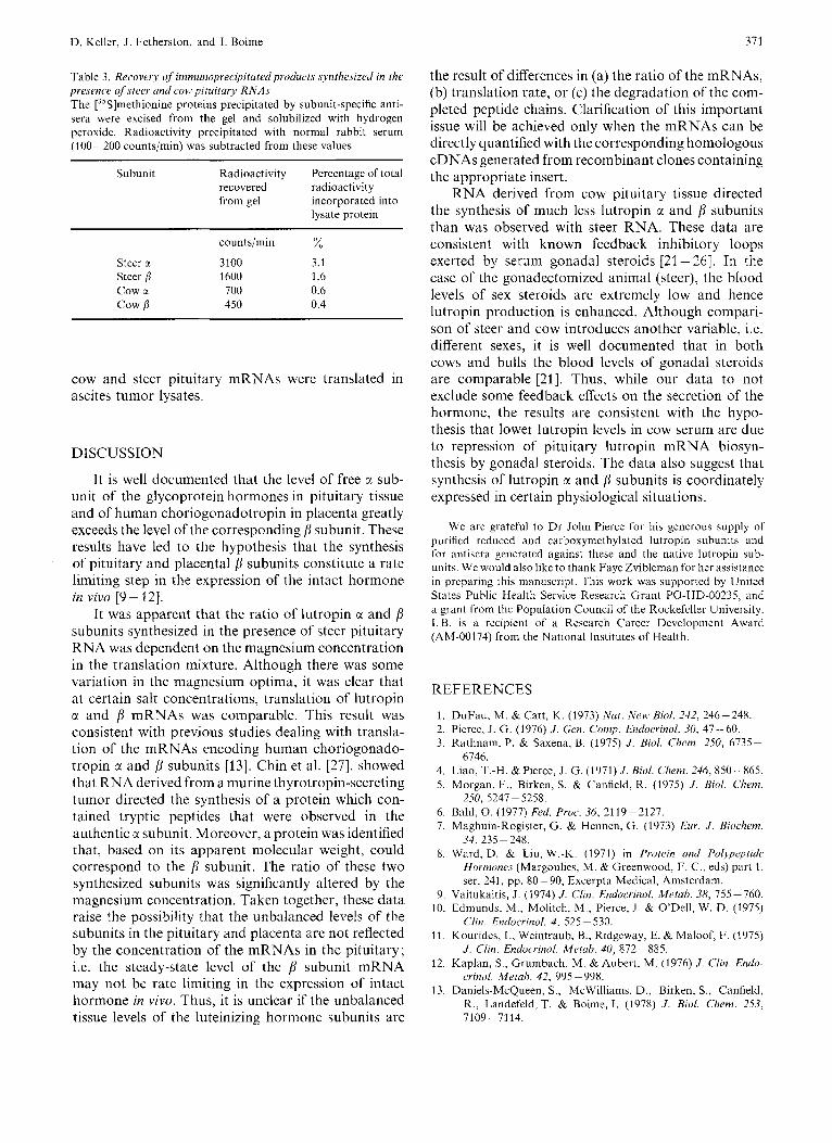
amount of cow pituitary RNA was added to the lysate and an equal quantity of radioactivity was immuno- precipitated. Cow pituitary RNA directed the syn- thesis of about five times less total lutropin subunits (Fig.5 and Table 3). This difference was not due to a lower overall translational efficiency of cow RNA, since synthesis of pre-prolactin in the presence of steer and cow RNA was comparable (Fig. 5). These changes were not significantly affected by altering the Mg2+ concentration in the translation mixtures (data not shown). Moreover, this difference (5 - 8-fold) was ob- served with several preparations of RNA and when
D. Keller, J . Fetherston, and I. Boime 371
Table 3 . Recovery of immunoprecipitutedproducts synthesized in tl7e presence of steer and cow pituitary RNAs The [35S]methionine proteins precipitated by subunit-specific anti- sera were excised from the gel and solubilized with hydrogen peroxide. Radioactivity precipitated with normal rabbit serum (100-200 counts/min) was subtracted from these values
Subunit Radioactivity Percentage of total recovered radioactivity from gel incorporated into
lysate protein
counts/min x Steer ti 3100 3.1 Steer ,8 1600 1.6 c o w Y. 700 0.6 cow /I 450 0.4
cow and steer pituitary mRNAs were translated in ascites tumor lysates.
DISCUSSION
It is well documented that the level of free CI sub- unit of the glycoprotein hormones in pituitary tissue and of human choriogonadotropin in placenta greatly exceeds the level of the corresponding j subunit. These results have led to the hypothesis that the synthesis of pituitary and placental b subunits constitute a rate limiting step in the expression of the intact hormone in vivo 19- f2].
It was apparent that the ratio of lutropin CI and j subunits synthesized in the presence of steer pituitary RNA was dependent on the magnesium concentration in the translation mixture. Although there was some variation in the magnesium optima, it was clear that at certain salt concentrations, translation of lutropin CI and /r mRNAs was comparable. This result was consistent with previous studies dealing with transla- tion of the mRNAs encoding human choriogonado- tropin a and j subunits [13]. Chin et al. [27], showed that RNA derived from a murine thyrotropin-secreting tumor directed the synthesis of a protein which con- tained tryptic peptides that were observed in the authentic a subunit. Moreover, a protein was identified that, based on its apparent molecular weight, could correspond to the subunit. The ratio of these two synthesized subunits was significantly altered by the magnesium concentration. Taken together, these data raise the possibility that the unbalanced levels of the subunits in the pituitary and placenta are not reflected by the concentration of the mRNAs in the pituitary; i.e. the steady-state level of the subunit mRNA may not be rate limiting in the expression of intact hormone in vivo. Thus, it is unclear if the unbalanced tissue levels of the luteinizing hormone subunits are
the result of differences in (a) the ratio of the mRNAs, (b) translation rate, or (c) the degradation of the com- pleted peptide chains. Clarification of this important issue will be achieved only when the mRNAs can be directly quantified with the corresponding homologous cDNAs generated from recombinant clones containing the appropriate insert.
RNA derived from cow pituitary tissue directed the synthesis of much less lutropin CI and f l subunits than was observed with steer RNA. These data are consistent with known feedback inhibitory loops exerted by serum gonadal steroids [21-261. In the case of the gonadectomized animal (steer), the blood levels of sex steroids are extremely low and hence lutropin production is enhanced. Although compari- son of steer and cow introduces another variable, i.e. different sexes, it is well documented that in both cows and bulls the blood levels of gonadal steroids arc comparable [21]. Thus, while our data to not exclude some feedback effects on the secretion of the hormone, the results are consistent with the hypo- thesis that lower lutropin levels in cow serum are due to repression of pituitary lutropin mRNA biosyn- thesis by gonadal steroids. The data also suggest that synthesis of lutropin CI and subunits is coordinately expressed in certain physiological situations.
We are grateful to D r John Pierce for his generous supply of purified reduced and carboxyinethylated lutropin subunits and for antiscra gencratcd against these and the native lutropin sub- units. We would also like to thank Faye Zvibleman for her assistance in preparing this manuscript. This work was supported by United States Public Health Service Reseal-ch Grant PO-HD-00235, and a grant from the Population Council of the Rockefeller University. I.B. is a recipient of a Research Career Development Award (AM-00174) from the National Institutes of‘ Health.
REFERENCES
1. DuFau, M. & Catt, K . (1973) Nat. New Biol. 242, 246-248. 2. Pierce, J. G. (1 976) J . Gen. Comp. Endocrinol. 30, 47 - 60. 3 . Kathnam, P. & Saxena, B. (1975) J . Bid. Clicm. 250, 6735-
4. Liao, T.-H. & Pierce, J . G. (1971) J . Bid . Clzem. 246, 850-865. 5. Morgan, F., Birken, S. & Canfield, R. (1975) J . Bid . Cl7enz.
6. Bahl, 0. (1977) Fed. Proc. 36, 2119-2127. 7. Maghuin-Rogister, G. & Hennen, G. (1973) Eur. J . Bioclzem.
8. Ward, D. & Liu, W.-K. (1971) in Protein and Pol-ypeptide Hormones (Margoulies, M. &Greenwood, F. C., eds) part 1, ser. 241, pp. 80- 90, Excerpla Medical, Amsterdam.
9. Vaitukaitis, J . (1974) J . Clin. Endocrznol. Metah. 38, 755-760. 10. Edmunds, M., Molitch, M., Pierce, J. & O’Dell, W. D. (1975)
c‘lin. Endoerinof. 4 , 525 - 530. 11. Kourides, I., Weintraub, B., Ridgeway, E. & Maloof, F. (1975)
J . Clin. Endoc.rmol. Merah. 40, 872-885. 12. Kaplan, S., Grurnbach, M. & Aubert, M. (1976) J . Clin. End(>-
crinnl. Melah. 42, 995-998. 13. Daniels-McQueen, S., McWilliams, D. , Birken, S., Canfield,
R., Landefeld, T. & Boime, I . (1978) J . Bid. Cliem. 253, 7109-7114.
6746.
250, 5247-5258.
34, 235 - 248.

372 D. Keller, J . Fetherston, and 1. Boime: Cell-Free Synthesis of Lutropin
14. Szczesna, E. & Boime, I. (1976) Proc. Nail Acad. Sci. U.S.A.
15. Boime, I., McWilliams, D., Szczesna, E. & Camel, M. (1976) J . Biol. Chem. 251, 820-825.
16. Morgan, F., Canfield, R., Vaitukaitis, J. & Ross, G . (1973) Methods in Investigative Endocrinology (Berson, S. & Yalow, R., eds) pp. 733-742, North-Holland Publishing Co., Am- sterdam.
17. Lasky, R. A. & Mills, A. D. (1975) Eur. J . Biochem. 56,
18. Birken, S., Fetherston, J., Desmond, J., Canfield, R. & Boime,
19. Landefeld, T. (1979) J . Bid. Chem. 254, 3685 -3688. 20. Swank, R. T. & Munkres, K. D. (1971) Anal. Biochem. 39,
73, 1179-1183.
335-341.
I. (1978) Biocliem. Biophys. Res. Commun. 85, 1247- 1253.
462-477.
21. McCarthy, M. S. & Swanson, L. V. (1976) J . Anim. Sci. 43, 151 -158.
22. Gay, V. & Midgley, A. R., Jr (1969) Endocrinol. 84, 1359- 1364.
23. O’Dell, W., Hescox, M. & Kiddy, C. (1970) in Gonadotropins and Ovarian Development (Butt, W., Crooke, A. & Ryze, M., eds) pp. 28-36, E. and S. Livingstone (Edinburgh).
24. Pelletier, J. (1970) Acta Endocrinol. 63, 290 - 298. 25. Walsh, P., Swerdloff, R. & O’Dell, W. (1973) Acta Endocrinol.
26. Bogdanove, E., N o h , J. & Campbell, G . (1975) in Recent Progress in Hormone Research (Greep, R. O., ed) pp. 567- 619, Academic Press, New York.
27. Chin, W., Habener, J., Kieffer, J. D. & Maloof, F. (1978) J . Biol. Chem. 253, 7985 - 7988.
74,449 - 460.
D. Keller, J. Fetherston, and I . Boime, Department of Obstetrics and Gynecology, Washington University School of Medicine, 4911 Barnes Hospital Plaza, Saint Louis, Missouri, U.S.A. 63110