issn: 2320-3528 research and reviews: journal of ... · research and reviews: journal of...
TRANSCRIPT
ISSN: 2320-3528
RRJMB| Volume 2 | Issue 3 | July – September, 2013 19
Research and Reviews: Journal of Microbiology and Biotechnology
Optimization and Isolation of Dermatophytes from Clinical Samples and In Vitro
Antifungal Susceptibility Testing By Disc Diffusion Method.
Amodkumar Yadav1*, AD Urhekar1, Vijay Mane1, Mahesh Singh Danu1, Nitin Goel1 and Ajit KG2
1Department of Microbiology, MGM Medical College & Hospitals Kamothe-Navi Mumbai - 410209, Maharashatra, India.
2Department of Biotechnology, RCOE.Mumbai, Maharashatra, India.
Research Article
Received: 02/06/2013
Revised: 15/06/2013
Accepted: 26/06/2013
*For Correspondence
Department of Microbiology, MGM
Medical College & Hospitals Kamothe-
Navi Mumbai - 410209,
Maharashatra.India.
Mobile: +91 9869375120
Keywords: Dermatophytosis, Disc
diffusion method and antifungal
agents.DTM.
ABSTRACT
India is a large subcontinent with remarkably varied topography, situated
within the tropical and subtropical belts of the world. In the study patients with Tinea
infections were examined clinically by dermatologist. Isolation, confirmatory test were
done as per the standard procedure, and Antifungal Susceptibility test was done by
Disc diffusion method.Other conditions such as seborrheic dermatitis, psoriasis,
alopecia areata, folliculitis and pseudopelade may mimic ringworm of head and other
tinea must be identified. A total of sixty six patients of dermatophytosis were studied.
Males were predominantly affected 51 (77%) cases as compared to females15 (23%)
cases. Male to female ratio was 3.4:1. Most common age group affected was 21-30
years with 20 cases (23%). Least common age group affected was 61-70 years with 3
cases (4%).Tinea corporis was more common in the age group 21-30 years with 13
cases (37.14%) and in males with 29 cases (82.85%) than females with 6 cases
(17.15%).Tinea unguium was more common in the age group of 31-40 years with 6
cases (37.5%) and in males with 10 cases (62.5%) than females with 6 cases
(37.5%).Tinea cruris was more common in the age group 51-60 years with 2 cases
(40%) and was more common in males with 5 cases (100%).In tinea pedis, one case
was seen in the age group of 11-20 years and the other in the age group of 41-50
and 51-60 years, and was more common in males with 3 cases (100%).Tinea barbae
was more common in the age group 21-30 years with 2 cases (66.66%) .Tinea capitis
was more common in the age group of 31-40 years with 2 cases (66.66%) and was
more common in females with 3 cases (100%). Tinea manuum was more common in
the age group of 31-40 years and in males with 1 case (100%). In males, commonest
infection was T. corporis while in female commonest infection was T.corporis.rate of
direct microscopy and culture (78.79%). About 89.47% of the dermatophytes grew
faster in DTM with compare to SDA, so the growth rate of dermatophyte is better in
DTM. A total of thirty five species of dermatophytes were isolated and identified.
T.rubrum 15(42.85%) is commonest among other isolates.Ketoconazole showed best
susceptibility i.e 26 (74.28%). The present study suggests that every patient of tinea
infection should be properly studied for mycological examination and should be
treated accordingly. This study revealed that Ketaconazole highest susceptibility.DTM
is better medium than SDA.
INTRODUCTION
The dermatophytes are a group of fungi that invade the superficial layer of the epidermis and degrade the keratinized
tissues of skin, hair, and nails in living animals including man [1,2]. Infections caused by these fungi are also known by the names
“Tinea” and “Ringworm.” It is important to emphasize that “ringworm” is not caused by a worm, but rather by a type of fungus called
„Dermatophyte‟ [3]. The species of dermatophytes are differentiated by Microconidia & Macroconidia. Clinically, ringworm can be
classified depending on the site involved. These include Tinea capitis (scalp), Tinea corporis (non-hairy skin of the body), Tinea cruris
(groin), Tinea pedis (foot) or athlete‟s foot and Tinea barbae or barber‟s itch (bearded areas of the face and neck). Favus is a chronic
ISSN: 2320-3528
RRJMB| Volume 2 | Issue 3 | July – September, 2013 20
type of ringworm involving the hair follicles [4]. The diseases caused by non-dermatophytic fungi infecting skin are called as
dermatomycoses whereas hair and nail are known as piedra and onychomycoses.An example of a very common dermatophyte
infection is athlete‟s foot, which is also called tinea pedis. Another common dermatophyte infection affecting the groin area is jock
itch, also known as tinea cruris [5].
The organisms are transmitted by either direct contact with infected host (human or animal) or by direct or indirect contact
with infected exfoliated skin or hair in combs, hair brushes, clothing, furniture, theatre seats, caps, bed linens, towels, hotel rugs, and
locker room floors. Depending on the species the organism may be viable in the environment for up to 15 months. There is an
increased susceptibility to infection when there is a pre-existing injury to the skin such as scars, burns, marching, excessive
temperature and humidity.
Depending on their habitat, dermatophytes are described as anthropophilic (human), zoophilic (animal) or geophilic (soil).
Anthropophilic dermatophytes are the most common sources of Tinea infections [6,7,8,9,10,11,12] Trichophyton rubrumand Trichophyton
tonsurans are two common dermatophytes. T. rubrum found in face, trunk, beard area, nails, feet and groin area infection.
T.tonsurans found in endothrix and black dot infection. These two species are usually transmitted from person to person. Another
common dermatophyte is Microsporum canis, which is transmitted from animals such as cats and dogs to humans [13,14,15,16,17,18,19,20,21].
Dermatophytes like to live on moist areas of the skin, such as places where there are skin folds. The dermatophyteinfection that
affects the scalp and hair is known as tinea capitis. It is especially common among school-aged children. For reasons that are not well
understood, tinea capitis does not usually occur after puberty. In the recent times few cases of subcutaneous and deep fungal
infections have been reported to be caused by dermatophytes. It has been noted that dermatophyte infections are more common in
adolescents and adults [3].
Cutaneous dermatophyte infections are common in the general population with up to 20% of people being infected at any
time. However, adults are generally less susceptible to skin infection than are children owing to the fungistatic properties of fatty
acids in the sebum. Most of these infections are not life threatening, but they can cause morbidity in immunocompromised, diabetic
patients, people who use communal baths, and people who are involved in contact sports such as wrestling.2 Outbreaks of infections
can occur in schools, households and institutional settings. Such infections can spread usually through direct contact with an infected
person or animal, clothing, bedding and towels can also become contaminated and spread the infection. Dermatophyte infections can
affect the skin on almost any area of the body, such as the scalp, legs, arms, feet, groin and nails. These infections are usually itchy,
redness, scaling, or fissuring of the skin, or a ring with irregular borders and a cleared central area may occur. If the infection involves
the scalp, an area of hair loss may result. More aggressive infections may lead to an abscess or cellulitis. Areas infected by
dermatophytes may become secondarily infected by bacteria. Symptoms typically appear between 4 and 14 days following exposure
[3]. Studies regarding use of culture media which are optimally suited for isolation of dermatophytes are very limited in Navi Mumbai.
The present study addresses the two points (1) Selection of an optimal medium for conidial formation by dermatophytes and
(2) the validations of the method with a large number of dermatophytes. Dermatophytosis, mycotic infection caused by
dermatophytes are commonly related in tropical countries and represent an important public health problem yet unresolved. Though
there are several antifungal drugs used to treat dermatophytosis, some infections respond well to topical antifungal therapy, whereas
others like tinea capitis, tinea unguium (nail infection), and more extensive or severe types may require systemic therapy.
Occasionally, in some cases, antifungal therapy is a failure because of resistance to theantifungal drugs by the fungi. Although recent
antifungal agents have high success rate in treating these conditions, lack of clinical response may occur in 20%.
Antifungal susceptibility testing is performed to provide information to allow clinicians to select appropriate antifungal
agents useful for treating a particular fungal infection.7For a definitive therapy also it is essential to evaluate the resistant
dermatophytes using a standardize, simple and reproducible in vitroassay to determine the antifungal activity of drugs against
isolates. In vitroantifungal susceptibility tests are now mainly used for epidemiological surveys, determination of the degree of
antifungal activity, and the prediction of clinical outcome based upon an optimization of antifungal therapy [5]. A few antifungal agents
are available and licensed for use in veterinary practice or human being treatment. The use of systemic drugs is limited to treat man
or animal due to their high toxicity and problems of residues in products intended for human consumption.Different treatments have
been recommended to control dermatophytes [6]. Several methods have been developed for testing antifungal agents against this
group of pathogens. Multicenter studies to develop a standardized antifungal susceptibility assay were initiated by the Clinical and
Laboratory Standards Institute (CLSI, formerly „National Committee for Clinical Laboratory Standards‟, NCCLS) in1983. Dilution tests
are widely used in macro- and micro-assays, but these methods are difficult to be used in most laboratories. Recently, studies were
done to establish a simple method to solve this problem. The agar-based disk diffusion (DD) susceptibility method for dermatophytes
is simple, inexpensive, and does not require specialized equipment. The disk diffusion method has a good correlation with the
reference dilution assay. The main aim of this study is to determine in vitro activity of four antifungal drugs that are most commonly
used to treat dermatophytosis; Miconazole (MIZ), Clotrimazole (CTZ), Fluconazole (FLZ) and Ketoconazole (KTZ). Hence the present
study is being undertaken to evaluate the optimum method for rapid isolation of dermatophytes and to study its resistance to
antifungal drugs [5].
ISSN: 2320-3528
RRJMB| Volume 2 | Issue 3 | July – September, 2013 21
MATERIAL AND METHOD
The infected skin, hair and nail sample were collected, KOH mount of the samples were prepared and cultured on SDA+CC
and DTM medium for isolation, after isolation slide culture and urease test is done for the identification of the dermatophytes.
Selection of cases
Inclusion criteria
Patients of all age groups and of both sexes, attending Skin and Venereology outpatient department of MGM hospital,
Kamothe, Navi Mumbai were taken for the study.
Exclusion criteria
Patients already under antifungal treatment were excluded from the study group.After the detailed history, clinical
examination of patient was made in good light which included site of lesion, number of lesions, types, presence of inflammatory
margin, etc.
Specimen collection
The affected area was cleaned with 70% ethyl alcohol, skin scales, crusts and pieces of nail were collected in clean black
paper and in the case of hair collected on clean white paper packets.Skin specimen was collected by scraping across the inflamed
margin of lesion into the apparently healthy tissue.Nail specimen was collected by taking clippings of the infected part and scrapings
beneath the nail. Hair specimen was collected by plucking with epilating forceps along with the base of the hair shaft around the
follicle.
Direct microscopic examination
KOH mount
a) Emulsify the specimen in a drop of 10% or 40% KOH on a microscopic slide with help of a straight wire.
b) Apply gentle heat by passing the slide over a Bunsen flame for 3-4 times.
c) Cover the smear with cover slip.
d) Leave it for 10-15 minutes. But in the case of hair or nail wait for overnight.
e) Examine the slide under low power (10X) and high power (40X) magnification.
f) Examine the slide for 15-20 minutes for demonstration of shining fungal elements.
Culture
After direct microscopic examination, irrespective of demonstration of fungal elements, the specimen was inoculated onto
two sets of test tubes, one containing Sabouraud‟s dextrose agar with 0.05% chloramphenicol and 0.5% cycloheximide and the other
to Dermatophyte test medium.
Sabouraud‟s dextrose agar with chloramphenicol and cycloheximide
The standard medium for growing dermatophyte is Sabouraud‟s dextrose agar containing chloramphenicol and
cycloheximide, which inhibit bacteria and saprophytic fungi respectively. The cycloheximide (Actidione) in a concentration of 0.1 to
0.4 mg per ml suppresses the growth of most saprophytic fungi such as Scytalidium, Hendersonula, Aspergillus, Candida species
without deterring the growth of dermatophytes.
Dermatophyte test medium
Specimen from skin, hair or nail were inoculated directly onto DTM and incubated at room temperature with the cap of the
culture tube loose. Dermatophytes change the medium from yellow to red within 14 days. Care must be taken in specimen collection
and interpretation of result, as many contaminants and other fungi increase the number of false positive changes in color. DTM does
not interfere with macroscopic morphology and microscopic characteristics of the dermatophytes, but it cannot be used to study
pigment production because of the intense red color of the indicator.
Macroscopic examination of culture
The growth on Sabouraud‟s dextrose agar was examined to study the colony morphology based on following characteristics.
ISSN: 2320-3528
RRJMB| Volume 2 | Issue 3 | July – September, 2013 22
Colony characters on obverse
The colour (white, pearl, ivory) and consistency (cottony, velvety, fluffy, suede).
Colony characters on the reverse
Presence or absence of pigment, whether diffusing or not.
Microscopic examination of culture
Tease mount
The tease mount was observed under low and high power objective of microscope, for the presence of hyphae,
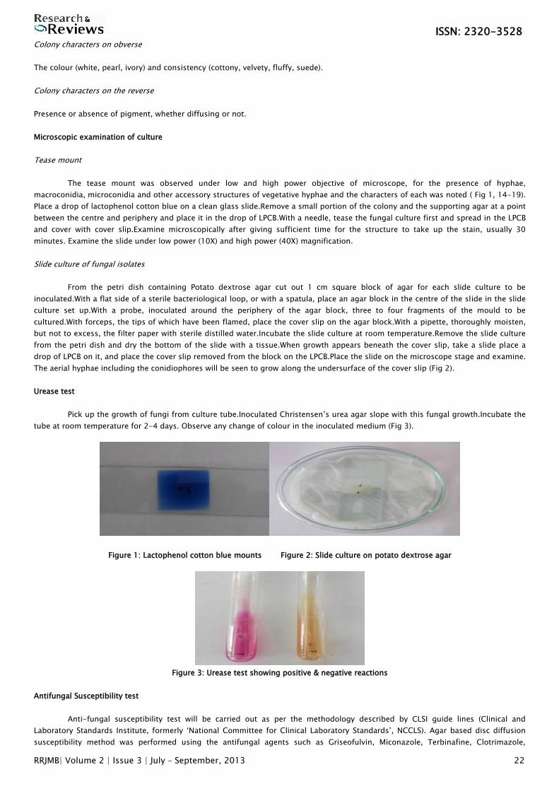
macroconidia, microconidia and other accessory structures of vegetative hyphae and the characters of each was noted ( Fig 1, 14-19).
Place a drop of lactophenol cotton blue on a clean glass slide.Remove a small portion of the colony and the supporting agar at a point
between the centre and periphery and place it in the drop of LPCB.With a needle, tease the fungal culture first and spread in the LPCB
and cover with cover slip.Examine microscopically after giving sufficient time for the structure to take up the stain, usually 30
minutes. Examine the slide under low power (10X) and high power (40X) magnification.
Slide culture of fungal isolates
From the petri dish containing Potato dextrose agar cut out 1 cm square block of agar for each slide culture to be
inoculated.With a flat side of a sterile bacteriological loop, or with a spatula, place an agar block in the centre of the slide in the slide
culture set up.With a probe, inoculated around the periphery of the agar block, three to four fragments of the mould to be
cultured.With forceps, the tips of which have been flamed, place the cover slip on the agar block.With a pipette, thoroughly moisten,
but not to excess, the filter paper with sterile distilled water.Incubate the slide culture at room temperature.Remove the slide culture
from the petri dish and dry the bottom of the slide with a tissue.When growth appears beneath the cover slip, take a slide place a
drop of LPCB on it, and place the cover slip removed from the block on the LPCB.Place the slide on the microscope stage and examine.
The aerial hyphae including the conidiophores will be seen to grow along the undersurface of the cover slip (Fig 2).
Urease test
Pick up the growth of fungi from culture tube.Inoculated Christensen‟s urea agar slope with this fungal growth.Incubate the
tube at room temperature for 2-4 days. Observe any change of colour in the inoculated medium (Fig 3).
Figure 1: Lactophenol cotton blue mounts Figure 2: Slide culture on potato dextrose agar
Figure 3: Urease test showing positive & negative reactions
Antifungal Susceptibility test
Anti-fungal susceptibility test will be carried out as per the methodology described by CLSI guide lines (Clinical and
Laboratory Standards Institute, formerly „National Committee for Clinical Laboratory Standards‟, NCCLS). Agar based disc diffusion
susceptibility method was performed using the antifungal agents such as Griseofulvin, Miconazole, Terbinafine, Clotrimazole,
ISSN: 2320-3528
RRJMB| Volume 2 | Issue 3 | July – September, 2013 23
Fluconazole and Ketoconazole.Sixty –six sample of skin, hair or nail from clinically suspected case of dermatophytosis attending
O.P.D at MGM hospital, Kamothe, Navi Mumbai.Commercially available discs from HiMedia Laboratories, preloaded with fluconazole
(25µ g/disk), Clotrimazole (10µg/disk), ketoconazole (15 µg/disk), and Miconazole (10µg/disk) will be used.The isolates were
transferred from distilled water stocks and sub-cultured to potato dextrose agar (Hi Media, India) to enhance sporulation. Seven day-
old cultures were covered with 1ml distilled water and the colonies probed with the tip of a sterile Pasteur pipette to obtain a mixture
of mycelium and conidia. The suspensions were transferred to sterile tubes and allowed to sediment for 30 minutes. The inocula will
be evenly spread on the surface of Petri dishes containing Sabouraud dextrose agar, Mueller-Hinton (MH) agar medium. Then, the
antifungal disks were applied to the plates, after which the plates were incubated at 25°C for 5-10 days. After the incubation colonies
grow and the zones of inhibition around the disks were measured and recorded.
RESULTS
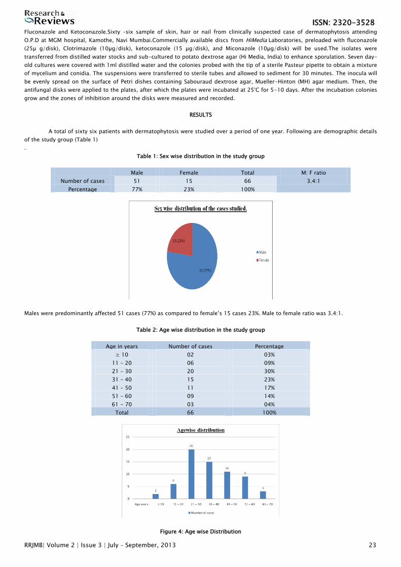
A total of sixty six patients with dermatophytosis were studied over a period of one year. Following are demographic details
of the study group (Table 1)
.
Table 1: Sex wise distribution in the study group
Male Female Total M: F ratio
Number of cases 51 15 66 3.4:1
Percentage 77% 23% 100%
Males were predominantly affected 51 cases (77%) as compared to female‟s 15 cases 23%. Male to female ratio was 3.4:1.
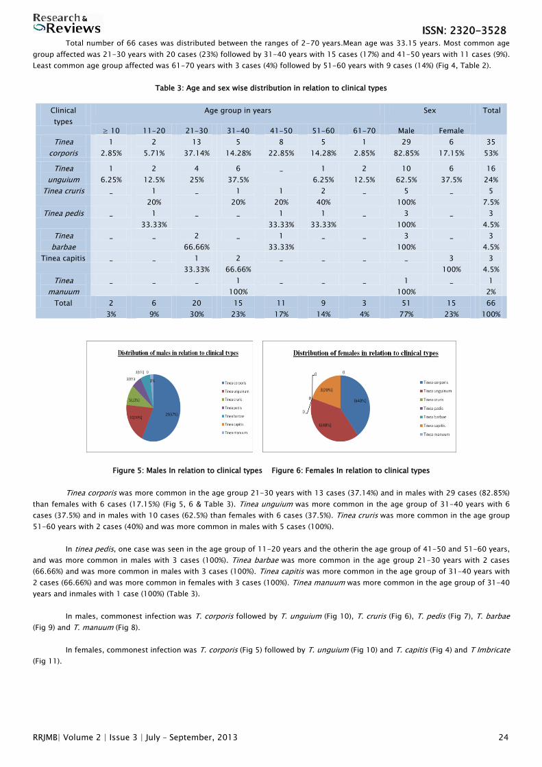
Table 2: Age wise distribution in the study group
Age in years Number of cases Percentage
≥ 10 02 03%
11 – 20 06 09%
21 – 30 20 30%
31 – 40 15 23%
41 – 50 11 17%
51 – 60 09 14%
61 - 70 03 04%
Total 66 100%
Figure 4: Age wise Distribution
ISSN: 2320-3528
RRJMB| Volume 2 | Issue 3 | July – September, 2013 24
Total number of 66 cases was distributed between the ranges of 2-70 years.Mean age was 33.15 years. Most common age
group affected was 21-30 years with 20 cases (23%) followed by 31-40 years with 15 cases (17%) and 41-50 years with 11 cases (9%).
Least common age group affected was 61-70 years with 3 cases (4%) followed by 51-60 years with 9 cases (14%) (Fig 4, Table 2).
Table 3: Age and sex wise distribution in relation to clinical types
Clinical
types
Age group in years Sex Total
≥ 10 11-20 21-30 31-40 41-50 51-60 61-70 Male Female
Tinea
corporis
1
2.85%
2
5.71%
13
37.14%
5
14.28%
8
22.85%
5
14.28%
1
2.85%
29
82.85%
6
17.15%
35
53%
Tinea
unguium
1
6.25%
2
12.5%
4
25%
6
37.5%
_ 1
6.25%
2
12.5%
10
62.5%
6
37.5%
16
24%
Tinea cruris _ 1
20%
_ 1
20%
1
20%
2
40%
_ 5
100%
_ 5
7.5%
Tinea pedis _ 1
33.33%
_ _ 1
33.33%
1
33.33%
_ 3
100%
_ 3
4.5%
Tinea
barbae
_ _ 2
66.66%
_ 1
33.33%
_ _ 3
100%
_ 3
4.5%
Tinea capitis _ _ 1
33.33%
2
66.66%
_ _ _ _ 3
100%
3
4.5%
Tinea
manuum
_ _ _ 1
100%
_ _ _ 1
100%
_ 1
2%
Total 2
3%
6
9%
20
30%
15
23%
11
17%
9
14%
3
4%
51
77%
15
23%
66
100%
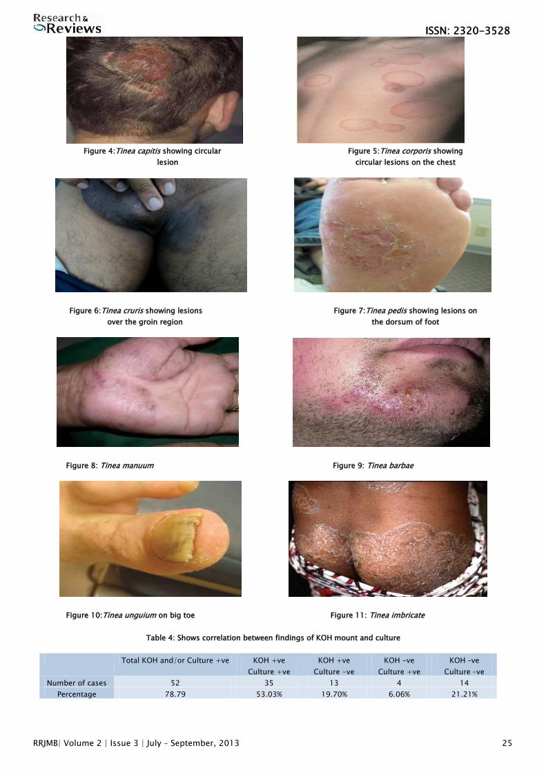
Figure 5: Males In relation to clinical types Figure 6: Females In relation to clinical types
Tinea corporis was more common in the age group 21-30 years with 13 cases (37.14%) and in males with 29 cases (82.85%)
than females with 6 cases (17.15%) (Fig 5, 6 & Table 3). Tinea unguium was more common in the age group of 31-40 years with 6
cases (37.5%) and in males with 10 cases (62.5%) than females with 6 cases (37.5%). Tinea cruris was more common in the age group
51-60 years with 2 cases (40%) and was more common in males with 5 cases (100%).
In tinea pedis, one case was seen in the age group of 11-20 years and the otherin the age group of 41-50 and 51-60 years,
and was more common in males with 3 cases (100%). Tinea barbae was more common in the age group 21-30 years with 2 cases
(66.66%) and was more common in males with 3 cases (100%). Tinea capitis was more common in the age group of 31-40 years with
2 cases (66.66%) and was more common in females with 3 cases (100%). Tinea manuum was more common in the age group of 31-40
years and inmales with 1 case (100%) (Table 3).
In males, commonest infection was T. corporis followed by T. unguium (Fig 10), T. cruris (Fig 6), T. pedis (Fig 7), T. barbae
(Fig 9) and T. manuum (Fig 8).
In females, commonest infection was T. corporis (Fig 5) followed by T. unguium (Fig 10) and T. capitis (Fig 4) and T Imbricate
(Fig 11).
ISSN: 2320-3528
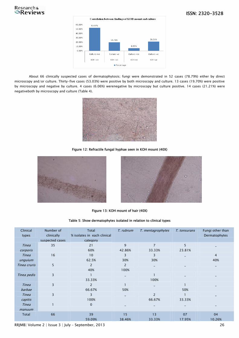
RRJMB| Volume 2 | Issue 3 | July – September, 2013 25
Figure 4:Tinea capitis showing circular Figure 5:Tinea corporis showing
lesion circular lesions on the chest
Figure 6:Tinea cruris showing lesions Figure 7:Tinea pedis showing lesions on
over the groin region the dorsum of foot
Figure 8: Tinea manuum Figure 9: Tinea barbae
Figure 10:Tinea unguium on big toe Figure 11: Tinea imbricate
Table 4: Shows correlation between findings of KOH mount and culture
Total KOH and/or Culture +ve KOH +ve
Culture +ve
KOH +ve
Culture -ve
KOH -ve
Culture +ve
KOH –ve
Culture –ve
Number of cases 52 35 13 4 14
Percentage 78.79 53.03% 19.70% 6.06% 21.21%
ISSN: 2320-3528
RRJMB| Volume 2 | Issue 3 | July – September, 2013 26
About 66 clinically suspected cases of dermatophytosis; fungi were demonstrated in 52 cases (78.79%) either by direct
microscopy and/or culture. Thirty-five cases (53.03%) were positive by both microscopy and culture. 13 cases (19.70%) were positive
by microscopy and negative by culture. 4 cases (6.06%) werenegative by microscopy but culture positive. 14 cases (21.21%) were
negativeboth by microscopy and culture (Table 4).
Figure 12: Refractile fungal hyphae seen in KOH mount (40X)
Figure 13: KOH mount of hair (40X)
Table 5: Show dermatophytes isolated in relation to clinical types
Clinical
types
Number of
clinically
suspected cases
Total
% isolates in each clinical
category
T. rubrum T. mentagrophytes T. tonsurans Fungi other than
Dermatophytes
Tinea
corporis
35 21
60%
9
42.86%
7
33.33%
5
23.81%
_
Tinea
unguium
16 10
62.5%
3
30%
3
30%
_ 4
40%
Tinea cruris 5 2
40%
2
100%
_ _ _
Tinea pedis 3 1
33.33%
_ 1
100%
_ _
Tinea
barbae
3 2
66.67%
1
50%
_ 1
50%
_
Tinea
capitis
3 3
100%
_ 2
66.67%
1
33.33%
_
Tinea
manuum
1 0 _ _ _ _
Total 66 39
59.09%
15
38.46%
13
33.33%
07
17.95%
04
10.26%
ISSN: 2320-3528
RRJMB| Volume 2 | Issue 3 | July – September, 2013 27
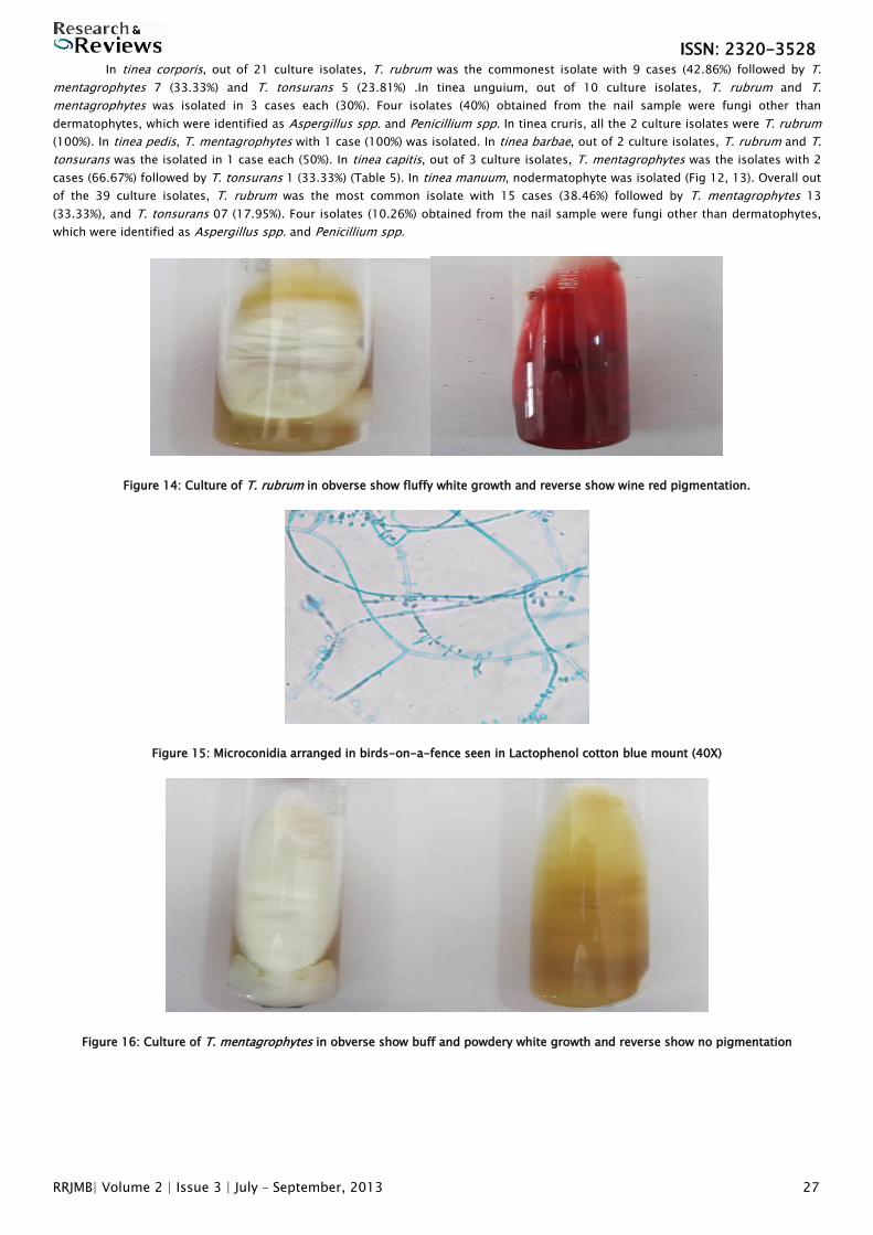
In tinea corporis, out of 21 culture isolates, T. rubrum was the commonest isolate with 9 cases (42.86%) followed by T.
mentagrophytes 7 (33.33%) and T. tonsurans 5 (23.81%) .In tinea unguium, out of 10 culture isolates, T. rubrum and T.
mentagrophytes was isolated in 3 cases each (30%). Four isolates (40%) obtained from the nail sample were fungi other than
dermatophytes, which were identified as Aspergillus spp. and Penicillium spp. In tinea cruris, all the 2 culture isolates were T. rubrum
(100%). In tinea pedis, T. mentagrophytes with 1 case (100%) was isolated. In tinea barbae, out of 2 culture isolates, T. rubrum and T.
tonsurans was the isolated in 1 case each (50%). In tinea capitis, out of 3 culture isolates, T. mentagrophytes was the isolates with 2
cases (66.67%) followed by T. tonsurans 1 (33.33%) (Table 5). In tinea manuum, nodermatophyte was isolated (Fig 12, 13). Overall out
of the 39 culture isolates, T. rubrum was the most common isolate with 15 cases (38.46%) followed by T. mentagrophytes 13
(33.33%), and T. tonsurans 07 (17.95%). Four isolates (10.26%) obtained from the nail sample were fungi other than dermatophytes,
which were identified as Aspergillus spp. and Penicillium spp.
Figure 14: Culture of T. rubrum in obverse show fluffy white growth and reverse show wine red pigmentation.
Figure 15: Microconidia arranged in birds-on-a-fence seen in Lactophenol cotton blue mount (40X)
Figure 16: Culture of T. mentagrophytes in obverse show buff and powdery white growth and reverse show no pigmentation
ISSN: 2320-3528
RRJMB| Volume 2 | Issue 3 | July – September, 2013 28
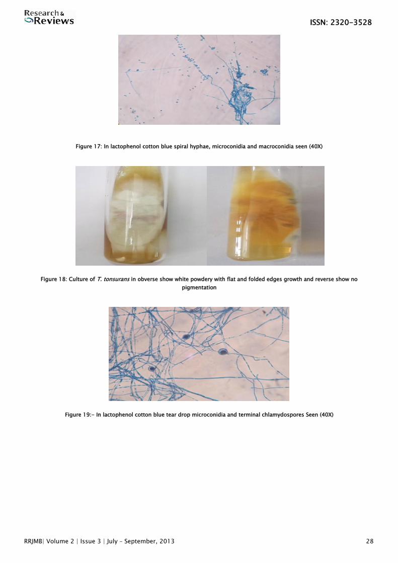
Figure 17: In lactophenol cotton blue spiral hyphae, microconidia and macroconidia seen (40X)
Figure 18: Culture of T. tonsurans in obverse show white powdery with flat and folded edges growth and reverse show no
pigmentation
Figure 19:- In lactophenol cotton blue tear drop microconidia and terminal chlamydospores Seen (40X)
ISSN: 2320-3528
RRJMB| Volume 2 | Issue 3 | July – September, 2013 29
Table6: Comparison of growth rate on SDA & DTM
Comparison of growth rate on SDA & DTM
Growth on DTM Growth on SDA
Day No. of samples Percentage (%) Day No. of samples Percentage (%)
3rd 7 36.84% 3rd 2 10.53%
4th 3 15.79% 4th 2 10.53%
5th 7 36.84% 5th 7 36.84%
6th 2 10.53% 6th 4 21.05%
7th 0 0% 7th 4 21.05%
Table 7: Criteria of susceptibility and resistance of antifungal disks
Antifungal drugs Potency Zone diameter in mm
Sensitive Intermediate Resistant
Clotrimazole 10 μg ≥20 19-21 ≤11
Fluconazole 25 μg ≥22 21-15 ≤14
Ketoconazole 15 μg ≥30 29-23 ≤22
Miconazole 10 μg ≥20 19-12 11
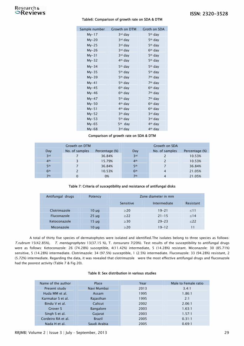
A total of thirty five species of dermatophytes were isolated and identified.The isolates belong to three species as follows:
T.rubrum 15(42.85%), T. mentagrophytes 13(37.15 %), T. tonsurans 7(20%). Test results of the susceptibility to antifungal drugs
were as follows: Ketoconazole: 26 (74.28%) susceptible, 4(11.42%) intermediate, 5 (14.28%) resistant. Miconazole: 30 (85.71%)
sensitive, 5 (14.28%) intermediate. Clotrimazole: 34 (97.5%) susceptible, 1 (2.5%) intermediate. Fluconazole: 33 (94.28%) resistant, 2
(5.72%) intermediate. Regarding the data, it was revealed that clotrimazole were the most effective antifungal drugs and fluconazole
had the poorest activity (Table 7 & Fig 20).
Table 8: Sex distribution in various studies
Name of the author Place Year Male to Female ratio
Present study Navi Mumbai 2013 3.4:1
Huda MM et al. Assam 1995 1.86:1
Karmakar S et al. Rajasthan 1995 2:1
Bindu V et al. Calicut 2002 2.06:1
Grover S Bangalore 2003 1.63:1
Singh S et al. Gujarat 2003 1.57:1
Cordeiro RA et al. Brazil 2005 0.31:1
Nada H et al. Saudi Arabia 2005 0.69:1
Sample number Growth on DTM Groth on SDA
My-17 3rd day 5th day
My-20 3rd day 5th day
My-25 3rd day 5th day
My-26 3rd day 6th day
My-31 3rd day 5th day
My-32 4th day 5th day
My-34 5th day 5th day
My-35 5th day 5th day
My-39 5th day 7th day
My-41 5th day 7th day
My-45 6th day 6th day
My-46 6th day 7th day
My-47 5th day 7th day
My-50 4th day 6th day
My-51 4th day 6th day
My-52 3th day 3rd day
My-53 5th day 3rd day
My-65 5th day 4th day
My-68 3rd day 4th day
ISSN: 2320-3528
RRJMB| Volume 2 | Issue 3 | July – September, 2013 30
Figure 20: Sensitivity T.mentagrophytes to tested antifungal drugs
K, ketoconazole; C, clotrimazole; M, miconazole; F, fluconazole
In the present study, males (77%) were more commonly affected than females (23%). Male to female ratio was 3.4:1, which is
comparable with other studies doneby Huda MM [29,30], Karmakar S [31], Bindu V [35], Grover S [36], Singh S [37], whereas Cordeiro RA [54]
andNada H [55] reported that females were more commonly affected than males, with male tofemale ratio being 0.31:1 and 0.69:1
respectively [32, 33
Male predominance may be due to increased outdoor physical activities andincreased opportunity for exposure to infection
than females. Also in rural India, males may visit the hospital to a greater extent than females who may not be very open for hospital
visit for dermatological infections especially in rural areas (Table 8).
Table 9: Age distribution as found in various studies (in percentage)
Name of the author Place Year Commonest age group (percentage)
Present study Navi Mumbai 2013 21-30 years (28%)
Karmakar S et al. Rajasthan 1995 0-30 years (64%)
Mishra M et al. Sambalpur 1998 15-35 years (30%)
Bokhari MA et al. Lahore 1999 20-40 years (36%)
Singh S et al. Baroda 2003 16-45 yeas (31.36%)
Sen SS et al. Assam 2006 21-30 years (44%)
Veer P et al. Aurangabad 2007 31-40 years (39.4%)
The present study shows that dermatophytosis was more common in the age group of 21-30 years (30%) followed by 31-40
years (23%), which is comparable with other studies done byMishra M [34], Sen SS [39,40]. However Veer P hasreported that the most
common age group affected was 31-40 years followed by 41-50 years (Table 9).The highest incidence in young adults aged 21-30
years may be due toincreased physical activity and increased opportunity for exposure.
Tinea corporis
In the present study, tinea corporis was the commonest clinical type encountered (53%) followed by tinea unguium (24%) and
the commonest age group affected was 21-30 years (30%). Males were predominantly affected with male to female ratio being 3.4:1,
which is comparable with other studies done by Bindu V(54.6%) [35], Singh S (58.8%) [37], Sen SS (48%) [40] and Venkatesan G (64.8%)
[41,42,43].
Tinea unguium
In the present study, onychomycosis was second commonest clinical type and more common in males. Male to female ratio
was 1.6:1, which is comparable with other studies done by Grover S [36] and Vijaya D[38]; whereas Cordeiro RA [54] and Nada H [55] in their
study reported that female‟s were commonly affected than males, with male to female ratio being 0.31:1 and 0.69:1 respectively
[44,45,46,47,48,49,50.51,52].
Tinea cruris
In the present study, tinea cruris showed prevalence of (7.5%) and commonest age group affected was 51-60 years (40%).
Males (100%) were more commonly affected than females, which is comparable withother studies done by Siddappa K [22], Mishra M [34]
and Sen SS [40].
ISSN: 2320-3528
RRJMB| Volume 2 | Issue 3 | July – September, 2013 31
Tinea pedis
In the present study, out of 66 cases, tinea pedis was seen in 4.5% cases, which is comparable with the study done by
Siddappa K22, whereas Chimelli PAV [53] in their study on dermatophytosis, reported tinea pedis in 9.9% cases respectively.
Tinea barbae
In the present study, out of 66 cases, tinea barbae was seen in 4.5% cases, which is comparable with the study done by Singh S [37],
Sen SS [40] and Keyvan Pakshir [56,57,58,59,60].
Tinea capitis
In the present study, out of 66 cases, tinea capitis was seen in 4.5% cases, more common age group of 31-40 years
(66.66%), which iscomparable with other studies done by Siddappa K [22], Kumar AG [23], Reddy BSN [25] and Kalla G [26,27,28].
Tinea manuum
In the present study, out of 66 cases of dermatophytosis tinea manuum was 1 case (2%), which is comparable with other
studies done by Siddappa K (1.53%)22 andChimelli PAV (1.9%) [53].
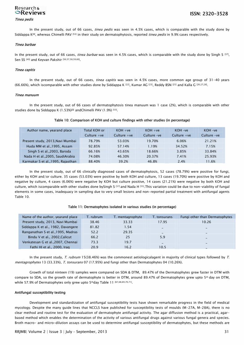
Table 10: Comparison of KOH and culture findings with other studies (in percentage)
Author name, yearand place
Total KOH or
Culture +ve
KOH +ve
Culture +ve
KOH +ve
Culture –ve
KOH –ve
Culture +ve
KOH –ve
Culture -ve
Present study, 2013,Navi Mumbai 78.79% 53.03% 19.70% 6.06% 21.21%
Huda MM et al.,1995, Assam 92.85% 57.14% 1.19% 34.52% 7.15%
Singh S et al.,2003, Baroda 66.16% 43.65% 18.66% 3.85% 33.84%
Nada H et al.,2005, SaudiArabia 74.08% 46.30% 20.37% 7.41% 25.93%
Karmakar S et al.,1995, Rajasthan 88.40% 39.2% 46.8% 2.4% 11.6%
In the present study, out of 66 clinically diagnosed cases of dermatophytosis, 52 cases (78.79%) were positive for fungi,
either by KOH and/or culture. 35 cases (53.03%) were positive by both KOH and culture, 13 cases (19.70%) were positive by KOH and
negative by culture, 4 cases (6.06%) were negative by KOH but culture positive, 14 cases (21.21%) were negative by both KOH and
culture, which iscomparable with other studies done bySingh S [37] and Nada H [55].This variation could be due to non-viability of fungal
elements in some cases, inadequacy in sampling due to very small lesions and non-reported partial treatment with antifungal agents
Table 10.
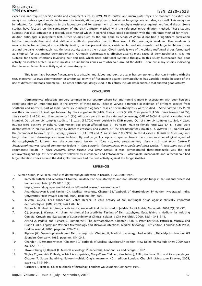
Table 11: Dermatophytes isolated in various studies (in percentage)
Name of the author, yearand place T. rubrum T. mentagrophyte T. tonsurans Fungi other than Dermatophytes
Present study, 2013, Navi Mumbai 38.46 33.33 17.95 10.26
Siddappa K et al., 1982, Davangere 81.82 1.54 _ _
Ranganathan S et al.,1995, Madras 52.2 29.35 _ _
Bindu V et al., 2002,Calicut 66.2 25 5.9 _
Venkatesan G et al.,2007, Chennai 73.3 19.7 _ _
Fathi HI et al., 2000, Iraq 20.9 16.2 10.5 _
In the present study, T. rubrum 15(38.46%) was the commonest aetiologicalagent in majority of clinical types followed by T.
mentagrophytes 13 (33.33%), T. tonsurans 07 (17.95%) and fungi other than Dermatophytes 04 (10.26%).
Growth of total ninteen (19) samples were compared on SDA & DTM, 89.47% of the Dermatophytes grew faster in DTM with
compare to SDA, so the growth rate of dermatophyte is better in DTM, around 89.47% of Dermatophytes grew upto 5th day on DTM,
while 57.9% of Dermatophytes only grew upto 5thday Table 11 [67,68,69,70,71].
Antifungal susceptibility testing
Development and standardization of antifungal susceptibility tests have shown remarkable progress in the field of medical
mycology. Despite the many guide lines that NCCLS have published for susceptibility tests of moulds (M-27A, M-28A), there is no
clear method and routine test for the evaluation of dermatophyte antifungal activity. The agar diffusion method is a practical, agar-
based method which enables the determination of the activity of various antifungal drugs against various fungal genera and species.
Broth macro- and micro-dilution assays can be used to determine antifungal susceptibility of dermatophytes, but these methods are
ISSN: 2320-3528
RRJMB| Volume 2 | Issue 3 | July – September, 2013 32
expensive and require specific media and equipment such as RPMI, MOPS buffer, and micro plate trays. The standard disk diffusion
assay constitutes a good model to be used for investigational purposes to test other fungal genera and drugs as well. This assay can
be adapted for routine diagnosis in the laboratory and for assessment of dermatophyte resistance against antifungal drugs. Some
studies have focused on the comparison of the disk diffusion method with the reference micro-dilution method. These studies
suggest that disk diffusion is a reproducible method which in general shows good correlation with the reference method for micro-
dilution antifungal susceptibility test. Other studies such as the one done by Singh et al could not find a significant correlation
between micro-dilution and disk diffusion methods, probably due to their use of Dermasel agar medium. This medium is
unacceptable for antifungal susceptibility testing. In the present study, clotrimazole, and miconazole had large inhibition zones
around the disks; clotrimazole had the best activity against the isolates. Clotrimazole is one of the oldest antifungal drugs formulated
as a topical for use against dermatophytosis. Although clotrimazole is effective against most cases of dermatophytosis, it is not
suitable for severe infections involving hair and nail, which need additional systemic therapy. In this study fluconazole had poor
activity on isolates tested. In most isolates, no inhibition zones were observed around the disks. There are many studies indicating
that fluconazole had less activity against dermatophytes.
This is perhaps because fluconazole is a triazole, and Sabouraud dextrose agar has components that can interfere with the
test. Moreover, in vitro determination of antifungal activity of fluconazole against dermatophytes has variable results because of the
use of different methods and media. The other antifungal drugs used in this study include ketoconazole that showed good activity.
CONCLUSION
Dermatophyte infections are very common in our country where hot and humid climate in association with poor hygienic
conditions play an important role in the growth of these fungi. There is varying difference in isolation of different species from
southern and northern part of India. Sixty-six clinically diagnosed cases of dermatophytosis were studied. Tinea corporis 35 (53%)
was the commonest clinical type followed by tinea unguium 16 (24%), tinea cruris 5 (7.5%), tinea pedis 3 (4.5%), tinea barbae 3 (4.5%),
tinea capitis 3 (4.5%) and tinea manuum 1 (2%). All cases were from the skin and venerology OPD of MGM Hospital, Kamothe, Navi
Mumbai. Out ofsixty six samples studied, 13 cases (19.70%) were positive by KOH mount. Out of sixty six samples studied, 4 cases
(6.06%) were positive by culture. Commonest age group affected was 21-30 years. Male to female ratio was 3.4:1. Fungi were
demonstrated in 78.89% cases, either by direct microscopy and culture. Of the dermatophytes isolated, T. rubrum 15 (38.46%) was
the commonest followed by T. mentagrophytes 13 (33.33%) and T. tonsurans 7 (17.95%). In the 4 cases (10.26%) of tinea unguium
fungi other than dermatophytes were isolated. By and large Trichophyton species forms the commonest aetiological agent of
dermatophytosis.T. Rubrum was the commonest isolate in tinea corporis, tineaunguium, tinea cruris and tinea barbae.T.
Mentagrophytes was second commonest isolate in tinea corporis, tineaunguium, tinea pedis and tinea capitis. T. tonsurans was third
commonest isolate in tinea corporis, tinea barbae and tinea capitis. It was demonstrated thatclotrimazole was the best
antimycoticagent against dermatophytes followed by miconazole and ketoconazole. Clotrimazole, miconazole and ketoconazole had
large inhibition zones around the disks; clotrimazole had the best activity against the fungal isolates.
REFERENCES
1. Suman Singh, P. M. Been. Profile of dermatophyte infection in Baroda. IJDVL.2003;69(4).
2. Ramesh Putheti and Amachree Otomba. Incidence of dermatophytes and non-dermatophytic fungi in natural and processed
human scalp hair. IJCAS.2010; 1(7).
3. http://www.cdc.gov/nczved/divisions/dfbmd/diseases/dermatophytes/.
4. Ananthanarayan R and Paniker CK. Medical mycology, Chapter 65.Textbook of Microbiology; 8th edition. Hyderabad, India:
Universities Press Private Limited, 2009, page no. 604-607.
5. Keyvan Pakshir, Leila Bahaedinie, Zahra Rezaei. In vitro activity of six antifungal drugs against clinically important
dermatophytes. IJMM. 2009; 2(4):158-163.
6. Fardos M. Bokhari. Antifungal activity of some medicinal plants used in Jeddah. Saudi Arabia, Mycopath. 2009;7(1):51-57.
7. C.J. Jessup, J. Warner, N. Isham. Antifungal Susceptibility Testing of Dermatophytes: Establishing a Medium for Inducing
Conidial Growth and Evaluation of Susceptibility of Clinical Isolates. J Clin Microbiol. 2000. 38(1); 341-344.
8. Arvind A. Padhye and Richard C. Summerbell. The dermatophytes. Chapter 13.In: S. Peter Borriello, Patrick R. Murray, and
Guido Funke. Topley and Wilson‟s Microbiology and Microbial Infections, Medical Mycology. 10th edition. London: ASM Press,
Hodder Arnold; 2005, page no. 220-239.
9. Rippon JW. Dermatophytosis and Dermatomycosis. Chapter 8, Medical mycology, 2nd edition. Philadelphia, London: WB
Saunders Company; 1982, page no. 154-241.
10. Chander J. Dermatophytoses. Chapter 10.Textbook of Medical Mycology.3rd edition. New Delhi: Mehta Publisher; 2009.page
no. 122-142.
11. Kwon Chung KJ, Bennet JE. Medical mycology. Philadelphia, London: Lea and Febiger; 1992.
12. Wigley C, Jeremiah C Healy, W Niall A Kirkpatrick, Mary-Clare C Miller, Nanchahal J, E Birgitte Lane. Skin and its appendages.
Chapter 7. Susan Standring. Editor-in-chief. Gray‟s Anatomy. 40th edition London: Churchill Livingstone Elsevier; 2008,
page no. 145-164.
13. Gartner LP, Hiatt JL. Color textbook of histology. London: WB Saunders Company; 1997.
ISSN: 2320-3528
RRJMB| Volume 2 | Issue 3 | July – September, 2013 33
14. Valia RG, Valia AR, Siddappa K. Textbook and Atlas of Dermatology. Bombay: Bhalani Publishing House; 1994.
15. Washington C. Winn, Stephen D. Allen, William M. Janda, Koneman W.Elmer, Gary W. Procop, Paul C. Schreckenberger, Gail L.
Woods. Identification of dermatophytes. Koneman‟s Color Atlas and Textbook of Diagnostic Microbiology, 6th edition.
Baltimore: Lippincott Williams & Wilkins; 2006 page no. 1187-1195.
16. L.J.R. Milne. Fungi. Chapter 41. In: Collee JG, Fraser AG, Marmion BP, Simmons A, editors. Mackie and McCartney Practical
Medical Microbiology. 14th edition. New Delhi. India Churchill Livingstone; 2008, page no. 695-717.
17. Cheesbrough Monica. Dermatophytes. District laboratory practice in tropical countries. Part 2, 2nd edition. United Kingdom:
Cambridge University Press; 2005, page no. 234-238.
18. Parija SC. Mycology. Textbook of Practical Microbiology, 1st edition. New Delhi, India: Aph Ahuja Publishing House; 2011.
Page no. 211-237.
19. Paul O. Gubbins, Elias J Anaissie. Historical perspective on the development of antifungal drugs. Clinical Mycology; 11 Dec
2009.
20. History of antifungals. Department of Dermatology. J Am Acad. 1990;(4 pt 2):776-8.
21. Mahmoud A, Ghannoum, Louis B. Mode of Action of Antifungal Agents. Clin Microbiol Rev. 1999; 12(4):501-517.
22. Siddappa K, Mahipal OA. Dermatophytosis in Davangere. Indian J Dermatol Venereol Leprol. 1982; 48:254-9.
23. Kumar AG, Lakshmi N. Tinea capitis in Tirupati. Indian J Pathol. Microbiol. 1990; 33:360-3.
24. Siddaramappa B, Hemashettar BM, Patil CS. Favus from South India – A case report. Indian J Dermatol Venereol Leprol. 1991;
57:43-4.
25. Reddy BNS, Swaminathan G, Kanungo R, D‟Souza M, Garg BR, Shantharaman R. Clinico-mycological study of tinea capitis in
Pondicherry. Indian J Dermatol Venereol Leprol. 1991; 57:180-2.
26. Rai MK. Tinea capitis due to Trichophyton rubrum in an adult woman. Indian J Dermatol Venereol Leprol. 1992; 58:213-4.
27. Ghorpade A, Ramanan C. Trichophyton tonsurans infection in a 12-day old infant. Indian J Dermatol Venereol Leprol. 1995;
61:52-3.
28. Kalla G, Begra B, Solanki A, Goyal A, Batra A. Clinico-mycological study of tinea capitis in desert district of Rajasthan. Indian J
Dermatol Venereol Leprol. 1995; 61:342-5.
29. Ranganathan S, Menon T, Selvi, GS, Kamalam A. Effect of socio-economic status on the prevalence of dermatophytosis in
Madras. Indian J Dermatol Venereol Leprol. 1995; 61:16-8.
30. Huda MM, Chakraborthy N, Bordoloi JNS. A clinico-mycological study of superficial mycoses in upper Assam. Indian J
Dermatol Venereol Leprol. 1995; 61:329-32.
31. Karmakar S, Kalla G, Joshi KR. Dermatophytosis in a desert district of Western Rajasthan. Indian J Dermatol Venereol Leprol.
1995; 61:280-3.
32. Mittal RR, Shivali. Tinea faciei and tinea capitis in a 15-day old infant. Indian J Dermatol Venereol Leprol. 1996; 62:41-2.
33. Gupta AK, Summerbell RC. Increased incidence of T. tonsurans, tinea capitis in Ontario, Canada between 1985 and 1996.
Medical Mycology. 1998; 36:55-60.
34. Mishra M, Mishra S, Singh PC, Mishra BC. Clinico-mycological profile of superficial mycoses. Indian J Dermatol Venereol
Leprol. 1998; 64:283-5.
35. Bindu V, Pavithran K. Clinico-mycological study of dermatophytosis in Calicut. Indian J Dermatol Venereol Leprol. 2002;
68:259-61.
36. Grover S. Study of onychomycosis in Bangalore. Indian J Dermatol Venereol Leprol. 2003; 69:284-6.
37. Singh S, Beena PM. Profile of dermatophyte infections in Baroda. Indian J Dermatol Venereol Leprol. 2003; 69:281-3.
38. Vijaya D, Anandkumar BH, Geetha SH. Study of onychomycosis. Indian J Dermatol Venereol Leprol. 2004; 70:185-6.
39. Garg, Venkatesh, Vimala MD, Singh, Mastan MD, Agarwal. Onychomycosis in Central India: a clinicoetiological correlation.
International J Dermatol. 2004; 43:7.
40. Sen S.S., ES Raul. Study in Dermatophytosis in Assam. Indian J Med Microbiol. 2006; 24; 77-8.
41. Kannan P, Janaki C, Selvi GS. Prevalence of dermatophytes and other fungal agents isolated from clinical samples. Indian J
Med Microbiol. 2006; 24:212-5.
42. Veer P, Patwardhan NS, Danle AS. Study of onychomycosis: prevailing fungi and pattern of infection. Indian J Med Microbiol.
2007; 25:53-6.
43. Venkatesan G, Singh AJAR, Murugesan AG, Janaki C, Shankar SG. Trichophyton rubrum – the predominant aetiological agent
in human dermatophytosis in Chennai, India. Afr J Microbiol Res. 2007; 9-12.
44. Neetu Jain, Meenakshi Sharma. Study in clinicomycological profile of Dermatophytosis in Jaipur. Indian J Dermatol Venereol.
Leprol, 2008; 74; 274-5.
45. Grover Chander, Arora Pooja, Manchanda Vikas. Tinea capitis in the pediatric population. A study from north India. Indian J
Dermatol Venereol Leprol. 2010; 76:527-32.
46. V Pankajalakshmi Venugopal, Taralakshmi Venugopal.In vitro susceptibility of dermatophytes to itraconazole. Indian J
Dermatol. 1992; 58(6):368-371.
47. PV Venugopal, TV.venugopal. In Vitro susceptibility testing of dermatophytes with itraconazole by Disk Diffusion. Indian J
Dermatol. 1993; 38(1):8-12.
48. Venugopal PV, Venugopal TV. Disk Diffusion susceptibility testing of dermatophytes with allylamines. Int J Dermatol. 1994;
33(10):730-732.
ISSN: 2320-3528
RRJMB| Volume 2 | Issue 3 | July – September, 2013 34
49. Venugopal PV, Venugopal TV. Disk diffusion susceptibility testing of dermatophytes with imidazoles.India J Pathol Microbiol.
1995;38(4); 369-74.
50. Bokhari MA. Study of onychomycosis. Int J Dermatol. 1999; 38:591-5.
51. Fathi HI, Alsamarai AM. Tinea capitis in Iraq. East Mediterr Health J. 2000; 6:138-48.
52. Perea S, Garau M, Gonzalez A. Study of prevalence and risk factors of tinea unguium and tinea pedis in the general
population of Spain. J Clin Microbiol. 2000; 38:3226-30.
53. Chimelli PAV, Sofiatti AA, Nunes RS, Martins JC. Dermatophyte agents in the city of Sao Paulo, from 1992 to 2002. Rev Inst
Med Trop S Paulo. 2003; 45:36-46.
54. Cordeiro RA, Medrano DJA, Rocha MFG, Monteiro AJ, Meireles TEF, Sidrim JJC Onychomycosis in Ceara (North-east Brazil).
Mem Inst Oswaldo Cruz Rio de Janeiro 2005; 100:131-5.
55. Nada H, Allah SS, Mokhtar M. Yeast infections as a cause of nail disease in the Western province of Saudi Arabia. Egypt J Med
Lab Sci. 2005; 14:2.
56. Keyvan Pakshir, Jamal Hashemi study in Dermatophytosis in Karaj, Iran. Int J Dermatol. 2006; 51; 262-4.
57. Lange M, Roszkiewicz J, Dobosz AS, Bykowsa B. Onychomycosis is no longer a rare finding in children. Mycoses 2006;
49:55-8.
58. Mebazaa Amel, Oumari E.L Kamel, Said Ben Moncef. Tinea capitis in adults in Tunisia. Int J Dermatol .2010; 49:513-16.
59. Fernandez-Torres B, Vazquez-Veiga H, LIovo l. In vitro susceptibility to itraconazole, clotrimazole, ketoconazole and
terbinafine of 100 isolates of Trichophyton rubrum. Chemotherapy. 2000; 46(6):390-4.
60. Carrillo AJ, Guarro j. In vitro activities of four novel triazoles against Scedosporium spp. Antimicrob Agents Chemother.
2001; 45(7); 2151-3.
61. Fernandez-Torres B, carrillo AJ, Martin E . In vitro activities of 10 antifungal drugs against 508 dermatophyte strains.
Antimicrob Agents Chemother. 2001; 45(9): 2524-8.
62. Karaca N, Koc AN. In vitro susceptibility testing of dermatophytes. Diagn Microbiol Infect Dis. 2004;48(4):259-64.
63. M Ghannoum, N Isham, D Sheehan. Voriconazole Susceptibilities of Dermatophyte Isolates Obtained from a Worldwide Tinea
Capitis Clinical Trial. J Clin Microbiol. 2006; 44(7): 2579–2580.
64. Saima A.Girgis, Nehal M.Zu El-Fakkar, Hala Badr, Genotypic identification and antifungal susceptibility pattern of
dermatophuytes in Egptian patients. IJJM, Dec-2006.
65. Singh J, Zaman M, Gupta AK. Evaluation of microdilution and disk diffusion methods for antifungal susceptibility testing of
dermatophytes. Med Mycol. 2007; 45(7):595-602.
66. EI Nweze, CC Ogbonna, JI Okafor. In vitro susceptibility testing of dermatophytes isolated from pediatric cases in Nigeria
against five antifungals. Revista do Instituto de Medicina Tropical de Sao Palo; Oct, 2007.
67. Ozkutuk A, Ergon C, Yulung N. Species distribution and antifungal susceptibilities of dermatophytes during a one year
period at a university hospital in Turkey. Mycoses. 2007; 50(2):125-9.
68. Crystiane Rodrigues Araujo, Karla Carvalho Miranda, Fernandes. In vitro Susceptibility testing of Dermatophytes isolated in
Goiania, Brazil, against five antifungal agents by broth microdilution method. 9-12, Jan-Feb, 2009.
69. Hilda Conceicao Diogo, Marcia Melhem, Aldo Sarpieri. Evaluation of the disk-diffusion method to determine the in vitro
efficacy of terbinafine against subcutaneous and superficial mycoses agents. An Bras Dermatol; vol-85; no. 3.
70. EI Nweze, PK Mukherjee, MA Ghannoum from Nigeria. Agar-Based Disk Diffusion Assay for Susceptibility Testing of
Dermatophytes. J Clin Microbiol. 2010:3750-3752.
71. Cibele Massotti Magagnin, Cheila Denise Ottonelli Stopiglia, Fabiane Jamono Vieira. Antifungal susceptibility of
dermatophytes isolated from patients with chronic renal failure. An Bras Dermatol. 86(4).