isuog basic training training skull , falx, csp, lv biometry bpd, ofd, hc biometry- tcd cerebellum/...
TRANSCRIPT

Basic Training
ISUOG Basic Training Distinguishing Between Normal & Abnormal
Appearances of the Skull & Brain
Basic Training
At the end of the lecture you will be able to:
• Describe how to obtain the 3 planes required to assess,
including measuring, the fetal head correctly
• Recognise the differences between the normal & most
common abnormal ultrasound appearances of the 3 planes
of the fetal brain
Learning objectives
Basic Training
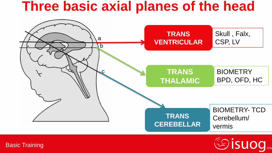
Skull , Falx,
CSP, LV
BIOMETRY
BPD, OFD, HC
BIOMETRY- TCD
Cerebellum/
vermis
TRANS
VENTRICULAR
TRANS
THALAMIC
TRANS
CEREBELLAR
Three basic axial planes of the head
Basic Training
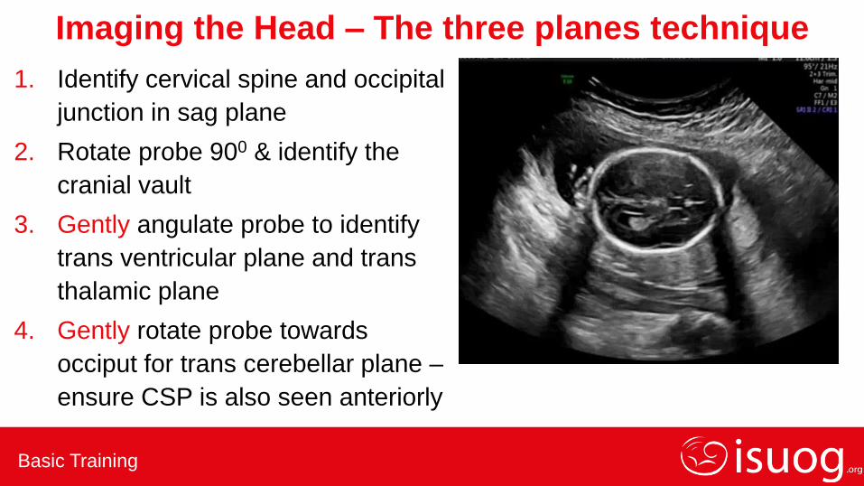
Imaging the Head – The three planes technique
1. Identify cervical spine and occipital
junction in sag plane
2. Rotate probe 900 & identify the
cranial vault
3. Gently angulate probe to identify
trans ventricular plane and trans
thalamic plane
4. Gently rotate probe towards
occiput for trans cerebellar plane –
ensure CSP is also seen anteriorly
Basic Training
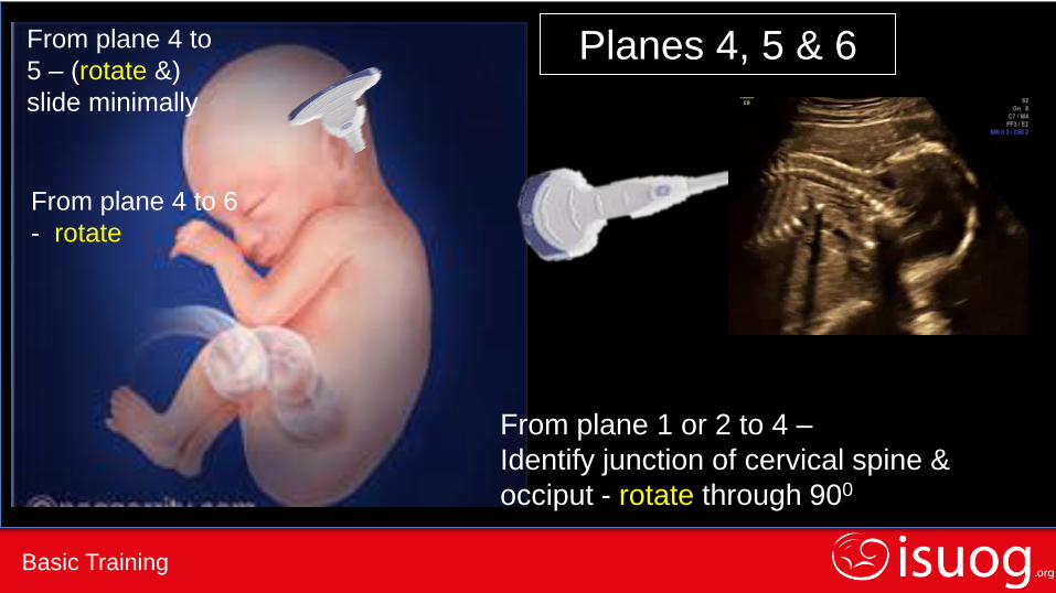
from plane 1 or 2 to 4 - rotate through 900
Planes 4, 5 & 6
From plane 1 or 2 to 4 –
Identify junction of cervical spine &
occiput - rotate through 900
From plane 4 to
5 – (rotate &)
slide minimally
From plane 4 to 6
- rotate
Basic Training
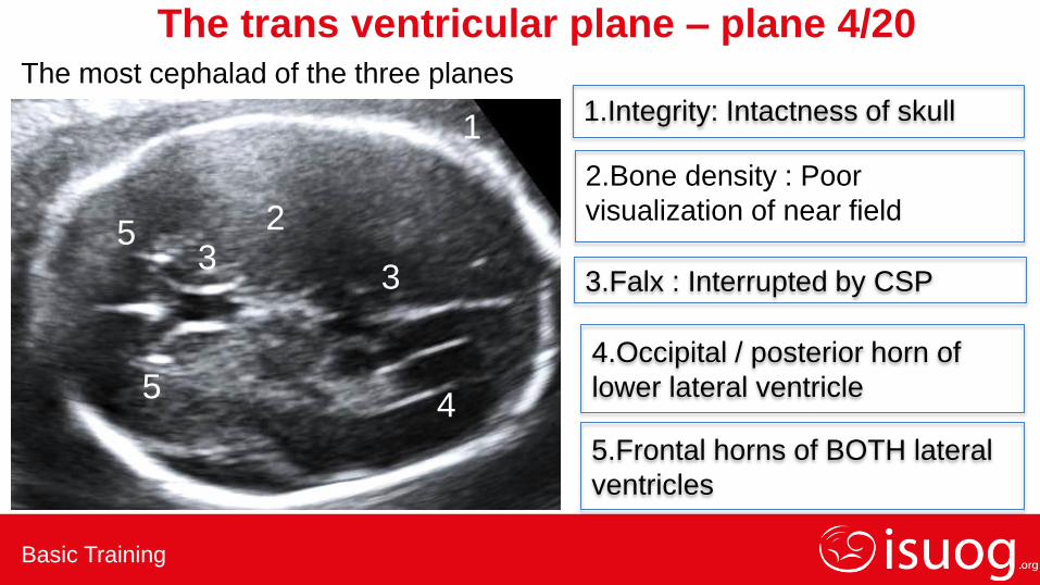
The trans ventricular plane – plane 4/20
1.Integrity: Intactness of skull
3.Falx : Interrupted by CSP
4.Occipital / posterior horn of
lower lateral ventricle
5.Frontal horns of BOTH lateral
ventricles
2
3 3
4
5
5
1
The most cephalad of the three planes
2.Bone density : Poor
visualization of near field
Basic Training
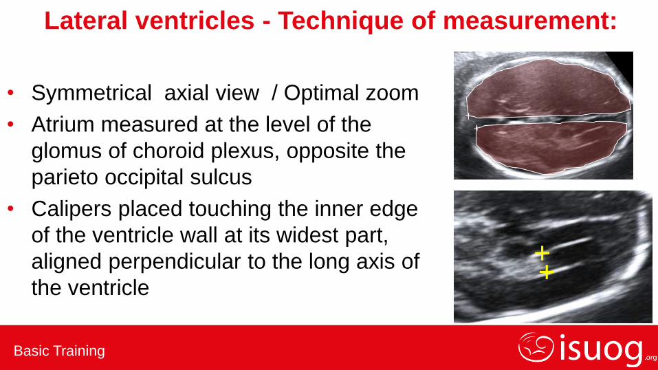
Lateral ventricles - Technique of measurement:
• Symmetrical axial view / Optimal zoom
• Atrium measured at the level of the
glomus of choroid plexus, opposite the
parieto occipital sulcus
• Calipers placed touching the inner edge
of the ventricle wall at its widest part,
aligned perpendicular to the long axis of
the ventricle
+ +
Basic Training
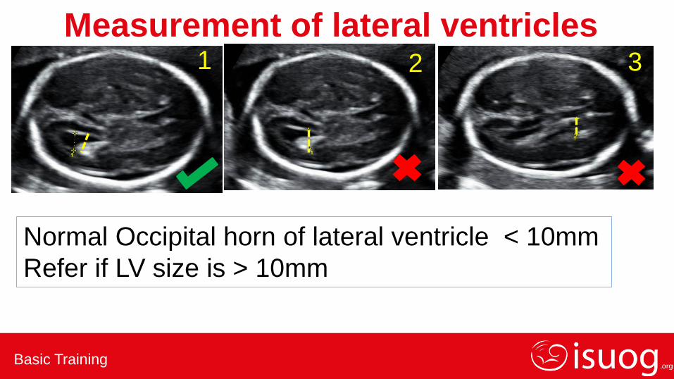
1 2 3
Measurement of lateral ventricles
Normal Occipital horn of lateral ventricle < 10mm
Refer if LV size is > 10mm
Basic Training
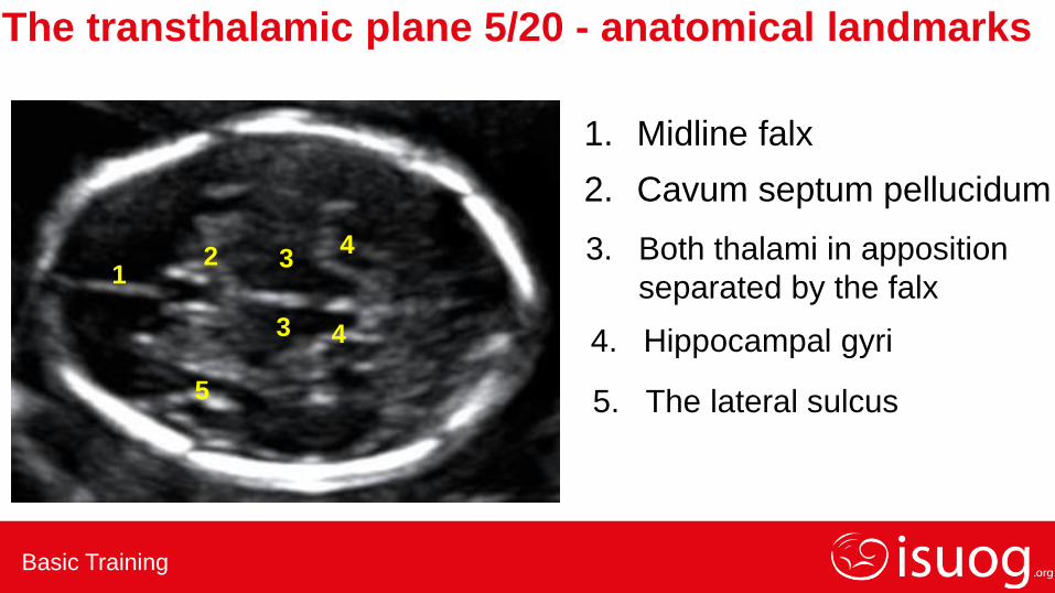
The transthalamic plane 5/20 - anatomical landmarks
1
1. Midline falx
2
2. Cavum septum pellucidum
4
4 4. Hippocampal gyri
5 5. The lateral sulcus
3
3. Both thalami in apposition
separated by the falx 3

Basic Training
1. Trans thalamic plane
2. Angle of insonation 90 deg to
midline echoes
3. Symmetric hemispheres
4. Falx with CSP & thalamus
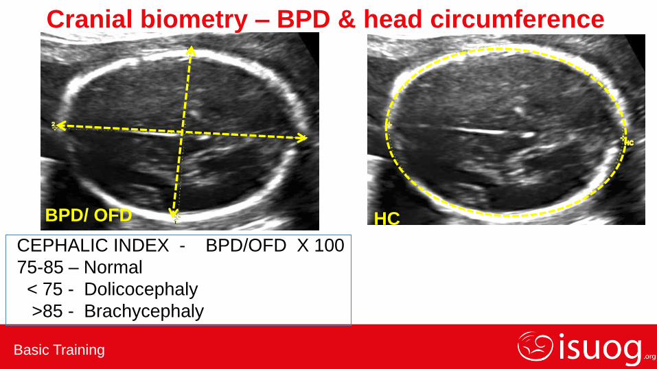
Cranial biometry – BPD & head circumference
Cerebellum NOT to be visualised
CALIPERS: Outer to inner
Use appropriate charts
Basic Training
HC BPD/ OFD
Cranial biometry – BPD & head circumference
CEPHALIC INDEX - BPD/OFD X 100
75-85 – Normal
< 75 - Dolicocephaly
>85 - Brachycephaly
Basic Training
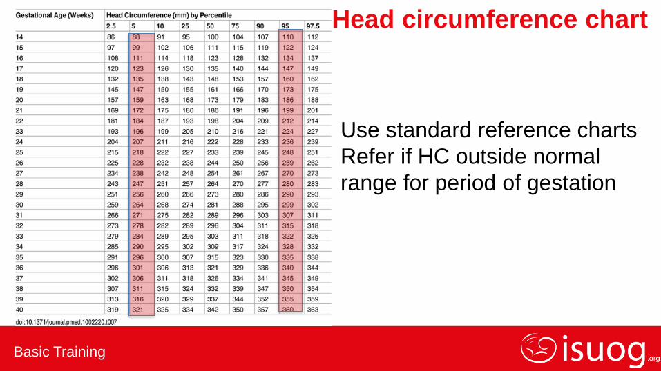
Head circumference chart
Use standard reference charts
Refer if HC outside normal
range for period of gestation
Basic Training
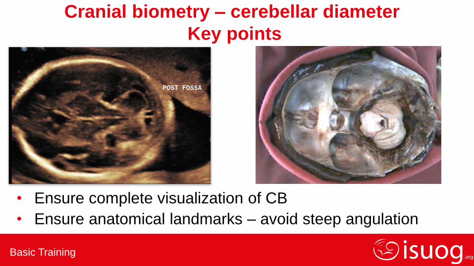
Cranial biometry – cerebellar diameter
Key points
• Ensure complete visualization of CB
• Ensure anatomical landmarks – avoid steep angulation
Basic Training
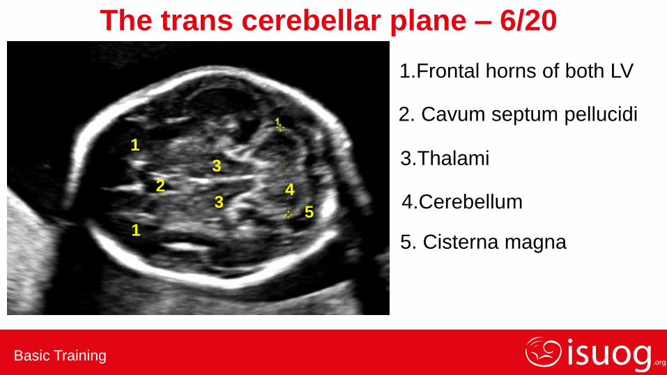
The trans cerebellar plane – 6/20
1.Frontal horns of both LV
1
1
2. Cavum septum pellucidi
2
3.Thalami
3
3
4.Cerebellum 4
5. Cisterna magna
5
Basic Training
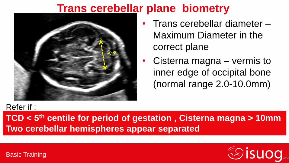
Trans cerebellar plane biometry
• Trans cerebellar diameter –
Maximum Diameter in the
correct plane
• Cisterna magna – vermis to
inner edge of occipital bone
(normal range 2.0-10.0mm)
TCD < 5th centile for period of gestation , Cisterna magna > 10mm
Two cerebellar hemispheres appear separated
Refer if :

Basic Training
Common abnormalities to be excluded in
the three planes
( 4, 5 ,6)
Basic Training
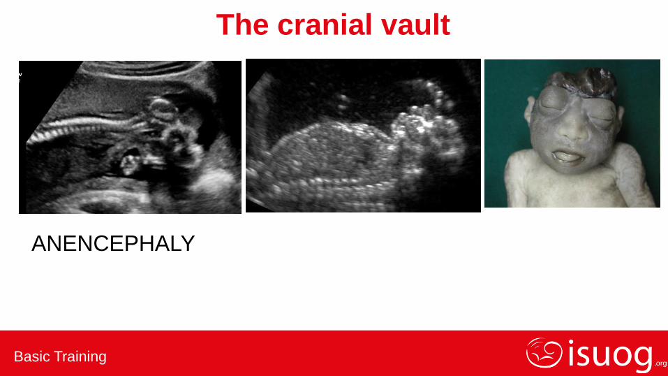
The cranial vault
ANENCEPHALY
Basic Training
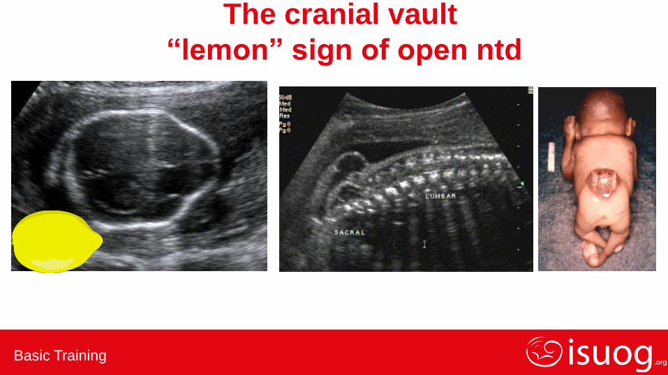
The cranial vault
“lemon” sign of open ntd
Basic Training
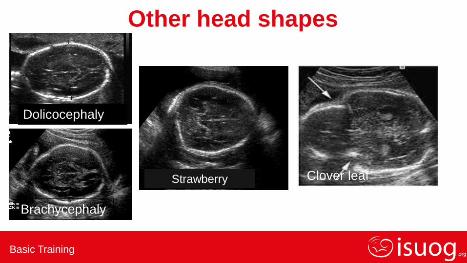
Other head shapes
Dolicocephaly
Brachycephaly
Clover leaf Strawberry
Basic Training
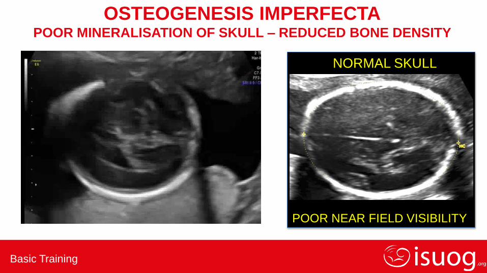
OSTEOGENESIS IMPERFECTA POOR MINERALISATION OF SKULL – REDUCED BONE DENSITY
NORMAL SKULL
POOR NEAR FIELD VISIBILITY

Basic Training
The cranial vault- skull integrity
Cephaloceles
• Can occur anywhere
• Most common in the occipital regaion
• Meningocele / meningo encephalocele
• Varying sizes
Basic Training
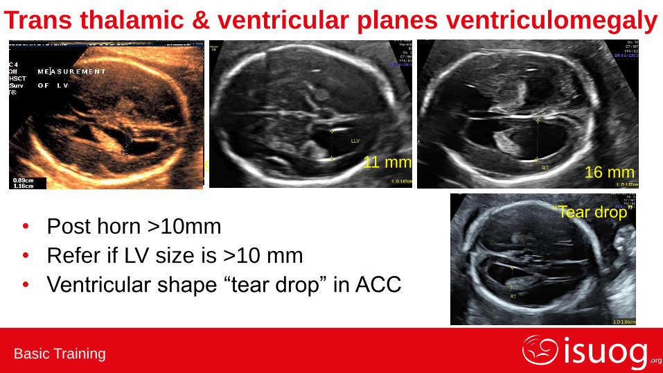
Trans thalamic & ventricular planes ventriculomegaly
• Post horn >10mm
• Refer if LV size is >10 mm
• Ventricular shape “tear drop” in ACC
0.91 11 mm 16 mm
“Tear drop”
Basic Training
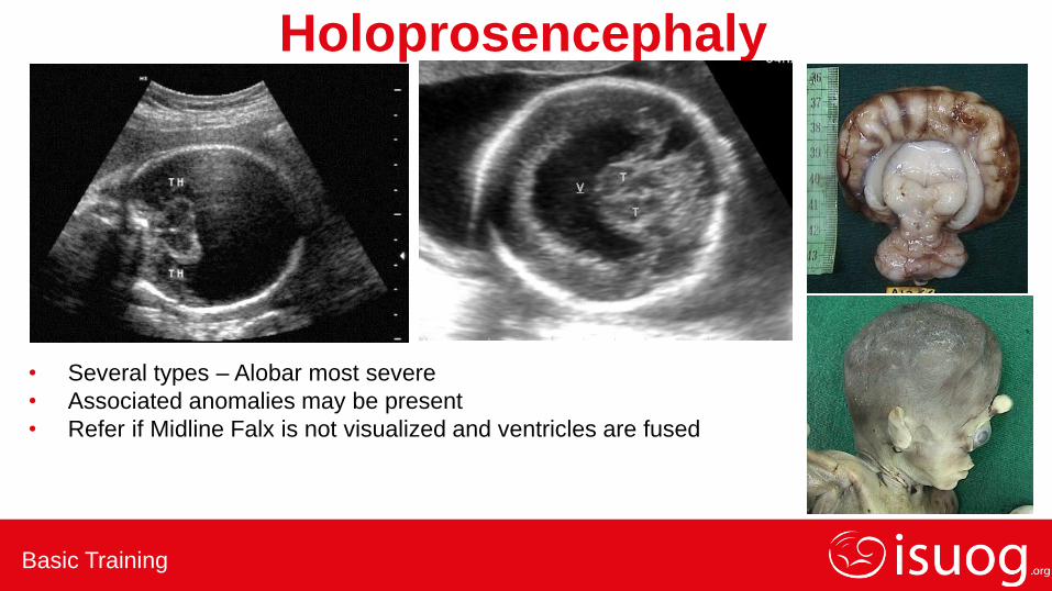
Holoprosencephaly
• Several types – Alobar most severe
• Associated anomalies may be present
• Refer if Midline Falx is not visualized and ventricles are fused
Basic Training
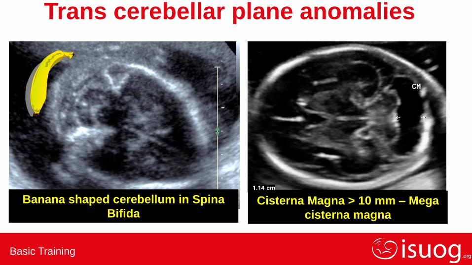
Trans cerebellar plane anomalies
Cisterna Magna > 10 mm – Mega
cisterna magna
Banana shaped cerebellum in Spina
Bifida
Basic Training
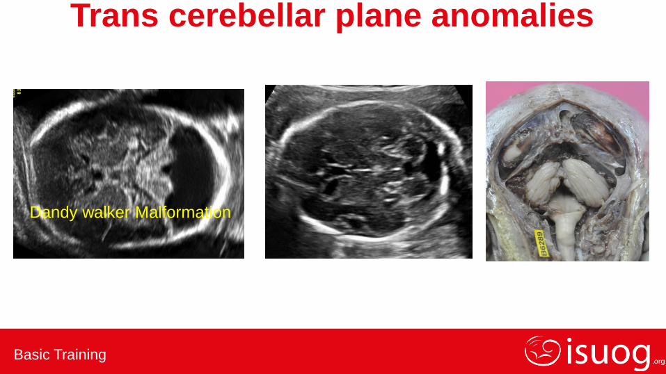
Trans cerebellar plane anomalies
Dandy walker Malformation
Basic Training
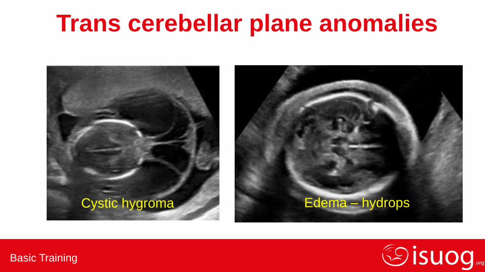
Trans cerebellar plane anomalies
Cystic hygroma Edema – hydrops
Basic Training
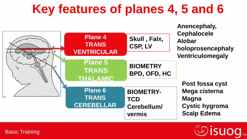
Skull , Falx,
CSP, LV
BIOMETRY
BPD, OFD, HC
BIOMETRY-
TCD
Cerebellum/
vermis
Plane 4
TRANS
VENTRICULAR
Plane 5
TRANS
THALAMIC
Plane 6
TRANS
CEREBELLAR
Anencephaly,
Cephalocele
Alobar
holoprosencephaly
Ventriculomegaly
Post fossa cyst
Mega cisterna
Magna
Cystic hygroma
Scalp Edema
Key features of planes 4, 5 and 6
Basic Training
Key Take Home Points • Head is imaged in three planes – Lateral ventricular plane ,
Trans thalamic plane & Trans cerebellar plane
• It is important to identify the specific landmarks
• Any variation in the appearances should raise suspicion of an anomaly
• Lateral ventricle > 10mm, Cisterna magna > 10mm – refer
• Head circumference < 5th centile / > 95th Centile – refer
• Trans cerebellar diameter < 5th centile or altered shape – refer

Basic Training
THANK YOU