it is a conglomeration of billions of cells forming nerves ... · sensory (afferent) neurons organs...
TRANSCRIPT
It is a conglomeration of billions of cells forming nerves that are specifically designed to provide a communication network within the human body The Nervous System
What are the three pimary functions of the nervous system? Sensory, Integrative and Motor function
The ability of the nervous system to sense changes in either the internal or external environment. Sensory Function
The ability of the nervous system to analyze and interpret the sensory information to allow for proper decision making and produce the appropriate response. Integrative Function
The neuromuscular response to the sensory information, such as causing the muscle to initally contract when stretched. Motor Function
The functional unit of the nervous system The Neuron
The three main parts of the neuron - Cell Body - Axon - Dendrites
The three main functional classifications of neurons - Sensory (Afferent) - Interneurons - Motor (Efferent)
Transmit nerve impulses from effector sites via receptors to the brain and spinal cord. Sensory Neurons
Transmit nerve impulses from one neuron to another. Interneurons
Transmit nerve impulses from the brain and spinal cord to the effector sites such as muscles or glands. Motor Neurons
Consistes of the brain and the spinal cord.Serves mainly to interpret information.

Central Nervous System
Consists of 12 cranial nerves, 31 pairs of spinal nerves and sensory receptors. Peripheral Nervous System
Two Functions of peripheral nerves 1. They provide a connection for the nervous system to activate different effector sites such as muscles. 2. Relay information from the effector sites back to the brain via sensory receptors providing a constant update on the relationship between body and environment.
Specialized structures that are designed to transform environmental stimuli (heat, light, sound, taste, motion) into sensory information that the brain and spinal cord can interpret and produce a response. Sensory Receptors
Specialized structures that are responsible for sensing distortion in tissues. Mechano Receptors
The major sensory organs of the muscle and sit parallel to the muscles fibers. They are sensetive to change in length and rate of length change. Muscle Spindles
At the point where the muscle and tendon meet and are sensitive to changes in muscular tension and rate of the tension change. Golgi Tendon Organs
Made up of three systems. The Muscular, Nervous and Skeletal systems. Kenetic Chain
A framework for our structure and movement. The Skeletal System
Form junctions that are connected by muscles and connective tissue. Bones
Sites where movement occurs as a result of muscle contraction. Joints
Made up of the skull, the rib cage, and the vertebral column. 80 Bones Axial Skeleton
The upper and lower extremeties as well as the shoulder and pelvic girdles. 126 Bones
Appendicular Skeleton
The number of joints in the body 300
Flattened or indented portions of the bone that are attachment sites for the supraspinatus and infraspinatous muscles, respectively. Bone Depressions
Projections protruding from the bone to which muscles, tendons, and ligaments can attach. Bone Processes
Joint motion with three major motion types. Roll, slide and spin. Arthrokinematics
Classifications of Joints Synovial and Nonsynovial
Comprising 80% of the joints in the body and are most associated with movement and have the greatest capacity for movement. (The Knee) Synovial Joints
No joint cavity and fibrous connective tissue; Little or no movement. Nonsynovial Joints
No axis of rotation; Moves by sliding side-to-side or back and forth. (Carpals of the hand) Gliding Joint
Formed by fitting of condyles of one bone into elliptical cavities of another; moves predominantly in one plane. (Knee) Condloid Joint
Uniaxial; moves in one plane of motion - sagittal (Elbow) Hinge
One bone fits like a saddle on another bone; moves predominantly in two planes - sagittal frontal (Joint of the thumb) Saddle
Only one axis; moves in one plane of motion - transverse (radioulnar) Pivot
Most mobile of joints; Moves in all three planes of motion (Shoulder) Ball-and-socket
Made up of collagen and is the primary conective tissue for a joint. Connect bone to bone and provide static and dynamic stability. Ligament
Muscles generate internal tension that, under the control of the nervous system, manipulates the bones of our body to produce movements. The Muscular System
Structures that attach muscles to bone and provide the anchor from which the muscle can exert force. Tendons
The wrapped outter layer of the muscle The Epimysium
Structures that atttach muscle to bone and provide the anchor from which the muscle can exert force. The Tendons
A plasma membrane that encases muscle fibers. The Sarcolemma
The functional unit of muscle that produces muscular contration and consists of repeating sections of actin and myosin. Sarcomere
The contraction of a muscle generated by neural stimulation Neural Activation
A motor neuron and all the muscle fibers it innervates Motor Unit
Chemical messengers that cross synapses to transmit electrical impulses from the nerve to the muscle. Neurotransmitter
Muscle Fiber Types Type 1 (Slow Twitch) Type 2 (Fast Twitch)
Muscle fiber with; -More Capillaries, mitochondria and myoglobin -Increased Oxygen delivery -Smaller in size -Less force produced -Slow to fatigue Type 1 (Slow Twitch)
Muscle Fiber: -Fewer capillaries, mitochondrea, and myoglobin -Decreased oxygen delivery -Larger in size -More force produced -Quick to fatigue Type II (Fast Twitch)
Four Muscle Types - Agonist - Synergist - Stabilizer - Antagonist
Muscles that are the primary movers in a joint motion. Also known as Prime Movers. Agonists
Muscles that act in direct opposition to agonists Antogonist
Muscles that assist Prime Movers (agonists) during functional movement patterns Synergists
Muscles that support or stabilize the body while the prime movers and the synergists perform the movement patterns. Stabilizers Cranial and spinal nerves that spread throughout the body and serve to relay information from bodily organs to the brain and from the brain to bodily organs Peripheral nervous system
Neurons that transmit nerve impulses from effector sites to the brain or spinal cord. Sensory (afferent) neurons
Organs sensitive to change in tension of the muscle and the rate of that change.
Golgi tendon organs
The neuromuscular response to sensory information Motor function
The combination and interrelation of the nervous, skeletal, and muscular systems. Kinetic chain
The functional unit of the nervous system Neuron
Large groups of cells that form nerves, which provide a communication network within the body. Nervous system
The ability of the nervous system to sense changes in either internal or external environments Sensory function
Neurons that transmit impulses from one neuron to another Interneurons
Consists of the brain and spinal cord and serves mainly to interpret information Central nervous system
THe ability of the nervous system to analyze and interpret sensory information to allow for proper decision making, which produces the appropriate response Integrative function
Neurons that transmit nerve impulses from the brain or spinal cord to the effector sites. Motor (efferent) neurons
Sensory receptors responsible for sensing distortion in bodily tissues Mechanoreceptors
Receptors sensitive to pressure, acceleration, and deceleration in the joint Joint receptors
Fibers sensitive to change in length of the muscles and the rate of that change Muscle spindles
________ attach muscles to bone and provide the anchor from which the muscle can exert force and control the bone and joint

Tendons
_______ is tissue consisting of long cells that contract when stimulated to produce motion Muscle
_______ are chemical messengers that transmit electrical impulses from the nerve to the muscle Neurotransmitters
The ________ is a series of muscles that the nervous system commands to move teh skeletal system Muscular system
The functional unit of muscle that produces muscular contraction (which consists of repeating sections of actin and myosin) is called the _______. Sarcomere
_______ is the contraction of a muscle generated by the communication between the nervous system and muscular system. Neural activation
The movable places where two or more bones meet Joints
Portion of the skeletal system that includes the upper and lower extremities Appendicular skeleton
Projection protruding from the bone where muscles, tendons, and ligaments are attach. Process
The body's frame, which is comprised of bones and joints Skeletal system
Portion of the skeletal system that consists of the skull, rib cage, and vertebral column Axial skeleton
The movements of the joints Arthrokinematics
Connective tissue that connects bone to bone Ligament

Joints that are held together by a joint capsule and ligaments and are most associated with movement in the body Synovial joints
Hard connective tissues taht connect to create a skeletal framework. Bones
Flattened or indented portion of bone, which can be a muscle attachment site Depression
Joints that do not have a joint cavity, connective tissue, or cartilage Nonsynovial joints
Lower in capillaries, mitochondria, and myoglobin____ Type II Is this a characteristic of Type I or Type II Muscle Fiber?
Smaller in size____ Type I Is this a characteristic of Type I or Type II Muscle Fiber?
Produce less force___ Type I Is this a characteristic of Type I or Type II Muscle Fiber?
Slow in fatigue___ Type I Is this a characteristic of Type I or Type II Muscle Fiber?
Long-term contractions___ Type I Is this a characteristic of Type I or Type II Muscle Fiber?
Produce more force___ Type II Is this a characteristic of Type I or Type II Muscle Fiber?
Quick to fatigue___ Type II Is this a characteristic of Type I or Type II Muscle Fiber?
Short-term contractions (force and power)___ Type II

Is this a characteristic of Type I or Type II Muscle Fiber? Three primary functions of the nervous system sensory, integrative, and motor funtions
Sensory Functions ability of the nervous system to sense changes in either internal or external environment
Integrative function ability of the nervous system to analyze and interpret the sensory information to allow for proper decision making, which produced the appropriate response
Motor Function the neuromuscular response to the sensory information
Neuron Functional until of the nervous system
Neuron are composed of three main part the .... cell body, axon and dentrites
list the three main classifications of neurons Sensory (afferent) neurons Interneurons Motor (efferent) neurons
Sensory Neurons transmit nerve impulses from effector sites to the brain or spinal cord
Interneurons Transmit nerve impulses from one neuron to another
Motor Neurons Transmit nerve impulses from the brain and spinal cord to effector sites
Central Nervous system Composed of the brain an spinal cord serves mainly to interpret information
Peripheral nervous system

Cranial and spinal nerves that spread throughout the body provides a connections for the nervous system to activate different effector sites and to relay information to the brain from the sensory receptors contain 12 cranial nerves, 31 pairs of spinal nerves
Four major categories of sensory receptors Mechanoreceptor- touch and pressure chemoreceptors- taste and smell photoreceptors- light
mechanorecptors receptor responsible for sensing distortions in body tissues located in muscles, tendons, ligaments, and joint capsules
Muscle spindles Receptors sensitive to change in length and rate of change
Golgi Tendon Organ Receptors sensitive to change in tension of the muscle and the rate of that change. They are at the point where muscle and tendon meet. When excited they will cause the muscle to relax
Joint Receptors Located in and around the joint capsule. They respond to pressure, acceleration, and deceleration in the joint
Skeletal system the bodys framework, composed of bones and joint
Bones provide a resting ground for muscles and protection of vital organs
Joint the movable junction where two or more bones meet
Axial Skeleton Skull, rib cage, and vertebral column 80 bones
Appedicular Skeleton upper and lower extremities as well as the shoulder and pelvic girdle 126 bones
There are ____bones in the body. ____ contribute to voluntary movement. ther are more that ____ joints roughly 206 177 voluntary movement more than 300 joints
The two main functions of bone are to act as levers and provide support
Depressions Flattened or indented portion of bone which can be a muscle attachment site. ex. supraspinus fossa, intertubercular sulcus pg 22
Process Projections protruding from the bone where tendons, muscles, and ligaments can attach
Common processes Condyle- located on the inner and outer portion of the femur and the top of the tibia Epicondyle- located on the inner and outer portion on the humerus to help form the elbow joint tubercle- located at the top of the humerus at the glenohumeral joint trochanter- located at the top of the femur and are attachment site for hip musculature
arthrokinematics joint motion. Three major motion types are roll, slide, and spin
Example of joint roll the femoral condyles moving over the tibial condyles during a squat
example of joint slide tibial condyles moving across femoral condyles during and knee extentions
example of joint spin radio-ulnar rotation

synovial joints joints that are held together by a joint capsule and ligaments and are move associated with movement in the body
types of synovial joints in the body Gliding Condyloid Hinge Saddle Pivot ball and Socket
Gliding joint moves back and forth or side to side. example: carpals in the hand
Condyloid joint one bone fits into the elliptical cavity of another bone for form the joint. movement in one plane with minimal movement in others.ex: in wrist between radius and capals and in the knee joint
Hinge joint uniaxial joint allowing movement in only one plane of motion ex. elbow, interphalangeal, ankle
Saddle Joint One bone looks like a saddle with the articulating bone straddling it ex: only in the thumb
piviot joint allow movement in predominantly one plane of motion (rotation, pronation and supination in transverse)
ball and socket most mobile, movement in all three planes hip, shoulder
Nonsynovial joints joints that do not have a joint cavity, connective tissue, or cartilage. little to no movement
ex: skull, distal joint of tibia and fibia, and symphysis pubis
muscle is wrapped by a connective tissue call_______and an inner layer immediately surrounding the tissue call fascia Epimysium
The bundle of muscle fibers with in a muscle is called a _____ . Each of these bundles is wrapped in a connective tissue called _________. fascicle perimysium
Individual muscle fibers are wrapped with a connective tissue called ________. endomysium
Tendons Connective tissues that attache muscle to bone and provide an anchor for the muscles to produce force.
Muscle fibers are encased by a plasma membrane known as a ______ sarcolema
Sarcomere The functional unit of muscle that produces muscular contraction and consists of repeating section of actin and myosin. It lies in the space between to z lines. Sarcomeres compose the myofibril
Tropomyosin located on the actin filament and block myosin binding sites located on the actin filament, keeping myosin from attaching to actin while the muscle is relaxed.
neural activation the contraction of a muscle generated by neural stimulation
Motor Unit A motor neuron and the muscle fibers it innervates
neuromuscular junction the point where the neuron meet and individual muscle fiber. the junction is actually an small gap between the nerve and muscle fiber often called a synapse
Neurotransmitter
Chemical messengers that cross synapses to transmit electrical impulses from the nerve to the muscle
Acetycholine ACh- the neurotransmitter used by the neuromuscular system. One attached to the receptor site ACh stimulates the muscle fiber to go through a series of step that produce muscle contractions
steps in sliding filament theory 1. A sercomere shortens as a result the of the Z lines moving closer together. 2. The Z line converge as the result of myosin heads attaching to the action filament and asynchronously pulling (power strokes) the actin filament across the myosin, resulting in shortening of muscle fiber
Type 1 muscle fibers slow twitch higher number of capillaries, mitochondria and myoglobin which allows for improved delivery of oxygen. smaller in size slower to produce maximal tension more resistant to fatigue important to stabilization "red fibers"
Type Two muscle fibers fast twitch contain fewer capillaries, mitochondria, and myoglobin. larger in size quick to produce maximal tension fatigue more quickly "white fibers" Type IIa have higher oxidative capacity and fatigue more slowly than type IIb Type IIb have a low oxidative capacity and fatigue quickly
Agonist in: Chest Press Overhead Press Row Squat Hip extension prime mover: Pectoralis major Deltoid Latissimus dorsi
Gluteus maximus, quadraceps gluteus maximus
Synergist in: Chest Press Overhead Press Row Squat Hip extension assist prime mover: Anterior deltoid, triceps Triceps Posterior deltoid, biceps hamstrings hamstring and erector spinae
Stabilizer in: Chest Press Overhead Press Row Squat Hip extension Stabilize while primer mover and synergist work; rotator cuff rotator cuff rotator cuff transverse abdominis transverse abdominis
Antagonist in: Chest Press Overhead Press Row Squat Hip extension Oppose prime mover: Posterior deltoid Latissimus dorsi Pectoralis major Psoas psoas
When the neuromuscular system responds to stimulus it is called? Motor Function

The combination and interrelation of nervous, skeletal and muscular systems is known as ... THE KINETIC CHAIN p. 16 (NASM)
Match the following terms of the nervous system with their respective functions: 1. Sensory function 2. Integrative function 3. Motor function A. Sense changes B. Respond to changes C. Analyze & interpret 1. SENSORY FUNCTION - sense changes in either the internal or external environment 2. INTEGRATIVE FUNCTION - analyze and interpret sensory information 3. MOTOR FUNCTION - neuromuscular response to sensory information p. 16 (NASM)
The Central Nervous System (CNS) consists of ... THE BRAIN & SPINAL CORD p. 17 (NASM)
The Peripheral Nervous System consists of ... CRANIAL/SPINAL NERVES AND SENSORY RECEPTORS p. 17 (NASM)
It becomes important to train the nervous system efficiently to ensure that _____________, which enhances performance and decreases the risk of injuries. PROPER MOVEMENT PATTERNS ARE BEING DEVELOPED p. 16 (NASM)
The function of the Peripheral Nervous System is to ... - PROVIDE A CONNECTION FOR THE NERVOUS SYSTEM TO ACTIVATE DIFFERENT EFFECTOR SITES (e.g., muscles) - RELAY INFORMATION FROM EFFECTOR SITES BACK TO THE BRAIN VIA SENSORY RECEPTORS

pp. 17, 19 (NASM)
What are the 3 main functions of the nervous system? - SENSE CHANGES IN EITHER INTERNAL OR EXTERNAL ENVIRONMENT (SENSORY) - ANALYZE & INTERPRET SENSORY INFORMATION (INTEGRATIVE) - PROVIDE THE NEUROMUSCULAR RESPONSE TO THE SENSORY INFORMATION (MOTOR) p. 16 (NASM)
Transmit nerve impulses from effector sites (e.g., hand) to the brain/spinal cord. SENSORY (AFFERENT) NEURONS p. 17 (NASM)
Transmit nerve impulses from one neuron to another. INTERNEURONS p. 17 (NASM)
Transmit nerve impulses from the brain/spinal cord to various effector sites (e.g., muscles, glands). MOTOR (EFFECTOR) NEURONS p. 17 (NASM)
Sensory receptors are specialized structures located throughout the body that are designed to transform ______________________ into ______________________ that the brain & spinal cord can then interpret to produce __________. - ENVIRONMENTAL STIMULI (heat, light, sound, taste, motion) - SENSORY INFORMATION - A RESPONSE There are 4 types of sensory receptors: - mechano-receptors - noci-receptors - chemo-receptors - photo-receptors p. 19 (NASM)
Mechano-receptors are responsible for sensing ________ in body tissues. DISTORTION p. 19 (NASM)
What are the three types of mechano-receptors, and to what are they sensitive? - MUSCLE SPINDLES: sense changes in muscle length and rate of length change - GOLGI TENDON ORGANS (GTO): sense changes in tension and rate of tension change - JOINT RECEPTORS: sense pressure and acceleration/deceleration of the joint pp. 19-20 (NASM)
LOCATION: Located in or around the joint capsule JOINT RECEPTORS p. 20 (NASM)
LOCATION: Located where the muscle and tendon meet (musculotendinous junction)
GOLGI TENDON ORGAN (GTO) p. 19 (NASM)
LOCATION: Are parallel to a muscle's fibers
MUSCLE SPINDLES p. 19 (NASM)
If one system of the _____________ is not working properly, it will affect the other systems and ultimately affect _____________. - KINETIC CHAIN - MOVEMENT p. 16 (NASM)
Name the parts of a neuron.
- CELL BODY - AXON - DENDRITES p. 17 (NASM)
The framework for both structure and __________ is called: - MOVEMENT - SKELETAL SYSTEM p. 21 (NASM)
Growth, maturation and functionality of the skeletal system are greatly affected by ... - POSTURE - ACTIVITY - NUTRITION p. 21 (NASM)
The _________ junction where 2 or more bones meet is called a ... - MOVABLE - JOINT Movement occurs here as a result of muscle contraction. p. 21 (NASM)
What causes movement at a joint?
MUSCLE CONTRACTION p. 21 (NASM)
This consists of some 80 bones in the skull, rib cage and vertebral column. AXIAL SKELETON p. 22 (NASM)
This consists of some 126 bones in the upper/lower extremities as well as the shoulder and pelvic girdles. APPENDICULAR SKELETON p. 22 (NASM)

What does this image depict? THE APPENDICULAR SKELETON p. 22 (NASM)
How many joints are there in the body? > 300 There are closer to 230 to 250 movable or semi-movable joints. This does not include non-movable joints in the skull. p. 22 (NASM)
Why do bones have surface markings? TO PROVIDE INCREASED STABILITY AND ATTACHMENT SITES FOR MUSCLES There are 2 types of surface marking: depressions and processes. p. 22 (NASM)
What is the purpose of bone depressions? - PROVIDE SITES FOR ATTACHMENT OF MUSCLES (fossa) - ALLOW SOFT TISSUE TO PASS THROUGH (sulcus) Examples: - Fossa - supraspinous and infraspinous fossa on the scapula (shoulder) - to attach the supraspinatus and infraspinatus muscles respectively p. 22 (NASM)
What type of process is located above a condyle, such as in the humerus to form the elbow joint or in the femur to form the knee joint?
EPICONDYLE p. 23 (NASM)
Condyles are _________ projections at a joint that usually articulate with a another bone, such as in ... (provide example)

- LARGE ROUNDED - THE BOTTOM (INSIDE/OUTSIDE) OF THE FEMUR & TOP OF THE TIBIA TO FORM THE KNEE JOINT p. 23 (NASM)
_________ are large rounded projections at a joint that usually articulate with another bones, such as in ... (provide example)
- CONDYLES - THE BOTTOM (INSIDE/OUTSIDE) OF THE FEMUR & TOP OF THE TIBIA TO FORM THE KNEE JOINT p. 23 (NASM)
These joint types comprise about 80% of all joints in the body, are most associated with movement, and have the greatest capacity for motion.
SYNOVIAL JOINTS p. 25 (NASM)
What are the 3 major motion types? - ROLL - SLIDE - SPIN Arthrokinematics = joint motion p. 24 (NASM)
What is arthrokinematics? - JOINT MOTION p. 24 (NASM)

During a squat, what major type of joint motion is exhibited as the femoral condyles move over the tibial condyles? ROLL MOVEMENT p. 24 (NASM)
During a knee extension, what major type of joint motion is exhibited as the tibial condyles move across the femoral condyles? SLIDE MOVEMENT p. 24 (NASM)
During pronation and supination of the forearm, what major type of joint motion is exhibited as the head of the radius moves on the end of the humerus? SPIN MOVEMENT p. 24 (NASM)
What type of joint is exhibited by the following? - Simplest movement of all joints - back & forth/side-to-side movement - Example: carpals of the hand
GLIDING (plane) JOINT
Also see "The Anatomy Lesson" at http://home.comcast.net/~wnor/ p. 25 (NASM)
What type of joint is exhibited by the following? - Fitting of the condyle of one bone into the elliptical cavity of another to form the joint - Movement occurs mostly in 1 plane: flexion/extension in the sagittal plane - Movement occurs minimally in the transverse plane (rotation) and frontal plane (adduction/abduction) - Example: in the wrist between the radius and carpals and in the knee joint CONDYLOID (ellipsoidal) JOINT Also see "The Anatomy Lesson" at http://home.comcast.net/~wnor/ p. 25 (NASM)
What type of joint is exhibited by the following? - uni-axial - movement mostly in the sagittal plane - Examples: elbow, ankle
HINGE JOINT Also see "The Anatomy Lesson" at http://home.comcast.net/~wnor/ p. 26 (NASM)
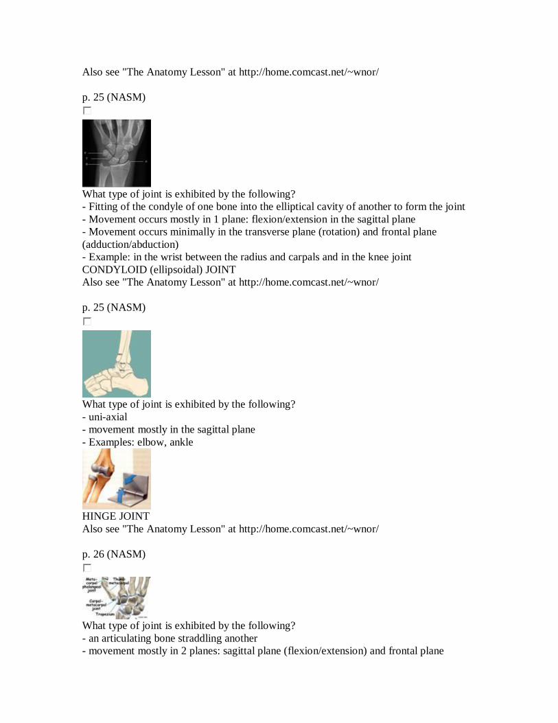
What type of joint is exhibited by the following? - an articulating bone straddling another - movement mostly in 2 planes: sagittal plane (flexion/extension) and frontal plane
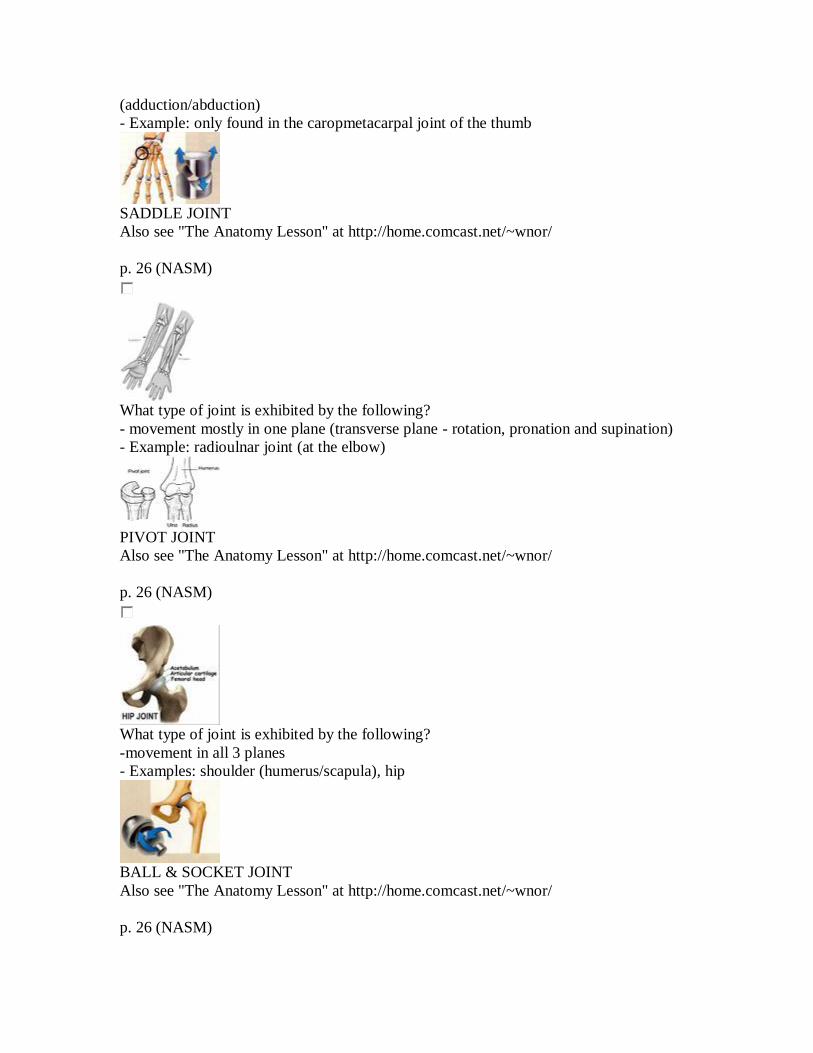
(adduction/abduction) - Example: only found in the caropmetacarpal joint of the thumb
SADDLE JOINT Also see "The Anatomy Lesson" at http://home.comcast.net/~wnor/ p. 26 (NASM)
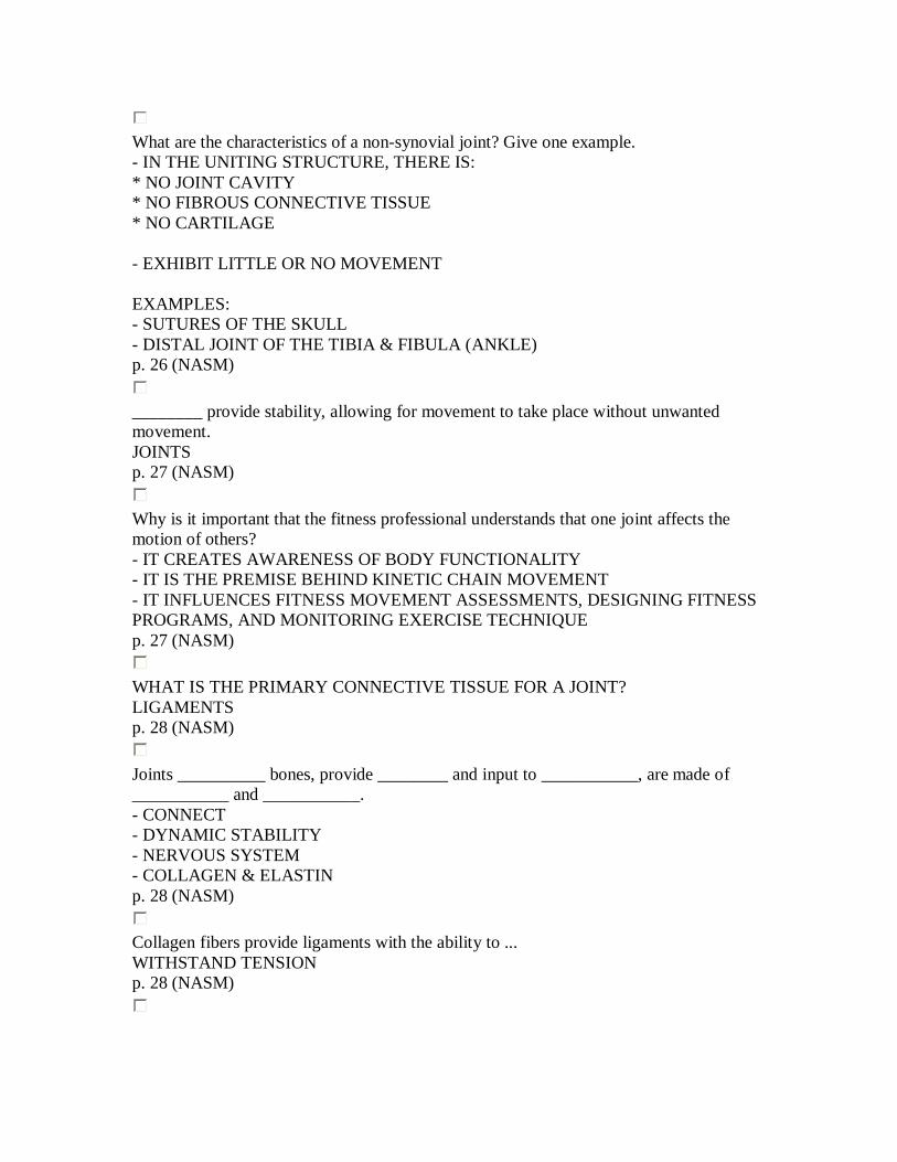
What type of joint is exhibited by the following? - movement mostly in one plane (transverse plane - rotation, pronation and supination) - Example: radioulnar joint (at the elbow)
PIVOT JOINT Also see "The Anatomy Lesson" at http://home.comcast.net/~wnor/ p. 26 (NASM)
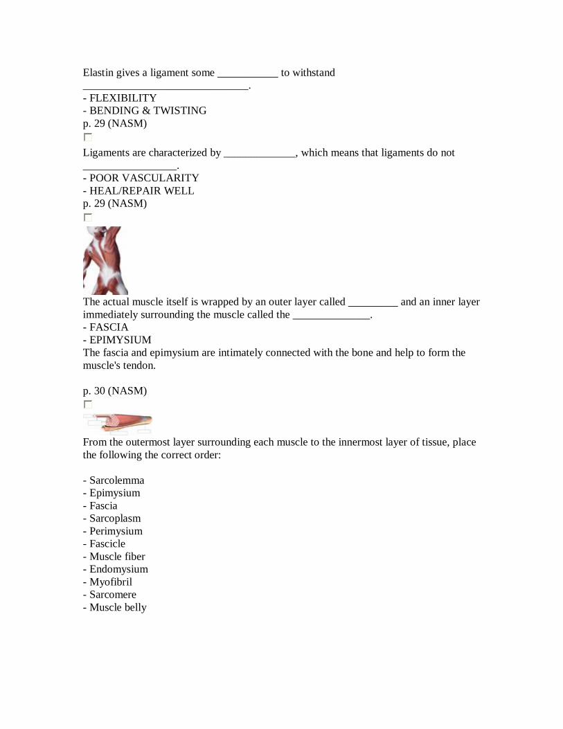
What type of joint is exhibited by the following? -movement in all 3 planes - Examples: shoulder (humerus/scapula), hip
BALL & SOCKET JOINT Also see "The Anatomy Lesson" at http://home.comcast.net/~wnor/ p. 26 (NASM)
What are the characteristics of a non-synovial joint? Give one example. - IN THE UNITING STRUCTURE, THERE IS: * NO JOINT CAVITY * NO FIBROUS CONNECTIVE TISSUE * NO CARTILAGE - EXHIBIT LITTLE OR NO MOVEMENT EXAMPLES: - SUTURES OF THE SKULL - DISTAL JOINT OF THE TIBIA & FIBULA (ANKLE) p. 26 (NASM)
________ provide stability, allowing for movement to take place without unwanted movement. JOINTS p. 27 (NASM)
Why is it important that the fitness professional understands that one joint affects the motion of others? - IT CREATES AWARENESS OF BODY FUNCTIONALITY - IT IS THE PREMISE BEHIND KINETIC CHAIN MOVEMENT - IT INFLUENCES FITNESS MOVEMENT ASSESSMENTS, DESIGNING FITNESS PROGRAMS, AND MONITORING EXERCISE TECHNIQUE p. 27 (NASM)
WHAT IS THE PRIMARY CONNECTIVE TISSUE FOR A JOINT? LIGAMENTS p. 28 (NASM)
Joints __________ bones, provide ________ and input to ___________, are made of ___________ and ___________. - CONNECT - DYNAMIC STABILITY - NERVOUS SYSTEM - COLLAGEN & ELASTIN p. 28 (NASM)
Collagen fibers provide ligaments with the ability to ... WITHSTAND TENSION p. 28 (NASM)
Elastin gives a ligament some ___________ to withstand ______________________________. - FLEXIBILITY - BENDING & TWISTING p. 29 (NASM)
Ligaments are characterized by _____________, which means that ligaments do not _________________. - POOR VASCULARITY - HEAL/REPAIR WELL p. 29 (NASM)
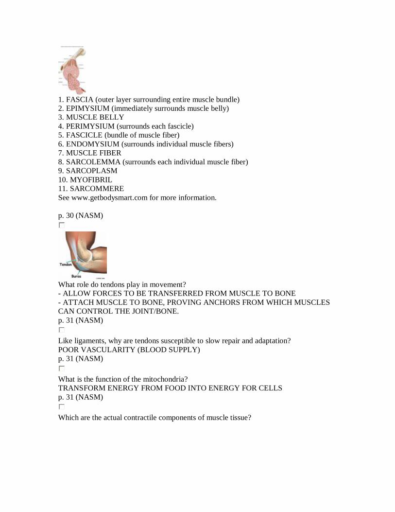
The actual muscle itself is wrapped by an outer layer called _________ and an inner layer immediately surrounding the muscle called the ______________. - FASCIA - EPIMYSIUM The fascia and epimysium are intimately connected with the bone and help to form the muscle's tendon. p. 30 (NASM)
From the outermost layer surrounding each muscle to the innermost layer of tissue, place the following the correct order: - Sarcolemma - Epimysium - Fascia - Sarcoplasm - Perimysium - Fascicle - Muscle fiber - Endomysium - Myofibril - Sarcomere - Muscle belly
1. FASCIA (outer layer surrounding entire muscle bundle) 2. EPIMYSIUM (immediately surrounds muscle belly) 3. MUSCLE BELLY 4. PERIMYSIUM (surrounds each fascicle) 5. FASCICLE (bundle of muscle fiber) 6. ENDOMYSIUM (surrounds individual muscle fibers) 7. MUSCLE FIBER 8. SARCOLEMMA (surrounds each individual muscle fiber) 9. SARCOPLASM 10. MYOFIBRIL 11. SARCOMMERE See www.getbodysmart.com for more information. p. 30 (NASM)
What role do tendons play in movement? - ALLOW FORCES TO BE TRANSFERRED FROM MUSCLE TO BONE - ATTACH MUSCLE TO BONE, PROVING ANCHORS FROM WHICH MUSCLES CAN CONTROL THE JOINT/BONE. p. 31 (NASM)
Like ligaments, why are tendons susceptible to slow repair and adaptation? POOR VASCULARITY (BLOOD SUPPLY) p. 31 (NASM)
What is the function of the mitochondria? TRANSFORM ENERGY FROM FOOD INTO ENERGY FOR CELLS p. 31 (NASM)
Which are the actual contractile components of muscle tissue?
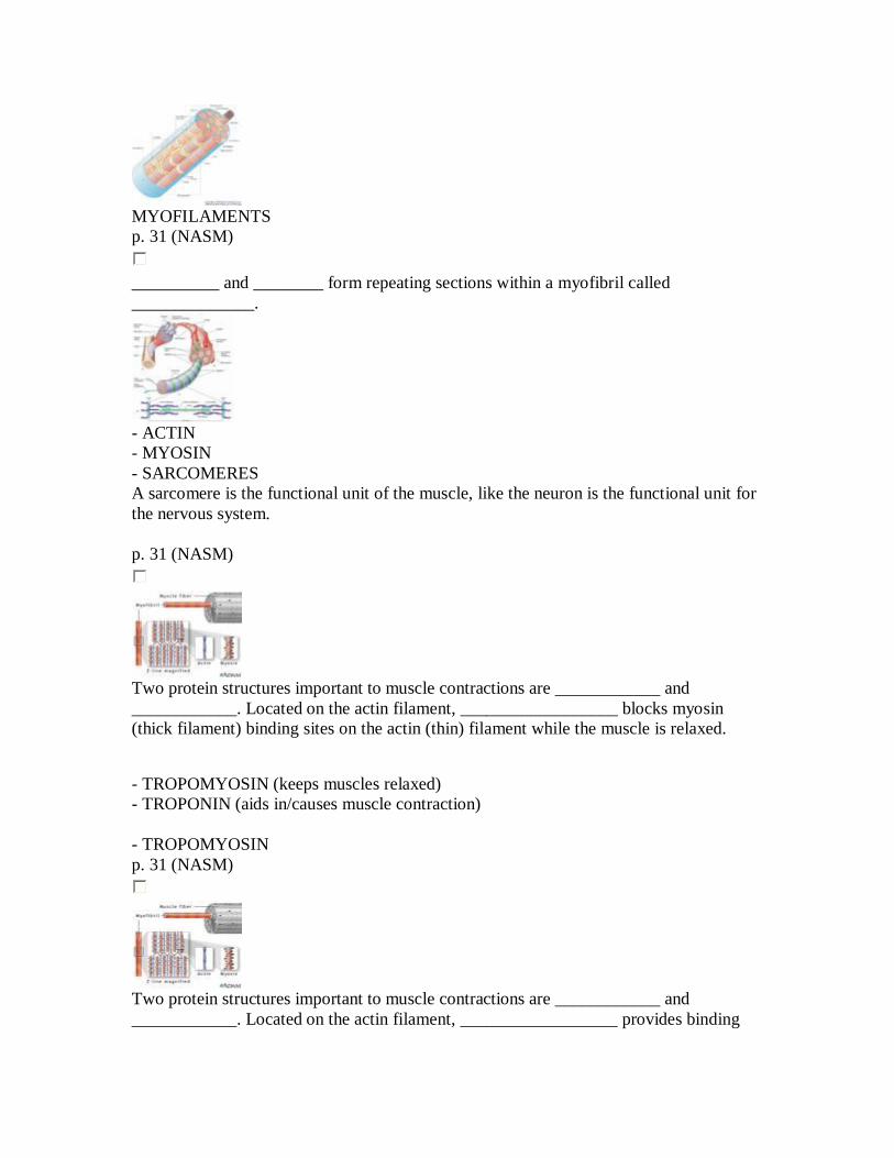
MYOFILAMENTS p. 31 (NASM)
__________ and ________ form repeating sections within a myofibril called ______________.
- ACTIN - MYOSIN - SARCOMERES A sarcomere is the functional unit of the muscle, like the neuron is the functional unit for the nervous system. p. 31 (NASM)
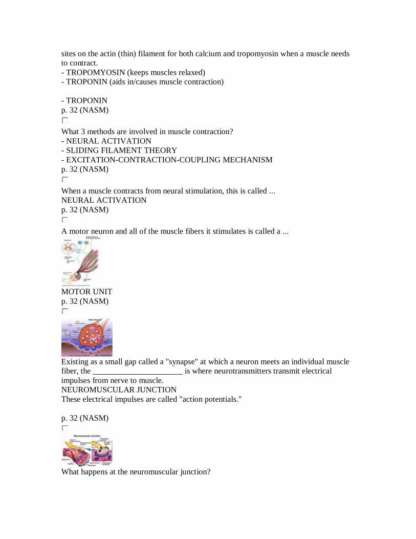
Two protein structures important to muscle contractions are ____________ and ____________. Located on the actin filament, __________________ blocks myosin (thick filament) binding sites on the actin (thin) filament while the muscle is relaxed.
- TROPOMYOSIN (keeps muscles relaxed) - TROPONIN (aids in/causes muscle contraction) - TROPOMYOSIN p. 31 (NASM)
Two protein structures important to muscle contractions are ____________ and ____________. Located on the actin filament, __________________ provides binding
sites on the actin (thin) filament for both calcium and tropomyosin when a muscle needs to contract. - TROPOMYOSIN (keeps muscles relaxed) - TROPONIN (aids in/causes muscle contraction) - TROPONIN p. 32 (NASM)
What 3 methods are involved in muscle contraction? - NEURAL ACTIVATION - SLIDING FILAMENT THEORY - EXCITATION-CONTRACTION-COUPLING MECHANISM p. 32 (NASM)
When a muscle contracts from neural stimulation, this is called ... NEURAL ACTIVATION p. 32 (NASM)
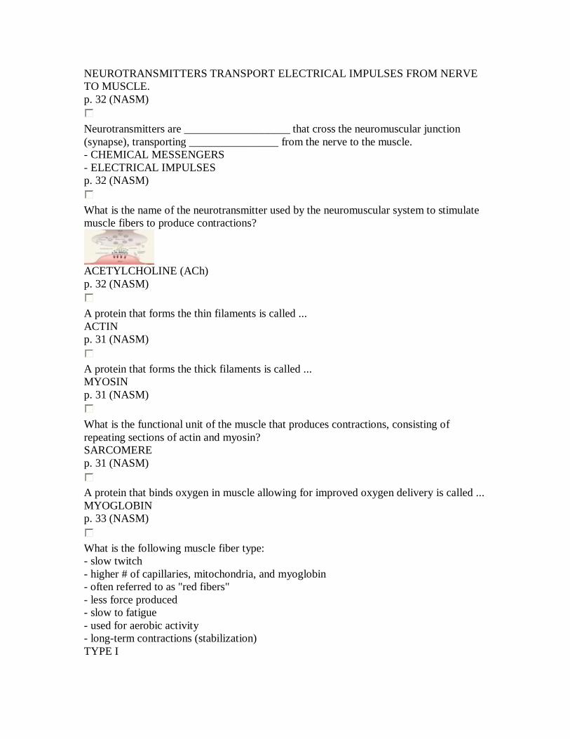
A motor neuron and all of the muscle fibers it stimulates is called a ...
MOTOR UNIT p. 32 (NASM)
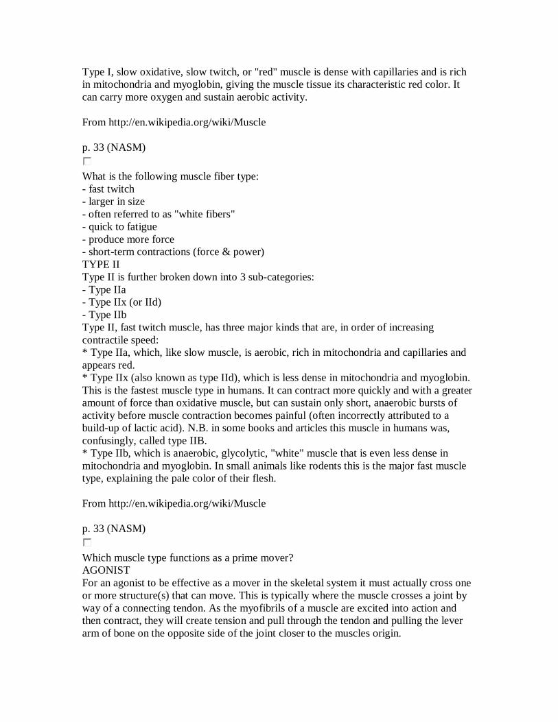
Existing as a small gap called a "synapse" at which a neuron meets an individual muscle fiber, the ______________________ is where neurotransmitters transmit electrical impulses from nerve to muscle. NEUROMUSCULAR JUNCTION These electrical impulses are called "action potentials." p. 32 (NASM)
What happens at the neuromuscular junction?
NEUROTRANSMITTERS TRANSPORT ELECTRICAL IMPULSES FROM NERVE TO MUSCLE. p. 32 (NASM)
Neurotransmitters are ___________________ that cross the neuromuscular junction (synapse), transporting ________________ from the nerve to the muscle. - CHEMICAL MESSENGERS - ELECTRICAL IMPULSES p. 32 (NASM)
What is the name of the neurotransmitter used by the neuromuscular system to stimulate muscle fibers to produce contractions?
ACETYLCHOLINE (ACh) p. 32 (NASM)
A protein that forms the thin filaments is called ... ACTIN p. 31 (NASM)
A protein that forms the thick filaments is called ... MYOSIN p. 31 (NASM)
What is the functional unit of the muscle that produces contractions, consisting of repeating sections of actin and myosin? SARCOMERE p. 31 (NASM)
A protein that binds oxygen in muscle allowing for improved oxygen delivery is called ... MYOGLOBIN p. 33 (NASM)
What is the following muscle fiber type: - slow twitch - higher # of capillaries, mitochondria, and myoglobin - often referred to as "red fibers" - less force produced - slow to fatigue - used for aerobic activity - long-term contractions (stabilization) TYPE I
Type I, slow oxidative, slow twitch, or "red" muscle is dense with capillaries and is rich in mitochondria and myoglobin, giving the muscle tissue its characteristic red color. It can carry more oxygen and sustain aerobic activity. From http://en.wikipedia.org/wiki/Muscle p. 33 (NASM)
What is the following muscle fiber type: - fast twitch - larger in size - often referred to as "white fibers" - quick to fatigue - produce more force - short-term contractions (force & power) TYPE II Type II is further broken down into 3 sub-categories: - Type IIa - Type IIx (or IId) - Type IIb Type II, fast twitch muscle, has three major kinds that are, in order of increasing contractile speed: * Type IIa, which, like slow muscle, is aerobic, rich in mitochondria and capillaries and appears red. * Type IIx (also known as type IId), which is less dense in mitochondria and myoglobin. This is the fastest muscle type in humans. It can contract more quickly and with a greater amount of force than oxidative muscle, but can sustain only short, anaerobic bursts of activity before muscle contraction becomes painful (often incorrectly attributed to a build-up of lactic acid). N.B. in some books and articles this muscle in humans was, confusingly, called type IIB. * Type IIb, which is anaerobic, glycolytic, "white" muscle that is even less dense in mitochondria and myoglobin. In small animals like rodents this is the major fast muscle type, explaining the pale color of their flesh. From http://en.wikipedia.org/wiki/Muscle p. 33 (NASM)
Which muscle type functions as a prime mover? AGONIST For an agonist to be effective as a mover in the skeletal system it must actually cross one or more structure(s) that can move. This is typically where the muscle crosses a joint by way of a connecting tendon. As the myofibrils of a muscle are excited into action and then contract, they will create tension and pull through the tendon and pulling the lever arm of bone on the opposite side of the joint closer to the muscles origin.
from http://en.wikipedia.org/wiki/Agonist_(muscle) p. 35 (NASM)
An agonist acts as a ... PRIME MOVER For an agonist to be effective as a mover in the skeletal system it must actually cross one or more structure(s) that can move. This is typically where the muscle crosses a joint by way of a connecting tendon. As the myofibrils of a muscle are excited into action and then contract, they will create tension and pull through the tendon and pulling the lever arm of bone on the opposite side of the joint closer to the muscles origin. from http://en.wikipedia.org/wiki/Agonist_(muscle) p. 35 (NASM)
A synergist acts as to ... ASSIST A PRIMER MOVER Synergist is a kind of muscle which performs, or assist in performing, the same set of joint motion as the agonists. Synergists are muscles that act on movable joints . Synergists are sometimes referred to as "neutralizers" because they help cancel out, or neutralize, extra motion from the agonists to make sure that the force generated works within the desired plane of motion. from http://en.wikipedia.org/wiki/Synergist p. 35 (NASM)
Which muscle type functions to assist a prime mover? SYNERGIST Synergist is a kind of muscle which performs, or assist in performing, the same set of joint motion as the agonists. Synergists are muscles that act on movable joints . Synergists are sometimes referred to as "neutralizers" because they help cancel out, or neutralize, extra motion from the agonists to make sure that the force generated works within the desired plane of motion. from http://en.wikipedia.org/wiki/Synergist p. 35 (NASM)
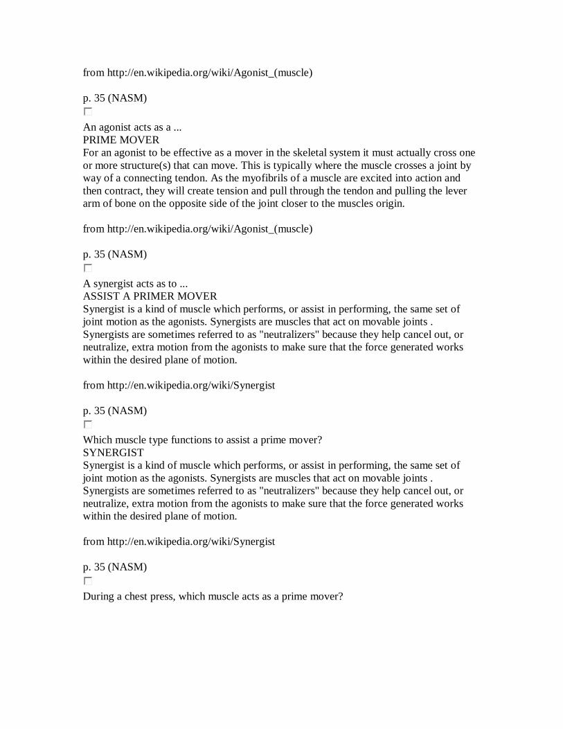
During a chest press, which muscle acts as a prime mover?
PECTORALIS MAJOR p. 35 (NASM)
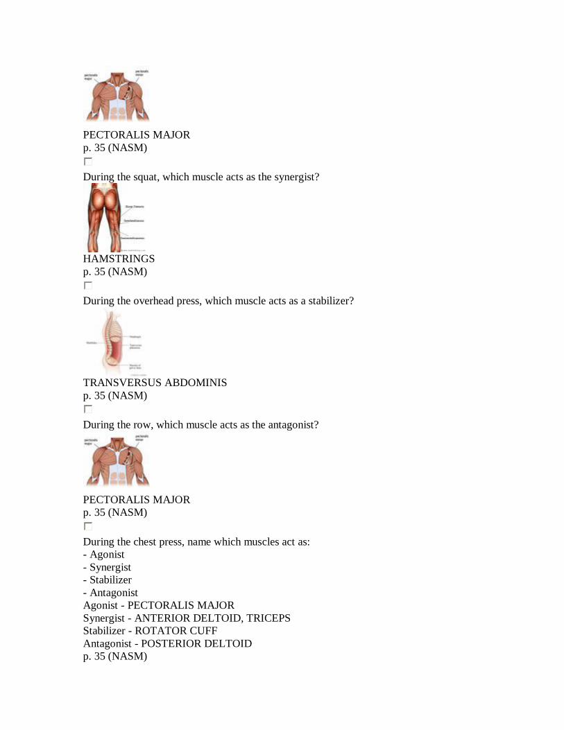
During the squat, which muscle acts as the synergist?
HAMSTRINGS p. 35 (NASM)
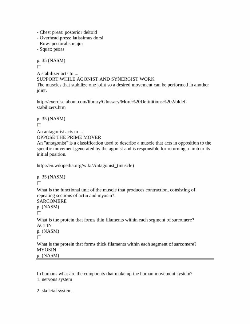
During the overhead press, which muscle acts as a stabilizer?
TRANSVERSUS ABDOMINIS p. 35 (NASM)
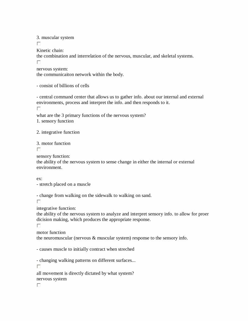
During the row, which muscle acts as the antagonist?
PECTORALIS MAJOR p. 35 (NASM)
During the chest press, name which muscles act as: - Agonist - Synergist - Stabilizer - Antagonist Agonist - PECTORALIS MAJOR Synergist - ANTERIOR DELTOID, TRICEPS Stabilizer - ROTATOR CUFF Antagonist - POSTERIOR DELTOID p. 35 (NASM)

During the overhead press, name which muscles act as: - Agonist - Synergist - Stabilizer - Antagonist Agonist - DELTOID Synergist - TRICEPS Stabilizer - ROTATOR CUFF Antagonist - LATISSIMUS DORSI p. 35 (NASM)
During the row, name which muscles act as: - Agonist - Synergist - Stabilizer - Antagonist Agonist - LATISSIMUS DORSI Synergist - POSTERIOR DELTOID, BICEPS Stabilizer - ROTATOR CUFF Antagonist - PECTORALIS MAJOR p. 35 (NASM)
During the squat, name which muscles act as: - Agonist - Synergist - Stabilizer - Antagonist Agonist - GLUTEUS MAXIMUS, QUADRICEPS Synergist - HAMSTRINGS Stabilizer - TRANSVERSUS ABDOMINIS Antagonist - PSOAS p. 35 (NASM)
Which muscle type functions to support while agonist and synergist work? STABILIZERS - Chest press: rotator cuff - Overhead press: rotator cuff - Row: rotator cuff - Squat: transversus abdominis p. 35 (NASM)
Which muscle type functions to oppose the prime mover? ANTAGONIST
- Chest press: posterior deltoid - Overhead press: latissimus dorsi - Row: pectoralis major - Squat: psoas p. 35 (NASM)
A stabilizer acts to ... SUPPORT WHILE AGONIST AND SYNERGIST WORK The muscles that stabilize one joint so a desired movement can be performed in another joint. http://exercise.about.com/library/Glossary/More%20Definitions%202/bldef-stabilizers.htm p. 35 (NASM)
An antagonist acts to ... OPPOSE THE PRIME MOVER An "antagonist" is a classification used to describe a muscle that acts in opposition to the specific movement generated by the agonist and is responsible for returning a limb to its initial position. http://en.wikipedia.org/wiki/Antagonist_(muscle) p. 35 (NASM)
What is the functional unit of the muscle that produces contraction, consisting of repeating sections of actin and myosin? SARCOMERE p. (NASM)
What is the protein that forms thin filaments within each segment of sarcomere? ACTIN p. (NASM)
What is the protein that forms thick filaments within each segment of sarcomere? MYOSIN p. (NASM) In humans what are the compoents that make up the human movement system? 1. nervous system 2. skeletal system
3. muscular system
Kinetic chain: the combination and interrelation of the nervous, muscular, and skeletal systems.
nervous system: the communicaiton network within the body. - consist of billions of cells - central command center that allows us to gather info. about our internal and external environments, process and interpret the info. and then responds to it.
what are the 3 primary functions of the nervous system? 1. sensory function 2. integrative function 3. motor function
sensory function: the ability of the nervous system to sense change in either the internal or external environment. ex: - stretch placed on a muscle - change from walking on the sidewalk to walking on sand.
integrative function: the ability of the nervous system to analyze and interpret sensory info. to allow for proer dicision making, which produces the appropriate response.
motor function the neuromuscular (nervous & muscular system) response to the sensory info. - causes muscle to initially contract when streched - changing walking patterns on different surfaces...
all movement is directly dictated by what system? nervous system
neuron
the functional unit of the nervous system
what are the three main parts that a neuron is composed of? 1. the cell body (soma) 2. axon 3. dendrites
what does the soma (cell body) contain? a nucleus and other organelles such as lysosomes, mitochondria, and a golgi complex
what are axons? - cylindrical projection from teh cell body that transmits nervous impousles to toher neurons or effector sites (muscles, organs, and other neurons) - provides communication from the brain and spinal cord to other parts of the body
what are dendrites? responsible for gathering info. from other structures back into the neuron.
sensory (afferent) neurons: transmit nerve impulses from effector sites (such as muscles or organs) to the brain or spinal cord.
interneurons: transmit nerve impulses from one neuron to another.
motor (efferent) neurons: transmit nerve impulses from the brain and spinal cord to effector sites (such as muclses or glands)
central nervous system:
composed of the brain and spinal cord - serves mainly to interpret info.
peripheral nervous system: consist of cranial and spinal nerves that branch/ spread throughout the body. - provide a connection for the nervous system to activate different effector sites such as muscles (motor function) - relay info. from the effector sites back to the brain via sensory recepors (sensory function) providing constant updates on the relationship between the body and the environment.
peripheral nervous system:
sensory receptors: specdialized structures located throughout the body that are designed to transform environmental stimuli (heat, light, sound, taste, motion) into sensory info.
4 major categories of sensory receptors and their response: 1. mechanoreceptors - respond to mechanical force (touch pressure) 2. Nociceptors: - respond to pain (pain receptors) 3. chemoreceptors: - respond to chemical interaction (smell and taste) 4. Photoreceptors: - respond to light (vision)
Muscles spindles: receptors sensitive to change in length of the muscle and the rate of the change. - major organ of the muscles - sits parallel to the muscle's fibers - sensitive to change in length and rate of length change
- prevents muscles from strentching too far or too fast (avoid injury)
picture of muscle spindle:
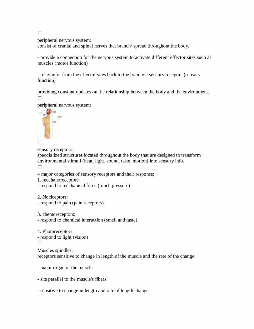
Golgi tendon orgnans (GTO): receptors sensitive to change in tension of the muscle and the rate of that change. - located where the muscle and tendon meet and are sensitive to changes in muscular tension and rate of the tension change - will cause muscles to relax when excited. - prevent muscles from being placed under excessive stress and sustaining injury.
Golgi tendon orgnans (GTO):
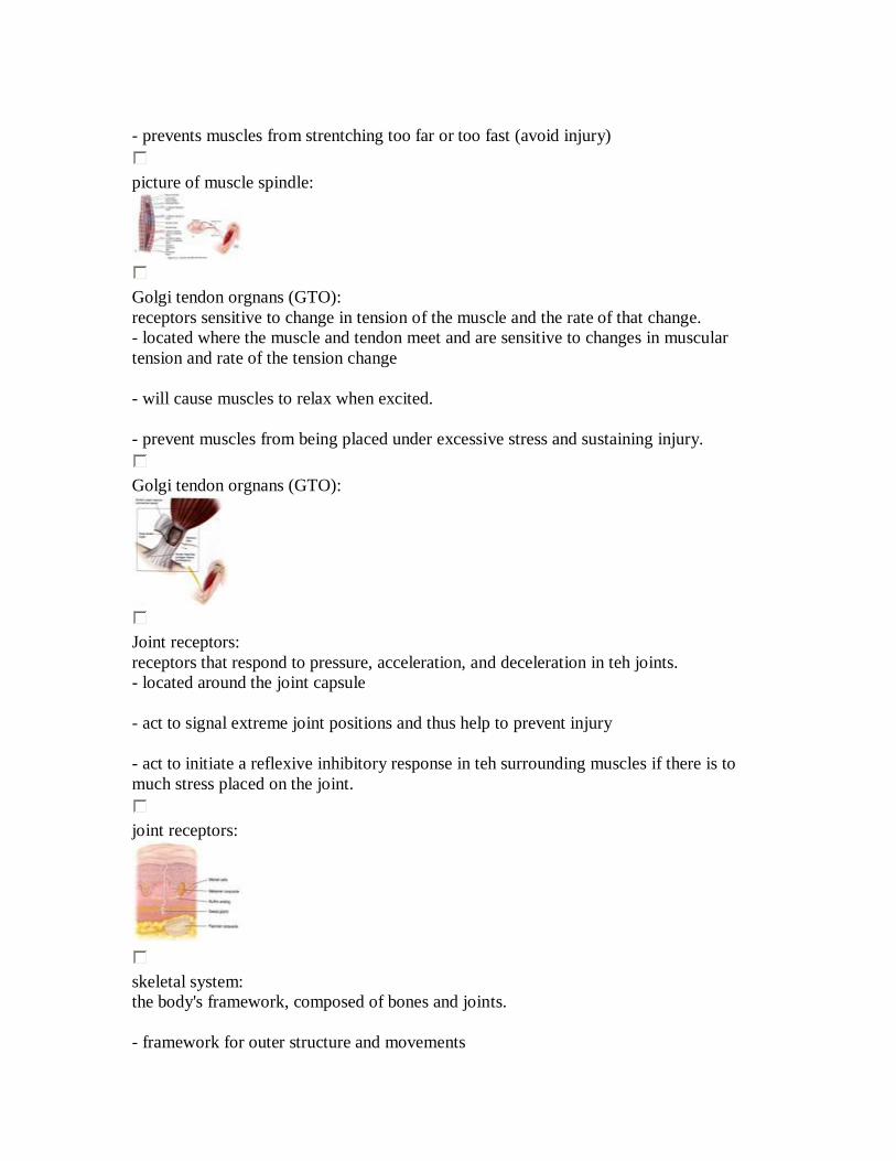
Joint receptors: receptors that respond to pressure, acceleration, and deceleration in teh joints. - located around the joint capsule - act to signal extreme joint positions and thus help to prevent injury - act to initiate a reflexive inhibitory response in teh surrounding muscles if there is to much stress placed on the joint.
joint receptors:
skeletal system: the body's framework, composed of bones and joints. - framework for outer structure and movements
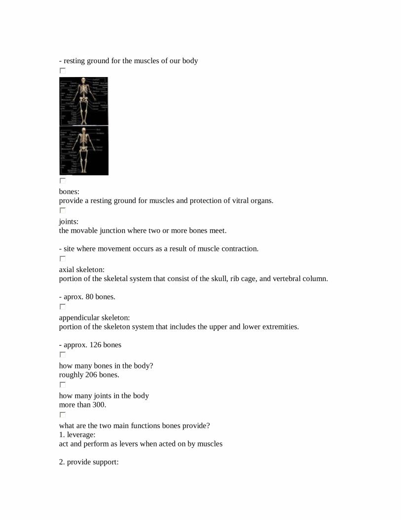
- resting ground for the muscles of our body
bones: provide a resting ground for muscles and protection of vitral organs.
joints: the movable junction where two or more bones meet. - site where movement occurs as a result of muscle contraction.
axial skeleton: portion of the skeletal system that consist of the skull, rib cage, and vertebral column. - aprox. 80 bones.
appendicular skeleton: portion of the skeleton system that includes the upper and lower extremities. - approx. 126 bones
how many bones in the body? roughly 206 bones.
how many joints in the body more than 300.
what are the two main functions bones provide? 1. leverage: act and perform as levers when acted on by muscles 2. provide support:
this translates into posture, which is necessary for the efficient distribution of forces acting on teh body.
depressions: flattened or inented portion of bone, which can be a muscle attachment site
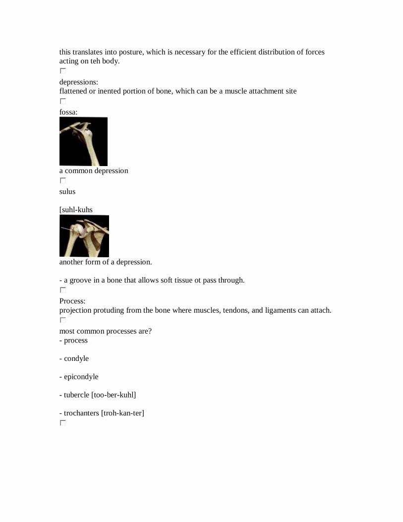
fossa:
a common depression
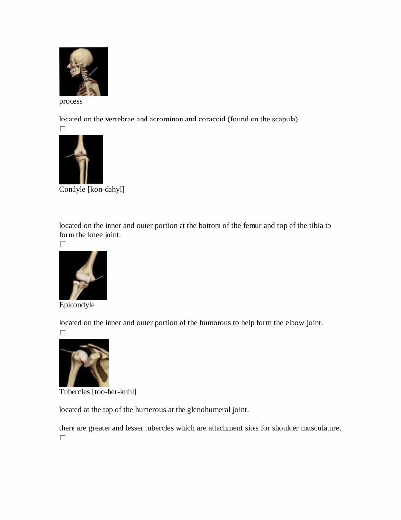
sulus [suhl-kuhs
another form of a depression. - a groove in a bone that allows soft tissue ot pass through.
Process: projection protuding from the bone where muscles, tendons, and ligaments can attach.
most common processes are? - process - condyle - epicondyle - tubercle [too-ber-kuhl] - trochanters [troh-kan-ter]
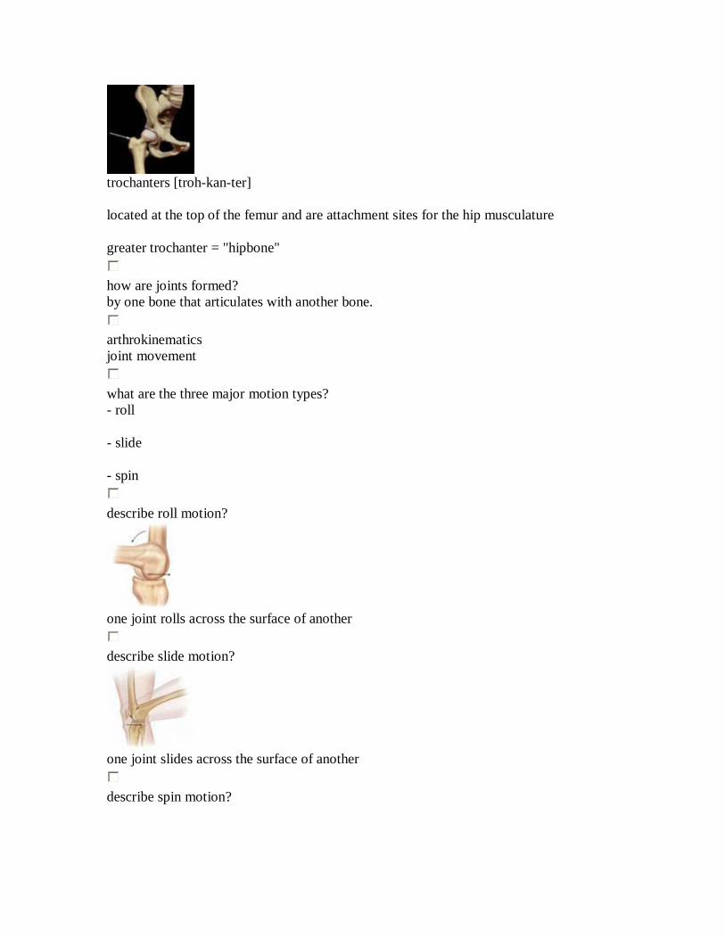
process located on the vertebrae and acrominon and coracoid (found on the scapula)
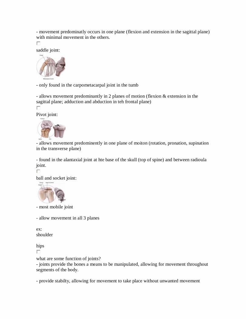
Condyle [kon-dahyl] located on the inner and outer portion at the bottom of the femur and top of the tibia to form the knee joint.
Epicondyle located on the inner and outer portion of the humorous to help form the elbow joint.
Tubercles [too-ber-kuhl] located at the top of the humerous at the glenohumeral joint. there are greater and lesser tubercles which are attachment sites for shoulder musculature.
trochanters [troh-kan-ter] located at the top of the femur and are attachment sites for the hip musculature greater trochanter = "hipbone"
how are joints formed? by one bone that articulates with another bone.
arthrokinematics joint movement
what are the three major motion types? - roll - slide - spin
describe roll motion?
one joint rolls across the surface of another
describe slide motion?
one joint slides across the surface of another
describe spin motion?

one joint surface rotates on another
synovial joints: joints that are held together by a joint capsule and ligaments and are more associated with movement in the body. - produce synovial fluid which lubricates joint surfaces to reduce excessive wear and to nourish the cartilage cells
what are the several types of synovial joints? - gliding (plane) - Condyloid (condylar or ellipsoidal) - hinge - saddle - pivot - ball and socket
gliding joint?
a nonaxial joint tha thas the simplest movement of all joints - moves either back and forth or side to side.
Condyloid (condylar or ellipsoidal joint:
called because the condyle of one bone fits into the elliptical cavity of another bone to form joint.
- movement predominatly occurs in one plane (flexion and extension in the sagittal plane) with minimal movement in the others.
saddle joint:
- only found in the carpometacarpal joint in the tumb - allows movement predominantly in 2 planes of motion (flexion & extension in the sagittal plane; adduction and abduction in teh frontal plane)
Pivot joint:
- allows movement predominently in one plane of moiton (rotation, pronation, supination in the transverse plane) - found in the alantaxial joint at hte base of the skull (top of spine) and between radioula joint.
ball and socket joint:
- most mobile joint - allow movement in all 3 planes ex: shoulder hips
what are some function of joints? - joints provide the bones a means to be manipulated, allowing for movement throughout segments of the body. - provide stabilty, allowing for movement to take place without unwanted movement

- all joints are linked together meaning that movement of one joint directly affects movement of the others.
nonsynovial joints:
- have no joint cavity, fibrous connective tissue, or cartilage in teh uniting structure. - can be structured in either a fibrous/cartilaginous manner - exhibit little/ no movement. ex: - sutures of the skull - distal joint of tibia & fibula - symphysis pubis
ligaments:
- primary connective tissue that connects bone together and provides stabilty, input to the nervous system, guidance, and the limitaion of improper joint movement.
what are ligaments made out of? - primarily collagen and elastin (protein)
collagen fibers: situated in a more parallel fashion providing ligaments to withstand tension.
elastin fibers: gives ligaments some flexibility to withstand bending and twisting
ligaments are characterized by poor vascularity (or blood supply), they do not heal or repair very well and may be slower to adapt.
skeletal system: body's framework and is made up of bone and joints in two divisions: - axial - appendicular
4 main types of bone? - long - short - flat - irregular (all have markings of depressions/ processes)
how are bones connected? (via ligaments) by either synovial/ nonsynovial joints which provide movement as well as stability
muscular system: series of muscles that moves the skeleton.
muscles are the _________ & _________ of our body movers, stabilizers
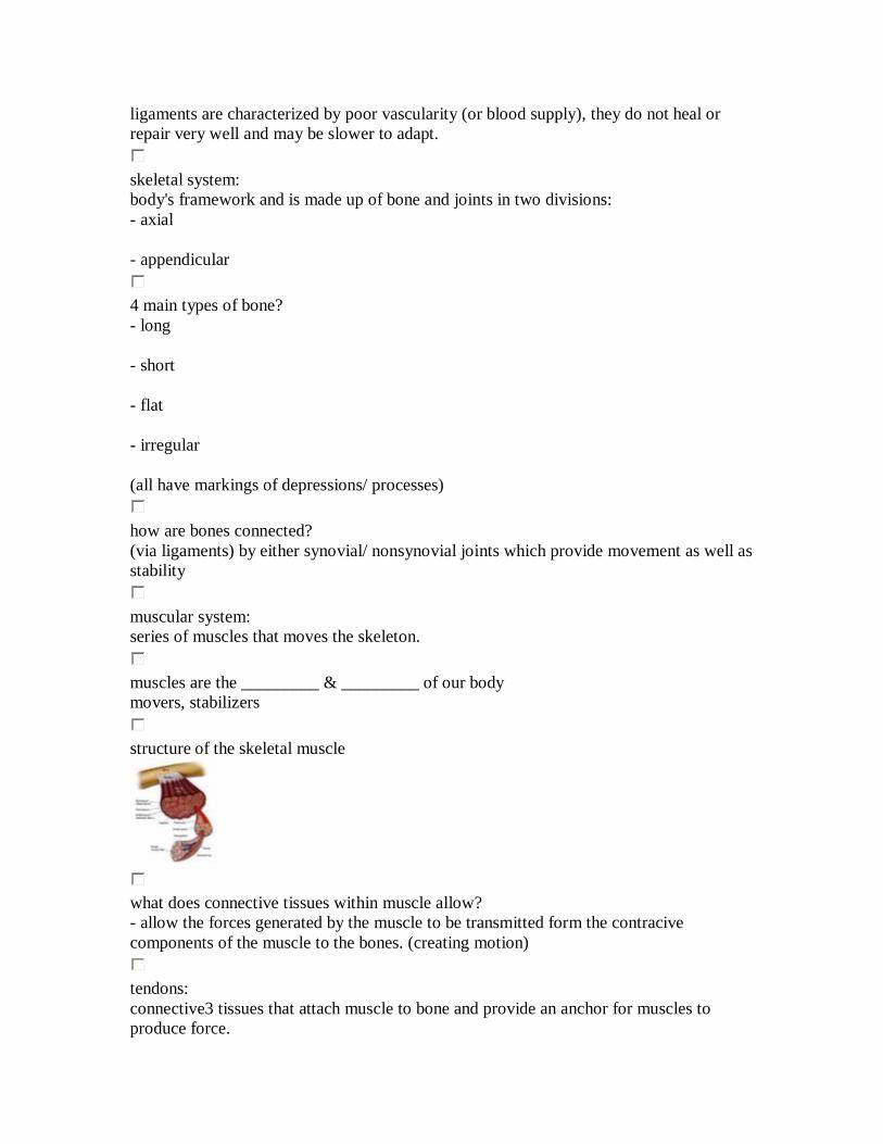
structure of the skeletal muscle
what does connective tissues within muscle allow? - allow the forces generated by the muscle to be transmitted form the contracive components of the muscle to the bones. (creating motion)
tendons: connective3 tissues that attach muscle to bone and provide an anchor for muscles to produce force.

have poor vascularity (blood suply), which leaves them susceptible to slower repair and adaption.
sarcoplasm: cellular plasma membrane surounding muscle fibers contains: -glycogen - fats - minerals - oxygen-binding myoglobin
Sarcomere
tropomyosin: located on teh actin filament and blocks myosin binding sites located on teh actin filaments keeping myosin from attaching to actin (relax state)
Troponin: - located on teh actin - plays role in muscle contraction by providing binding sites for both calcium and tropomyosin when muscles need to contract.
neural activation: the contraction of a muscle gnerated by neural stimulation. - essential for a muscle to contract, for movement and stabilization
motor unit: a motor neuron and all of the muscle fibers it innervates.
neuromuscluar junction: the point at which the neuron meets an individual muscle fiber. (acually a small gap between the nerve and muscle fiber "synapse")
action potentials: electrical impulses which are transported from the central nervous system down the axon of the neuron
neurotransmitter: chemical messengers that cross synapses to transmit electrical impulses from the nerve to the muscle. - activate a series of steps that produce muscle contraction.
sliding filament theory:
Excitation-contraction coupling: the process of neural stimualtion creating muscle contraction.
what are the main group of muscle fibers? - type I (slow twitch) - type II (fast twitch) - type IIa - type IIb
type I muscle fibers? - slow twitch - contain a high # of capillaires, mitochondria, and myoglobin (allows for improved delivery of oxygen - often referd to as red fibers - smaller in size (diameter) - slower to produce maximal tension and more resistant to fatigue - important for muscles producing long-term contractions necessary for stabilization and postural control.
type II muscle fibers: - fast twitch - genreally contail fewer capillaries, mitochondria, and myoglobin

- often refered to as white fibers - larger in size - quick to produce maximal tension, and fatigue more quickly - important for muscle producing movements requiring force and power such as performing a sprint.
type IIa: higher oxidative capacity and fatigue slower.
type IIb: - low oxidative capacity and fatigue quickly
steps in the initial contraction and at the end:
.
agonist muscles: muscles that act as prime movers (they are muscles most responsible for a particular movement)
synergist muscle: assist prime movers during movement ex: hamstring and erector spinae with gluteus max. during hip extension.
stabilizer muscles: support/ stabilize the body while the prime movers and synergists perform movement patterns. ex:
transversus abdominis, internal oblize, multifiduc (deep back muscle) during hip extension.
antagonist muscle: perform opposite action of the prime mover. ex: psoas (deep hip flexor) to gluteus max. during hip extension.