j. 1991 defined in intracellular doxorubicin of p-glycoprotein
TRANSCRIPT
Br. J. Cancer (1991), 64, 857-861 1991
Early multidrug resistance, defined by changes in intracellular doxorubicindistribution, independent of P-glycoprotein
G.J. Schuurhuis', H.J. Broxterman', J.H.M. de Lange2, H.M. Pinedo"3, T.H.M. vanHeijningen', C.M. Kuiperl, G.L. Scheffer2, R.J. Scheper2, C.K. van Kalken', J.P.A. Baak2 & J.Lankelmal
Departments of 'Medical Oncology and 2Pathology, de Boelelaan 1117, 1081 HV Amsterdam; 3Netherlands Cancer Institute,Amsterdam, The Netherlands.
Summary Resistance to multiple antitumour drugs, mostly antibiotics or alkaloids, has been associated witha cellular plasma membrane P-glycoprotein (Pgp), causing energy-dependent transport of drugs out of cells.However, in many common chemotherapy resistant human cancers there is no overexpression of Pgp, whichcould explain drug resistance. In order to characterise early steps in multidrug resistance we have derived aseries of P-glycoprotein-positive (Pgp/+) and P-glycoprotein-negative (Pgp/-) multidrug resistant cell lines,from a human non-small cell lung cancer cell line, SW-1573, by stepwise selection with increasing concentra-tions of doxorubicin. These cells were exposed to doxorubicin and its fluorescence in nucleus (N) andcytoplasm (C) was quantified with laserscan microscopy and image analysis. The fluorescence N/C ratio inparent cells was 3.8 and decreased both in Pgp/+ and Pgp/- cells with increasing selection pressure to1.2-2.6 for cells with a resistance factor of 7-17. N/C ratios could be restored partly with verapamil only inPgp/+ cells. N/C ratio measurements may define a general Pgp-independent type of defense of mammaliancells against certain anticancer agents which may precede Pgp expression in early doxorubicin resistance.
The development of resistance of human cancers to potentanticancer agents has been classically ascribed to the selectionand outgrowth of pre-existing or newly occurring subpopula-tions of resistant tumour cells (Carl, 1989; Coldman & Goldie,1985; Skipper, 1986). Great progress in the understanding ofthe mechanism of one type of in vitro derived resistance, theso-called multidrug resistance (MDR), was recently obtainedfrom the successful cloning and transfection of the mdrlgene, which codes for a 170kDa plasmamembrane protein,called P-glycoprotein (Pgp) (Gottesman & Pastan, 1988;Hammond et al., 1989; Juranka et al., 1989; Lincke et al.,1990). The role of this glycoprotein was elucidated by study-ing the phenotype of cell lines, transfected with mdrl (Gottes-man & Pastan, 1989; Hammond et al., 1989; Juranka et al.,1989; Lincke et al., 1990) or selected in vitro for resistance toanticancer agents, such as anthracyclines and vinca alkaloids,colchicine and actinomycin D (Biedler et al., 1988; Bradley etal., 1988). Thus Pgp was proven to confer multidrug resis-tance. Further it was proposed that Pgp functions as aplasma membrane pump for several classes of lipophilicdrugs. Evidence for this was based on the predicted aminoacid sequence of Pgp, which contained two consensus ATPbinding sites, and on the demonstration of ATP-dependentdrug binding to Pgp and energy-dependent drug efflux fromPgp/+ cells (Broxterman et al., 1988; Choi et al., 1988; Groset al., 1986). However, it has been shown that in somePgp/ + cells with high levels of doxorubicin resistance the netchange in cellular accumulation of drug was relativelymodest, which was taken as argument against an importantrole of such a drug export protein in the resistance mecha-nism in these cells (Siegfried et al., 1983). Recently weshowed that in the Pgp/ + cells 2780AD and CHRC5, theresistance to doxorubicin could be quantitatively accountedfor, when, in addition to doxorubicin accumulation, thedistribution of doxorubicin between nuclear sites and cyto-plasmic localisations was taken into consideration (Schuur-huis et al., 1989). The data suggested that these Pgp/+ cellshad developed a resistance mechanism characterised by a
decrease in net cellular doxorubicin accumulation, includingthe nuclear target sites, while accumulation in cytoplasmicstructures was relatively unaffected.We have now investigated whether such a defence mechan-
ism of mammalian cells against certain types of anticanceragents is necessarily dependent on Pgp overexpression andwe have therefore especially focussed on low level resistantcells. Hereto we used two series of variants selected bydoxorubicin exposure from the human non-small cell lungcancer cell line, SW-1573, which both displayed multidrugresistance.
Materials and methods
Cells and cell cultureThe human non-small cell lung cancer cell line was obtainedfrom Dr H. Joenje (Department of Anthropogenetics, FreeUniversity, Amsterdam; Keizer et al., 1989) and was culturedin Dulbecco's modified Eagle's medium + 7.5-10% foetalcalf serum (GIBCO Europe, Paisley, Scotland). The resistantsublines were derived by continuous exposure to increasingdoxorubicin concentrations: e.g. 2R50 (2R80 etc) means thatthese cells were continuously cultured in 50 (80 etc) nMdoxorubicin, until they were harvested 7-14 days before theexperiments by short trypsinisation. There was no significantdifference in cell size between all the SW-1573 sublines (dia-meter SW-1573: 16.2 ± 1.2 gsm in nine experiments). Cellcycle distribution as measured with flow cytometry (GI, Sand G2 + M phase, resp., expressed as % of total number ofcells and determined in two independent experiments) was asfollows. SW-1573: 54, 37 and 9, resp.; SW-1573/2R50: 59,33and 9, resp.; SW-1573/2R120: 62, 20 and 19, resp.; SW-1573/4R50: 41, 40 and 19, resp.; SW-1573/4R120: 54, 28 and 18,resp. Cell doubling times were 22 ± 1 (SW-1573), 32 ± 3(SW-1573/2R50), 38 ± 3 (SW-1573/2R120), 43 ± 5 (SW-1573/2R160), 26 ± 1 (SW-1573/4R50) and 28 ± 2 (SW-1573/4R120),determined in 3-5 independent experiments (M ± s.d.).
Drug cytotoxicityDoxorubicin resistance factor (RF) and dose-modifying fac-tor (DMF) were calculated from 50% cell-growth inhibitory
Correspondence: G.J. Schuurhuis.Received 19 March 1991; and in revised form 12 July 1991.
Br. J. Cancer (1991), 64, 857-861 17" Macmillan Press Ltd., 1991
858 G.J. SCHUURHUIS et al.
concentrations of doxorubicin (IC") determined in a cellproliferation assay; IC50 of SW-1 573 cells was 0.12 ± 0.02 gM(mean ± s.d. of 5 exp., 2 h drug exposure in a waterbath at37°C in culture medium lacking NaHCO3 but containing20 mM HEPES). After 2 h exposure to doxorubicin and32 jM verapamil (for DMF), the cell culture medium wasrefreshed and after another 4 h with or without verapamil inthe same medium the cells were allowed to grow in NaHCO3containing medium for three cell doubling-times, again inthe presence of 32 gM verapamil for DMF determinations(Schuurhuis et al., 1987). The 2 h exposure was carried out inmedium lacking NaHCO3 in order to be able to compare thecytotoxicity data directly with drug accumulation and N/Cratio measurements (see below). The IC50 value for SW-1573cells was lower (factor 2) in medium lacking NaHCO3 thanin growth medium but no major differences in resistancefactors were found when the two media were compared.Resistance factors for the other drugs were determined ina cell proliferation assay by a continuous incubation withdrugs as described (Broxterman et al., 1989; Mosman, 1983).A concentration of 32 tLM verapamil was used in order toobtain maximal effects on doxorubicin accumulation, cyto-toxicity and intracellular drug distribution, since in case lowlevel MDR cells are used these parameters do not differmuch from those in sensitive cells. Previously dose-dependenteffects of verapamil on these parameters have been shown(Schuurhuis et al., 1987, 1989, 1990).
Drug accumulationDoxorubicin accumulation and accumulation-enhancementfactor by co-incubation with 32JM verapamil (AEF) weredetermined by exposure of adhered cells to 0.5 fLM [14C]-doxorubicin (2 h, 37°C) in culture medium lacking NaHCO3,three rapid cold washes and subsequent trypsinisation ofthe cells; further procedures were essentially as described(Schuurhuis et al., 1987).
Flow cytometryFor quantification of Pgp, 106 unfixed cells were incubatedwith the monoclonal antibody MRK-16 (4Agml-') or anirrelevant mouse IgG (51igml-) for 1 h in a volume of200 1l at 20°C. Samples were washed three times with phos-phate buffered saline + 1% bovine serum albumin (PBS-BSA)and incubated with 100 gl rabbit-antimouse IgG-fluores-ceinisothiocyanaat (100 lg ml1'), DAKO immunoglobulins,Copenhagen, Denmark) for 45 min at 20°C. After washingthe cells three times with PBS-BSA, the cells were resus-
Table I Doxorubicin resistance and accumulation in SW- 1573variants
doxDMFa accumulation AEFb
Cell line RF (dox) (32jom Vp) (% of SW-1573) (32gMVp)SW-1573 1 1.6±0.3d 100 1.16±0.09c4R50 7.7±3.8 7.3±1.4d 12.2±0.5d 5.9 ±0.lc4R80 10.0± 1.2 12.8± 1.4d 10.2±0.2d 7.4 ±0.7d4R120 13.2± 1.9 lo.o±o.9d 10.9± l.od 5.2 ±0.4d4R160 16.7±0 16.1 ±0.9d 7.1±0.2d 10.0 ± .lC2R50c 6.9±0.3 2.5±0.1c 34.0±6.od 1.3 ±0.12R80F 9.4±2.0 5.5±1.4d 35.4±2.5d 1.3 ±0.22R120c 11.1±2.8 6.3 ± 0.4d 41.9±8.od 1.4 ±0.22R160 63 ± 12 17.2± 1.3d 4.7±od 9.3 ± 1.7daDMF, dose modifying factor = IC50 minus Vp/IC50 plus Vp. bAEF,
accumulation enhancement factor = drug accumulation plus Vp/drugaccumulation minus Vp. Data are means± s.d. of2-3 experiments eachperformed at least in duplicate (cytotoxicity) or triplicate (accumula-tion). c,d Significantly different from I (DMF), from SW-1573 (-Vp)levels (fourth column) or from accumulation -Vp (fifth column):cP< 0.02; dP<0.01 (Student's t-test). enon-Pgp cell lines, as measuredwith RNAase protection assay (Baas et al., 1990; Zinn et al., 1983). Vp,verapamil.
pended in 500 ttl PBS-BSA and fluorescence was measuredwith a FACSTAR Plus, Becton Dickinson Medical Systems(Sharon, Ma).
Determination ofN/C doxorubicin fluorescence ratiosTrypsinised cells were allowed to adhere on tissue culturepetri dishes (Costar, Cambridge, Ma) for 24h. Cells wereincubated with doxorubicin for 1 h under the same condi-tions as described for drug accumulation and cytotoxicityexperiments in Table I and quickly washed with PBS toreduce background fluorescence. PBS was lacking glucose toprevent drug efflux and redistribution. Dox concentrationswere chosen to obtain equal net cellular drug amounts in allcell lines (4pM in SW-1573 cells). Thirty to fifty cells wererecorded for each treatment using laserscan microscopy andfluorescence ratios were quantified by delineating nuclei andcytoplasms interactively using digital image analysis as des-cribed (Schuurhuis et al., 1989).
Determination of the ratio intercalated doxorubicin/non-intercalatedfluorescent doxorubicinThe ratios of intercalated doxorubicin vs non-intercalatedfluorescent doxorubicin were determined as described (Lan-kelma et al., 1990). SW-1573 and SW-1573/2R160 cells wereloaded for 1 h at 37°C with 4 and 20 gM doxorubicin, respec-tively, in order to obtain about equal intracellular drugamounts.
RNAase protection assayTen jg RNA samples were hybridised with a 32P-labelled 301nucleotide human mdrl cDNA specific probe, obtained fromF. Baas, Neth. Cancer Inst., and analysed by RNAase pro-tection assay essentially as described (Baas et al., 1990; Zinnet al., 1983). A T-actin probe was used to control for equalamounts of analysed RNA.
Results and discussion
Two separate series of resistant cells were selected by con-tinuous exposure to 50, 80, 120 and 160 nM doxorubicin.The cross-resistance pattern of the 2R series has recentlybeen described (Baas et al., 1990; Kuiper et al., 1990); it wasshown that all sublines from this series had a decreaseddoxorubicin and vincristine accumulation, while Pgp expres-sion, which was detectable with a sensitive RNAase protec-tion assay in the parent cell line, was lost during an earlyselection step, but reappeared, strongly overexpressed, at alater selection step (Baas et al., 1990; Kuiper et al., 1990). In
Table II Cross-resistance in SW-1573 variantsRF RF RF RF
Cell line (dauno) (vincr) (gramD) (etoposide) pgpaSW-1573 1 1 1 1 1.08±0.054R50 3.0±1.3 213± 80 161±53 6.6±2.0 21.4 ±4.24R80 3.4± 1.6 222± 76 185±70 5.0±2.1 37.6 ±4.34R120 4.9±3.5 299± 55 149±45 5.2± 1.2 20.8 ±2.4A Yk I -If I4RI60 6.6± 3.7
22.4 ±5.42RSOb 3.3±0.4 5.8± 1.1 3.2± 1.1 14± 6 1.10±0.152R80b 5.9±1.6 16± 3 3.1 (n= 1) 21±17 ND2R120b 3.7±0.1 17± 3 2.3± 1.7 45± 13 1.02±02R160 35±9 480± 170 146±33 120±36 20.4 ±8.2
Resistance factors (RF) were determined in a continuous incubationassay with drug as described (Broxterman et al., 1989; Mosmann, 1983).Data are means ± s.d. from 3 -4 separate experiments. The RF data fordaunorubicin, vincristine, gramicidin D and etoposide for the 2R seriesare from Kuiper et al., 1990 and are shown for comparison. aData aregiven as mean fluorescence of MRK-16, divided by mean fluorescenceIgG (mean ± s.d. of 2 experiments). Fluorescence ratio for 2780AD cells(Schuurhuis et al., 1987) was 37 (mean of seven experiments). bpgpnegative cell lines (see Table I). ND, not determined.
413± 108 5.4± 2-202± 55
INTRACELLULAR DRUG DISTRIBUTION IN MULTIDRUG RESISTANCE 859
:4A2
M.DRR1
.-o 242
which was more prominent, however, in the 4R series (TableI). Especially in the 2R series the accumulation defect wouldnot be sufficient to fully account for the observed resistancefactors. Doxorubicin accumulation, like doxorubicin resis-tance, could be modulated more effectively in the Pgp/ + cells(Table I). Thus, while interaction of verapamil with Pgpseems to allow an effective modulation of doxorubicin resis-tance and accumulation, it does not prove that doxorubicinresistance and impairment of accumulation in these Pgp/+cells is directly caused by Pgp.We have shown before by a quantitative approach using
laserscan microscopy and image analysis that a decreasein doxorubicin fluorescence nucleus/cytoplasm (N/C) ratiocould be measured in the low level mdr Pgp/ + cell line8226/dox 4 (Broxterman et al., 1990). Those data suggested acorrelation of doxorubicin fluorescence N/C ratio with Pgpexpression. The present SW-1573 experimental system allow-ed us to study the doxorubicin fluorescence N/C ratio's incells with increasing levels of multidrug resistance in Pgp/+as well as Pgp/- cells, derived from the same parent cells.
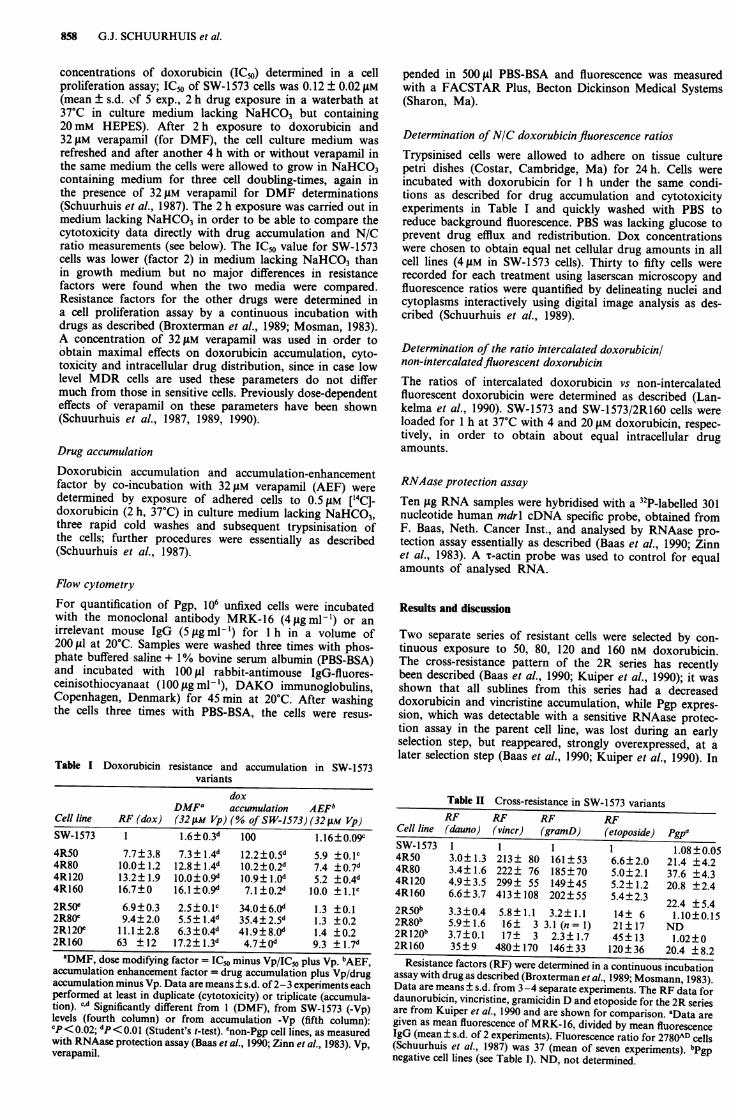
Figure 2 shows a decrease in N/C ratio with increasingselection pressure in Pgp/+ as well as in Pgp/- cells. Inter-estingly, N/C ratios were slightly lower in Pgp/- cells com-pared to Pgp/+ cells at the same levels of resistance, whileon the other hand doxorubicin accumulation was less in thePgp/+ cells (Table I). This suggests that the relative contri-bution of decreased drug accumulation and altered drug
4.0
Actin
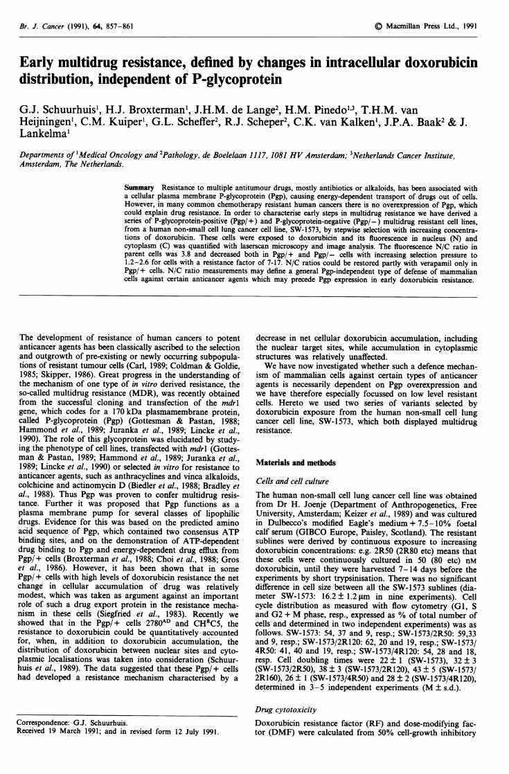
Figure 1 mdrl overexpression is an all or non phenomenon inthe present cell lines. The RNAase protection assay was carriedout as described under Materials and methods. From left to rightbands corresponding to RNA from SW-1573, 4R50, 4R80, 4R120and 4R160 are shown. The parent SW-1573 cells have a justdetectable signal, which varies somewhat with proliferation stateof the cells (Baas et al., 1990). It was shown before that thesimilarity in intensity was not due to saturation phenomenabecause that a further increase of intensity occurs with a furtherincrease of drug selection pressure (Baas et al., 1990).
a separate selection with doxorubicin, the 4R series was
derived (Tables I and II) which appeared to have overexpres-sion of Pgp from the first selection step on, as determined atprotein level with the monoclonal antibody MRK-16 (TableII) and with a RNAase protection assay using a mdrl specificprobe (Figure 1). Remarkably the 4R50, 4R120, 4R160 as
well as the 2R160 cells have similar amounts of Pgp, whilethe 4R80 cells for unknown reasons have higher amounts(Table II). In separate experiments it was shown that cellswith a higher degree of resistance (2780AD, Schuurhuis et al.,1987) have a higher amount of Pgp (see legends of Table II),indicating non-saturability of the method used. Both the 2Rand 4R series display a MDR-like phenotype with, relative todoxorubicin and daunorubicin resistance (Tables I and II),high vincristine and gramicidin D resistance in Pgp/+ cellsand high etoposide resistance in Pgp/- cells (Table II).Doxorubicin resistance could be modulated by coincubationwith verapamil in 4R.Pgp/+ cells to a large extent (Table I).In 2R.Pgp/- cells, however, the resistance modulation withverapamil was less effective (Table I). Both the 2R and 4Rseries show an impairment of doxorubicin accumulation,
0
X. 3.00za)
o 2.0uoa)0
x0 1.0
n
O 2R-series
* 4R-series
I
. I
10Resistance factor
Figure 2 Doxorubicin fluorescence nucleus/cytoplasm (N/C)ratios as a function of doxorubicin resistance in Pgp/+ andPgp/- cells. Fluorescence N/C ratios were determined as des-cribed under Materials and methods. Data are from three inde-pendent experiments each performed in duplicate (means±s.d.).
Table III Effect of verapamil on doxorubicin fluorescence ratios inSW- 1573 variants
Cell line N/C N/C ( + verapamil)
SW- 1573 3.77 ±0.18 3.87±0.44 [1.0]4R50 2.60±+0.18b 3.27±0.25c [1.3]4R80 1.89±0.37b 4.02±0.18e [2.1]4R120 2.02±0.17b 3.04±0.10d [1.5]4R160 1.16±0.24b 2.61 ±0.llc [2.3]2R50a 2.18 +±0.16b 1.70±0.03 [0.8]2R80a 1.72±0.24b 1.81 ±0.23 [1.1]2R120a 1.75±+0.13b 1.77±0.37 [1.0]2R160 0.67±0.12b 2.39±0.64e [3.6]
N/C ratios (doxorubicin fluorescence in nucleus/doxorubicin fluores-cence in cytoplasm) were measured as described for Figure 2. a pgpnegative cell lines. bSignificantly different from 3.77 (P <0.01, Student'st-test). cdeSignificantly different from N/C ratios minus verapamil:cP< 0.05; dp <0.02; 'P<0.01. Values in brackets represent factorincrease of N/C ratios by verapamil. Data are from 2-6 independentexperiments each performed in duplicate.
100^ - - ^ - - - - - ^ - - - - - - -
vi
1I
r- ,. I.
860 G.J. SCHUURHUIS et al.
distribution to the resistance phenotype may differ dependingon the presence of Pgp. The fact that 4R160 cells have alower N/C ratio than e.g. 4R50 cells, despite similar amountsof Pgp, indicates that the non-Pgp resistance mechanismmight also be present in the 4R cells. However, it cannot beexcluded that the high resistance to VP-16 in Pgp/- cellswould be caused in part by additional changes such as analtered topoisomerase II activity (Baas et al., 1990).
Again, like for doxorubicin accumulation, for an effectivemodulation of doxorubicin fluorescence N/C ratio byverapamil the presence of Pgp seems to be a prerequisite(Table III, compare e.g. 2R120 with 4R120 cells). Remark-ably, verapamil-induced changes in accumulation (Table I)together with changes in N/C ratios (Table III) can accountfor resistance modulation (DMF, Table I) in Pgp/ + cells butnot in Pgp/- cells, which leaves the possibility of additionalactions of verapamil which affect doxorubicin cytotoxicity. Inthe 4R series the 4R80 cells show a somewhat exceptionalbehaviour: the amount of Pgp as estimated with flow cyto-metry is higher than in the other 4R cells (Table II). In linewith this stimulation of doxorubicin accumulation as well asreversal of resistance by verapamil was more prominent inthe 4R80 cells (Table I). Also the N/C ratio in 4R80 cells wasrelatively low, while its modification with verapamil wasrelatively high (Table III).
In this study doxorubicin fluorescence N/C ratios are usedoperationally to probe mechanisms of drug resistance, relatedto changes in intracellular drug distribution. These ratios assuch do not reflect the actual concentrations of doxorubicinin each compartment, since they do not take into account thequenching of doxorubicin fluorescence at several differentlocalisations of the drug: quenching of doxorubicin fluore-scence due to DNA intercalation has been estimated at 95%(Chaires et al., 1982), while cytoplasmic fluorescence may belargely unaffected (Budge & Tritton, 1985; Tarasiuk et al.,1989). We have used an independent technique to show thatN/C ratio changes reflect changes in the actual compartmen-tal amounts of doxorubicin. With this technique the ratiointercalated drug/non-intercalated fluorescent drug can bemeasured (Lankelma et al., 1990). It was found that thisratio was 4.2 ± 0.6 in SW-1573 cells and 1.2 ± 0.2 in SW-1573/2R160 cells (M ± s.e.m. of two independent experi-ments). N/C ratio changes might also be found if certaindrug binding sites in the cell are saturated at the relativelyhigh drug concentrations which were used in the fluorescenceassays. This is unlikely, however, since N/C ratios wereshown to be independent of doxorubicin concentrations inthe medium (J.H.M. de Lange, N.W. Schipper, G.J. Schuur-huis et al., manuscript submitted).The molecular mechanism(s) responsible for the induction
of decreases in doxorubicin N/C ratio are not yet known; inthe SW-1573 cell lines the presence of Pgp is not required forthis decrease to occur. One explanation would be the
presence of a pump protein present at vesicular membranesoriented to pump drug inside such vesicles. However, in thecase of Pgp, the antigenic determinant of MRK-16 could bedetected only on the Golgi stack but not in other vesicles(Willingham et al., 1987). Moreover, similar amounts of Pgp,as estimated with MRK-16 binding, do not seem to correlatewith resistance factor or N/C ratios (compare e.g. 2R160 and4R160 cells in the Tables I-III). Thus, even after the induc-tion of Pgp overexpression, the early, non-Pgp mediatedmechanism, may still be operative. Since cytoplasmic pH waselevated in some resistant SW-1573 isolates, compared toSW-1573 parent cells as measured in medium without bicar-bonate (Keizer & Joenje, 1989), one possibility to be con-sidered is that anthracyclines are forced to accumulate to ahigher extent in an acidic vesicular compartment in the cell(Beck, 1987; Keizer et al., 1989; Sehested et al., 1987). Sinceeven in steady-state the 2R.Pgp/- cells accumulated lessdoxorubicin (Table I) or vincristine (Kuiper et al., 1990), anactive extrusion process could be present. Active drug ext-rusion processes for different types of drugs, not related toPgp overexpression, have in fact been reported recently(Henderson & Tsuji, 1990; Hindenburg et al., 1989; McGrathet al., 1989a,b). In such highly resistant cells anthracyclineresistance could also be related to differences in drug bindingto nuclear and cytoplasmic bindings sites (Hindenburg et al.,1989). Since different independently selected, lowly resistantisolates from SW-1573 cells showed the Pgp/- multidrugresistance (Baas et al., 1990), which now has been shown tobe related to decreased doxorubicin N/C ratio's, a mutationin one gene product could be responsible for the observedresistance pattern in Pgp/- cells. The remarkably low resis-tance factors of 4R160 compared to 2R160 for both anthra-cyclines and etoposide, despite a similar Pgp expression fur-ther suggests that such a mechanism still prevails in theresistance of 2R160 Pgp/+ cells for these drugs. The com-bined measurement of doxorubicin accumulation and fluore-scence N/C ratio's in tumour cells, isolated directly frompatient's tumours would allow to investigate the role of drugtransport resistance in failure of chemotherapy. Further,applying immunohistochemical staining techniques after N/Cratio measurements would allow a direct comparison of func-tional changes (reflected by N/C ratio changes) and thepresence of resistance-related antigenic determinants (e.gPgp) in individual cells.
This study was supported by the Dutch Cancer Society (grant IKA-VU 88-22) and by a grant from the Bristol-Myers Squibb Company.H.J.B. is a fellow of the Royal Netherlands Academy of Arts andSciences. We thank H.S Miilder for determination of the ratiosintercalated drug/non-intercalated drug. Dr P. Borst and Dr F. Baasare acknowledged for their stimulating discussions of this work. Wethank Dr T. Tsuruo for the generous supply of MRK-16.
References
BAAS, F., JONGSMA, A.P.M., BROXTERMAN, H.J. & 7 others (1990).Non-P-glycoprotein mediated mechanism for multidrug resistanceprecedes P-glycoprotein expression during in vitro selection fordoxorubicin resistance in a human lung cancer cell line. CancerRes., 50, 5392.
BECK, W.T. (1987). The cell biology of multiple drug resistance.Biochem. Pharmacol., 36, 2879.
BIEDLER, J.L., CHANG, T-d., SCOTTO, K.W., MELERA, P.W. & SPEN-GLER, B.A. (1988). Chromosomal organization of amplified genesin multidrug-resistant Chinese hamster cells. Cancer Res., 48,3179.
BRADLEY, G., JURANKA, P.F. & LING, V. (1988). Mechanism ofmultidrug resistance. Biochim. Biophys. Acta, 948, 87.
BROXTERMAN, H.J., PINEDO, H.M., KUIPER, C.M., KAPTEIN, L.C.M.,SCHUURHUIS, G.J. & LANKELMA, J. (1988). Induction of vera-pamil of a rapid increase in ATP consumption in multidrug-resistant tumor cells. FASEB J., 2, 2278.
BROXTERMAN, H.J., PINEDO, H.M., KUIPER, C.M. & 7 others (1989).Immunohistochemical detection of P-glycoprotein in humantumour cells with a low degree of drug resistance. Int. J. Cancer,43, 340.
BROXTERMAN, H.J., SCHUURHUIS, G.J., LANKELMA, J., BAAK,J.P.A. & PINEDO, H.M. (1990). Towards functional screening formultidrug resistant cells in human malignancies. In Drug Resis-tance: Mechanisms and Reversal, Mihich, E. (ed.), p. 309, JohnLibbey CIC: Roma.
BUDGE, T.G. & TRITTON, T.R. (1985). Location and dynamics ofanthracyclines bound to unilamellar phosphatidylcholine vesicles.Biochemistry, 24, 5972.
CARL, J. (1989). Drug-resistance patterns assessed from tumormarker analysis. J. Natl Cancer Inst., 81, 1631.
INTRACELLULAR DRUG DISTRIBUTION IN MULTIDRUG RESISTANCE 861
CHAIRES, J.B., DATTAGUPTA, N. & CROTHERS, D.M. (1982). Studieson interaction of anthracycline antibiotics and deoxyribonucleicacid: equilibrium binding studies on interaction of daunomycinwith deoxyribonucleic acid. Biochemistry, 21, 3933.
CHOI, K., CHEN, C-j., KRIEGLER, M. & RONINSON, I.B. (1988). Analtered pattern of cross-resistance in multidrug-resistant humancells results from spontaneous mutations in the mdrl (P-glyco-protein) gene. Cell, 53, 519.
COLDMAN, A.J. & GOLDIE, J.H. (1985). Role of mathematical model-ing in protocol formulation in cancer chemotherapy. CancerTreat. Rep., 69, 1041.
GOTTESMAN, M.M. & PASTAN, I. (1988). Resistance to multiplechemotherapeutic agents in human cancer cells. Tr. Pharmacol.Sci., 9, 54.
GROS, P., CROOP, J. & HOUSMAN, D. (1986). Mammalian multidrugresistance gene: complete cDNA sequence indicates strong homo-logy to bacterial transport proteins. Cell, 47, 371.
HAMMON, J.R., JOHNSTONE, R.M. & GROS, P. (1989). Enhancedefflux of [3H] vinblastine from Chinese hamster ovary cells trans-fected with a full-length complementary DNA clone for the mdrlgene. Cancer Res., 49, 3867.
HENDERSON, G.B. & TSUJI, J.M. (1990). Identification of cholate as ashared substrate for the unidirectional efflux systems for metro-trexate in L1210 mouse cells. Biochem. Biophys. Acta, 1051, 60.
HINDENBURG, A.A., GERVASONI, J.E., KRISHNA, S. & 6 others(1989). Intracellular distribution and pharmacokinetics of dauno-rubicin in anthracycline-sensitive and -resistant HL-60 cells.Cancer Res., 49, 4607.
JURANKA, P.F., ZASTAWNY, R.L. & LING, V. (1989). P-glycoprotein:multidrug-resistance and a superfamily of membrane-associatedtransport proteins. FASEB J., 3, 2583.
KEIZER, H.G. & JOENJE, H. (1989). Increased cytosolic pH in multi-drug-resistant human lung tumor cells: effect of verapamil. J.Nat! Cancer Inst., 81, 706.
KEIZER, H.G., SCHUURHUIS, G.J., BROXTERMAN, H.J. & 5 others(1989). Correlation of multidrug resistance with decreased drugaccumulation, altered subcellular drug distribution, and increasedP-glycoprotein expression in cultured SW-1 573 human lungtumor cells. Cancer Res., 49, 2988.
KUIPER, C.M., BROXTERMAN, H.J., BAAS, F. & 5 others (1990).Drug transport variants without P-glycoprotein overexpressionfrom a human squamous lung cancer cell line after selection withdoxorubicin. J. Cell Pharmacol., 1, 35.
LANKELMA, J., MULDER, H.S., VAN MOURIK, F., WONG FONGSANG, H.W., KRAAYENHOF, R. & VAN GRONDELLE, R. (1991)Cellular daunomycin fluorescence in multidrug resistant 2780ADcells and its relation to cellular drug localisation. Biochim.Biophys. Acta, 1093, 147.
LINCKE, C.R., VAN DER BLIEK, A.M., SCHUURHUIS, G.J., VAN DERVELDE-KOERTS, T., SMIT, J.J.M. & BORST, P. (1990). Multidrugresistance phenotype of human BRO melanoma cells transfectedwith a wild-type human mdrl complementary DNA. Cancer Res.,50, 1779.
MCGRATH, T., LATOUD, C., ARNOLD, S.T., SAFA, A.R., FELSTED,R.L. & CENTER, M.S. (1989a). Mechanisms of multidrug resis-tance in HL60 cells. Analysis of resistance associated membraneproteins and levels of mdr gene expression. Biochem. Pharmacol.,38, 3611.
MCGRATH, T., MARQUARDT, D. & CENTRE, M.S. (1989b). Multiplemechanisms of adriamycin resistance in the human leukemia cellline CCRF-CEM. Biochem. Pharmacol., 38, 497.
MOSMANN, T. (1983). Rapid colometric assay for cellular growthand survival: application to proliferation and cytotoxicity assays.J. Immunol. Meth., 65, 55.
SCHUURHUIS, G.J., BROXTERMAN, H.J., CERVANTES, A. & 5 others(1989). Quantitative determination of factors contributing todoxorubicin resistance in multidrug-resistant cells. J. Natl CancerInst., 81, 1887.
SCHUURHUIS, G.J., BROXTERMAN, H.J., PINEDO, H.M. & 5 others(1990). The polyoxyethylene castor oil Cremophor EL modifiesmultidrug resistance. Br. J. Cancer, 62, 591.
SCHUURHUIS, G.J., BROXTERMAN, H.J., VAN DER HOEVEN, J.J.M.,PINEDO, H.M. & LANKELMA, J. (1987). Potentiation of doxo-rubicin cytotoxicity by the calcium antagonist bepridil in anthra-cycline-resistant and -sensitive cell lines. A comparison withverapamil. Cancer Chemother. Pharmacol., 20, 285.
SEHESTED, M., SKOVSGAARD, T., VAN DEURS, B. & WINTER-NIEL-SEN, H. (1987). Increased plasma membrane traffic in dauno-rubicin resistant P388 leukaemic cells. Br. J. Cancer, 56, 747.
SIEGFRIED, J.M., TRITTON, J.R. & SARTORELLI, A.C. (1983). Com-parison of anthracycline concentrations in SI 80 cell lines ofvarying sensitivity. Eur. J. Cancer Clin. Oncol., 19, 1133.
SKIPPER, H.E. (1986). On mathematical modeling of critical variablesin cancer treatment. Goals: better understanding of the past andbetter planning in the future. Bull. Math. Biol., 48, 253.
TARASIUK, J., FREZARD, F., GARNIER-SUILLEROT, A. & GATTEG-NO, L. (1989). Anthracycline incorporation in human lympho-cytes. Kinetics of uptake and nuclear concentration. Biochim.Biophys. Acta, 1013, 109.
WILLINGHAM, M.C., RICHERT, N.D., CORNWELL, M.M. & 4 others(1987). Immunocytochemical localization of P170 at the plasmamembrane of multidrug-resistant human cells. J. Histochem.Cytochem., 35, 1451.
ZINN, K., DIMAIO, D. & MANIATIS, T. (1983). Identification of twodistinct regulatory regions adjacent to the human b-interferongene. Cell, 34, 865.