j. r. soc. interface published online changes in reflectin...
TRANSCRIPT

J. R. Soc. Interface
on June 21, 2018http://rsif.royalsocietypublishing.org/Downloaded from
*Author for c
doi:10.1098/rsif.2009.0299Published online
Received 18 JAccepted 14 A
Changes in reflectin proteinphosphorylation are associated with
dynamic iridescence in squidMichi Izumi1,2, Alison M. Sweeney1,2, Daniel DeMartini1,2,James C. Weaver1,2, Meghan L. Powers1,2, Andrea Tao1,2,
Tania V. Silvas1,2, Ryan M. Kramer3, Wendy J. Crookes-Goodson3,Lydia M. Mathger4, Rajesh R. Naik3, Roger T. Hanlon4
and Daniel E. Morse1,2,*1Institute for Collaborative Biotechnologies, Materials Research Laboratory, California
Nano-Systems Institute, and 2Department of Molecular, Cellular and Developmental Biology,University of California, Santa Barbara, Santa Barbara, CA 93106, USA
3Materials and Manufacturing Directorate, Air Force Research Laboratory,Wright-Patterson AFB, OH 45433, USA
4Marine Biological Laboratory, Woods Hole, MA 02543, USA
Many cephalopods exhibit remarkable dermal iridescence, a component of their complex,dynamic camouflage and communication. In the species Euprymna scolopes, the light-organ iridescence is static and is due to reflectin protein-based platelets assembled intolamellar thin-film reflectors called iridosomes, contained within iridescent cells callediridocytes. Squid in the family Loliginidae appear to be unique in which the dermis possessesa dynamic iridescent component with reflective, coloured structures that are assembled anddisassembled under the control of the muscarinic cholinergic system and the associatedneurotransmitter acetylcholine (ACh). Here we present the sequences and characterizationof three new members of the reflectin family associated with the dynamically changeableiridescence in Loligo and not found in static Euprymna iridophores. In addition, we show thatapplication of genistein, a protein tyrosine kinase inhibitor, suppresses ACh- and calcium-induced iridescence in Loligo. We further demonstrate that two of these novel reflectins areextensively phosphorylated in concert with the activation of iridescence by exogenous ACh.This phosphorylation and the correlated iridescence can be blocked with genistein. Ourresults suggest that tyrosine phosphorylation of reflectin proteins is involved in the regulationof dynamic iridescence in Loligo.
Keywords: iridescence; biochemistry; dynamic; phosphorylation
1. INTRODUCTION
Structural coloration, or iridescence, is widespread acrossmetazoa, from insects and marine invertebrates to fishesand birds (Denton 1970; Osorio & Ham 2002; Mathgeret al. 2003; Vukusic & Sambles 2003). Reflective cells,called iridocytes, typically contain stacks of platelets, oriridosomes, composed of different high refractive indexmaterials in different organisms. These stacks of plateletsfunction as iridescent Bragg reflectors (Huxley 1968;Land 1972; Mirow 1972; Cloney & Brocco 1983). Theiridosomes in fish, amphibians and reptiles containoblong, extracellular purine crystals nested in spaces inreticulated cells (Rohrlich & Rubin 1975; Menter et al.1979; Kobelt & Linsenmair 1986; Mathger et al. 2003),whereas in insects layers of microstructured cuticularchitin serve the same optical function (Vigneron et al.2005). In organisms such as lizards, iridescence can be
orrespondence ([email protected]).
uly 2009ugust 2009 1
modulated with mobile chromatophore cells acting asshutters, but the iridescent structures themselvesremain immobile (Carlton 1903). Uniquely, loliginidsquid can actuate the structures responsible for irides-cence, turning the optical effect off and on under thecontrol of muscarinic acetylcholine receptors (mAChRs).Accordingly, exogenous acetylcholine (ACh) andcalcium ionophores induce iridescence in these species(Mathger et al. 2004; figure 1). We know of no otherorganisms in which the iridescent structures themselvesare assembled and disassembled under fast control,rather than slightly modulated or shuttered (Mathgeret al. 2003).
In contrast to the purine crystals found in ver-tebrates and layered chitin found in insects, squidiridophore platelets are proteinaceous. Crookes and col-leagues characterized the proteinaceous constituents ofthe static light-organ iridosomes of the bobtail squidEuprymna scolopes, and named this group of proteins
This journal is q 2009 The Royal Society

(a) (b)
0 s
30 s
60 s
0 s
120 s
300 s
Figure 1. Effect of ACh and calcium ionophore on Loligodynamic iridescence. (a) Reflectivity of squid skin increasesupon addition of exogenous 10 mM ACh. (b) The same effectis observed upon addition of a calcium ionophore (A23187,15 mM final concentration) supplemented with calcium chlor-ide (4 mM). Time following addition indicated in lower left ofeach panel. Each panel shows approximately a 1 cm2 patch ofskin.
2 Reflectin phosphorylation M. Izumi et al.
on June 21, 2018http://rsif.royalsocietypublishing.org/Downloaded from
‘reflectins’ (Crookes et al. 2004). Subsequent work hasshown that the dynamic iridosomes of the loliginidsare also proteinaceous, and that their dynamic behav-iour is regulated by ACh-dependent muscarinic-typeG-protein-coupled receptors (Mathger et al. 2004).Activation of the loliginid iridophore layer by the neuro-transmitter ACh leads to a dramatic increase inreflectance (Cooper et al. 1990). Because nicotinic-type receptors that characterize the neuromuscularjunction do not affect dynamic iridescence, this opticalchange is not attributed to muscular contraction, as isthe case for chromatophore signalling in squid (Messenger2001; Mathger et al. 2004). In addition, isolatedreflectin proteins exhibit unusual solubility and self-association properties (Kramer et al. 2007). All thesedata suggest that the proteinaceous nature of irido-somes in loliginids may contribute to their dynamicability to modulate their iridescent effects. To betterunderstand the biochemical and biophysical bases ofdynamic iridescence in loliginids, we have sought tocharacterize the protein targets of the muscarinic AChsignal transduction cascade as well as the downstreameffects on reflectins resulting from this cascade.
Here we report that ACh-mediated signalling induceschanges in the phosphorylation state of Loligo reflectinproteins, and that phosphorylation is concomitant withthe dynamic changes in the optical properties of Loligoskin. These changes in phosphorylation can be inhibitedwith genistein, a potent kinase inhibitor. We hypoth-esize that the mechanism leading to reflectinaggregation and increased skin iridescence may belinked to protein charge neutralization via addition ofphosphates upon ACh activation, suggesting a bio-chemical and biophysical mechanism for dynamic
J. R. Soc. Interface
iridescence in loliginid squid. Although it has longbeen known that post-translational protein modifi-cations can lead to aggregation, opacity and disease inocular and neuronal tissues (e.g. Haass & Selkoe 1993;Takemoto & Boyle 2000), this is the first suggestionof a functional and reversible post-translational modifi-cation leading to aggregation and the consequentcontrol of an important optical function.
2. MATERIAL AND METHODS
2.1. Biological material
Specimens of the Longfin Inshore Squid, Loligo pealeii,collected from coastal waters at Woods Hole, MA, wereused for all the experiments. Escherichia coli strainTOP10 (Invitrogen, Carlsbad, CA, USA) cells wereused for all recombinant DNA manipulations.
2.2. Protein preparation
Freshly isolated and physically responsive iridophorelayers from L. pealeii were homogenized using mortarand pestle in liquid nitrogen. Tissue homogenateswere resuspended in suspension buffer (phosphate-buffered saline containing 1 mM dithiothreitol (DTT),Phosphatase Inhibitor Cocktails I and II (EMD Bio-science, San Diego, CA, USA) and complete proteaseinhibitor (Roche, Indianapolis, IN, USA), and then cen-trifuged at 13 500 r.p.m. for 15 min at 48C in a F45-30-11 rotor (Eppendorf, Westbury, NY, USA). The result-ing pellet containing the reflectin proteins wasresuspended and washed with suspension buffer toremove any residual soluble proteins. This crude reflec-tin extract was further processed by dissolving thepellet in buffer A (8 M urea, 2 mM DTT, complete pro-tease inhibitor and 30 mM Z-(N-morpholino)ethanesulphonic acid (MES)-NaOH pH 5.5) and centri-fuged as mentioned above. The residual insolublematerial was hydrolysed with 6 N hydrochloric acid(1108C; 16 h) and analysed with a Beckman System6300 amino acid analyser with a cation exchangecolumn, HiTrapSP, connected to an AKTA purifier(GE Healthcare, NY, USA) with 0–50 % of buffer B(8 M urea, 2 mM DTT, complete protease inhibitor(Roche), 2 M NaCl and 30 mM MES-NaOH pH 5.5).Fractions containing reflectin A1 and A2 were identifiedby Western blotting with an antibody against thereflectin first purified and identified from Euprymnascolopes (Crookes et al. 2004) and collected for furtherpurification. Pooled fractions were concentrated by cen-trifugation (Ultracon; Millipore Corp., Billerica, MA,USA) and further purified with a C18 reverse-phasecolumn equilibrated with buffer C (0.1% TFA inwater) and a 30–50 % gradient of buffer D (0.1%TFA in 95% acetonitrile). The resulting purified pro-teins were lyophilized and stored at 2808C until use.Purification was monitored by sodium dodecylsulphate–polyacrylamide gel electrophoresis (SDS–PAGE) with a 10–20 % acrylamide gradient gel.Prior to SDS–PAGE, the proteins were dissolved inSDS–PAGE sample buffer (Invitrogen). Protein
Reflectin phosphorylation M. Izumi et al. 3
on June 21, 2018http://rsif.royalsocietypublishing.org/Downloaded from
concentration was measured using a Bio-Rad ProteinAssay Kit (Bio-Rad) or a two-dimensional Quant Kit(GE Healthcare).
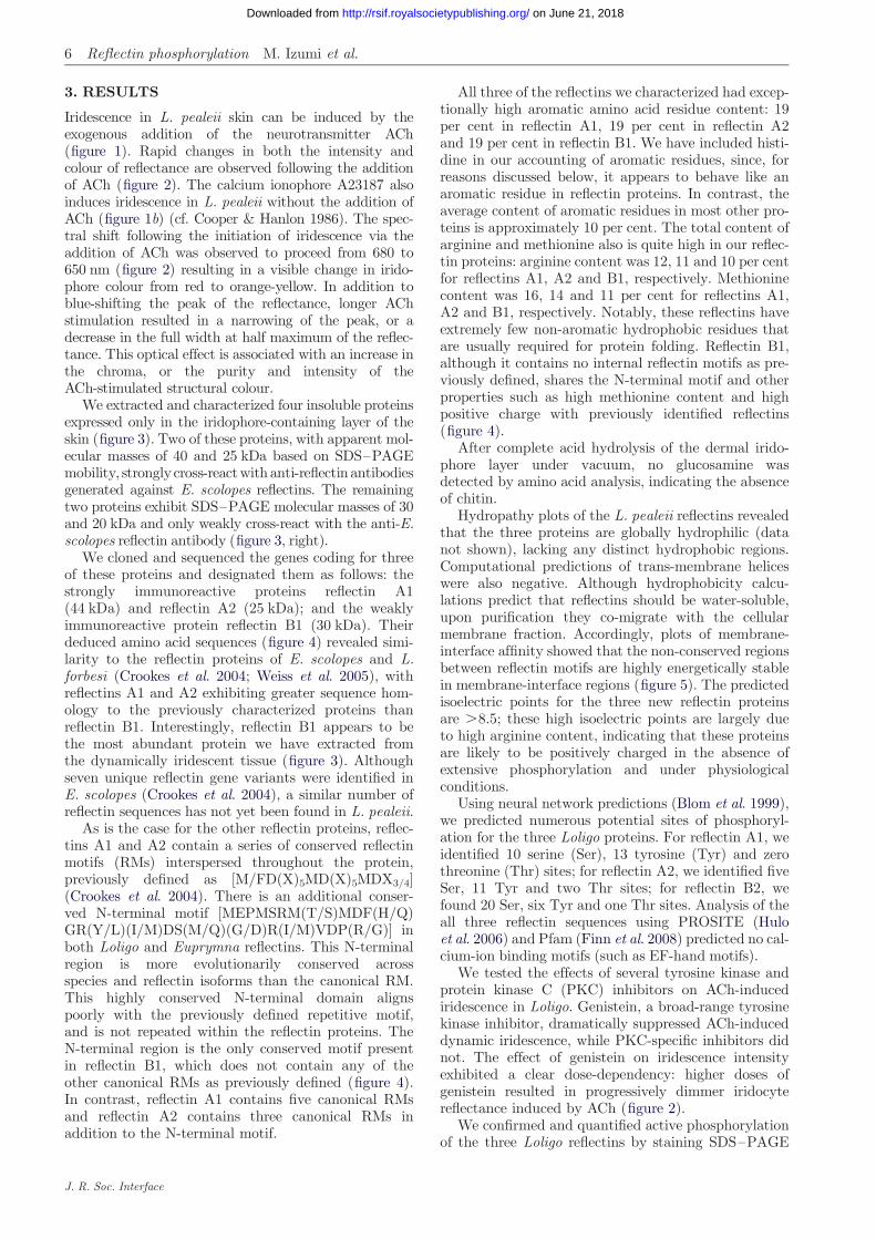
To prepare samples for quantification of phosphoryl-ation level, powdered frozen iridophore layers weredirectly dissolved in SDS–PAGE sample buffer con-taining DTT, protease inhibitor and phosphataseinhibitors. Following incubation at 988C for 10 min,the samples were centrifuged as mentioned above, andthe resulting supernatants were recovered for analysis.Preparation of RNA and cDNA messenger RNA waspurified from the freshly isolated iridophore layer ofL. pealeii. Iridophore layers were dissected from livesquid and stored in RNAlater (Qiagen, Valencia, CA,USA) at 48C or 2808C until use. Total RNA was iso-lated using TriZol (Invitrogen) followed by RNeasy(Qiagen) for further purification. First-strand cDNAwas synthesized using Super-Script III RT (Invitrogen)with an oligo(dT)20 primer. A BD SMART RACEcDNA amplification kit (Clontech, Mountain View,CA, USA) was used to prepare rapid amplification ofcomplementary DNA ends (RACE)-ready cDNA. Allthe procedures were performed according to themanufacturers’ instructions.
2.3. Amino acid sequencing and gene cloning
To clone reflectin A2, several primers were designedbased on the consensus sequence information obtainedfrom E. scolopes and L. forbesi reflectins, and the fol-lowing primer was successfully used to obtain thesequence in this manuscript (Lp2-1, ATGAACCGCTCTATGAACAGATACC). PCR using primer Lp2-1and oligo-dT was performed using single-strand cDNAas a template. The resulting PCR product containedthe 30 region of the reflectin A2 gene. To clone the 50
end of the gene, an internal primer from the resultingPCR product was designed for rapid amplification ofcDNA ends (RACE) (GTCCATCCAGCGTCCCTGCATGTCCATCTGATAGCCGGACAT).
To obtain the nucleotide sequence of reflectin A1, aninternal amino acid sequence of protein purified usingthe method above was determined by Edman degra-dation following trypsin digestion and high-performance liquid chromatography (HPLC) purification.This procedure was performed at the PAN facility atStanford University (Stanford, CA, USA). Based onthe resulting internal amino acid sequences, primerswere designed for RACE PCR (for 30 RACE, TAYAAYAAYGCNTTYTCNCARATGTGGCA and TAYAAYAAYGCNTTYAGNCARATGTGGCA; for 50 RACE,TGCCACATYTGNGARAANGC). For RACE PCR, aSMART RACE cDNA Amplification Kit was used.Additional sets of primers were designed to annealregions (Lp1-F, GTCTCCTTCGAGAACGCATGCCCTGTTGTCCGGG; Lp1-R, GGAAAATGTCCGGTTTCATTTTGAACATGCCACGCCC; Lp2-F, GTCTCACACGAGAACAAAACCTCGAAGCCACC; Lp2-R,CCCACAATTCCCAGTTGACATATATCCGACCGCC). PCR products were cloned into pCR2.1 vector(Invitrogen) using TA cloning. The sequence of theinserted fragment was analysed with an ABI 3730Capillary Electrophoresis Genetic Analyser (Applied
J. R. Soc. Interface
Bioscience) at the DNA sequencing facility at the Uni-versity of California, Davis. Sequence data wereanalysed using MACVECTOR (Accelrys Software Inc.).
To determine the sequence of reflectin B1, weobtained de novo sequence using LCMS/MS (liquidchromatogtraphy followed by two-dimensional massspectrometry). Following HPLC, purified reflectin B1was digested with sequence-grade trypsin (Roche) in0.02 per cent SDS and 50 mM Tris–HCl, pH 8.5, at aratio of 50 : 1. Peptides derived from this digestionwere separated with a PepMapC18 HPLC column.Each fraction was applied to a Waters MicromassQTOF2 tandem mass spectrometer equipped with ananoflow electrospray ionization source coupled to anAgilent 1100 nano LC system with a Zorbax 300SB-C18column that was 150 � 0.075 mm with 3.5 mm particlesize. Based on the obtained amino acid sequences(TTYGCNGAYGGNATGTAYMG) and (TTYATGGAYATGCAYTAYGAYGGNATGGGNATG) were usedas primers for RACE-PCR performed against single-strand iridophore cDNA as described above. Theresulting DNA sequences were deposited in GenBankunder accession numbers FJ824804, FJ824805 andFJ824806.
2.4. Computational methods
We computationally predicted the potential phos-phorylation sites of our novel reflectin sequences usingNetPhos 2.0 (Blom et al. 1999, 2004). Hydropathy pro-files were generated and transmembrane regions werepredicted using TMHMM (Sonnhammer et al. 1998).Calcium-binding sites were predicted using Pfam(Finn et al. 2008). The cytoplasm–membrane interfaceaffinities of the new reflectin protein sequences wereanalysed using MPEX software (Jaysinghe et al. 2006)with water-to-lipid partitioning and a 13-amino acidwindow.
2.5. One- and two-dimensional polyacrylamidegel electrophoresis
Protein samples were prepared from either ACh-treated(iridescent) or untreated (not iridescent) iridophorelayers of L. pealeii. Iridophore layers were powderedusing a mortar-and-pestle under liquid nitrogen, andthen solubilized with DeStreak Rehydration Solution(GE Healthcare), followed by centrifuging to removethe residual insoluble materials. Following treatmentwith DeStreak Rehydration Solution, the protein con-centration was determined with a two-dimensionalQuant Kit (GE Healthcare). For first-dimension iso-electric focusing (IEF), the soluble component wasloaded onto an Immobiline DryStrip gel (GE Health-care) (pH 6–11; 60 mg of total protein), followed bySDS–PAGE separation with a 10–20 % gradient gel(Bio-Rad). For IEF, a Multiphor II ElectrophoresisUnit (GE Healthcare) was used. Following two-dimensional PAGE, gels were analysed either bystaining for phospho-amino acids with Pro-Q Diamond(Molecular Probes, Inc.) or by immunodetection ofphosphotyrosine. All the experiments were performedin triplicate and the results reported are representative

4 Reflectin phosphorylation M. Izumi et al.
on June 21, 2018http://rsif.royalsocietypublishing.org/Downloaded from
of those obtained. One-dimensional PAGE was per-formed using only the second step of this two-dimensional PAGE protocol.
2.6. Immunoblotting
Immunoblotting was performed with either anti-reflectin polyclonal antibodies or horseradish peroxidase(HRP)-conjugated anti-phosphotyrosine primary anti-bodies (HRP-PY20) (BD Transduction Laboratories,San Jose, CA, USA). Following one-dimensionalSDS–PAGE or two-dimensional PAGE, proteins wereelectroblotted from the acrylamide gel to a polyvinyli-dene fluoride (PVDF) membrane. This PVDFmembrane was blocked with 1 per cent BSA in PBST(0.1% Tween-20 in PBS) for 1 h at room temperature,then incubated in PBST containing a 1 : 5000 dilutionof primary antibody for at least 1 h at room tempera-ture on a rocking platform. The membrane was thenwashed three times with PBST to remove unboundand non-specifically bound antibodies. For the use ofanti-reflectin polyclonal antibodies, a secondary anti-body (alkaline phosphatase-conjugated antirabbit)was used to identify immunopositive bands. For alkalinephosphatase detection, a 1 : 50 dilution of NBT/BCIPstock solution (Roche) in alkaline phosphatase buffer(10 mM MgCl2, 100 mM Tris–HCl, pH 9.0) was used.Following sufficient development, the reaction was termi-nated by adding stop solution (10 mM Tris–HCl, 1 mMEDTA, pH 8). For HRP detection, an ECL kit wasused (GE Healthcare). All the experiments were per-formed in triplicate and the results reported areidentified either as the averages of the normalized resultsor as representative of those obtained.
2.7. Imaging analyses with fluorescent dye
For analysis of staining with fluorescent dyes a Molecu-lar Imager FX with QUANTITY ONE software for imageanalysis (Bio-Rad Laboratories, Hercules, CA, USA)was used. IMAGEJ (NIH) was used to quantify theimmunoblot signal intensity.
2.8. Quantification of phosphorylation level
To quantify relative levels of reflectin phosphorylation,two different techniques were used: signal intensities ofreflectin staining obtained with Pro-Q Diamond stainor normalized signal intensities obtained by Westernblotting with HRP-PY20 were measured and normal-ized to the respective signal intensities obtained fromprotein quantification with SYPRO Ruby (MolecularProbes) stain, thereby providing the relative phos-phorylation level per protein. Values for samplestreated with ACh and/or genistein were furthernormalized to those obtained for the correspondingnon-ACh-treated samples.
2.9. Mass spectroscopy mapping forphosphorylation sites
Mass spectroscopy was used to identify the specificallyphosphorylated residues. Partially purified reflectinA1 and A2 were subjected to trypsin digestion followed
J. R. Soc. Interface
by matrix-assisted laser desorption/ionization-time offlight mass spectroscopy (MALDI-TOF MS). All themapping analyses were performed at the StanfordProteomics and Integrative Research Facility,Stanford University.
2.10. Activation of adaptive iridescenceand the effects of protein kinaseinhibitors on iridescence
Squid tissue samples were prepared identically for alladaptive iridescence experiments. The superficial chro-matophore- and iridophore-containing layers of skinwere surgically removed from freshly killed squid andthe iridophore-containing layer trimmed to size to fitthe Sylgard-lined incubation tray used in eachexperiment.
To activate iridescence, specimens of explanted irido-phore-containing layers were transferred either toartificial sea water (500 mM NaCl, 10 mM CaCl2,10 mM KCl, 12 mM MgCl2 and 10 mM HEPES, pH8.0) or to 0.2 mm-filtered natural sea water followedby the addition of ACh (Sigma, St Louis, MO, USA)to a final concentration of 10 mM. To investigate theeffects of various kinase inhibitors, iridophore layersamples were pre-soaked with individual kinase inhibi-tors prior to the addition of ACh for at least 20 minat room temperature at concentrations as indicated infigure 2. All the kinase inhibitors and the calcium iono-phore A23187 were purchased from EMD Bioscience.
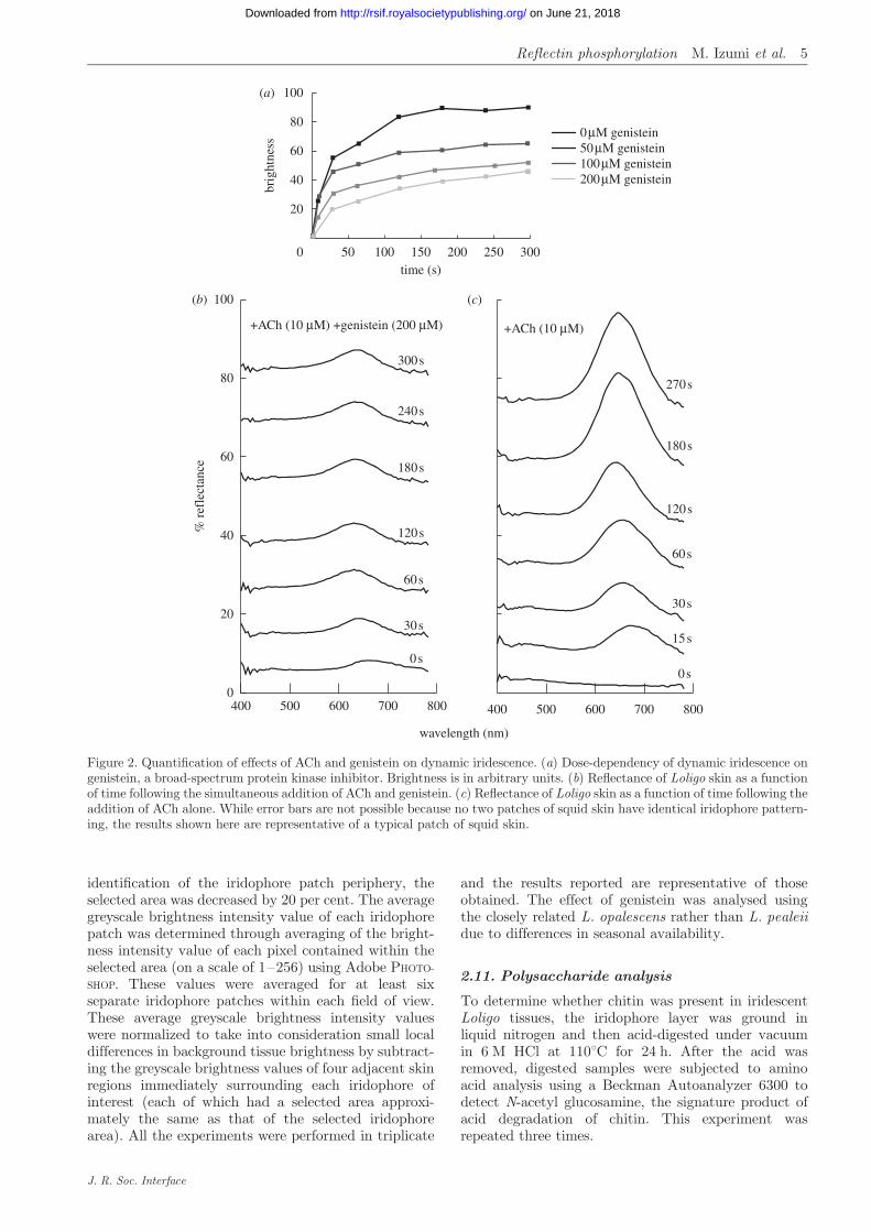
Spectral reflectance measurements before and afterthe addition of ACh were obtained with a fibre-opticspectrometer (USB2000, Ocean Optics, USA; spectrarecorded on PC, using OOIBASE 32 software) connectedvia a 1 mm diameter optical fibre, to the c-mount of adissecting microscope (Zeiss; angle of acceptance: 138;distance to specimen approx. 9 cm). At the highestmagnification of the microscope, the area of themeasured field was approximately 0.3 mm in diameter.Illumination was provided by a Schott fibre-opticmicroscope-light source, illuminating the iridophoresat an angle of incidence of approximately 108. Thislamp uses a halogen bulb with useable output for ourreflectance experiments from 380 to 800 nm, effectivelycovering the visible range of wavelengths. A diffusereflection standard (WS-1, Ocean Optics) was used tostandardize measurements. For iridophore reflectancemeasurements, squid were killed by decapitation.Small specimens of skin from the dorsal side of themantle, containing active iridophores with known opti-cal properties (see Mathger & Hanlon 2006, 2007), weredissected, streched to original size and pinned onto aSylgard-covered Petri dish. The chromatophore layerwas then peeled away, exposing the underlying irido-phore layer. To quantify the intensity of iridescencefollowing genistein treatment, the average digitalimage greyscale brightness values of individual irido-phore patches were normalized to the non-iridescentbackground and measured as a function of time. Sinceeach iridophore patch is largely elliptical, an ellipticalpixel selection tool was used to sample the interiorregion of each iridophore. In order to eliminate pro-blems associated with edge effects, following
0 µM genistein50 µM genistein100 µM genistein200 µM genistein
50 100 150 200 250 3000
20
40
60
80
100(a)
(b) (c)
400 500 600 700 8000
20
40
60
80
100
400 500 600 700 800
wavelength (nm)
time (s)
300 s
240 s
180 s
120 s
60 s
30 s
0 s
270 s
180 s
120 s
60 s
30 s
15 s
0 s
+ACh (10 µM) +genistein (200 µM) +ACh (10 µM)
brig
htne
ss
% r
efle
ctan
ce
Figure 2. Quantification of effects of ACh and genistein on dynamic iridescence. (a) Dose-dependency of dynamic iridescence ongenistein, a broad-spectrum protein kinase inhibitor. Brightness is in arbitrary units. (b) Reflectance of Loligo skin as a functionof time following the simultaneous addition of ACh and genistein. (c) Reflectance of Loligo skin as a function of time following theaddition of ACh alone. While error bars are not possible because no two patches of squid skin have identical iridophore pattern-ing, the results shown here are representative of a typical patch of squid skin.
Reflectin phosphorylation M. Izumi et al. 5
on June 21, 2018http://rsif.royalsocietypublishing.org/Downloaded from
identification of the iridophore patch periphery, theselected area was decreased by 20 per cent. The averagegreyscale brightness intensity value of each iridophorepatch was determined through averaging of the bright-ness intensity value of each pixel contained within theselected area (on a scale of 1–256) using Adobe PHOTO-
SHOP. These values were averaged for at least sixseparate iridophore patches within each field of view.These average greyscale brightness intensity valueswere normalized to take into consideration small localdifferences in background tissue brightness by subtract-ing the greyscale brightness values of four adjacent skinregions immediately surrounding each iridophore ofinterest (each of which had a selected area approxi-mately the same as that of the selected iridophorearea). All the experiments were performed in triplicate
J. R. Soc. Interface
and the results reported are representative of thoseobtained. The effect of genistein was analysed usingthe closely related L. opalescens rather than L. pealeiidue to differences in seasonal availability.
2.11. Polysaccharide analysis
To determine whether chitin was present in iridescentLoligo tissues, the iridophore layer was ground inliquid nitrogen and then acid-digested under vacuumin 6 M HCl at 1108C for 24 h. After the acid wasremoved, digested samples were subjected to aminoacid analysis using a Beckman Autoanalyzer 6300 todetect N-acetyl glucosamine, the signature product ofacid degradation of chitin. This experiment wasrepeated three times.
6 Reflectin phosphorylation M. Izumi et al.
on June 21, 2018http://rsif.royalsocietypublishing.org/Downloaded from
3. RESULTS
Iridescence in L. pealeii skin can be induced by theexogenous addition of the neurotransmitter ACh(figure 1). Rapid changes in both the intensity andcolour of reflectance are observed following the additionof ACh (figure 2). The calcium ionophore A23187 alsoinduces iridescence in L. pealeii without the addition ofACh (figure 1b) (cf. Cooper & Hanlon 1986). The spec-tral shift following the initiation of iridescence via theaddition of ACh was observed to proceed from 680 to650 nm (figure 2) resulting in a visible change in irido-phore colour from red to orange-yellow. In addition toblue-shifting the peak of the reflectance, longer AChstimulation resulted in a narrowing of the peak, or adecrease in the full width at half maximum of the reflec-tance. This optical effect is associated with an increase inthe chroma, or the purity and intensity of theACh-stimulated structural colour.
We extracted and characterized four insoluble proteinsexpressed only in the iridophore-containing layer of theskin (figure 3). Two of these proteins, with apparent mol-ecular masses of 40 and 25 kDa based on SDS–PAGEmobility, strongly cross-reactwith anti-reflectin antibodiesgenerated against E. scolopes reflectins. The remainingtwo proteins exhibit SDS–PAGE molecular masses of 30and 20 kDa and only weakly cross-react with the anti-E.scolopes reflectin antibody (figure 3, right).
We cloned and sequenced the genes coding for threeof these proteins and designated them as follows: thestrongly immunoreactive proteins reflectin A1(44 kDa) and reflectin A2 (25 kDa); and the weaklyimmunoreactive protein reflectin B1 (30 kDa). Theirdeduced amino acid sequences (figure 4) revealed simi-larity to the reflectin proteins of E. scolopes and L.forbesi (Crookes et al. 2004; Weiss et al. 2005), withreflectins A1 and A2 exhibiting greater sequence hom-ology to the previously characterized proteins thanreflectin B1. Interestingly, reflectin B1 appears to bethe most abundant protein we have extracted fromthe dynamically iridescent tissue (figure 3). Althoughseven unique reflectin gene variants were identified inE. scolopes (Crookes et al. 2004), a similar number ofreflectin sequences has not yet been found in L. pealeii.
As is the case for the other reflectin proteins, reflec-tins A1 and A2 contain a series of conserved reflectinmotifs (RMs) interspersed throughout the protein,previously defined as [M/FD(X)5MD(X)5MDX3/4](Crookes et al. 2004). There is an additional conser-ved N-terminal motif [MEPMSRM(T/S)MDF(H/Q)GR(Y/L)(I/M)DS(M/Q)(G/D)R(I/M)VDP(R/G)] inboth Loligo and Euprymna reflectins. This N-terminalregion is more evolutionarily conserved acrossspecies and reflectin isoforms than the canonical RM.This highly conserved N-terminal domain alignspoorly with the previously defined repetitive motif,and is not repeated within the reflectin proteins. TheN-terminal region is the only conserved motif presentin reflectin B1, which does not contain any of theother canonical RMs as previously defined (figure 4).In contrast, reflectin A1 contains five canonical RMsand reflectin A2 contains three canonical RMs inaddition to the N-terminal motif.
J. R. Soc. Interface
All three of the reflectins we characterized had excep-tionally high aromatic amino acid residue content: 19per cent in reflectin A1, 19 per cent in reflectin A2and 19 per cent in reflectin B1. We have included histi-dine in our accounting of aromatic residues, since, forreasons discussed below, it appears to behave like anaromatic residue in reflectin proteins. In contrast, theaverage content of aromatic residues in most other pro-teins is approximately 10 per cent. The total content ofarginine and methionine also is quite high in our reflec-tin proteins: arginine content was 12, 11 and 10 per centfor reflectins A1, A2 and B1, respectively. Methioninecontent was 16, 14 and 11 per cent for reflectins A1,A2 and B1, respectively. Notably, these reflectins haveextremely few non-aromatic hydrophobic residues thatare usually required for protein folding. Reflectin B1,although it contains no internal reflectin motifs as pre-viously defined, shares the N-terminal motif and otherproperties such as high methionine content and highpositive charge with previously identified reflectins(figure 4).
After complete acid hydrolysis of the dermal irido-phore layer under vacuum, no glucosamine wasdetected by amino acid analysis, indicating the absenceof chitin.
Hydropathy plots of the L. pealeii reflectins revealedthat the three proteins are globally hydrophilic (datanot shown), lacking any distinct hydrophobic regions.Computational predictions of trans-membrane heliceswere also negative. Although hydrophobicity calcu-lations predict that reflectins should be water-soluble,upon purification they co-migrate with the cellularmembrane fraction. Accordingly, plots of membrane-interface affinity showed that the non-conserved regionsbetween reflectin motifs are highly energetically stablein membrane-interface regions (figure 5). The predictedisoelectric points for the three new reflectin proteinsare .8.5; these high isoelectric points are largely dueto high arginine content, indicating that these proteinsare likely to be positively charged in the absence ofextensive phosphorylation and under physiologicalconditions.
Using neural network predictions (Blom et al. 1999),we predicted numerous potential sites of phosphoryl-ation for the three Loligo proteins. For reflectin A1, weidentified 10 serine (Ser), 13 tyrosine (Tyr) and zerothreonine (Thr) sites; for reflectin A2, we identified fiveSer, 11 Tyr and two Thr sites; for reflectin B2, wefound 20 Ser, six Tyr and one Thr sites. Analysis of theall three reflectin sequences using PROSITE (Huloet al. 2006) and Pfam (Finn et al. 2008) predicted no cal-cium-ion binding motifs (such as EF-hand motifs).
We tested the effects of several tyrosine kinase andprotein kinase C (PKC) inhibitors on ACh-inducediridescence in Loligo. Genistein, a broad-range tyrosinekinase inhibitor, dramatically suppressed ACh-induceddynamic iridescence, while PKC-specific inhibitors didnot. The effect of genistein on iridescence intensityexhibited a clear dose-dependency: higher doses ofgenistein resulted in progressively dimmer iridocytereflectance induced by ACh (figure 2).
We confirmed and quantified active phosphorylationof the three Loligo reflectins by staining SDS–PAGE

6 7 8 9 10 11
Pro-Q stain
PY-20 AB
reflectin A1
reflectin B1reflectin A2
reflectin B2
reflectin A1reflectin B1reflectin A2
reflectin B2
SDS–PAGE
64–49–
37–
26–19–
15–
64–49–
37–
26–19–
15–
anti-reflectinimmunoblot
(a)
(b)
Figure 3. Electrophoresis, immunoreactivity and phosphorylation of reflectins. Centre panels: overlay of two artificially colouredtwo-dimensional electrophoretic blots of reflectin proteins stained with Pro-Q Diamond stain for phosphoamino acids (a) or PY-20antibody against phosphotyrosine (b). Grey blot shows protein from unactivated control skin. Red blot shows protein fromACh-activated, iridescent skin. Populations of multiple phosphorylation states exist for each of the major reflectins in boththe control and the treated state. Upon ACh activation, these populations of phosphorylation states apparently shift towardscharge neutrality. Left panel: SDS–PAGE of iridophore layer. Right panel: immunoblot of iridophore layer with antibodyto E. scolopes reflectins, showing that A-group reflectins are strongly cross-reactive, while B-group reflectins are weaklycross-reactive.
Reflectin phosphorylation M. Izumi et al. 7
on June 21, 2018http://rsif.royalsocietypublishing.org/Downloaded from
separated reflectins with Pro-Q Diamond, a stainspecific for phosphoamino acids. We compared stainingintensities of ACh-treated tissue with those in theACh þ genistein-treated tissue. These intensities werenormalized to those of untreated control tissue. Wefound that both reflectin A1 and reflectin A2 showedhigher phosphorylation levels in the ACh-only treat-ment (figure 6) than in either the ACh þ genisteintreatment or the control treatment. The ratio of nor-malized staining intensity between the ACh-only andthe ACh þ genistein-treated tissues increased by 130and 240 per cent for reflectins A1 and A2, respectively(figure 6). In contrast, the net phosphorylation stainingintensity of reflectin B2 decreased by 25 per cent(figure 6).
Since Pro-Q Diamond cannot differentiate betweendifferent phosphorylated amino acids, we specificallyquantified levels of tyrosine phosphorylation via Wes-tern blotting with PY-20, a phosphotyrosine-specificantibody. Consistent with the results from Pro-Q stain-ing, phosphotyrosine levels for the ACh-treated tissueincreased by 170 and 290 per cent for reflectins A1and A2, respectively, when compared with the ACh þgenistein treatment (figure 6). The control values areincluded in the data presented in figure 5, as the datafor the experimental treatments are plotted as the percent change relative to the control values obtainedfrom the corresponding (control) lane of the gel. Wewere unable to detect reflectin B1 with PY-20 in anytissue or treatment, indicating that tyrosine phos-phorylation is low, not present, or especially labile for
J. R. Soc. Interface
this protein. PY-20 sensitivity is known to be depen-dent on sequence context, possibly contributing tothis observation. Alternatively, this result may suggestthat phosphorylation of serine and/or threonine domi-nates in the active state for reflectin B1, in contrast tothe phosphorylation of tyrosine residues in the reflectinA sequences.
To identify the locations and identities of the reflec-tin residues phosphorylated in the ACh-activatedtissue, MALDI-TOF MS was performed on trypsindigests of reflectins A1 and reflectin A2 (table 1).This technique revealed that Tyr14 and Tyr127 ofreflectin A1 and Tyr12, Tyr 214, Ser218 and Tyr223of reflectin A2 were phosphorylated in the active state(figure 4), consistent with our stain-based analysis ofphosphorylation. Reflectin A1 had an additional pep-tide (WMDAQGRFNNQFGQMWHGR) that showeda mass shift consistent with phosphorylation despitethe absence of tyrosine, serine or threonine. This mayindicate a histidine phosphorylation, as indicated infigure 4. In those cases in which more than one phos-phorylatable amino acid was present in a mass-shiftedpeptide, we used the congruence of our computationalphosphorylation predictions and our MS data to ident-ify the specific residues that were phosphorylated.Interestingly, all but one phoshorylation event occurredoutside the conserved reflectin domains. It is possiblethat additional non-tyrosine phosphoamino acids mayhave been missed by our analyses because of their labilityduring protein isolation and purification (Sickmann &Meyer 2001; Mikesh et al. 2006).

Figure 4. Sequence alignment and MALDI-TOF confirmed phosphorylation sites of novel reflectin proteins. The three novelreflectin proteins characterized here are aligned with reflectin 1B. Phosphorylated residues identified in MALDI-TOF MSanalysis are highlighted in red. The dark grey box and white text indicates the conserved N-terminal peptide common to allreflectins. The lighter grey boxes with black text indicate the conserved reflectin motif found in all E. scolopes reflectins, butonly the reflectin A sequences of Loligo. E.S.E. scolopes.
8 Reflectin phosphorylation M. Izumi et al.
on June 21, 2018http://rsif.royalsocietypublishing.org/Downloaded from
Two-dimensional PAGE revealed the populations ofphosphorylated states of the reflectins before and afteriridescence activation (figure 3). The multiple phos-phorylated states of each isoform reflect the fact thateach reflectin contains multiple residues that can bephosphorlyated. However, because reflectin A1 hasvery limited solubility in the IEF buffer in the absenceof detergent (as required for two-dimensional PAGE),
J. R. Soc. Interface
and the different reflectin isoforms exhibit variable solu-bilities in detergent-free buffer, it proved difficult toquantitatively compare phosphorylation levels betweenreflectin species in the same two-dimensional PAGEexperiment. In contrast, we found that reproduciblecomparisons of the levels of phosphorylation can beobtained from the analyses of one-dimensional gelelectrophoresis conducted in the presence of SDS.

ener
gy o
f in
terf
ace
inte
ract
ion
(kca
l mol
–1)
reflectin A1
reflectin A2
reflectin B1
0
0
0
Figure 5. Interface affinity of reflectin proteins. Traces indi-cate the calculated energy of interface interaction between ahypothetical membrane surface and 13-residue ‘windows’ ofthe reflectin proteins as a function of position in the proteins.Values are calculated by the method of Jaysinghe et al.(2006). Grey scale bar shows 2 kcal mol21; negative valuesindicate regions of potential membrane association, whilepositive values indicate regions predicted to be cytosolic.Blue regions are the conserved ‘reflectin motifs’. Black regionsare the intervening non-conserved regions. Dashed lines showthe hypothesized interface interaction energy after phos-phorylation. Red dots indicate residues phosphorylatedconcomitant with the activation of iridescence by ACh.When a residue becomes phosphorylated, the surroundingregions of the protein are predicted to shift from being mem-brane-associated to cytosolic. Sites of phosphorylation inreflectin 2B have not yet been identified.
pro-Q stain PY-20 immunoblotA1 A2 B1 A1 A2
0
30
60
90
120
–30
chan
ge in
sta
inin
g in
tens
ityre
lativ
e to
non
-iri
desc
ing
skin
(%
)
Figure 6. Changes in phosphorylation with ACh treatmentand genistein þ ACh treatment of iridophores. Left, changesin Pro-Q Diamond staining intensity of reflectin proteinsupon treatment with 10 mM ACh (red columns) or 10 mMACh þ genistein at indicated concentrations (grey columns).Data are per cent differences relative to an untreated control,here shown as zero. All the analyses were done in triplicate,and error bars show+ s.d. Right, changes in PY-20 anti-phos-photyrosine antibody upon treatment with ACh or ACh þgenistein. Colours and values are the same as in left panel.
Reflectin phosphorylation M. Izumi et al. 9
on June 21, 2018http://rsif.royalsocietypublishing.org/Downloaded from
Our two-dimensional PAGE data enabled us to evalu-ate the shifts in the populations of differentiallyphosphorylated states of the reflectins from untreatedand ACh-treated samples. Thus, staining with Pro-QDiamond and PY-20 revealed that reflectin A1 andreflectin A2 each consist of populations with several dis-crete phosphorylated states for both activated andinactivated tissues (figure 3). Immunodetection withPY-20 (and, to a lesser degree, staining with Pro-QDiamond) revealed that the most acidic, extensivelyphosphorylated reflectin A2 molecules possess an isoelec-tric point of 7, and are in significantly higher abundancein the activated tissue. Reflectin B2 also appeared toexhibit a higher degree of phosphorylation, as deter-mined with Pro-Q Diamond, following the addition ofACh (figure 3). In contrast, Pro-Q Diamond stainingshows that reflectin B1 became more basic (less phos-phorylated) upon activation, and also is present inseveral distinct phosphorylated populations (figure 3).In general, the reflectin proteins extracted from theACh-treated tissues appeared to migrate slightly fartherin the first dimension of two-dimensional PAGE thandid the proteins from the controls. Although we cannot
J. R. Soc. Interface
rule out imperfect alignment of gel images, it seems prob-able that this is caused by increased negative charge onthe activated proteins due to phosphorylation.
4. DISCUSSION
We tested the hypothesis that, in loliginid squid, themuscarinic ACh cascade leading to dynamic iridescencehas direct biochemical effects on the reflectin proteinsconstituting the squid iridosomes. We characterizedthe reflectins found in the dynamically tunable irido-somes and found that these reflectin proteins appearto be progressively phosphorylated or dephosphorylatedupon stimulation of iridocytes with ACh. These changesin phosphorylation are concurrent with the activationand deactivation of iridescence. The dose-dependentability of genistein, a broad-spectrum tyrosine kinaseinhibitor, to quench the stimulation of iridescence aswell as prevent the phosphorylation of reflectin furthersupports our hypothesis.
Genistein lowered the phosphorylation levels of reflec-tin proteins below those of non-iridescent, untreatedcontrols. Because baseline protein phosphorylationstates are maintained by active homeostasis in cells,this finding supports an active role for reflectin phos-phorylation in iridescence modulation. In general, cellsexert multiple mechanistic layers of intricate controlover protein phosphorylation state (Ubersax & Ferrell2007). This phenomenon, together with our findingsshowing that reflectin phosphorylation is dynamic, pro-vides a model for the intricate homeostatic control bywhich iridocytes regulate the brightness and colour ofiridescence.
Because PKC inhibitors did not achieve the sameiridescence-suppressing effect as genistein, we hypothesizea key role for tyrosine kinases in iridescence activation.However, because commercially available PKC inhibi-tors may be specific only to mammalian proteins, andbecause their effects on cephalopod protein kinases areunknown, we cannot yet identify the specific protein

Table 1. Reflectin phosphorylations predicted from MALDI-TOF MS. Peptide sequences identified by MALDI-TOF MS, withasterisks indicating identified phosphorylated residues.
protein ID residues mass (a.m.u.) predicted phoshorylated tryptic peptide
Refl A1 10–17 1181.564 LYNMY*RNK102–120 2415.548 WMDAQGRFNNPFGQMWH*GR121–136 1936.058 QGHYPGY*MSSHSMYGR
Refl A2 4–17 2015.897 YMMRHRPMY*SNMYR121–136 1936.058 QGHYPGY*MSSHSMYGR207–230 3350.899 WMDTQGRY*MDPS*WSNMY*DNYNSWY
10 Reflectin phosphorylation M. Izumi et al.
on June 21, 2018http://rsif.royalsocietypublishing.org/Downloaded from
kinases that mediate the ACh-induced phosphorylationof the reflectins studied here.
Phosphorylation is known to trigger dramaticchanges in the conformations and assembly of manyproteins and extensive tyrosine phosphorylation is lar-gely responsible for the many rapid changes in cellphysiology, such as in insulin signaling (Karim et al.2006; Schmelzle et al. 2006). This signaling paradigmis a possible precedent for the dynamic iridescencemechanism outlined below.
We suggest three possible mechanisms that may workindependently or in concert to account for the observedchanges in iridescence. First, phosphorylation mayinduce changes in the net charge of the reflectins,which may directly alter reflectin protein–protein inter-action and assembly. These changes could play a role inaltering the refractive index, thickness and/or spacingof the reflectin-containing iridosomes, leading to theobserved changes in reflectance. Second, phosphorylationof reflectins may trigger changes in the Gibbs–Donnanequilibrium across the iridosome membranes, affecting anumber of variables such as protein aggregation, proteinmobility, turgor pressure or thickness of the membrane-bound iridosomes. Third, phosphorylation of reflectinscould modulate the affinity of reflectins for cellmembranes, shifting assembly dynamics toward auto-aggregation and hierarchical self-assembly (cf. Hanlonet al. 1990). Consistent with the third hypothesis, reflec-tins are characterized, in part, by their extremely highcontent of aromatic and arginine residues. These residuesare both preferentially soluble in membrane–cytoplasminterfaces (Wimley & White 1996).
Membrane–cytoplasm interfaces are known to exhi-bit unique solubility properties relative to aqueouscytoplasm. Polar aromatic residues are particularlystable in this microenvironment, while very hydro-phobic and very hydrophilic residues are not (Yauet al. 1998; Granseth et al. 2005). Arginine, because ofits positive charge and long hydrophobic stem, exhibits‘snorkeling’ behaviour from the interface into the mem-brane (Killian & von Heijne 2000). Reflectinsare, indeed, characterized by their high content ofpolar aromatic residues and arginines, and severallines of evidence suggest that they are intrinsicallyunstructured, with no likely transmembrane, alpha-helix or beta-sheet regions. E. scolopes reflectins alsohave high arginine, hydrophobic and aromatic content(Crookes et al. 2004). The first study to characterizethe reflectins described them as ‘methionine-richmembrane-associated proteins’ and demonstrated that
J. R. Soc. Interface
they have a high affinity for assembly with microsomalmembranes when translated in vitro (Weiss et al. 2005).Given these data, and what is known about thebiophysics of membrane interfaces, we hypothesizethat reflectins may exhibit energetically favourableinteractions with membrane interfaces.
This suggestion is consistent with our observationthat the reflectins co-purify with cell membranes, yetlack any traditionally recognized transmembranedomains and small hydrophobic amino acids. ReflectinsA1 and A2 exhibit an interesting periodicity of domainspotentially capable of interacting with membrane inter-faces, with the proteins apparently alternating betweeninterface-stable regions and hydrophilic domainsapproximately every 50 amino acids (figure 5). ReflectinB2, which exhibits the opposite phosphorylation pat-tern upon ACh stimulation from reflectins A1 and A2,does not show the same periodicity, consistent withthe possibility of a different assembly dynamic for thisprotein. Interestingly, the calculated affinity of specificdomains of reflectins A1 and A2 for membrane is pre-dicted to be abolished upon the observedphosphorylation of those sites (figure 5). Phosphoryl-ations occur in regions of the protein that prior tophosphorylation are predicted to be highly stable inmembrane interfaces, while the conserved RMs arenot phosphorylated, and are predicted to be morestable in the cytoplasmic compartment of the cell.After phosphorylation (mimicked for these calculationsby substitution of the original residues with negativelycharged glutamate residues), the phosphorylatedprotein regions are predicted to change from beinginterface-stable to being stable in the cytoplasm.
Although iridescence is induced by both ACh andelevation of intracellular calcium concentrations viaadministration of a calcium ionophore (see figure 1), wefind no evidence for any calcium ion-binding motifs inthe newly identified reflectins. Instead, our result suggeststhat calcium functions as a second messenger in thesignal transduction cascade rather than as a direct modu-lator of the reflectin proteins. Support for our suggestionof a receptor-activated G protein-dependent signal trans-duction pathway is provided by our observation thatiridescence is activated by cholera toxin (an activator ofthe G protein) in the absence of ACh (data not shown).
Reflectins A1 and A2 from Loligo are most similar tothe reflectin 1 group from the static iridophores inEuprymna, in both primary sequence and immunoreac-tivity to antibodies against Euprymna reflectin.Therefore, the protein reflectin B1 may be especially
Reflectin phosphorylation M. Izumi et al. 11
on June 21, 2018http://rsif.royalsocietypublishing.org/Downloaded from
interesting in discerning important functional and evol-utionary differences between static iridescence inE. scolopes and dynamic iridescence in Loligo. Unlikethe reflectins A1 and A2, reflectin B1 has no conservedRMs and instead contains only the initial N-terminalpeptide characteristic of reflectins. Reflectin B1 ismore acidic than reflectins A1 or A2, and it exhibitsthe opposite phosphorylation response to ACh stimu-lation relative to reflectins A1 or A2. In addition, acomparison of Pro-Q staining with PY-20 stainingsuggests that reflectin B1 may be predominantlyserine/threonine-phosphorylated, rather than tyrosine-phosphorylated. Given that reflectins A1 and A2 andreflectin B1 have differing phosphorylation responsesto ACh, the reflectin As and reflectin Bs could operateunder separate control mechanisms within a singlecell. These characteristics may provide importantclues for future work in understanding how RMs, phos-phorylation and charge modulation of reflectinscontribute to the ability of loliginid iridocytes to modu-late their reflectance.
Our data demonstrate changes in the relative levelsof phosphorylation of the reflectin proteins upon AChstimulation of the adaptive iridophores of Loligo.Detailed quantitation and mapping of the sites of phos-phorylation by MS are now in progress to help provide adeeper mechanistic understanding of the biophysicalrole of phosphorylation in tuning the Bragg reflectorsin this system.
Finally, in contrast to prior reports (see Denton &Land 1971; Cloney & Brocco 1983), we have shownthat chitin is not present in measurable quantities in iri-descent squid skin, as indicated by the completeabsence of glucosamine after complete acid hydrolysis(data not shown). This should help end historical specu-lation that small movements of chitin platelets could beresponsible for dynamic iridescence in squid.
Our observation of ACh-dependent changes inreflectin phosphorylation accompanying increasedreflectance suggests a molecular mechanism for adap-tive control in this biophotonic system. The reciprocalnature of the phosphorylation of reflectins A1 and A2versus reflectin B presents interesting possibilities forfurther probing the biophysical phenomena that pro-mote dynamic iridescence. We have traced the signaltransduction cascade in this system from the transmit-ter to a likely final target: the reflectin proteins thatapparently change their conformation or assembly toreversibly create the photonic structure. Further eluci-dation of the underlying molecular mechanismsregulating the intensity and colour of the tunableBragg reflectors in this system can be expected toshed new insight into the evolutionary origin of adap-tive reflectance in cephalopods, and may also inspirenovel synthesis strategies for production of the nextgeneration of tunable photonic materials for advancedapplications.
We gratefully acknowledge support from Anteon contractF33615-03-D-5408 to the Marine Biological Laboratory,Woods Hole, MA, and grant no. W911NF-06-1-0285 fromthe Army Research Office to D.E.M. We also thankMargaret McFall-Ngai (NIH AI50611) and Edward G. Ruby
J. R. Soc. Interface
(NIH RR 12294) for providing the anti-reflectin antibodies,Tim Athens for providing live squid for analysis, Dr JamesPavlovich for de novo amino acid sequence analyses andDrs Paul Hansma, Igor Mezic, Yoshiko Okamura, KathyFoltz and Daniel Alkon for their helpful discussions andsuggestions.
REFERENCES
Blom, N., Gammeltoft, S. & Brunak, S. 1999 Sequence- andstructure-based prediction of eukaryotic protein phos-phorylation sites. J. Mol. Biol. 294, 1351–1362. (doi:10.1006/jmbi.1999.3310)
Blom, N., Sicheritz-Ponten, T., Gupta, R. & Gammeltoft, S.2004 Prediction of posttranslational glycosylation andphosphorylation of proteins from the amino acid sequence.Proteomics 4, 1633–1649. (doi:10.1002/pmic.200300771)
Carlton, F. C. 1903 The color changes in the skin of theso-called Florida Chameleon, Anolis Carolinensis Cuv.Proc. Am. Acad. Arts Sci. 39, 259–276.
Cloney, R. A. & Brocco, S. L. 1983 Chromatophore organs,reflector cells, iridocytes and leucophores in cephalopods 1.Integr. Comp. Biol. 23, 581–592. (doi:10.1093/icb/23.3.581)
Cooper, K. M. & Hanlon, R. T. 1986 Correlation of iridescencewith changes in iridophore platelet ultrastructure in thesquid. Lolliguncula brevis. J. Exp. Biol. 121, 451–455.
Cooper, K. M., Hanlon, R. T. & Budelmann, B. U. 1990Physiological color change in squid iridophores. II. Ultra-structural mechanisms in Lolliguncula brevis. Cell TissueRes. 259, 15–24. (doi:10.1007/BF00571425)
Crookes, W. J., Ding, L., Huan, Q. L., Kimbell, J. R.,Horwitz, J. & McFall-Ngai, M. J. 2004 Reflectins: theunusual proteins of squid reflective tissues. Science 303,235–238. (doi:10.1126/science.1091288)
Denton, E. J. 1970 Review lecture: on the organization ofreflecting surfaces in some marine animals. Phil.Trans. R. Soc. Lond. B 258, 285–312. (doi:10.1098/rstb.1970.0037)
Denton, E. J. & Land, M. F. 1971 Mechanism of reflexion insilvery layers of fish and cephalopods. Proc. R. Soc.Lond. B 178, 43–61. (doi:10.1098/rspb.1971.0051)
Finn, R. D. et al. 2008 The Pfam protein families database.Nucl. Acids Res. 36, D281. (doi:10.1093/nar/gkm960)
Granseth, E., von Heijne, G. & Elofsson, A. 2005 A study ofthe membrane-water interface region of membrane pro-teins. J. Mol. Biol. 346, 377–385. (doi:10.1016/j.jmb.2004.11.036)
Haass, C. & Selkoe, D. J. 1993 Cellular processing of b-amy-loid precursor protein and the genesis of amyloid b-peptide. Cell 75, 1039–1042.
Hanlon,R.T.,Cooper, K.M., Budelmann,B.U.& Pappas,T. C.1990 Physiological color change in squid iridophores I.Behavior, morphology and pharmacology in Lolligunculabrevis. Cell Tissue Res. 259, 3–14. (doi:10.1007/BF00571424)
Hulo, N., Bairoch, A., Bulliard, V., Cerutti, L., De Castro, E.,Langendijk-Genevaux, P., Pagni, M. & Sigrist, C. J. A.2006 The PROSITE database. Nucl. Acids Res. 34,D227–D230. (doi:10.1093/nar/gkj063)
Huxley, A. F. 1968 A theoretical treatment of the reflexion oflight by multilayer structures. J. Exp. Biol. 48, 227–245.
Jaysinghe, S., Hristova, K., Wimley, W., Snider, C. & White,S. H. 2006. See http://blanco.biomol.uci.edu/mpex.
Karim, C. B., Zhang, Z., Howard, E. C. & Torgersen, K. D.2006 Phosphorylation-dependent conformational switchin spin-labeled phospholamban bound to SERCA. J.

12 Reflectin phosphorylation M. Izumi et al.
on June 21, 2018http://rsif.royalsocietypublishing.org/Downloaded from
Mol. Biol. 358, 1032–1040. (doi:10.1016/j.jmb.2006.02.051)
Killian, J. A. & von Heijne, G. 2000 How proteins adapt to amembrane-water interface. TIBS 25, 429–434.
Kobelt, F. & Linsenmair, K. E. 1986 Adaptations of the reedfrog Hyperolius viridiflavus (Amphibia, Anura, Hyperolii-dae) to its arid environment. Oecologia 68, 533–541.(doi:10.1007/BF00378768)
Kramer, R. M., Crookes-Goodson, W. J. & Naik, R. R. 2007The self-organizing properties of squid reflectin protein.Nat. Mater. 6, 533–538. (doi:10.1038/nmat1930)
Land, M. F. 1972 The physics and biology of animal reflectors.Progr. Biophys. Mol. Biol. 24, 75–106. (doi:10.1016/0079-6107(72)90004-1)
Mathger, L. M. & Hanlon, R. T. 2006 Anatomical basis forcamouflaged polarized light communication in squid.Biol. Lett. 2, 494–496. (doi:10.1098/rsbl.2006.0542)
Mathger, L. M. & Hanlon, R. T. 2007 Malleable skin color-ation in cephalopods: selective reflectance, transmissionand absorbance of light by chromatophores and irido-phores. Cell tissue Res. 329, 179–186.
Mathger, L. M., Land, M. F., Siebeck, U. E. & Marshall, N. J.2003 Rapid colour changes in multilayer reflecting stripesin the paradise whiptail, Pentapodus paradiseus. J. Exp.Biol. 206, 3607–3613. (doi:10.1242/jeb.00599)
Mathger, L. M., Collins, T. F. T. & Lima, P. A. 2004 The roleof muscarinic receptors and intracellular Ca2þ in the spec-tral reflectivity changes of squid iridophores. J. Exp. Biol.207, 1759–1769. (doi:10.1242/jeb.00955)
Menter, D. G., Tchen, T. T., Obika, M. & Taylor, J. D. 1979Leucophores and Iridophores of Fundulus heteroclitus.J. Morphol. 160, 102–120.
Messenger, J. B. 2001 Cephalopod chromatophores:neurobiology and natural history. Biol. Rev. 76, 473–528.
Mikesh, L.M.,Ueberheide,B., Chi,A.,Coon, J. J.&Syka, J. E.P.2006 The utility of ETD mass spectrometry in proteomicanalysis. BBA-Proteins Proteom. 1764, 1811–1822. (doi:10.1016/j.bbapap.2006.10.003)
Mirow, S. 1972 Skin color in the squids Loligo pealii andLoligo opalescens. Cell Tissue Res. 125, 176–190.
J. R. Soc. Interface
Osorio, D. & Ham, A. D. 2002 Spectral reflectance and direc-tional properties of structural coloration in bird plumage.J. Exp. Biol. 205, 2017–2027.
Rohrlich, S. T. & Rubin, R. W. 1975 Biochemical characteriz-ation of crystals from the dermal iridophores of achameleon Anolis carolinensis. J. Cell Biol. 66, 635–645.(doi:10.1083/jcb.66.3.635)
Schmelzle, K., Kane, S., Gridley, S. & Lienhard, G. E. 2006 Tem-poral dynamics of tyrosine phosphorylation in insulinsignaling. Diabetes 55, 2171–2179. (doi:10.2337/db06-0148)
Sickmann, A. & Meyer, H. E. 2001 Phosphoamino acidanalysis. Proteomics 1, 200–206. (doi:10.1002/1615-9861(200102)1:2,200::AID-PROT200.3.0.CO;2-V)
Sonnhammer, E. L., von Heijne, G. & Krogh, A. 1998 Ahidden Markov model for predicting transmembranehelices in protein sequences. Proc. Int. Conf. Intell. Syst.Mol. Biol. Proc. 6, 175–182.
Takemoto, L. & Boyle, D. 2000 Increased deamidation ofasparagine during human senile caractogenesis. Mol.Vision 6, 164–168.
Ubersax, J. A. & Ferrell Jr, J. E. 2007 Mechanisms of speci-ficity in protein phosphorylation. Nat. Rev. Mol. CellBiol. 8, 530–541. (doi:10.1038/nrm2203)
Vigneron, J. P., Colomer, J. F., Vigneron, N. & Lousse, V.2005 Natural layer-by-layer photonic structure in the squa-mae of Hoplia coerulea (Coleoptera). Phys. Rev. E 72,061904. (doi:10.1103/PhysRevE.72.061904)
Vukusic, P. & Sambles, J. R. 2003 Photonic structures inbiology. Nature 424, 852–855. (doi:10.1038/nature01941)
Weiss, J. L., Evans, N. A., Ahmed, T., Wrigley, J. D. J. &Khan, S. 2005 Methionine-rich repeat proteins: a familyof membrane-associated proteins which contain unusual.BBA Biomembr. 1668, 164–174. (doi:10.1016/j.bbamem.2004.11.014)
Wimley, W. C. & White, S. H. 1996 Experimentally determinedhydrophobicity scale for proteins at membrane interfaces.Nat. Struct. Biol. 3, 842–848. (doi:10.1038/nsb1096-842)
Yau, W., Wimley, W. C., Gawrisch, K. & White, S. H. 1998The preference of tryptophan for membrane interfaces.Biochemistry 37, 14713–14718. (doi:10.1021/bi980809c)