j. virol. doi:10.1128/jvi.01534-07 2 3 4 5 accepted...
TRANSCRIPT

1
Title: 1
Broad Anti-Retroviral Activity and Resistance Profile of a Novel Human 2
Immunodeficiency Virus Integrase Inhibitor, Elvitegravir (JTK-303/GS-9137) 3
4
Authors: 5
Kazuya Shimura1, Eiichi Kodama
1*, Yasuko Sakagami
1, Yuji Matsuzaki
2, Wataru 6
Watanabe2†
, Kazunobu Yamataka2, Yasuo Watanabe
2, Yoshitsugu Ohata
2, Satoki Doi
3, 7
Motohide Sato2, Mitsuki Kano
2, Satoru Ikeda
2, and Masao Matsuoka
1 8
9
Affiliations: 10
1 Laboratory of Virus Immunology, Institute for Virus Research, Kyoto University, 11
53 Kawaramachi, Shogoin, Sakyo-ku, Kyoto 606-8507, Japan 12
2 Japan Tobacco Inc., Central Pharmaceutical Research Institute, 1-1 Murasaki-cho, 13
Takatsuki, Osaka 569-1125, Japan 14
3 Japan Tobacco Inc., Central Pharmaceutical Research Institute, Pharmaceutical 15
Frontier Research Laboratories, 1-13-2 Fukuura, Kanazawa-Ku, Yokohama, 16
Kanagawa 236-0004, Japan 17
† Present address: Kyushu University of Health and Welfare, 1714-1 Yoshinomachi, 18
Nobeoka, Miyazaki 882-8508, Japan 19
20
Running title: Novel Retroviral Integrase Inhibitor, Elvitegravir 21
22
ACCEPTED
Copyright © 2007, American Society for Microbiology and/or the Listed Authors/Institutions. All Rights Reserved.J. Virol. doi:10.1128/JVI.01534-07 JVI Accepts, published online ahead of print on 31 October 2007
on July 10, 2018 by guesthttp://jvi.asm
.org/D
ownloaded from
2
*Correspondence: 1
E. Kodama, Laboratory of Virus Immunology, Institute for Virus Research, Kyoto 2
University, 53 Kawaramachi, Shogoin, Sakyo-ku, Kyoto 606-8507, Japan. Phone : 3
81-75-751-3986. Fax : 81-75-751-3986. E-mail : [email protected] 4
5
Manuscript information: 242 words for the abstract 6
5,884 words for the text 7
8
9
10
11
12
13
14
15
16
17
18
19
20
21
22
ACCEPTED
on July 10, 2018 by guesthttp://jvi.asm
.org/D
ownloaded from
3
Abstract 1
Integrase (IN), an essential enzyme of human immunodeficiency virus 2
(HIV), is an attractive antiretroviral drug target. The antiviral activity and 3
resistance profile in vitro of a novel IN inhibitor, elvitegravir (EVG, also known as 4
JTK-303/GS-9137), currently being developed for the treatment of HIV-1 infection, 5
are described. EVG blocked the integration of the HIV-1 cDNA through inhibition 6
of DNA strand transfer. EVG inhibited replication of HIV-1 including various 7
subtypes and multiple drug-resistant clinical isolates, and HIV-2 strains with a 8
50% effective concentration in the sub-nanomolar to nanomolar range. 9
EVG-resistant variants were selected in two independent inductions and a total of 10
eight amino acid substitutions in the catalytic core domain of IN were observed. 11
Among the observed IN mutations, T66I and E92Q substitutions mainly 12
contributed to EVG resistance. These two primary resistance mutations are 13
located in the active site and other secondary mutations identified are proximal to 14
these primary mutations. The EVG-selected IN mutations, some of which 15
represent novel IN inhibitor resistance mutations, conferred reduced susceptibility 16
to other IN inhibitors suggesting that a common mechanism is involved in 17
resistance and potential cross-resistance. The replication capacity of 18
EVG-resistant variants was significantly reduced relative to both wild-type virus 19
and other IN inhibitor-resistant variants selected by L-870,810. EVG and 20
L-870,810 both inhibited replication of murine leukemia virus and simian 21
immunodeficiency virus, suggesting that IN inhibitors bind to a conformationally 22
ACCEPTED
on July 10, 2018 by guesthttp://jvi.asm
.org/D
ownloaded from
4
conserved region of various retroviral IN enzymes and are an ideal drug for a 1
range of retroviral infections. 2
3
ACCEPTED
on July 10, 2018 by guesthttp://jvi.asm
.org/D
ownloaded from
5
Introduction 1
Three unique and essential HIV enzymes, protease (PR), reverse transcriptase 2
with RNase H (RT), and integrase (IN), appear to be ideal targets for the development 3
of inhibitors of human immunodeficiency virus (HIV) replication. Anti-HIV drugs 4
targeting PR (protease inhibitors, PIs) and RT (nucleoside/tide and non-nucleoside RT 5
inhibitors, NRTIs and NNRTIs, respectively) have been approved for use in the 6
treatment of HIV infection. Combinations of these drugs used in highly active 7
anti-retroviral therapy (HAART) can effectively suppress HIV replication in vivo to 8
undetectable levels and have led to significant declines in HIV-associated mortality (28, 9
40). However, emergence of drug resistant HIV variants can attenuate the efficacy of 10
antiretroviral treatment. Some primary infections also result from transmission of HIV 11
strains that possess drug resistant genotypes and phenotypes (9). To suppress these drug 12
resistant variants, new anti-HIV drugs that block new targets are urgently needed. 13
IN, a 32-kDa protein resulting from the proteolytic cleavage of the gag-pol 14
precursor, plays an essential role in the integration of proviral DNA into the host 15
genome. As LaFemina et al. reported that there is no human homologue of HIV IN (31), 16
therefore it is an attractive target for the development of new antiretroviral therapeutic 17
agents without adverse effects. IN consists of three domains: a N-terminal zinc-finger 18
domain and C- terminal DNA-binding domain flank a central catalytic core domain 19
(CCD) that plays a critical role in its enzymatic activity (13, 14). Following reverse 20
transcription, IN exerts at least two functions: cleavage of two conserved nucleotides 21
from the 3’ end of both strands of the viral cDNA (3’-processing) (1) and subsequently, 22
ACCEPTED
on July 10, 2018 by guesthttp://jvi.asm
.org/D
ownloaded from

6
ligation of the viral cDNA into the host genome (strand transfer) (14). Gap filling of the 1
interfaces between the viral and host genomic DNA is then completed using the host 2
DNA repair machinery via a mechanism that is not yet fully understood. The 3
completion of integration results in a fully functional provirus which can then be used to 4
initiate viral DNA transcription. 5
Several compounds have been described that inhibit IN activity, including 6
diketo acid (DKA) derivatives such as L-731,988 (24) and S-1360 (16), both of which 7
have potent antiviral activity. Crystal structure analysis has indicated that 8
1-(5-chloroindol-3-yl)-3-hydroxy-3-(2H-tetrazol-5-yl)-propenone (5CITEP), an S-1360 9
derivative, binds to the CCD, the putative active site of IN (19). In vitro resistance 10
selection experiments with several IN inhibitors have demonstrated that mutations in the 11
CCD of IN play a significant role in the generation of IN inhibitor-resistant viral 12
variants. In vitro selection of HIV-1 in the presence of the DKA IN inhibitors 13
L-731,988 and S-1360, resulted in the emergence of viral variants carrying IN 14
mutations associated with resistance. These mutations, including T66I, S153Y, and 15
M154I, are located in close proximity to the catalytic triad residues (D64, D116, and 16
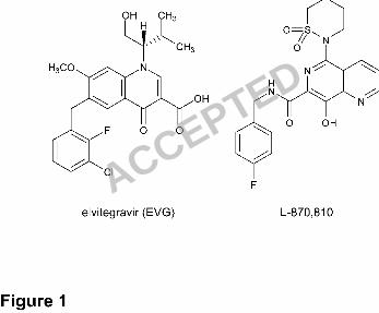
E152) in the CCD of IN (16, 24). In contrast, L-870,810 (Figure 1), which has 17
previously demonstrated potent antiviral activity in HIV-1 infected patients in a 18
monotherapy study (33), induced unique IN mutations, including V72I, F121Y, T125K, 19
and V151I, when HIV was selected with the compound in vitro (23). These mutations 20
are also located in the active site of IN, suggesting that a common mechanism may be 21
involved in the acquisition of resistance to IN inhibitors. 22
ACCEPTED
on July 10, 2018 by guesthttp://jvi.asm
.org/D
ownloaded from
7
Although no IN inhibitors have currently been approved for clinical use (41), 1
two IN inhibitors, elvitegravir (EVG, formerly known as JTK-303/GS-9137, being 2
co-developed by Gilead Sciences and Japan Tobacco, Figure 1) (43, 56) and raltegravir 3
(MK-0518, developed by Merck) (22) are currently being investigated in clinical studies 4
in HIV-1 infected patients. In a phase II study, antiretroviral treatment-experienced 5
patients using 125 mg EVG (boosted with ritonavir) along with an active optimized 6
background regimen showed a > 2 log10 decline in their viral load that was durable 7
through week 24 (56). 8
Here, we describe the antiviral activity, mechanism of action, and resistance 9
profile of EVG in vitro. EVG exerted potent anti-HIV activity against not only 10
wild-type strains but also drug-resistant clinical isolates. Interestingly, EVG also 11
showed antiviral activity against murine leukemia virus (MLV) and simian 12
immunodeficiency virus (SIV). These results imply that IN inhibitors are ideal agents 13
for treatment of a range of retroviral infections. During the selection of EVG-resistant 14
viral variants, novel IN mutations emerged. Combinations of these mutations conferred 15
resistance to EVG and reduced susceptibility to other IN inhibitors, suggesting there is a 16
common mechanism underlying the resistance to IN inhibitors. One such mechanism 17
may be conformational changes induced by multiple mutations located in the active site 18
of IN. 19
20
21
22
ACCEPTED
on July 10, 2018 by guesthttp://jvi.asm
.org/D
ownloaded from
8
Materials and Methods 1
Antiviral Agents. 2
Zidovudine (AZT) and dextran sulfate (DS5000; average molecular weight 3
5000) were purchased from Sigma (St. Louis, MO, USA). Efavirenz (EFV; NNRTI) 4
and nelfinavir (NFV; PI) were used for the control inhibitor. Elvitegravir (EVG) (43), 5
L-731,988 (42), L-870,810 (23), and S-1360 (16) were synthesized as described. The 6
structures of EVG and L-870,810 are depicted in Figure 1. 7
Cells and Viruses. 8
MT-2 and MT-4 cells were grown in RPMI 1640 medium. 293T cells were 9
grown in Dulbecco’s Modified Eagle medium (DMEM). These media were 10
supplemented with 10% fetal calf serum (FCS), 2 mM L-glutamine, 100 U/mL 11
penicillin, and 50 !g/mL streptomycin. HeLa-CD4/CCR5-LTR/!-gal cells (5) were 12
kindly provided by Dr. J. Overbaugh through the AIDS Research and Reference 13
Reagent Program, Division of AIDS, National Institute of Allergy and Infectious 14
Disease (NIAID) (Bethesda, MD, USA) and maintained in DMEM supplemented with 15
10% FCS, 200 !g/mL hygromycin B, 10 !g/mL puromycin, and 200 !g/mL geneticin. 16
PBMC were obtained from healthy HIV-1 seronegative donors by centrifugation 17
through Ficoll-Hypaque density gradients. PBMC were stimulated with 20 U/mL IL-2 18
(Shionogi, Osaka, Japan) and 0.5 !g/mL phytohemagglutinin (PHA; Sigma) for 3 days, 19
then used for assay as described previously (30). 20
Three laboratory strains, HIV-1IIIB, HIV-2EHO, and HIV-2ROD were used in 21
this study. Various subtypes of drug-naïve clinical isolates of HIV-1 (4 isolates of 22
ACCEPTED
on July 10, 2018 by guesthttp://jvi.asm
.org/D
ownloaded from
9
subtype B and 7 isolates of non-B subtypes) were employed. Four drug-resistant clinical 1
isolates of HIV-1, including IVR401, 409, 411, and 415, were kindly provided by Dr. S. 2
Oka, (AIDS Clinical Center, International Medical Center of Japan, Tokyo, Japan). 3
Determination of HIV Drug Susceptibility. 4
Inhibitory effect of compounds on HIV infection was determined using 5
multinuclear activation of a galactosidase indicator (MAGI) assay, as previously 6
described (37). Inhibitory effect on HIV-1 clinical isolates was measured by p24 7
production and cytotoxicity was measured by 8
[3-(4,5-dimethylthiazol-2-yl)-2,5-diphenyltetrazolium bromide] (MTT) colorimetric 9
assay, as described previously (30). Antiviral activity and cytotoxicity of inhibitors are 10
presented as the concentration that blocks viral replication by 50% (EC50) and that 11
suppresses viability of target cells by 50% (CC50), respectively. 12
Quantification of HIV-1 DNA Species. 13
MT-2 cells (5 x 10⁵ cells) were infected with HIV-1IIIB at a multiplicity of 14
infection (MOI) of 0.1 in the absence or presence of various inhibitors. Infected cells 15
were washed after incubation for 2 h at 37°C. At 24 h post-infection, DNA was 16
extracted using DNAzol reagent (Invitrogen, Carlsbad, CA, USA). 17
Quantification of integrated HIV-1 DNA and 2-LTR circle was performed by 18
real-time quantitative PCR as described previously (4). To normalize DNA species 19
among inhibitors, !-globin amplification was used as an internal control (51). Reactions 20
were analyzed by using the ABI Prism 7500 sequence detector (PE Applied Biosystems, 21
Foster City, CA), then results were normalized and expressed as relative HIV-1 DNA 22
ACCEPTED
on July 10, 2018 by guesthttp://jvi.asm
.org/D
ownloaded from
10
species compared to a “no inhibitor” control. 1
In Vitro Strand Transfer Assay. 2
An oligonucleotide-based strand transfer assay was performed as previously 3
described (8) with some modifications. Briefly, preprocessed oligonucleotide 4
H-U5V1-2 (5’-ATGTGGAAAATCTCTAGCA-3’) derived from the U5 end of HIV-1 5
LTR was labeled at the 5’-end with ["-32
P] ATP. Radiolabeled H-U5V1-2 was annealed 6
to H-U5V2 (5’-ACTGCTAGAGATTTTCCACAT-3’), then used for assay. 7
Recombinant HIV-1 IN derived from HIV-1 NL4-3 (wild-type) or EVG-selected 8
mutants were prepared using an Escherichia coli expression system. The strand transfer 9
assay was performed with 1 !M IN and 150 nM substrate DNA in 20 mM MOPS buffer 10
with 30 mM MgCl2, and incubated in either the presence or absence of IN inhibitors, at 11
37°C for 60 min. Reaction products were analyzed by electrophoresis on 25% 12
polyacrylamide gels and quantified using a BAS-2500 imaging system (Fuji Photo Film, 13
Tokyo, Japan). The concentration of IN inhibitor that inhibited production of strand 14
transfer products by 50% (IC50) compared to control was determined. 15
Selection of EVG-Resistant HIV-1 Variants In Vitro. 16
MT-2 cells (2 x 10⁵) were infected with HIV-1IIIB, then cultured in the 17
presence of 0.5 nM (Figure 3A) or 0.1 nM (Figure 3B) EVG. Cultures were incubated at 18
37°C until extensive cytopathic effect (CPE) was observed, and then culture supernatant 19
was harvested for further passage in fresh MT-2 cells. The concentration of EVG was 20
increased when significant CPE was observed. At the indicated passages (Figure 3, A 21
and B), proviral DNA was extracted from infected MT-2 cells, then subjected to PCR 22
ACCEPTED
on July 10, 2018 by guesthttp://jvi.asm
.org/D
ownloaded from
11
followed by direct population-based sequencing. Susceptibility to EVG at the indicated 1
passages was determined using the MAGI assay (Figure 3A) or p24 production (Figure 2
3B). 3
Recombinant HIV-1 Clones. 4
An HIV-1 infectious clone, pNL101 (38), kindly provided by Dr. K-T. Jeang 5
(NIH, Bethesda, MD, USA), was used to generate recombinant HIV-1 clones. A 6
wild-type HIV-1, HIV-1WT, was constructed by replacing the pol-coding region 7
(nucleotide position 2,006 of the Apa I site to 5,122 of the Nde I site of pNL101) with 8
HIV-1 BH10 strain. The pol-coding region contains a silent mutation at nucleotide 9
4,232 (TTTAGA to TCTAGA) resulting in the generation of a unique Xba I site. 10
Recombinant HIV-1 IN infectious clones were generated using a modified pNL101 11
based vector, pNLRTWT. In brief, mutations were introduced into the Xba I-Nde I region 12
(891 bp) of pSLIntWT, which encodes nucleotides 4,232 to 5,122 of pNL101, using an 13
oligonucleotide-based site directed mutagenesis method (54). Next, the Xba I-Nde I 14
fragments were inserted into pBN#Int, which encodes nucleotides 5,122 (Nde I) to 15
5,785 (Sal I) of pNL101. Finally, the Xba I-Sal I region (1,554 bp) was inserted into 16
pNL101. Each infectious clone was transfected into 293T cells. The following day, 17
MT-2 cells were added and the supernatants were harvested when extensive CPE was 18
observed. 19
Replication Kinetics of Resistant HIV-1 Variants. 20
MT-2 cells (105 cells) were infected with each virus preparation (500 MAGI 21
units) for 4 h. The infected cells were then washed and cultured in the presence or 22
ACCEPTED
on July 10, 2018 by guesthttp://jvi.asm
.org/D
ownloaded from
12
absence of the EVG. The culture supernatants were harvested on day 5 after infection, 1
and p24 levels were quantified using a RETRO-TEK HIV-1 p24 antigen ELISA 2
(ZeptoMetrix, Buffalo, NY). 3
Evaluation of Antiretroviral Activity of IN inhibitors. 4
The MLV-based retroviral vector, pRCV/LIG (15), and plasmid, 5
pcDNA-VSVG encoding the vesicular stomatitis virus envelope glycoprotein, generous 6
gifts from H. Miyoshi, RIKEN Bioresource Center (Tsukuba, Japan), were employed to 7
generate viral particles. These plasmids were co-transfected into an 8
MLV-derived-gag-pol-expressing packaging cell line, GP293 (Clontech, Palo Alto, CA, 9
USA). After 48 h of transfection, culture supernatants were filtered through a 0.45 !m 10
membrane and stored at -80°C until use. 11
An HIV-1-based luciferase expression vector, pBC-LIG, and pCMV#8/9, 12
encoding the HIV-1 viral proteins including IN, and pcDNA-VSVG were transfected 13
into 293T cells to generate pseudotyped HIV-1. The viruses were used to infect 293T 14
cells (105 cells per well in 12-well plates) at a MOI of 0.02 in the absence or presence of 15
inhibitors. After 48 h of transduction, luciferase activity was determined using a 16
luciferase assay system (Promega, Madison, WI, USA) and an LB 9507 luminometer 17
(Berthold, Bad Wildbad, Germany). 18
An SIV molecular clone, pMA239 (46), containing the full SIVmac239 19
genome was a kind gift from Dr. E. Ido, Institute for Virus Research, Kyoto University. 20
pMA239 was used to generate viral stocks as previously described (6). Antiviral 21
activity of IN inhibitors against SIVmac239 was determined using the MAGI assay as 22
ACCEPTED
on July 10, 2018 by guesthttp://jvi.asm
.org/D
ownloaded from
13
described above. 1
Molecular Modeling Studies. 2
A 3D model of EVG in complex with HIV-1 IN CCD was prepared by 3
PyMOL software version 0.97 using published data (44). Amino acid residues involved 4
in resistance to EVG were displayed within this model. 5
6
7
8
9
10
11
12
13
14
15
16
17
18
19
20
21
22
ACCEPTED
on July 10, 2018 by guesthttp://jvi.asm
.org/D
ownloaded from
14
Results 1
Anti-HIV Activity of IN inhibitors. 2
Antiviral activity of EVG against HIV-1IIIB, HIV-2EHO, and HIV-2ROD was 3
first evaluated by the MAGI assay. EVG showed potent antiviral activity against three 4
laboratory strains of HIV with EC50 in the sub-nanomolar to nanomolar range (Table 1). 5
Next, we evaluated the activity of EVG against wild-type clinical isolates representing 6
various subtypes of HIV-1. EVG suppressed the replication of all HIV-1 subtypes tested 7
with an antiviral EC50 ranging from 0.10 to 1.26 nM (Table 2). Moreover, EVG 8
suppressed the replication of HIV-1 clinical isolates carrying NRTI, NNRTI and 9
PI-resistance associated genotypes, as did a control IN inhibitor, the compound 10
L-870,810 (Table S1). Cytotoxicity of these inhibitors was also determined using MTT 11
colorimetric assay. Mean values for CC50 of EVG and L-870,810 in peripheral blood 12
mononuclear cells (PBMC) obtained from three independent donors were 4.6 ± 0.5 !M 13
and 2.7 ± 0.6 !M, respectively. Thus, EVG can suppress various HIV strains, including 14
diverse HIV-1 subtypes and clinical isolates carrying multiple mutations associated with 15
resistance to currently approved antiretroviral drugs. 16
Mechanism of Anti-HIV Activity of EVG 17
First, we performed “time of addition” experiment as described previously 18
(30) with some modification. MT-4 cells were infected with HIV-1IIIB at a MOI of 0.5. 19
One hour after the infection, infected cells were extensively washed and compounds 20
were added, including an NNRTI (EFV, 100 nM), a PI (NFV, 500 nM), or EVG (100 21
nM). Amounts of p24 antigen were determined at 31 h post-infection. Antiviral activity 22
ACCEPTED
on July 10, 2018 by guesthttp://jvi.asm
.org/D
ownloaded from
15
of EFV gradually decreased from 6 h post-infection and disappeared at 12 h 1
post-infection, whereas antiviral activity of EVG decreased from 10 h post-infection 2
and was no longer detected by 12 h post-infection. On the other hand, the PI NFV 3
effectively blocked the infection up to 12 h post-infection and still exerted 4
approximately 20% inhibitory activity up to 24 h post-infection. These results strongly 5
suggest that EVG inhibits the HIV replication at a step which occurs after reverse 6
transcription but before proteolytic cleavage, consistent with the integration step. 7
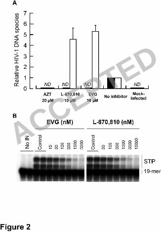
To elucidate the mode of action of EVG on HIV-1 replication, the levels of 8
intracellular HIV-1 DNA species were determined using real-time quantitative PCR 9
(Figure 2A). MT-2 cells were infected with HIV-1IIIB in the presence or absence of a 10
CD4-gp120 binding inhibitor, DS5000, an NRTI, AZT, an IN inhibitor, L-870,810, and 11
EVG. Unintegrated (2-long terminal repeat; 2-LTR), and integrated forms of reverse 12
transcribed HIV-1 genomic DNA were quantified after 24 h of infection, then 13
normalized with !-globin DNA. In the presence of 20 !M AZT, neither 2-LTR nor 14
integrated forms were detected as expected. Similar results were also observed with 20 15
!M of DS5000 (data not shown). In the presence of 10 !M L-870,810, integrated 16
provirus was undetectable while relative 2-LTR levels increased about five-fold (4.6 ± 17
1.0). Similar results were observed with 10 !M EVG (2-LTR, 5.3 ± 0.5), indicating that 18
EVG exerts anti-HIV activity by blocking the integration step. 19
To further characterize the mechanism by which EVG inhibits the integration 20
step, the effect of EVG on strand transfer was assessed by characterizing its ability to 21
inhibit the activity of recombinant wild-type HIV-1 IN enzyme in an 22
ACCEPTED
on July 10, 2018 by guesthttp://jvi.asm
.org/D
ownloaded from
16
oligonucleotide-based strand transfer assay (Figure 2B). EVG and L-870,810 both 1
inhibited synthesis of strand transfer products with 50% inhibitory concentrations (IC50) 2
of 54 nM and 118 nM, respectively. Taken together, these results indicate that like 3
L-870,810, EVG blocks integration via inhibition of IN-mediated strand transfer. 4
Selection of EVG-Resistant HIV-1 Variants In Vitro. 5
To determine the in vitro resistance profile of EVG, EVG-resistant viral 6
variants were selected using a dose-escalation method and the susceptibility to EVG 7
(EC50) of the resulting selected variants was determined. Selection of resistant HIV-1IIIB 8
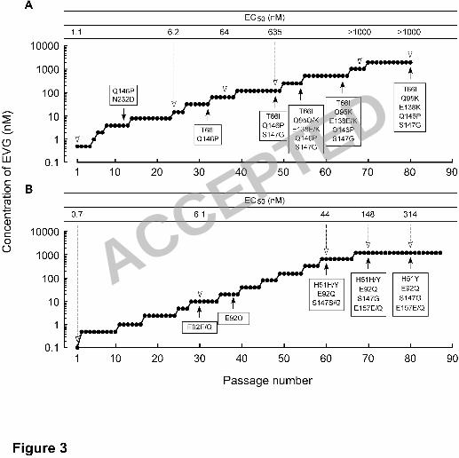
was initiated with 0.5 nM EVG (Figure 3A). At passage 12 (P-12), where concentration 9
of EVG was 4 nM, two amino acids substitutions, glutamine-to-proline at IN codon 146 10
(Q146P) and asparagine-to-aspartic acid at IN codon 232 (N232D) were observed 11
(Figure 3A). An N232D substitution has been reported as an IN polymorphism in 12
HIV-1 (34). The EVG EC50 of a P-24 variant containing a Q146P and N232D 13
substituted variant was 6.2 nM. At P-32 (32 nM EVG), a T66I IN substitution was 14
newly observed, whereas the N232D substitution had reverted to the baseline sequence. 15
The EVG EC50 against a P-36 variant was 64 nM. An S147G IN substitution was 16
detected at P-48 (128 nM EVG) and the EVG EC50 further increased to 635 nM. In 17
addition, a Q95Q/K IN substitution (mixture of Q and K) and an E138E/K IN 18
substitution were newly identified at P-54 (256 nM EVG). These mixtures, Q95Q/K and 19
E138E/K, fully emerged in the viral pools by P-64 and P-80, respectively. The EVG 20
EC50 at P-68 (1,024 nM EVG) was greater than 1,000 nM. 21
An independent EVG selection experiment, again using HIV-1IIIB was 22
ACCEPTED
on July 10, 2018 by guesthttp://jvi.asm
.org/D
ownloaded from
17
performed but beginning at 0.1 nM EVG (Figure 3B). An E92E/Q mixture in the IN 1
coding region was first detected at P-30 (10 nM EVG) and was predominantly E92Q by 2
P-38 (20 nM EVG). Additional IN substitutions H51H/Y and S147S/G emerged at P-60 3
(640 nM EVG), and an E157E/Q mixture emerged at P-70 (1,280 nM EVG); the viral 4
pools at the terminal passage P-80 (1,280 nM EVG) had the IN sequence 5
H51Y/E92Q/S147G/E157E/Q (Figure 3B). The emergence of each of these mutations 6
correlated with an increase in the EVG EC50 of the resulting viral pools (Figure 3). 7
Other than the N232D polymorphism, all of these mutations are located in the CCD of 8
IN. 9
Phenotypic Analysis of IN-Recombinant Viruses. 10
(i) EVG-Selected Mutations. 11
To characterize which mutations are responsible for EVG resistance, 12
infectious HIV-1 clones containing single IN substitutions (H51Y, T66I, E92Q, Q95K, 13
E138K, Q146P, S147G, or E157Q) that were observed to emerge under selection with 14
EVG were generated (Figure 3, Table 3). Mutations were classified into two groups 15
based on the level of resistance: mutations that conferred more than 10-fold reduced 16
susceptibility compared to wild-type were defined as primary mutations; mutations 17
conferring less than 10-fold reduced susceptibility were defined as secondary mutations. 18
T66I and E92Q substitutions conferred significantly reduced susceptibility to EVG (37 19
and 36-fold reduced, respectively, relative to wild-type) whereas the Q146P and S147G 20
substitutions conferred more moderate reductions in EVG susceptibility (11-fold 21
reduced) indicating that these four IN mutations are primary mutations involved in 22
ACCEPTED
on July 10, 2018 by guesthttp://jvi.asm
.org/D
ownloaded from

18
resistance to EVG. In contrast, H51Y, Q95K and E157Q substitutions all conferred 1
smaller reductions in EVG susceptibility (each less than 6-fold reduced compared to 2
wild-type), suggesting these substitutions are secondary resistance mutations. 3
Interestingly, the E138K mutation alone conferred no reduction in susceptibility to 4
either EVG or L-870,810. Thus, several distinct mechanisms of resistance may be 5
represented by these different IN mutations. 6
Multi-substituted clones observed during EVG selection experiments were 7
also generated. HIV-1T66I/Q146P showed high-level resistance to EVG (119-fold reduced 8
susceptibility) (Table 3). Combinations of S147G with T66I/Q146P or E92Q further 9
enhanced resistance, 412 and 356-fold, respectively. The triple mutant 10
HIV-1H51Y/E92Q/S147G showed high-level resistance to EVG (700-fold reduced 11
susceptibility). Interestingly, the addition of the secondary mutation H51Y, which on its 12
own reduced EVG susceptibility only 3.6-fold, substantially enhanced resistance 13
relative to that observed for the double mutant HIV-1E92Q/S147G. HIV-1T66I/Q95K/Q146P/S147G, 14
HIV-1T66I/Q95K/E138K/Q146P/S147G, and HIV-1H51Y/E92Q/S147G/E157Q mutants all showed 15
high-level resistance to EVG with EC50 values greater than 1,000 nM in all cases. These 16
results indicate that the T66I and E92Q mutations provided the highest fold change in 17
EVG susceptibility as individual resistance mutations, and that the additional 18
substitutions identified further enhance the level of resistance to EVG when combined 19
with these primary mutations. 20
(ii) L-870,810-Selected Mutations. 21
Infectious HIV-1 clones containing mutations (V72I, F121Y, T125K, and 22
ACCEPTED
on July 10, 2018 by guesthttp://jvi.asm
.org/D
ownloaded from
19
V151I) previously shown to be associated with resistance to L-870,810 (23) and two 1
mutations, L74M and G163R, observed in our selection using L-870,810 (data not 2
shown) were generated. Among these variants, HIV-1F121Y and HIV-1V151I demonstrated 3
reduced susceptibility to both L-870,810 and EVG (Table 3). V151I has been observed 4
in some HIV-1 clinical isolates and may be an IN polymorphism (34). Moreover, the 5
effect of V151I on susceptibility to L-870,810 has been controversial (23, 29). This 6
discrepancy might arise from the viral strain or plasmid backbone used, so further 7
experiments to clarify the effect of V151I on IN inhibitor susceptibility are needed. 8
HIV-1F121Y/T125K showed significant resistance to both L-870,810 and EVG (68-fold and 9
177-fold reduced susceptibility, respectively). HIV-1V72I/F121Y/T125K/V151I showed 10
high-level resistance to both IN inhibitors (EC50 greater than 1,000 nM). 11
(iii) DKA IN inhibitor-Selected Mutations. 12
Highlighting the potential for related mechanisms of IN inhibitor-resistance 13
and cross-resistance, the T66I mutation has also been observed to be selected by DKA 14
IN inhibitors, such as L-708,906 and S-1360. Additional mutations L74M and S153Y, 15
in combination with T66I, were also observed to be selected by these DKA IN 16
inhibitors (16, 17). L74M also emerged during L-870,810 selection in our studies (data 17
not shown) but conferred no change in susceptibility to L-870,810 when present alone 18
and only low-level resistance (3.0-fold) to EVG (Table 3). The combination of T66I and 19
L74M conferred slightly higher resistance to EVG (45-fold) compared to T66I alone but 20
only moderate resistance to L-870,810 (7-fold). Another IN mutant, HIV-1T66I/S153Y 21
observed in L-708,906 selection experiments (24), showed high-level resistance to EVG 22
ACCEPTED
on July 10, 2018 by guesthttp://jvi.asm
.org/D
ownloaded from
20
(260-fold) but low-level resistance to L-870,810 (5-fold). These results suggest that the 1
mechanism of EVG resistance may have some similarities to that of DKA IN inhibitors. 2
Taken together, these results suggest that a variety of IN mutations may be 3
selected by EVG and other IN inhibitors. Most of the IN inhibitor resistance mutations 4
are observed to cluster in the CCD of IN. The resulting mutations and their 5
combinations have the capacity to confer varying levels of resistance and potential 6
cross-resistance to EVG and other IN inhibitors. Given their location in the CCD, many 7
of these mutations may act via a common mechanism. The observed development of IN 8
inhibitor resistance mutations resembles that seen for other antiretroviral drugs such as 9
PIs, i.e., multiple mutations are introduced in a stepwise fashion and are required for 10
high-level resistance to the selecting inhibitors (10, 50). 11
Strand Transfer Assay. 12
To further characterize the effect of EVG-selected resistance mutations on IN 13
function, the effect of mutations on the enzymatic activity of recombinant IN was 14
evaluated in an in vitro strand transfer assay (Figure 4). IN enzymes carrying the 15
individual mutations H51Y, S147G and E157Q had reduced strand transfer activity 16
relative to wild-type (57%, 36%, and 79% of wild-type, respectively). Strand transfer 17
activities of E92Q, E92Q/S147G, and H51Y/E92Q/S147G IN enzymes decreased with 18
the accumulation of mutations from 57% to 29 and 22% of wild-type, respectively. 19
However, introduction of E157Q to H51Y/E92Q/S147Q partially restored strand 20
transfer activity to 46% of wild-type, suggesting that E157Q may play a role in 21
compensating for the loss of strand transfer activity resulting from the emergence of 22
ACCEPTED
on July 10, 2018 by guesthttp://jvi.asm
.org/D
ownloaded from
21
EVG resistance mutations. 1
The effect of EVG-selected mutations on the inhibition of strand transfer by 2
EVG and L-870,810 was also determined (Figure 4). Recombinant IN enzymes carrying 3
the individual H51Y, S147G, and E157Q substitutions remained susceptible to both 4
EVG and L-870,810 (0.7 to 2.1-fold reduced susceptibility). E92Q IN demonstrated 5
only moderate resistance to both IN inhibitors in the strand transfer assay (4.3-fold 6
reduced for both inhibitors). The combination of E92Q with S147G enhanced resistance 7
to both EVG and L-870,810 (7.6 and 8.5-fold reduced susceptibility, respectively). 8
However, unlike the IN-recombinant viruses in the antiviral assay, neither the 9
H51Y/E92Q/S147G nor H51Y/E92Q/S147G/E157Q IN enzymes showed further 10
enhancement of resistance in the strand transfer assay. This difference in results from 11
the strand transfer assay versus the antiviral assay may reflect differences in the 12
recombinant IN enzyme versus the viral IN enzyme in situ. Indeed, structure-activity 13
relationship experiments published in previously report (43) revealed that antiviral 14
activity and in vitro enzyme inhibition were well correlated. Nevertheless, this 15
biochemical analysis confirmed that the E92Q IN mutation confers significantly 16
reduced susceptibility to EVG at the level of inhibition of strand transfer, consistent 17
with its identification as a primary EVG resistance mutation in the virological analyses. 18
Replication kinetics of IN inhibitor-resistant variants. 19
The effects of IN mutations on the replication kinetics of HIV-1 variants were 20
assessed by comparing their p24 production in culture supernatants to that of wild-type 21
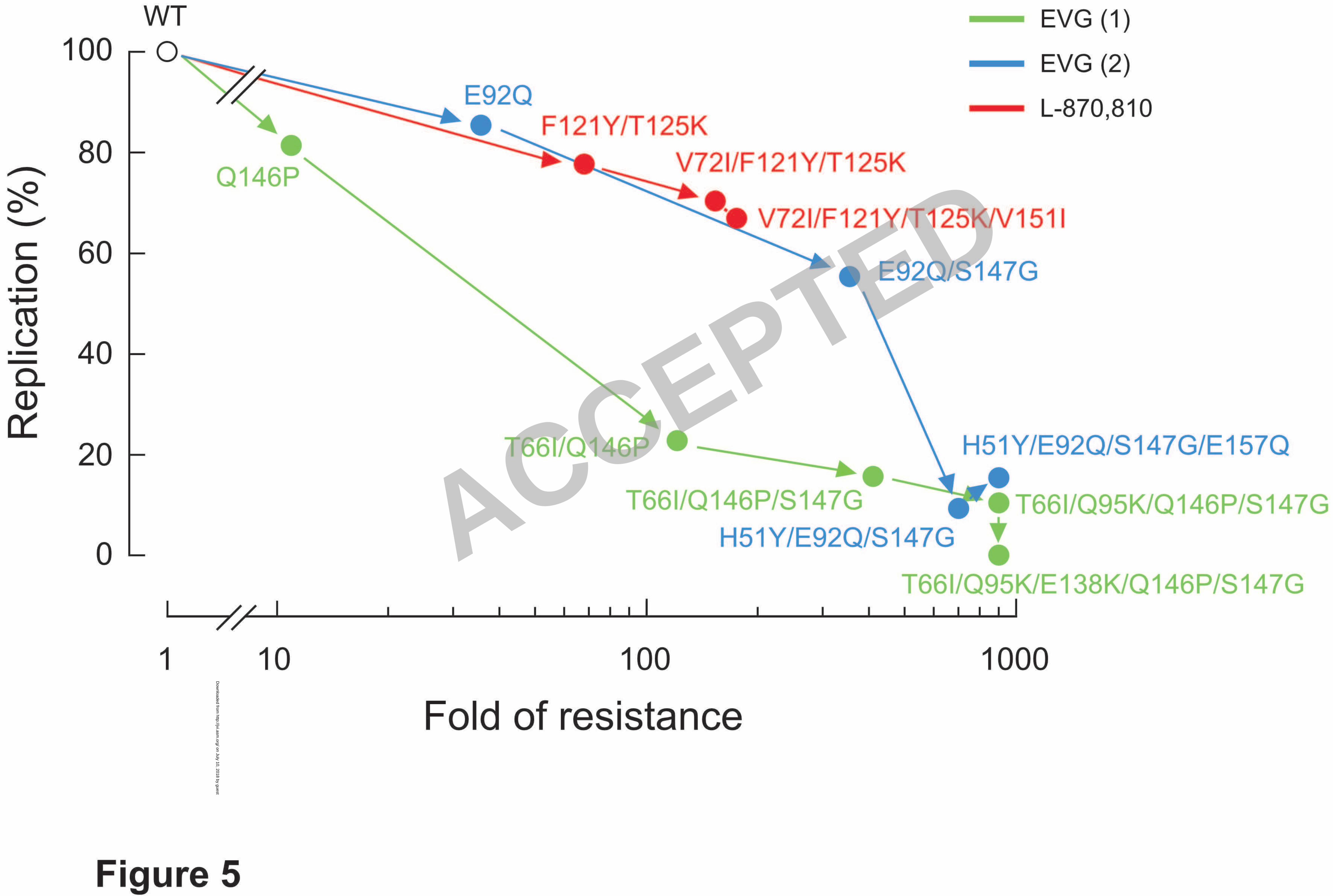
virus (Figure 5). At day 5 post-infection, p24 production by the HIV-1E92Q and 22
ACCEPTED
on July 10, 2018 by guesthttp://jvi.asm
.org/D
ownloaded from
22
HIV-1Q146P variants were 86% and 82% of HIV-1WT, respectively. These variants 1
showed high-level (36-fold) or moderate (11-fold) resistance to EVG (Table 3), whereas 2
replication levels of both were similar to wild-type. However, introduction of additional 3
EVG resistance mutations further decreased p24 production, indicative of a decline in 4
the levels of viral replication. In particular, HIV-1T66I/Q146P/S147G, 5
HIV-1T66I/Q95K/Q146P/S147G, HIV-1T66I/Q95K/E138K/Q146P/S147G, HIV-1H51Y/E92Q/S147G/, and 6
HIV-1H51Y/E92Q/S147G/E157Q all showed significantly reduced levels of p24 production (less 7
than 20% of wild-type by day 5 in all cases). Thus, there was an inverse correlation 8
between the levels of EVG resistance and the viral replication capacity, that is, as 9
resistance to EVG increased, viral replication decreased. Interestingly, viral variants 10
carrying L-870,810-selected mutations had more moderate reductions in replication 11
capacity, even in the case of the HIV-1V72I/F121Y/T125K/V151I variant that had high-level 12
resistance to both L-870,810 and to EVG (68% of wild-type). These results indicate that 13
mutations associated with resistance to IN inhibitors can have varying effects on viral 14
replication capacity. The reduced replication capacity of EVG-resistant variants was not 15
rescued in the presence of the inhibitor (data not shown), as has been observed for 16
NFV-resistant variants in the presence of NFV (35). Thus, reduced replication capacity 17
of IN inhibitor-resistant variants may present a barrier to their emergence in vivo. 18
Antiviral Effect of IN inhibitors on Retroviruses. 19
Antiviral activity of EVG against other retroviruses, including MLV and SIV, 20
was assessed. EVG and L-870,810 inhibited integration of the HIV-based vector used as 21
a positive control for the luciferase assay (EC50: 0.8 and 5.0 nM, respectively), as 22
ACCEPTED
on July 10, 2018 by guesthttp://jvi.asm
.org/D
ownloaded from
23
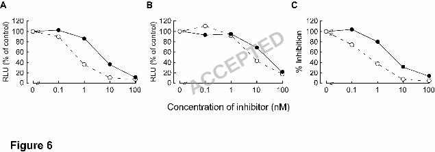
observed in the MAGI assay with HIV-1IIIB (Figure 6). EVG and L-870,810 suppressed 1
replication of MLV infection (EC50: 5.8 and 22 nM, respectively) as well as that of the 2
primate retroviruses, SIV (0.5 and 3.2 nM, respectively), indicating that IN inhibitors 3
have antiviral activity against a broad range of retroviruses. 4
5
6
7
8
9
10
11
12
13
14
15
16
17
18
19
20
21
22
ACCEPTED
on July 10, 2018 by guesthttp://jvi.asm
.org/D
ownloaded from
24
Discussion 1
The data described here show that EVG inhibits HIV replication by 2
specifically blocking the strand transfer reaction mediated by IN, as demonstrated by 3
intracellular accumulation of 2-LTR DNA products, a signature of non-productive 4
integration. Furthermore EVG directly blocked production of strand transfer products in 5
an in vitro strand transfer assay. Confirming that EVG is a bona fide IN inhibitor, we 6
selected EVG-resistant viral variants in vitro and demonstrated that the resulting viral 7
variants had acquired multiple mutations in the IN coding region and had 8
simultaneously acquired reduced phenotypic susceptibility to EVG. HIV-1 molecular 9
clones carrying the EVG-selected IN mutations had an EVG-resistant phenotype and in 10
many cases also had reduced susceptibility to another IN inhibitor, L-870,810. These 11
data provide formal proof that the observed IN mutations are indeed EVG-resistance 12
mutations and that EVG is an IN inhibitor. 13
Among the IN mutations observed to be selected by EVG, two mutations, 14
T66I and E92Q, appeared to provide the major contribution to EVG resistance. Both of 15
these individual mutations resulted in > 30-fold reduced susceptibility to EVG. The 16
T66I mutation conferred cross-resistance to S-1360 and L-731,988 (Table 3) and has 17
also been observed in an independent EVG selection by Jones et al. (26). The E92Q 18
mutation, when introduced into a recombinant IN enzyme, also reduced susceptibility of 19
the resulting mutant IN enzyme to EVG, as measured by reduced EVG inhibition of the 20
in vitro strand transfer assay (Figure 4). The other IN mutations identified, including 21
H51Y, Q95K, E138K, Q146P, S147G, and E157Q, individually resulted in lower fold 22
ACCEPTED
on July 10, 2018 by guesthttp://jvi.asm
.org/D
ownloaded from
25
changes in EVG susceptibility (1.0 to 11.0-fold) but when added to either the T66I or 1
E92Q mutations, further increased resistance to EVG to varying degrees, relative to 2
either mutation alone. Interestingly, accumulation of these EVG-selected IN mutations 3
resulted in significant attenuation of viral replication-kinetics. Thus emergence of 4
resistance to IN inhibitors may be associated with reductions in viral fitness, which may 5
provide a barrier to the emergence of these mutations in vivo or be associated with 6
lower viral loads if they do emerge. 7
Of the three HIV enzymes, PR, RT and IN, the structure and mechanism of 8
IN is the least well understood and despite extensive efforts, the structure of the 9
complete IN enzyme remains to be determined. Only partial two-domain crystal 10
structures of the IN apoenzyme are available and no structure has been published 11
showing full-length IN bound to its viral cDNA substrate. During integration in vivo, IN 12
functions in the preintegration complex, which also includes RT and the viral DNA (2, 13
3). Some limited evidence suggests that RT interacts with the active site of IN (39). IN 14
has also been proposed to function with several cellular factors including IN interactor 1 15
(Ini1) (27) and lens-epithelium-derived growth factor (LEDGF/p75) (7). In the context 16
of these associated cellular factors, IN may retain a different conformation to that of the 17
recombinant enzyme alone. This may be one of the reasons that only moderate EVG 18
resistance was observed in the oligonucleotide-based strand transfer assay compared to 19
a cell-based antiviral assay. 20
Alignment of several deposited IN CCD structures in the Protein Data Bank 21
(PDB) indicates that there are two regions with poorly defined or disordered structure, 22
ACCEPTED
on July 10, 2018 by guesthttp://jvi.asm
.org/D
ownloaded from
26
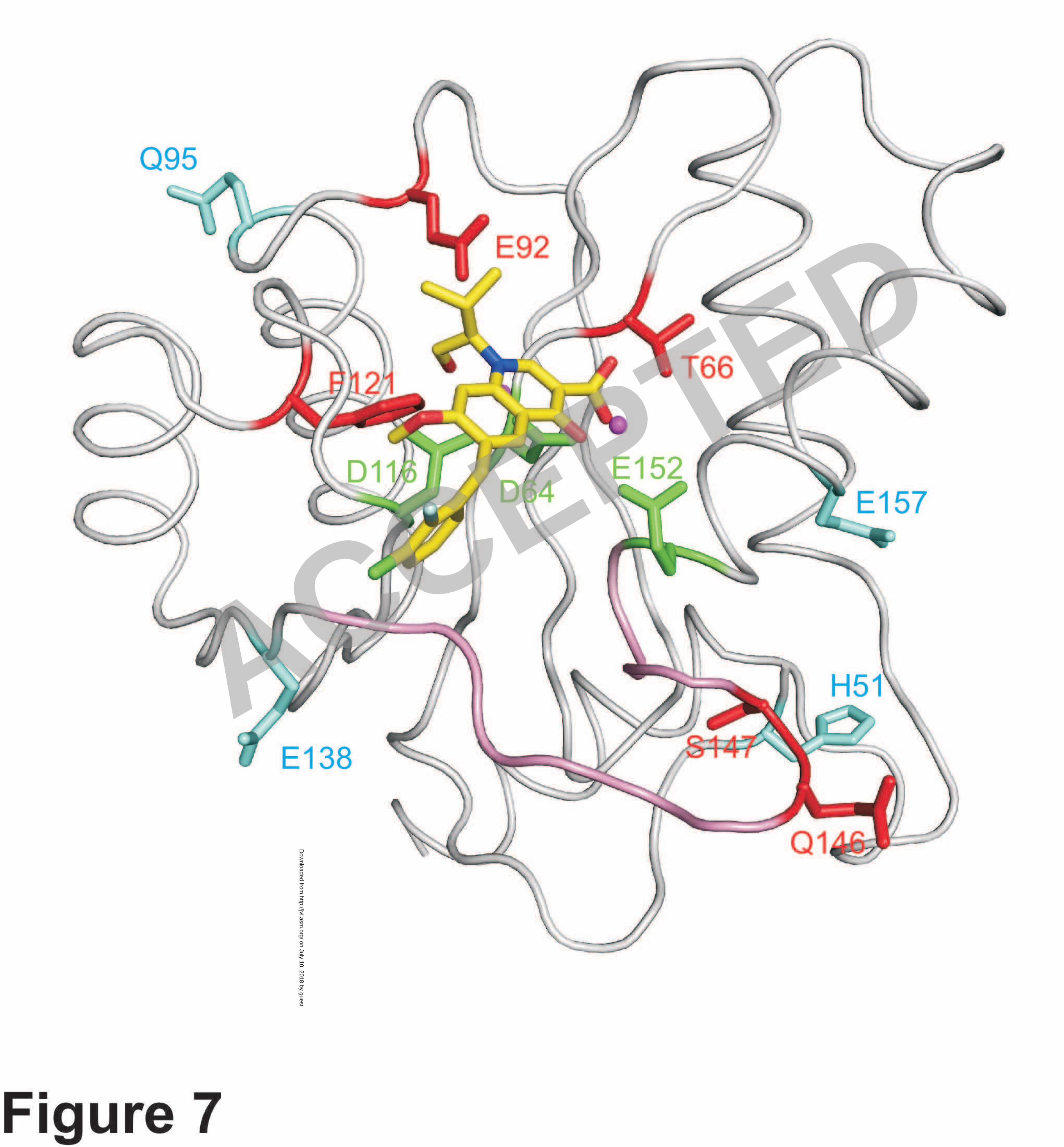
including residues 47-56 and 140-152 (Figure 7 and S1). Of these two disordered 1
regions, residues 140-152 have been implicated as a flexible loop involved in viral 2
cDNA binding (20, 21, 53). Although the precise structural details are unknown, the 3
flexible loop has been proposed to adopt different conformations in the presence or 4
absence of the viral cDNA (12). Notably, several of the EVG-selected mutations that we 5
observed are located on or adjacent to this proposed flexible loop, including E138K, 6
Q146P and S147G. The flexible loop is important for the catalytic activity of IN (21, 7
32), and as shown in Figure 4, introduction of mutations in these residues, especially 8
S147G, drastically reduced the catalytic activity of IN. Previously published data have 9
also demonstrated that another mutation at codon 147 (S147I) resulted in HIV-1 that 10
was highly replication defective, including effects on viral DNA synthesis (47). Indeed, 11
S147 is highly conserved among various retroviruses (Figure S2), highlighting the 12
importance of the loop for IN function. It is possible that IN inhibitor resistance mutants 13
may have additional pleiotropic effects on processes in viral replication other than 14
integration; in particular RT and IN have previously been suggested to interact 15
functionally (25). 16
Recently, an in silico docking simulation of HIV IN with several IN 17
inhibitors, including EVG was reported (44). Notably the author showed that in the best 18
fit model for EVG docked to IN, the isobutyl substituent on the quionolone moiety of 19
EVG orients directly towards IN residue E92. Interestingly, the hydroxyl component of 20
the isobutyl on the quinolone replaces a water molecule that is coordinated by residue 21
E92 between the two catalytic residues D64 and E152. This docking structure may 22
ACCEPTED
on July 10, 2018 by guesthttp://jvi.asm
.org/D
ownloaded from
27
provide insight into both the mechanism of IN inhibition by EVG and provides a 1
starting point for understanding the mechanism of EVG resistance mediated by the 2
E92Q substitution. However, it is uncertain whether this docking simulation represents 3
the precise binding mode of EVG with IN in vivo. Therefore, to accurately assess the 4
binding mode of IN inhibitors with IN, available structural data needs to be 5
supplemented by a variety of other approaches. In this study, a virological and 6
enzymatic approach was integrated to characterize the mechanism of action, antiviral 7
activity and resistance profile in vitro of EVG. 8
As shown in Figure 7, primary EVG resistance mutations are located around 9
the catalytic triad of the CCD of IN and are surrounded by the secondary mutations. 10
Among the residues affected by primary mutations, E92 and F121 are located close to 11
EVG on the model and might interact with the IN inhibitor. However, the mechanism 12
by which these mutations interact with the IN inhibitor or with the viral cDNA to 13
mediate resistance is currently unclear. Recently clinical isolate data was reported from 14
patients experiencing virologic failure in an ongoing phase III studies of another IN 15
inhibitor, raltegravir; E92Q was among the mutations noted to develop in these 16
raltegravir failure patients, usually in combination with another IN mutation, N155H 17
(11, 48). This preliminary clinical data and the data presented here with L-870,810, 18
indicate that the E92Q mutation may be able to mediate resistance and potential 19
cross-resistance to multiple IN inhibitors including EVG and raltegravir. Consistent 20
with the data described here, site-directed mutant HIV carrying the E92Q mutation have 21
been confirmed to show resistance to EVG and to have low-level (approximately 22
ACCEPTED
on July 10, 2018 by guesthttp://jvi.asm
.org/D
ownloaded from

28
6-fold) reduced susceptibility to raltegravir (26). 1
Several of the IN residues affected by primary mutations observed in 2
EVG-selected variants including T66, E92, and S147 are absolutely conserved among 3
the retroviruses tested (HIV-1, HIV-2, SIV, and MLV) and in retroviruses from 4
multiple mammalian species (Figure S2). The significant conservation of mammalian 5
retroviral IN CCDs at both the level of sequence homology and structure of the active 6
site was demonstrated by the ability of EVG to inhibit HIV, SIV and MLV IN activity. 7
This suggests that EVG, and probably other IN inhibitors bind to a conformationally 8
conserved region of all retroviral INs; binding of EVG and other IN inhibitors to IN is 9
also likely to involve the catalytic magnesium ion. Taken together, these results suggest 10
several distinct mechanisms may contribute to IN inhibitor resistance including 11
conformational changes in the structure of IN that affect the binding of the IN inhibitor, 12
charge effects, steric hindrance, loss of stabilizing binding interactions, or possibly 13
alteration of magnesium binding. 14
Similar reduction in viral replication capacity as a result of drug resistance 15
mutations has previously been reported for NRTI resistance mutations (K65R, L74V 16
and M184V) (45, 55) and for PI resistance mutations (D30N) (49). Mutations that act to 17
compensate for some of the loss of viral replication resulting from drug resistance, for 18
example GAG processing mutants, have also been described (18, 36, 52). At least one 19
of the EVG secondary mutations, E157Q, may have an analogous role as it partially 20
restored strand transfer activity that was attenuated by other EVG-selected mutations 21
and also further enhanced resistance to EVG (Figure 4). Some secondary IN mutations 22
ACCEPTED
on July 10, 2018 by guesthttp://jvi.asm
.org/D
ownloaded from
29
might act to compensate for the altered conformation of IN resulting from the structural 1
effects of primary resistance mutations. The E138K mutation may be such an example 2
as on it’s own it showed no effect on susceptibility to either EVG or L-870,810. The 3
clinical implications of the reduction in fitness resulting from selection of 4
EVG-resistant mutations are not yet understood. 5
In conclusion, EVG is a potent inhibitor of the HIV IN enzyme that acts by 6
blocking the strand transfer reaction, and is effective not only against HIV but also other 7
retroviruses. Moreover, the emergence of viral variants that were highly resistant to 8
EVG was associated with significant reductions in viral replication in vitro. These 9
results indicate that EVG should be highly effective for the treatment of HIV-1 infected 10
patients, including those who have had virologic failure of their HAART due to 11
emergence of HIV-1 drug resistance to approved antiretroviral drugs. 12
13
14
15
16
17
18
19
20
21
22
ACCEPTED
on July 10, 2018 by guesthttp://jvi.asm
.org/D
ownloaded from
30
Acknowledgments 1
We would like to thank Shinjiro Hino for technical advice and Mieko Ikeuchi 2
for technical assistance. We appreciate Damian McColl for critical reading and 3
comment on our manuscript. This work was supported in part by a grant for the 4
Promotion of AIDS Research from the Ministry of Health and Welfare of Japan (E.K. 5
and M.M.), a grant for Research for Health Science Focusing on Drug Innovation from 6
the Japan Health Science Foundation (E.K. and M.M.), and a grant from the Ministry of 7
Education, Culture, Sports, Science, and Technology of Japan (E.K.). K.S. is supported 8
by the 21st Century COE Program of the Ministry of Education, Culture, Sports, 9
Science, and Technology. 10
11
12
13
14
15
16
17
18
19
20
21
22
ACCEPTED
on July 10, 2018 by guesthttp://jvi.asm
.org/D
ownloaded from
31
Figure Legends 1
Figure 1. Structure of elvitegravir and L-870,810. 2
A dihydroquinoline carboxylic acid derivative elvitegravir (EVG) and a 3
naphthyridine carboxamide derivative L-870,810 (a representative IN inhibitor). 4
5
Figure 2. Mechanism of action of EVG. 6
(A) Quantification of HIV-1 DNA species. MT-2 cells were infected with 7
HIV-1IIIB in the presence or absence of AZT, L-870,810, and EVG. Unintegrated 8
(2-LTR; white bar) and integrated forms (black bar) of proviral DNA were quantified 9
by real-time PCR and normalized to the !-globin gene after 24 h of infection. The data 10
is represented as mean and standard deviation of relative value to no inhibitor control 11
from three independent experiments. ND means not detected the signals even after 40 12
cycles amplification. (B) Inhibitory effect of IN inhibitors on strand transfer activity. 13
Gel electrophoresis shows strand transfer products (STP) generated from preprocessed 14
donor DNA substrate (19-mer) covalently bound to acceptor DNA. 15
16
Figure 3. Induction of EVG-resistant HIV-1. 17
MT-2 cells. Initial concentration of EVG was 0.5 nM (Panel A) and 0.1 nM 18
(Panel B). Results are from two identical but independent experiments (Panels A and B). 19
At the indicated passage number (black arrowhead), proviral DNA extracted from 20
infected MT-2 cells was sequenced. Amino acid substitutions are shown. The EC50 of 21
HIV-1 variants selected by EVG at the indicated passage number (white arrowhead) 22
ACCEPTED
on July 10, 2018 by guesthttp://jvi.asm
.org/D
ownloaded from
32
were determined using MAGI assay (Panel A) or production of p24 in MT-2 cells 1
(Panel B). 2
3
Figure 4. Effect of EVG-selected mutations on IN strand transfer activity and on 4
inhibition of strand transfer by IN inhibitors. 5
Strand transfer activity of recombinant IN enzymes carrying EVG-selected 6
mutations was determined using an oligonucleotide-based strand transfer assay. Strand 7
transfer activity of IN mutants was compared to wild-type (WT); results are shown as 8
percentage of wild-type activity. Effect of IN inhibitors on strand transfer was also 9
determined for wild-type and mutant IN enzymes; results were expressed as 10
fold-increase in IC50 of inhibitors, relative to wild-type. 11
12
Figure 5. Replication kinetics of EVG- and L-870,810-resistant viral variants. 13
The replication kinetics of wild-type and IN inhibitor-resistant viral variants 14
were determined by p24 ELISA. The relationship of replication capacity and fold 15
change in susceptibility (shown in Table 3) is depicted. Variants are plotted according 16
to the observed order of their emergence during selection experiments in vitro. 17
Replication kinetics of EVG-selected mutants derived from the two independent 18
selection experiments (shown in Figure 3) are plotted in different colors. 19
20
Figure 6. Inhibitory effect of IN inhibitors on retroviruses. 21
Antiviral activity of EVG (open circle with dashed line) and L-870,810 22
ACCEPTED
on July 10, 2018 by guesthttp://jvi.asm
.org/D
ownloaded from

33
(closed circle with solid line) against (A) HIV- or (B) MLV-based vectors harboring the 1
luciferase gene were determined by measuring luciferase activity at 48 h 2
post-transduction. Results are expressed as percentage of relative luciferase units (RLU) 3
compared to no inhibitor control. (C) Anti-SIV activity was determined using the 4
MAGI assay. These results shown are one representative assay from three independent 5
experiments. 6
7
Figure 7. Location of IN mutations associated with resistance to EVG. 8
EVG in complex with the HIV-1 IN CCD is shown along with the catalytic 9
triad residues (D64, D116, and E152; green) and a magnesium ion (magenta). Amino 10
acid residues conferring resistance to EVG as primary mutations (T66, E92, F121, 11
Q146, and S147) or as secondary mutations (H51, Q95, E138, and E157) are shown in 12
red and cyan, respectively. The flexible loop (residues 140-152) is shown in pink. 13
14
15
16
17
18
19
20
21
22
ACCEPTED
on July 10, 2018 by guesthttp://jvi.asm
.org/D
ownloaded from
34
References 1
1. Asante-Appiah, E., and A. M. Skalka. 1997. Molecular mechanisms in 2
retrovirus DNA integration. Antiviral Res 36:139-56. 3
2. Brown, P. O., B. Bowerman, H. E. Varmus, and J. M. Bishop. 1987. Correct 4
integration of retroviral DNA in vitro. Cell 49:347-56. 5
3. Bukrinsky, M. I., N. Sharova, T. L. McDonald, T. Pushkarskaya, W. G. 6
Tarpley, and M. Stevenson. 1993. Association of integrase, matrix, and reverse 7
transcriptase antigens of human immunodeficiency virus type 1 with viral 8
nucleic acids following acute infection. Proc Natl Acad Sci U S A 90:6125-9. 9
4. Butler, S. L., M. S. Hansen, and F. D. Bushman. 2001. A quantitative assay 10
for HIV DNA integration in vivo. Nat Med 7:631-4. 11
5. Chackerian, B., E. M. Long, P. A. Luciw, and J. Overbaugh. 1997. Human 12
immunodeficiency virus type 1 coreceptors participate in postentry stages in the 13
virus replication cycle and function in simian immunodeficiency virus infection. 14
J Virol 71:3932-9. 15
6. Chakrabarti, L. A., K. J. Metzner, T. Ivanovic, H. Cheng, J. 16
Louis-Virelizier, R. I. Connor, and C. Cheng-Mayer. 2003. A truncated form 17
of Nef selected during pathogenic reversion of simian immunodeficiency virus 18
SIVmac239#nef increases viral replication. J Virol 77:1245-56. 19
7. Cherepanov, P., G. Maertens, P. Proost, B. Devreese, J. Van Beeumen, Y. 20
Engelborghs, E. De Clercq, and Z. Debyser. 2003. HIV-1 integrase forms 21
stable tetramers and associates with LEDGF/p75 protein in human cells. J Biol 22
Chem 278:372-81. 23
8. Chow, S. A. 1997. In vitro assays for activities of retroviral integrase. Methods 24
12:306-17. 25
9. Clavel, F., and A. J. Hance. 2004. HIV drug resistance. N Engl J Med 26
350:1023-35. 27
10. Condra, J. H., W. A. Schleif, O. M. Blahy, L. J. Gabryelski, D. J. Graham, J. 28
C. Quintero, A. Rhodes, H. L. Robbins, E. Roth, M. Shivaprakash, D. Titus, 29
T. Yang, H. Tepplert, K. E. Squires, P. J. Deutsch, and E. A. Emini. 1995. In 30
vivo emergence of HIV-1 variants resistant to multiple protease inhibitors. 31
Nature 374:569-71. 32
11. Cooper, D., J. Gatell, J. Rockstroh, C. Katlama, P. Yeni, A. Lazzarin, J. 33
ACCEPTED
on July 10, 2018 by guesthttp://jvi.asm
.org/D
ownloaded from
35
Chen, R. Isaacs, H. Teppler, B. Nguyen, and for the BENCHMRK-1 Study 1
Group. 2007. 14th Conference on Retroviruses and Opportunistic Infections, 2
abstr. 105aLB. 3
12. De Luca, L., G. Vistoli, A. Pedretti, M. L. Barreca, and A. Chimirri. 2005. 4
Molecular dynamics studies of the full-length integrase-DNA complex. 5
Biochem Biophys Res Commun 336:1010-6. 6
13. Dyda, F., A. B. Hickman, T. M. Jenkins, A. Engelman, R. Craigie, and D. R. 7
Davies. 1994. Crystal structure of the catalytic domain of HIV-1 integrase: 8
similarity to other polynucleotidyl transferases. Science 266:1981-6. 9
14. Engelman, A., K. Mizuuchi, and R. Craigie. 1991. HIV-1 DNA integration: 10
mechanism of viral DNA cleavage and DNA strand transfer. Cell 67:1211-21. 11
15. Fan, J., E. Kodama, Y. Koh, M. Nakao, and M. Matsuoka. 2005. 12
Halogenated thymidine analogues restore the expression of silenced genes 13
without demethylation. Cancer Res 65:6927-33. 14
16. Fikkert, V., A. Hombrouck, B. Van Remoortel, M. De Maeyer, C. 15
Pannecouque, E. De Clercq, Z. Debyser, and M. Witvrouw. 2004. Multiple 16
mutations in human immunodeficiency virus-1 integrase confer resistance to the 17
clinical trial drug S-1360. AIDS 18:2019-28. 18
17. Fikkert, V., B. Van Maele, J. Vercammen, A. Hantson, B. Van Remoortel, 19
M. Michiels, C. Gurnari, C. Pannecouque, M. De Maeyer, Y. Engelborghs, 20
E. De Clercq, Z. Debyser, and M. Witvrouw. 2003. Development of 21
resistance against diketo derivatives of human immunodeficiency virus type 1 22
by progressive accumulation of integrase mutations. J Virol 77:11459-70. 23
18. Gatanaga, H., Y. Suzuki, H. Tsang, K. Yoshimura, M. F. Kavlick, K. 24
Nagashima, R. J. Gorelick, S. Mardy, C. Tang, M. F. Summers, and H. 25
Mitsuya. 2002. Amino acid substitutions in Gag protein at non-cleavage sites 26
are indispensable for the development of a high multitude of HIV-1 resistance 27
against protease inhibitors. J Biol Chem 277:5952-61. 28
19. Goldgur, Y., R. Craigie, G. H. Cohen, T. Fujiwara, T. Yoshinaga, T. 29
Fujishita, H. Sugimoto, T. Endo, H. Murai, and D. R. Davies. 1999. Structure 30
of the HIV-1 integrase catalytic domain complexed with an inhibitor: a platform 31
for antiviral drug design. Proc Natl Acad Sci U S A 96:13040-3. 32
20. Goldgur, Y., F. Dyda, A. B. Hickman, T. M. Jenkins, R. Craigie, and D. R. 33
ACCEPTED
on July 10, 2018 by guesthttp://jvi.asm
.org/D
ownloaded from

36
Davies. 1998. Three new structures of the core domain of HIV-1 integrase: an 1
active site that binds magnesium. Proc Natl Acad Sci U S A 95:9150-4. 2
21. Greenwald, J., V. Le, S. L. Butler, F. D. Bushman, and S. Choe. 1999. The 3
mobility of an HIV-1 integrase active site loop is correlated with catalytic 4
activity. Biochemistry 38:8892-8. 5
22. Grinsztejn, B., B. Y. Nguyen, C. Katlama, J. M. Gatell, A. Lazzarin, D. 6
Vittecoq, C. J. Gonzalez, J. Chen, C. M. Harvey, and R. D. Isaacs. 2007. 7
Safety and efficacy of the HIV-1 integrase inhibitor raltegravir (MK-0518) in 8
treatment-experienced patients with multidrug-resistant virus: a phase II 9
randomised controlled trial. Lancet 369:1261-9. 10
23. Hazuda, D. J., N. J. Anthony, R. P. Gomez, S. M. Jolly, J. S. Wai, L. 11
Zhuang, T. E. Fisher, M. Embrey, J. P. Guare, Jr., M. S. Egbertson, J. P. 12
Vacca, J. R. Huff, P. J. Felock, M. V. Witmer, K. A. Stillmock, R. Danovich, 13
J. Grobler, M. D. Miller, A. S. Espeseth, L. Jin, I. W. Chen, J. H. Lin, K. 14
Kassahun, J. D. Ellis, B. K. Wong, W. Xu, P. G. Pearson, W. A. Schleif, R. 15
Cortese, E. Emini, V. Summa, M. K. Holloway, and S. D. Young. 2004. A 16
naphthyridine carboxamide provides evidence for discordant resistance between 17
mechanistically identical inhibitors of HIV-1 integrase. Proc Natl Acad Sci U S 18
A 101:11233-8. 19
24. Hazuda, D. J., P. Felock, M. Witmer, A. Wolfe, K. Stillmock, J. A. Grobler, 20
A. Espeseth, L. Gabryelski, W. Schleif, C. Blau, and M. D. Miller. 2000. 21
Inhibitors of strand transfer that prevent integration and inhibit HIV-1 22
replication in cells. Science 287:646-50. 23
25. Hehl, E. A., P. Joshi, G. V. Kalpana, and V. R. Prasad. 2004. Interaction 24
between human immunodeficiency virus type 1 reverse transcriptase and 25
integrase proteins. J Virol 78:5056-67. 26
26. Jones, G., R. Ledford, F. Yu, M. Miller, M. Tsiang, and D. McColl. 2007. 27
14th Conference on Retroviruses and Opportunistic Infections, abstr. 627. 28
27. Kalpana, G. V., S. Marmon, W. Wang, G. R. Crabtree, and S. P. Goff. 1994. 29
Binding and stimulation of HIV-1 integrase by a human homolog of yeast 30
transcription factor SNF5. Science 266:2002-6. 31
28. Kaufmann, G. R., and D. A. Cooper. 2000. Antiretroviral therapy of HIV-1 32
infection: established treatment strategies and new therapeutic options. Curr 33
ACCEPTED
on July 10, 2018 by guesthttp://jvi.asm
.org/D
ownloaded from
37
Opin Microbiol 3:508-14. 1
29. Kehlenbeck, S., U. Betz, A. Birkmann, B. Fast, A. H. Goller, K. Henninger, 2
T. Lowinger, D. Marrero, A. Paessens, D. Paulsen, V. Pevzner, R. 3
Schohe-Loop, H. Tsujishita, R. Welker, J. Kreuter, H. 4
Rubsamen-Waigmann, and F. Dittmer. 2006. Dihydroxythiophenes are novel 5
potent inhibitors of human immunodeficiency virus integrase with a diketo 6
acid-like pharmacophore. J Virol 80:6883-94. 7
30. Kodama, E. I., S. Kohgo, K. Kitano, H. Machida, H. Gatanaga, S. Shigeta, 8
M. Matsuoka, H. Ohrui, and H. Mitsuya. 2001. 4'-Ethynyl nucleoside 9
analogs: potent inhibitors of multidrug-resistant human immunodeficiency virus 10
variants in vitro. Antimicrob Agents Chemother 45:1539-46. 11
31. LaFemina, R. L., C. L. Schneider, H. L. Robbins, P. L. Callahan, K. 12
LeGrow, E. Roth, W. A. Schleif, and E. A. Emini. 1992. Requirement of 13
active human immunodeficiency virus type 1 integrase enzyme for productive 14
infection of human T-lymphoid cells. J Virol 66:7414-9. 15
32. Lee, M. C., J. Deng, J. M. Briggs, and Y. Duan. 2005. Large-scale 16
conformational dynamics of the HIV-1 integrase core domain and its catalytic 17
loop mutants. Biophys J 88:3133-46. 18
33. Little, S., G. Drusano, R. Schooley, D. Haas, P. Kumar, S. Hammer, D. 19
McMahon, K. Squires, R. Asfour, D. Richman, J. Chen, A. Saah, R. Leavitt, 20
D. Hazuda, B. Y. Nguyen, and Protocol 004 Study Team. 2005. 12th 21
Conference on Retroviruses and Opportunistic Infections, abstr. 161. 22
34. Los Alamos National Laboratory. Theoretical Biology and Biophysics 23
Group T-10. 2001. HIV sequence compendium, 2001. Theoretical Biology and 24
Biophysics Group Los Alamos National Laboratory, Los Alamos, N.M. 25
35. Matsuoka-Aizawa, S., H. Sato, A. Hachiya, K. Tsuchiya, Y. Takebe, H. 26
Gatanaga, S. Kimura, and S. Oka. 2003. Isolation and molecular 27
characterization of a nelfinavir (NFV)-resistant human immunodeficiency virus 28
type 1 that exhibits NFV-dependent enhancement of replication. J Virol 29
77:318-27. 30
36. Myint, L., M. Matsuda, Z. Matsuda, Y. Yokomaku, T. Chiba, A. Okano, K. 31
Yamada, and W. Sugiura. 2004. Gag non-cleavage site mutations contribute to 32
full recovery of viral fitness in protease inhibitor-resistant human 33
ACCEPTED
on July 10, 2018 by guesthttp://jvi.asm
.org/D
ownloaded from
38
immunodeficiency virus type 1. Antimicrob Agents Chemother 48:444-52. 1
37. Nameki, D., E. Kodama, M. Ikeuchi, N. Mabuchi, A. Otaka, H. Tamamura, 2
M. Ohno, N. Fujii, and M. Matsuoka. 2005. Mutations conferring resistance to 3
human immunodeficiency virus type 1 fusion inhibitors are restricted by gp41 4
and Rev-responsive element functions. J Virol 79:764-70. 5
38. Neuveut, C., and K. T. Jeang. 1996. Recombinant human immunodeficiency 6
virus type 1 genomes with tat unconstrained by overlapping reading frames 7
reveal residues in Tat important for replication in tissue culture. J Virol 8
70:5572-81. 9
39. Oz Gleenberg, I., O. Avidan, Y. Goldgur, A. Herschhorn, and A. Hizi. 2005. 10
Peptides derived from the reverse transcriptase of human immunodeficiency 11
virus type 1 as novel inhibitors of the viral integrase. J Biol Chem 12
280:21987-96. 13
40. Palella, F. J., Jr., K. M. Delaney, A. C. Moorman, M. O. Loveless, J. Fuhrer, 14
G. A. Satten, D. J. Aschman, and S. D. Holmberg. 1998. Declining morbidity 15
and mortality among patients with advanced human immunodeficiency virus 16
infection. HIV Outpatient Study Investigators. N Engl J Med 338:853-60. 17
41. Pommier, Y., A. A. Johnson, and C. Marchand. 2005. Integrase inhibitors to 18
treat HIV/AIDS. Nat Rev Drug Discov 4:236-48. 19
42. Reinke, R., D. J. Lee, and W. E. Robinson, Jr. 2002. Inhibition of human 20
immunodeficiency virus type 1 isolates by the integrase inhibitor L-731,988, a 21
diketo Acid. Antimicrob Agents Chemother 46:3301-3. 22
43. Sato, M., T. Motomura, H. Aramaki, T. Matsuda, M. Yamashita, Y. Ito, H. 23
Kawakami, Y. Matsuzaki, W. Watanabe, K. Yamataka, S. Ikeda, E. 24
Kodama, M. Matsuoka, and H. Shinkai. 2006. Novel HIV-1 Integrase 25
Inhibitors Derived from Quinolone Antibiotics. J Med Chem 49:1506-8. 26
44. Savarino, A. 2007. In-Silico docking of HIV-1 integrase inhibitors reveals a 27
novel drug type acting on an enzyme/DNA reaction intermediate. Retrovirology 28
4:21. 29
45. Sharma, P. L., and C. S. Crumpacker. 1997. Attenuated replication of human 30
immunodeficiency virus type 1 with a didanosine-selected reverse transcriptase 31
mutation. J Virol 71:8846-51. 32
46. Shibata, R., M. Kawamura, H. Sakai, M. Hayami, A. Ishimoto, and A. 33
ACCEPTED
on July 10, 2018 by guesthttp://jvi.asm
.org/D
ownloaded from
39
Adachi. 1991. Generation of a chimeric human and simian immunodeficiency 1
virus infectious to monkey peripheral blood mononuclear cells. J Virol 2
65:3514-20. 3
47. Shin, C. G., B. Taddeo, W. A. Haseltine, and C. M. Farnet. 1994. Genetic 4
analysis of the human immunodeficiency virus type 1 integrase protein. J Virol 5
68:1633-42. 6
48. Steigbigel, R., P. Kumar, J. Eron, M. Schechter, M. Markowitz, M. Loufty, 7
J. Zhao, R. Isaacs, B. Nguyen, H. Teppler, and for the BENCHMRK-2 8
Study Group. 2007. 14th Conference on Retroviruses and Opportunistic 9
Infections, abstr. 105bLB. 10
49. Sugiura, W., Z. Matsuda, Y. Yokomaku, K. Hertogs, B. Larder, T. Oishi, A. 11
Okano, T. Shiino, M. Tatsumi, M. Matsuda, H. Abumi, N. Takata, S. 12
Shirahata, K. Yamada, H. Yoshikura, and Y. Nagai. 2002. Interference 13
between D30N and L90M in selection and development of protease 14
inhibitor-resistant human immunodeficiency virus type 1. Antimicrob Agents 15
Chemother 46:708-15. 16
50. Tisdale, M., R. E. Myers, B. Maschera, N. R. Parry, N. M. Oliver, and E. D. 17
Blair. 1995. Cross-resistance analysis of human immunodeficiency virus type 1 18
variants individually selected for resistance to five different protease inhibitors. 19
Antimicrob Agents Chemother 39:1704-10. 20
51. Toossi, Z., H. Mayanja-Kizza, J. Baseke, P. Peters, M. Wu, A. Abraha, H. 21
Aung, A. Okwera, C. Hirsch, and E. Arts. 2005. Inhibition of human 22
immunodeficiency virus-1 (HIV-1) by beta-chemokine analogues in 23
mononuclear cells from HIV-1-infected patients with active tuberculosis. Clin 24
Exp Immunol 142:327-32. 25
52. Verheyen, J., E. Litau, T. Sing, M. Daumer, M. Balduin, M. Oette, G. 26
Fatkenheuer, J. K. Rockstroh, U. Schuldenzucker, D. Hoffmann, H. Pfister, 27
and R. Kaiser. 2006. Compensatory mutations at the HIV cleavage sites p7/p1 28
and p1/p6-gag in therapy-naive and therapy-experienced patients. Antivir Ther 29
11:879-87. 30
53. Wang, J. Y., H. Ling, W. Yang, and R. Craigie. 2001. Structure of a 31
two-domain fragment of HIV-1 integrase: implications for domain organization 32
in the intact protein. EMBO J 20:7333-43. 33
ACCEPTED
on July 10, 2018 by guesthttp://jvi.asm
.org/D
ownloaded from
40
54. Weiner, M. P., G. L. Costa, W. Schoettlin, J. Cline, E. Mathur, and J. C. 1
Bauer. 1994. Site-directed mutagenesis of double-stranded DNA by the 2
polymerase chain reaction. Gene 151:119-23. 3
55. White, K. L., N. A. Margot, T. Wrin, C. J. Petropoulos, M. D. Miller, and L. 4
K. Naeger. 2002. Molecular mechanisms of resistance to human 5
immunodeficiency virus type 1 with reverse transcriptase mutations K65R and 6
K65R+M184V and their effects on enzyme function and viral replication 7
capacity. Antimicrob Agents Chemother 46:3437-46. 8
56. Zolopa, A., M. Mullen, D. Berger, P. Ruane, T. Hawkins, L. Zhong, S. 9
Chuck, J. Enejosa, B. Kearney, and A. Cheng. 2007. 14th Conference on 10
Retroviruses and Opportunistic Infections, abstr. 143LB. 11
12
13
ACCEPTED
on July 10, 2018 by guesthttp://jvi.asm
.org/D
ownloaded from

2
References1
1. Bujacz, G., J. Alexandratos, Z. L. Qing, C. Clement-Mella, and A.2
Wlodawer. 1996. The catalytic domain of human immunodeficiency virus3
integrase: ordered active site in the F185H mutant. FEBS Lett 398:175-8.4
2. Chen, J. C., J. Krucinski, L. J. Miercke, J. S. Finer-Moore, A. H. Tang, A. D.5
Leavitt, and R. M. Stroud. 2000. Crystal structure of the HIV-1 integrase6
catalytic core and C-terminal domains: a model for viral DNA binding. Proc7
Natl Acad Sci U S A 97:8233-8.8
3. Cherepanov, P., A. L. Ambrosio, S. Rahman, T. Ellenberger, and A.9
Engelman. 2005. Structural basis for the recognition between HIV-1 integrase10
and transcriptional coactivator p75. Proc Natl Acad Sci U S A 102:17308-13.11
4. Goldgur, Y., R. Craigie, G. H. Cohen, T. Fujiwara, T. Yoshinaga, T.12
Fujishita, H. Sugimoto, T. Endo, H. Murai, and D. R. Davies. 1999. Structure13
of the HIV-1 integrase catalytic domain complexed with an inhibitor: a platform14
for antiviral drug design. Proc Natl Acad Sci U S A 96:13040-3.15
5. Goldgur, Y., F. Dyda, A. B. Hickman, T. M. Jenkins, R. Craigie, and D. R.16
Davies. 1998. Three new structures of the core domain of HIV-1 integrase: an17
active site that binds magnesium. Proc Natl Acad Sci U S A 95:9150-4.18
6. Greenwald, J., V. Le, S. L. Butler, F. D. Bushman, and S. Choe. 1999. The19
mobility of an HIV-1 integrase active site loop is correlated with catalytic20
activity. Biochemistry 38:8892-8.21
7. Maignan, S., J. P. Guilloteau, Q. Zhou-Liu, C. Clement-Mella, and V. Mikol.22
1998. Crystal structures of the catalytic domain of HIV-1 integrase free and23
complexed with its metal cofactor: high level of similarity of the active site with24
other viral integrases. J Mol Biol 282:359-68.25
8. Thompson, J. D., D. G. Higgins, and T. J. Gibson. 1994. CLUSTAL W:26
improving the sensitivity of progressive multiple sequence alignment through27
sequence weighting, position-specific gap penalties and weight matrix choice.28
Nucleic Acids Res 22:4673-80.29
30
31
ACCEPTED
on July 10, 2018 by guesthttp://jvi.asm
.org/D
ownloaded from
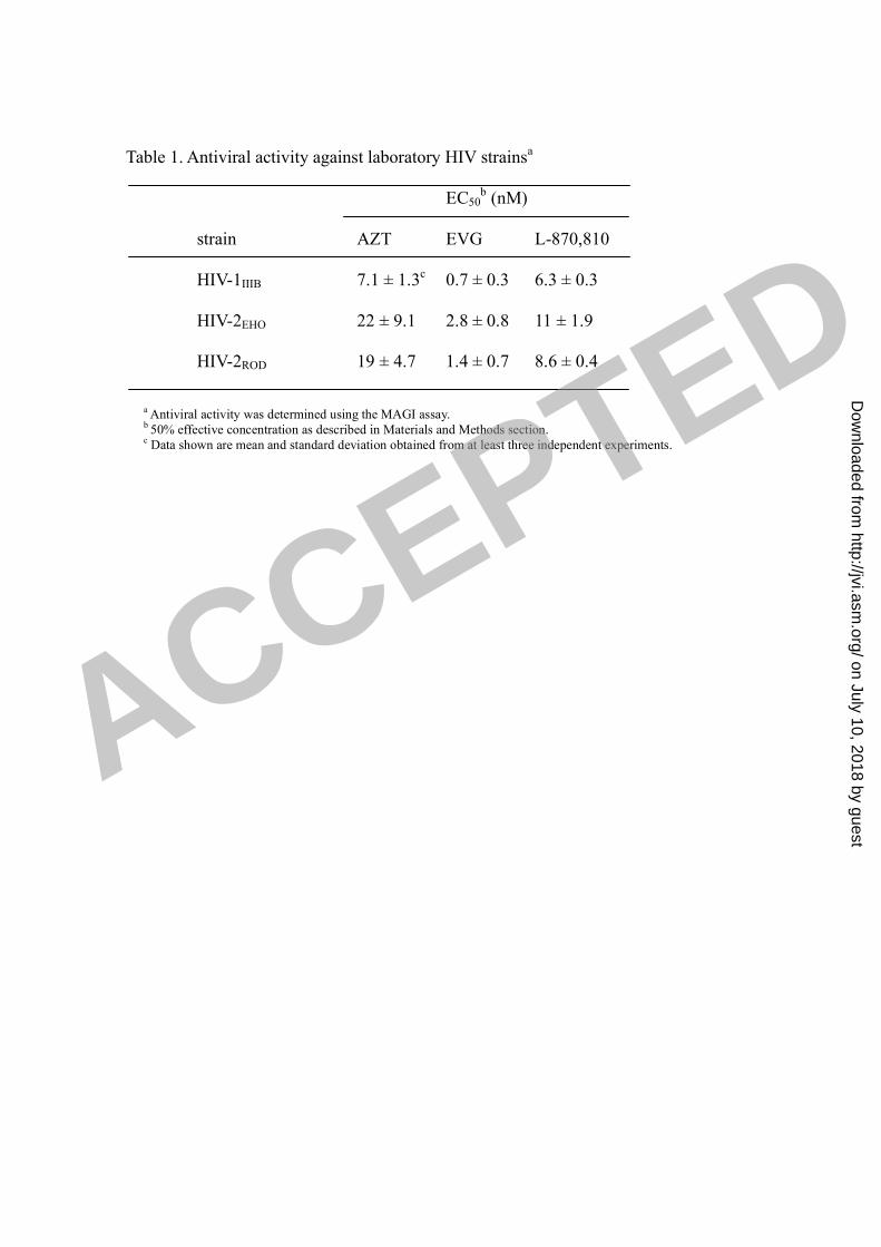
Table 1. Antiviral activity against laboratory HIV strainsa
EC50b (nM)
strain AZT EVG L-870,810
HIV-1IIIB 7.1 ± 1.3c
0.7 ± 0.3 6.3 ± 0.3
HIV-2EHO 22 ± 9.1 2.8 ± 0.8 11 ± 1.9
HIV-2ROD 19 ± 4.7 1.4 ± 0.7 8.6 ± 0.4
a Antiviral activity was determined using the MAGI assay.
b 50% effective concentration as described in Materials and Methods section.
c Data shown are mean and standard deviation obtained from at least three independent experiments.
ACCEPTED
on July 10, 2018 by guesthttp://jvi.asm
.org/D
ownloaded from

Table 2. Antiviral activity of EVG against various subtypes of HIV-1a
subtype isolate EC50b (nM)
AZT EVG
A RW/92/016 7.91 0.41
B 96USHIPS7 8.41 0.26
BR/92/021 2.13 0.76
BR/93/017 1.10 0.18
BR/93/022 11.7 1.13
C BR/92/025 2.84 0.10
D UG/92/046 7.26 0.50
E CMU02 9.07 1.26
F BR/93/020 25.3 0.74
G JV1083 11.1 0.35
O BCF01 1.52 1.17
a Antiviral activity was determined using p24 ELISA.
b 50% effective concentration as described in Materials and Methods section.
ACCEPTED
on July 10, 2018 by guesthttp://jvi.asm
.org/D
ownloaded from
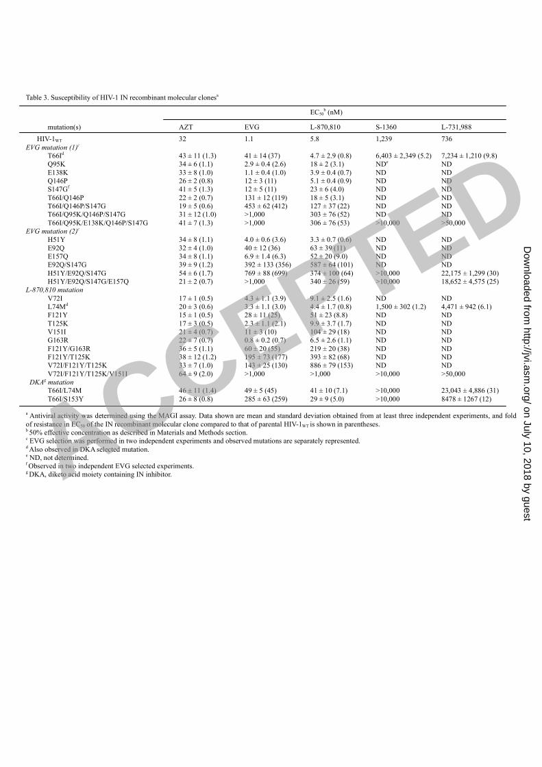
Table 3. Susceptibility of HIV-1 IN recombinant molecular clonesa
EC50b (nM)
mutation(s) AZT EVG L-870,810 S-1360 L-731,988
HIV-1WT 32 1.1 5.8 1,239 736
EVG mutation (1)c
T66Id 43 ± 11 (1.3) 41 ± 14 (37) 4.7 ± 2.9 (0.8) 6,403 ± 2,349 (5.2) 7,234 ± 1,210 (9.8)
Q95K 34 ± 6 (1.1) 2.9 ± 0.4 (2.6) 18 ± 2 (3.1) NDe
ND
E138K 33 ± 8 (1.0) 1.1 ± 0.4 (1.0) 3.9 ± 0.4 (0.7) ND ND
Q146P 26 ± 2 (0.8) 12 ± 3 (11) 5.1 ± 0.4 (0.9) ND ND
S147Gf
41 ± 5 (1.3) 12 ± 5 (11) 23 ± 6 (4.0) ND ND
T66I/Q146P 22 ± 2 (0.7) 131 ± 12 (119) 18 ± 5 (3.1) ND ND
T66I/Q146P/S147G 19 ± 5 (0.6) 453 ± 62 (412) 127 ± 37 (22) ND ND
T66I/Q95K/Q146P/S147G 31 ± 12 (1.0) >1,000 303 ± 76 (52) ND ND
T66I/Q95K/E138K/Q146P/S147G 41 ± 7 (1.3) >1,000 306 ± 76 (53) >10,000 >50,000
EVG mutation (2)c
H51Y 34 ± 8 (1.1) 4.0 ± 0.6 (3.6) 3.3 ± 0.7 (0.6) ND ND
E92Q 32 ± 4 (1.0) 40 ± 12 (36) 63 ± 39 (11) ND ND
E157Q 34 ± 8 (1.1) 6.9 ± 1.4 (6.3) 52 ± 20 (9.0) ND ND
E92Q/S147G 39 ± 9 (1.2) 392 ± 133 (356) 587 ± 64 (101) ND ND
H51Y/E92Q/S147G 54 ± 6 (1.7) 769 ± 88 (699) 374 ± 100 (64) >10,000 22,175 ± 1,299 (30)
H51Y/E92Q/S147G/E157Q 21 ± 2 (0.7) >1,000 340 ± 26 (59) >10,000 18,652 ± 4,575 (25)
L-870,810 mutation
V72I 17 ± 1 (0.5) 4.3 ± 1.1 (3.9) 9.1 ± 2.5 (1.6) ND ND
L74Md
20 ± 3 (0.6) 3.3 ± 1.1 (3.0) 4.4 ± 1.7 (0.8) 1,500 ± 302 (1.2) 4,471 ± 942 (6.1)
F121Y 15 ± 1 (0.5) 28 ± 11 (25) 51 ± 23 (8.8) ND ND
T125K 17 ± 3 (0.5) 2.3 ± 1.1 (2.1) 9.9 ± 3.7 (1.7) ND ND
V151I 21 ± 4 (0.7) 11 ± 3 (10) 104 ± 29 (18) ND ND
G163R 22 ± 7 (0.7) 0.8 ± 0.2 (0.7) 6.5 ± 2.6 (1.1) ND ND
F121Y/G163R 36 ± 5 (1.1) 60 ± 20 (55) 219 ± 20 (38) ND ND
F121Y/T125K 38 ± 12 (1.2) 195 ± 73 (177) 393 ± 82 (68) ND ND
V72I/F121Y/T125K 33 ± 7 (1.0) 143 ± 25 (130) 886 ± 79 (153) ND ND
V72I/F121Y/T125K/V151I 64 ± 9 (2.0) >1,000 >1,000 >10,000 >50,000
DKAg mutation
T66I/L74M 46 ± 11 (1.4) 49 ± 5 (45) 41 ± 10 (7.1) >10,000 23,043 ± 4,886 (31)
T66I/S153Y 26 ± 8 (0.8) 285 ± 63 (259) 29 ± 9 (5.0) >10,000 8478 ± 1267 (12)
a Antiviral activity was determined using the MAGI assay. Data shown are mean and standard deviation obtained from at least three independent experiments, and fold
of resistance in EC50 of the IN recombinant molecular clone compared to that of parental HIV-1WT is shown in parentheses.b 50% effective concentration as described in Materials and Methods section.
c EVG selection was performed in two independent experiments and observed mutations are separately represented.
d Also observed in DKA selected mutation.
e ND, not determined.
f Observed in two independent EVG selected experiments.
g DKA, diketo acid moiety containing IN inhibitor.ACCEPTED
on July 10, 2018 by guesthttp://jvi.asm
.org/D
ownloaded from