:j52 :^ a m;li& mutations affecting agonist sensitivity of the nicotinic
TRANSCRIPT
: J52 : ^ a M;li&Mutations affecting agonist sensitivity of the nicotinic acetylcholinereceptor
Gordon F. Tomaselli,* James T. McLaughlin,t11 Mark E. Jurman,t11 Edward Hawrot,§ and Gary YellentII¶'Howard Hughes Medical Institute and the I"Departments of Neuroscience, 'Biophysics and *Medicine, The Johns HopkinsSchool of Medicine, Baltimore, Maryland 21205; and 'The Section of Molecular and Biochemical Pharmacology, BrownUniversity, Providence, Rhode Island 02912 USA
ABSTRACT The nicotinic acetylcholine receptor (AChR) is a pentameric transmembrane protein (X2P3Y8) that binds theneurotransmitter acetylcholine (ACh) and transduces this binding into the opening of a cation selective channel. The agonist,competitive antagonist, and snake toxin binding functions of the AChR are associated with the a subunit (Kao et al., 1984; Tzartosand Changeux, 1984; Wilson et al., 1985; Kao and Karlin, 1986; Pederson et al., 1986). We used site-directed mutagenesis andexpression of AChR in Xenopus oocytes to identify amino acid residues critical for ligand binding and channel activation. Severalmutations in the a subunit sequence were constructed based on information from sequence homology and from previousbiochemical (Barkas et al., 1987; Dennis et al., 1988; Middleton and Cohen, 1990) and spectroscopic (Pearce and Hawrot, 1990;Pearce et al., 1990) studies. We have identified one mutation, Tyr'90 to Phe (Y190F), that had a dramatic effect on ligand binding andchannel activation. These mutant channels required more than 50-fold higher concentrations of ACh for channel activation than didwild type channels. This functional change is largely accounted for by a comparable shift in the agonist binding affinity, as assessedby the ability of ACh to compete with a-bungarotoxin binding. Other mutations at nearby conserved positions of the a subunit(H186F, P194S, Y198F) produce less dramatic changes in channel properties. Our results demonstrate that ligand binding andchannel gating are separable properties of the receptor protein, and that Tyr'9" appears to play a specific role in the receptor site foracetylcholine.
INTRODUCTION
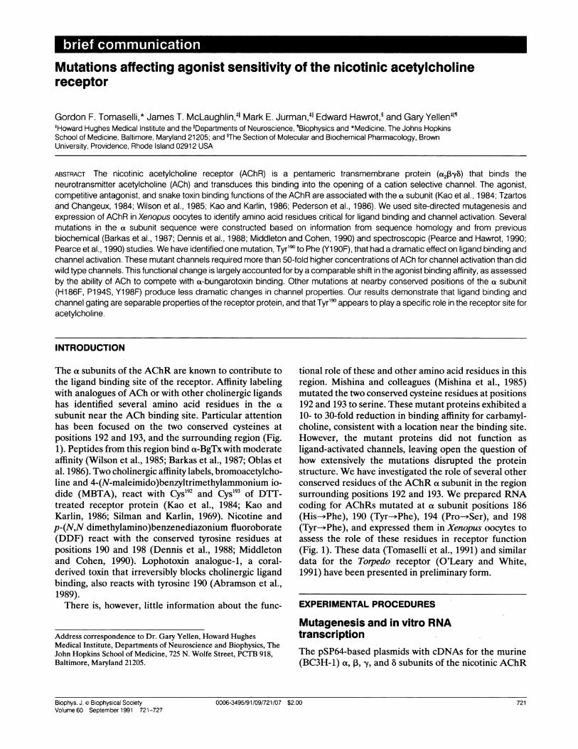
The at subunits of the AChR are known to contribute tothe ligand binding site of the receptor. Affinity labelingwith analogues of ACh or with other cholinergic ligandshas identified several amino acid residues in the cxsubunit near the ACh binding site. Particular attentionhas been focused on the two conserved cysteines atpositions 192 and 193, and the surrounding region (Fig.1). Peptides from this region bind a-BgTx with moderateaffinity (Wilson et al., 1985; Barkas et al., 1987; Oblas etal. 1986). Two cholinergic affinity labels, bromoacetylcho-line and 4-(N-maleimido)benzyltrimethylammonium io-dide (MBTA), react with Cys"92 and Cyst93 of DTT-treated receptor protein (Kao et al., 1984; Kao andKarlin, 1986; Silman and Karlin, 1969). Nicotine andp-(N,N dimethylamino)benzenediazonium fluoroborate(DDF) react with the conserved tyrosine residues atpositions 190 and 198 (Dennis et al., 1988; Middletonand Cohen, 1990). Lophotoxin analogue-1, a coral-derived toxin that irreversibly blocks cholinergic ligandbinding, also reacts with tyrosine 190 (Abramson et al.,1989).There is, however, little information about the func-
Address correspondence to Dr. Gary Yellen, Howard HughesMedical Institute, Departments of Neuroscience and Biophysics, TheJohn Hopkins School of Medicine, 725 N. Wolfe Street, PCTB 918,Baltimore, Maryland 21205.
tional role of these and other amino acid residues in thisregion. Mishina and colleagues (Mishina et al., 1985)mutated the two conserved cysteine residues at positions192 and 193 to serine. These mutant proteins exhibited a
10- to 30-fold reduction in binding affinity for carbamyl-choline, consistent with a location near the binding site.However, the mutant proteins did not function as
ligand-activated channels, leaving open the question ofhow extensively the mutations disrupted the proteinstructure. We have investigated the role of several otherconserved residues of the AChR a subunit in the regionsurrounding positions 192 and 193. We prepared RNAcoding for AChRs mutated at a subunit positions 186(His-*Phe), 190 (Tyr-*Phe), 194 (Pro- Ser), and 198(Tyr--Phe), and expressed them in Xenopus oocytes toassess the role of these residues in receptor function(Fig. 1). These data (Tomaselli et al., 1991) and similardata for the Torpedo receptor (O'Leary and White,1991) have been presented in preliminary form.
EXPERIMENTAL PROCEDURES
Mutagenesis and in vitro RNAtranscriptionThe pSP64-based plasmids with cDNAs for the murine(BC3H-1) a, ,B, y, and 8 subunits of the nicotinic AChR
Biophys. J. Biophysical SocietyVolume 60 September 1991 721-727
0006-3495/91/09/721/07 $2.00 7210006-3495/91/09/721/07 $2.00 721
MUSCLE BC3H-1 aTorpedo bHuman"ChickdXenopus
d
NEURONAL Chick a2dChick c3 dChick q4dRat ae2fRat a3 8Rat ct4 9Fly h
FIGURE 1 The amino acid sequences of a subunits from muscle andneuronal tissue. The sequences are compared with the murineBC3H-1 cell line nicotinic receptor sequence. Shaded areas indicateidentity of the amino acid with the corresponding residue in theBC3H-1 sequence. The numbered residues were mutated as follows:H186F, Y19OF, P194S, and Y198F. Abbreviations for the amino acidresidues are: A, ala; C, cys; D, asp; E, glu; F, phe; G, gly; H, his; I, ile;K. lys; L, leu; M, met; N, asn; P, pro; Q, gln; R, arg; S, ser; T, thr; V, val;W, trp; and Y, tyr. References for sequences: aBoulter et al., 1985;bNoda et al., 1982; cNoda et al., 1983; dNef et al., 1988; eBaldwin et al.,1988; fWada et al., 1988; 5Boulter et al., 1987; hBossy et al., 1988.
were generously provided by Dr. J. Boulter of the SalkInstitute. The a subunit cDNA was excised with EcoRIand subcloned into pBluescript KS- (Stratagene, LaJolla, CA) for oligonucleotide-directed mutagenesis.Missense mutations were introduced using dut- andung- selection by the method of Kunkel (Kunkel, 1985).The entire cDNA of the a subunit was sequenced(Sanger et al., 1977) to confirm the nucleotide changesand to guard against stray mutations. The EcoRI frag-ments of the a subunit mutants were cloned back intopSP64 for in vitro transcription.RNAs encoding the wild type and mutant receptor
subunits were transcribed in a similar fashion. Plasmidscontaining the wild type and mutant subunit cDNA were
linearized using Sca I. The linearized plasmid (2-4 ,ug)was incubated in 10 mM NaCl, 6 mM MgCl2, 40mM Tris(pH 7.5), 1 mM dithiothreitol, 2 mM spermidine, NTPs(1 mM each except GTP which was 0.2 mM), 0.6 mMdiguanosine triphosphate (Pharmacia, Piscataway, NJ),30U SP6 RNA polymerase, and 100 U RNasin (Promega,Madison, WI) at 37° for 1 h. An additional 30 U of SP6RNA polymerase and 100 U RNasin were added afteran hour and the incubatiorn was continued for anotherhour. The template DNA was removed with 4 U ofRNase-free RQ1 DNase (Promega, Madison, WI) for 15min at 37°. The RNA was extracted with phenolchloro-form and chloroform, precipitated from isopropanolthen resuspended in DEPC-treated water at a concentra-tion of - 0.2 Rg/4il. Subunit RNA was mixed in a ratio of2a:1 3:1y:18 for later injection into oocytes.
Preparation and microinjection ofoocytes
Adult female, HCG-primed Xenopus laevis (Xenopus I,
Ann Arbor, MI) were anesthetized by immersion in0.17% aminobenzoic acid ethyl ester and several lobes ofovary were removed through a small abdominal incision.Individual stage V or VI oocytes were isolated bymanual dissection followed by incubation in 0.5-2 mg/mlcollagenase (type IA; Sigma Chemical Co., St. Louis,MO) in calcium-free saline (82 mM NaCl, 2 mM KCl, 1mM MgCl2, 5 mM Hepes pH 7.6) for 1-2 h. The oocyteswere allowed to recover in amphibian saline (96 mMNaCl, 2 mM KCl, 1.8 mM CaCl2, 1 mM MgCl2, 5 mMHepes pH 7.6) before injection.
Selected oocytes were injected with 25-50 nl of RNAsolution using a positive displacement injector (Drum-mond Instruments, Broomhall, PA) through glass boreswith tip diameters of 20-40 ,um. The injected oocyteswere incubated for at least 48 h in amphibian salinesupplemented with 100 U/ml penicillin and 100 p,g/mlstreptomycin sulfate (Gibco BRL, Gaithersburg, MD),0.5 mM theophylline, and 2 mM Na pyruvate.
ElectrophysiologyThe oocytes were screened for ACh-induced current bytwo microelectrode voltage clamp as previously de-scribed (Tomaselli et al., 1990). Whole oocytes were
continuously perfused with ACh at a concentration thatelicited near maximal current response for the channelvariant being tested. Oocytes with sufficient whole cellcurrent (>0.5 ,uA at -30 mV holding potential) were
then patch clamped. The oocytes were devitellinized inpreparation for patch clamp recording as previouslydescribed (Methfessel et al., 1986). Excised outside-outpatches (Hamill et al., 1981) were obtained in symmetricpipette and bath solutions (in mM): 97 KCl, 1 MgCl2, 0.2mM EGTA, 5 K-Hepes, pH 7.5. Patches were continu-ously perfused and currents were elicited by the applica-tion of the bath solution containing the desired concen-
tration of ACh.The patch was positioned in a custom-designed bath at the convergence point of streams ofcontrol and ACh-containing solution. Rapid solutionchanges (1-2 ms) were made by computer-controlledsolenoid switches as previously described (Maconachieand Knight, 1989). ACh was applied in 400 ms pulsesbeing delivered no more frequently than every 5 s.
Currents were recorded by a List EPC7 patch clampamplifier (List-electronic, Darmstadt, West Germany).During pulsatile application of ACh the currents were
digitized at 2.5 kHz and filtered at 1 kHz (8-pole Bessel,-3 dB). The level of channel activity varied from patch
722 Biophysical Journal Volume 60 September 1991722 Biophysical Journal Volume 60 September 1991

to patch; ensemble averages were compiled from 5-32individual records. The peak of the ensemble averagecurrent at a given ACh concentration was corrected forACh block using the measured single channel current.The ensemble average currents were then normalized tothe maximal corrected current elicited from that patchand are plotted against the log of the ACh concentra-ti6n. The single channel current voltage relationshipswere obtained by continuous application of ACh at thedesired concentration with rampwise or steady-statevoltage changes. Under these conditions the currentswere digitized at 10 kHz and filtered at 2 kHz (8-poleBessel, -3dB). All recordings were done at room
temperature 22-25°C.
Toxin and competition bindingWhole oocyte binding was performed on 5-20 oocytesinjected with wild type or mutant AChR channel RNAand a similar number of naive oocytes. The cells were
incubated in (in mM): 96 NaCl, 2 KCl, 1.8 CaCl2, 1MgCl2, 5 Hepes pH 7.6, 0.1% bovine serum albumin andsaturating [1"I]-a-BgTx (1,000 counts/fmol) for 2-4 h,then washed with the same solution minus the labeledtoxin. The oocytes were counted on an LKB model 1282gamma counter. The number of counts per oocyte werecorrected for nonspecific binding by subtracting theaverage number of counts bound to uninjected oocytes.The density of receptors in the oocyte surface mem-
brane is estimated as fmols of bound toxin per oocyte.Membrane preparation and toxin binding assays were
performed as previously described (McLaughlin andHawrot, 1989). Briefly, oocyte membranes were pre-pared fresh or in some cases from previously frozencells. 100-200 oocytes were homogenized in 0.1 ml/oocyte of membrane binding buffer (in mM: 140 NaCl,20 Na phosphate pH 7.5, 1 EDTA, 1 EGTA, 0.5phenylmethylsulfonyl fluoride, and 200 U/ml aprotinin).Homogenates were centrifuged for 30 min at 20,000 x g,pellets were then resuspended in 0.04 ml/oocyte ofmembrane binding buffer and stored at - 80°C.Homogenized membranes (0.16 ml) were incubated
for 3 h at room temperature in a final volume of 0.22 mlwith 2.5 nM "2I labeled-a-BgTx and the desired concen-tration of ACh. The samples were then diluted to 6 mlwith cold Tris-buffered saline (in mM: 150 NaCl, 50Tris-HCl pH 7.6) and filtered through Whatman GF/Ffilters and washed. The filters were counted on an LKBmodel 1282 gamma counter to determine the level of['"I]-a-BgTx bound. Correction for nonspecific bindingwas made using a parallel protocol done in the presenceof 1.25 p,M unlabeled a-BgTx.
RESULTS AND DISCUSSION
Each mutant was assayed electrophysiologically to deter-mine its functional response to ACh. Outside-out patchesfrom injected oocytes were placed in a stream of bathingsolution that could be switched rapidly (within 1-2 ms)to a solution containing a known, constant concentrationofACh. Patches from oocytes expressing the wild type ormutant channels typically contained many channels( - 5-50) that opened rapidly in response to ACh appli-cation (Fig. 2). A dose-response curve for each channelvariant was made by compiling ensemble averages of theresponses to repeated presentations of each concentra-tion studied, and measuring the peak of the currentresponse (Fig. 2,A and B). All of the mutant channelsrespond to ACh, but the Y19OF mutant is dramaticallyaltered in its dose response and requires millimolarconcentrations of ACh for channel opening (Fig. 2 C;Table 1).
This change in the sensitivity of the Y19OF mutantcould be due either to a change in the binding affinity ofthe receptor for ACh or to a change in the energetics ofopening of the channel after binding. To distinguishbetween these possibilities, we assessed the ACh bind-ing affinity of the mutants by measuring the ability ofACh to compete for [1251]-t-bungarotoxin binding. Theaffinity of toxin itself is relatively unaffected by themutations (Table 1), but the ability of ACh to preventtoxin binding is dramatically altered in the Y19OFmutant. As for channel activation, the concentration ofACh required for competition in the Y19OF mutant isseveral hundred-fold higher than that for wild type (Fig.3). Thus, the entire physiological effect of the mutationcan be explained by its effect on binding, withoutpostulating an effect on transduction.
Binding of ACh to the AChR has not only theshort-term consequence of opening the channel, but alsothe longer-term consequence of receptor desensitiza-tion. Desensitization reflects the closure of channels inspite of prolonged exposure to ACh, and it is alsoaccompanied by an increase in ACh binding affinity(Katz and Thesleff, 1957; Sine and Taylor, 1979; Sineand Taylor, 1980). Two observations on the mutantchannels indicate that the Y19OF mutation affects AChbinding to the resting and the desensitized states of thechannel in parallel. The first indication is that AChcompetition with toxin binding, which is measured inlong incubations and thus reflects binding to the desensi-tized state, parallels the results on the rapid physiologi-cal effects of ACh. The second indication comes fromdirect observation of desensitization rates during the
Toasll et a. Muain of th ACh Aciato SitTomaselli et al. Mutations of the AChR Activation Site 723

A Wild type
W~~IIIrIrHw r1
w~~~11r 7Y~
10 pAL80 ms
Y198FYI9OF
z
z
C
O wild type* Y19OFv Y198F
[ACh] (M)
FIGURE 2 Physiological responses to a ACh. (A) Single channel current responses to the application of 1 ,uM (wild type), 10 F.M (Y198F), or 1mM (Y19OF) ACh. Patches were held at a potential of -100 mV. These records were selected to show resolved single channel currents; mostpatches contained many more channels. (B) Ensemble average currents obtained at a series of ACh concentrations for wild type (1, 2.5, 5, 10, and50 ,uM), Y19OF (0.1, 0.5, 1, 3, and 5 mM) and Y198F (1, 10, 50, 300, and 500 F.M). The vertical position of each of the averages is indicative of theACh concentration that elicited that current. The vertical scale bar for each of the channel variants is 200 pA (wild type), 5 pA (Y19OF), and 80 pA(Y198F). The horizontal scale bar represents 300 ms. (C) A plot of the normalized current corrected for ACh block against the log of the AChconcentration. Each point represents the mean of the peak of the ensemble average current corrected for ACh block and normalized to themaximal current elicited in that patch for wild type (0), Y19OF (0), and Y198F (v). The smooth curves are fits to the data generated as describedin Table 1. The Hill coefficients for each of the channel types were optimized and had values of 1.5 + 0.09 (wild type), 1.12 + 0.1 (Y19OF), and1.17 + 0.09 (Y198F).
physiological exposure to ACh. Desensitization is typi-cally faster at higher concentrations of agonist (Fig.2 B). Desensitization of the Y19OF mutant, however,remains slow even at concentrations that produce rapiddesensitization in the wild type channel; rapid desensiti-zation of the Y19OF mutant occurs only at the millimolarconcentrations required for maximal activation of themutant. Thus, desensitization appears to be affected inparallel with activation. This observation argues against
the proposed existence of separate ACh binding sites foractivation and desensitization (Dunn and Raftery, 1982).The Y19OF mutant specifically affects the ligand
binding properties of the AChR, and does not affect itsopen channel properties. The single-channel currentvoltage relationship is unaltered in the mutant (Fig. 4A).At high concentrations, ACh not only opens the AChRchannels, it also acts to block current through the openchannel (Sine and Steinbach, 1984). Such blockade
724 Biophysical Journal Volume 60 September 1991~~~~~~~~~~~~~~~~
B wild type-6 r
C:-4
-3
-2
aY19OF c&Y198F
-5 P
io-8 10-7 10-6 10-5 10-4 lo-_3 10-2 lo-'
724 Biophysical Journal Volume 60 September 1991

TABLE 1 Affinity constants of wild type and mutant channels
SurfaceReceptor binding oa-BgTx Kd EC50 IC50
finolloocyte nM pFM piMWild type 1.5 + 1.1 (7) 0.54 + 0.1 11.0 + 0.7 (6) 4.4 ± 2.0 (3)H186F 1.9 ± 0.8 (5) 0.37 ± 0.1 21.8 + 1.2 (7) 3.2 ± 1.4 (3)Y19OF 0.7 ± 0.3 (7) 0.49 ± 0.2 548 ± 52 (5) 338 ± 98 (3)P194S 2.1 ± 1.5 (3) 0.66 ± 0.2 10.2 + 0.1 (7) 3.6 ± 1.8 (3)Y198F 0.9 ± 0.4 (4) 0.44 ± 0.1 60.3 ± 8.1 (5) 23.8 ± 4.8 (3)
The EC50 and IC58s were estimated from a weighted least squares fit ofthe mean normalized current and percent of control a-BgTx binding,respectively, to the functionf = 1 - 1/(1 + ([ACh]/x)n), where x is theEC50 or IC,o and n is the Hill coefficient (see Figs. 2 and 3 legend). Thenormalized current and percent of control binding are determined asdescribed in Figs. 2 and 3, respectively. The EC50 and IC_Os arepresented as the mean and standard deviation. In parentheses are thenumber of patches or membrane preparations for each channelvariant.The affinity of the receptors for toxin is similar as illustrated by the K.s.The Kds were determined by competition of labeled a-BgTx byunlabeled toxin values are the mean of three determinations. The datawere normalized and fit as described for agonist competition of toxinbinding.
appears as a voltage-dependent reduction in the openchannel current at high ACh concentration. This reduc-tion is identical in the wild type and mutant channels(Fig. 4B).The other mutations of conserved residues in this
region have smaller or no effect on binding and activa-
100 K C_ O wild tvye
A-200
O *0 wild typeo * Y19OF
B
-150
Voltage (mV)
-100 -50 00
-1
-2
-3
-4 a
-5
-6
-7
-8
Log [ACh] (M)-8 -7 -6 -5 -4 -3 -2 -1
Vh -100 mVO wild typeA H186F* Y19OF* P194SV Y198F
-1
-2
-3
-4 <.
-5
-6
-7
FIGURE 4 Open channel properties of the AChR are not altered bythe mutations. (A) Representative single channel current voltagerelationships for the wild type (0, 0) and Y19OF (D, *). ACh at 100FLM (open symbols) and 1 mM ACh (solid symbols). The conductancesin the negative voltage range for both channel variants are identical.(B) Single channel current amplitude as a function of the AChconcentration for all channel variants at a holding potential of - 100mV: wild type (0), H186F (A), Y19OF (0), P194S (-), and Y198F(V).
.0 I. ..
108o o-7 1o-6 10 lo-3 1o 2 o 1
[ACh]
FIGURE 3 Concentration dependence of ACh inhibition of steady-state cL-BgTx binding for the wild type receptor and the mutantreceptor channels. Toxin binding at each ACh concentration wasnormalized to the [1"I]-a-BgTx binding in the absence of agonist. Thenormalized binding is the mean of at least three determinations ateach ACh concentration for wild type (0), H186F (E), Y19OF (0),P194S (A), and Y198F (V). The data were fit and the IC50s weredetermined as described in Table 1. Hill coefficients ranged from 0.7 to1.3 for both wild type and mutant receptors.
tion by ACh. For each of the other mutants, the EC50sand IC50s are within an order of magnitude of the wildtype values. The conserved proline residue at position194 may conformationally constrain this region of thereceptor, but the absence of an effect of the P194Smutation rules out a critical role for this residue. Thebasic residue His'86 has been suggested to function in a
charge relay system essential to ligand binding (Pearceand Hawrot, 1990; Pearce et al., 1990); however, muta-tion of this residue to Phe produces no change in binding
Tomaselli et al. Mutations of the AChR Activation Site 725
80
60
40
cr
-o
c
ia0vx
20
I
725Mutations of the AChR Activation SiteTomaselli et al.

and only a small change in physiological response. Thusit is possible to alter conserved residues in this regionwithout producing a nonspecific effect on binding andactivation, supporting the notion that Tyr"9 plays aparticular and important role in these processes. Thesmaller but significant effect of the Y198F mutation alsoimplicates this residue in binding and channel activa-tion.Another suggestion that either Tyr"9 or Tyr'98 might
play an interesting role in ACh binding comes fromspectroscopic studies of synthetic peptides from thisregion of the AChR that bind a-BgTx. The intrinsicfluorescence spectra of the peptides suggest the pres-ence of a negatively charged tyrosinate (phenolate) ionat neutral pH (Pearce and Hawrot, 1990; Pearce et al.,1990). The authors of this work have suggested that oneof these tyrosines might therefore be a good candidatefor the hypothetical "anionic subsite" of the AChRbinding site proposed to interact with the positivelycharged, quaternary ammonium moiety common tomost cholinergic ligands. Is this proposal consistent withour observation that mutation of Tyr'" to Phe (whichcannot ionize) produces a several hundred-fold shift inACh binding? Perhaps. One might expect at first thatremoving a critical charge-charge interaction mighthave an even greater effect on binding affinity. Theobserved shift in the dose response corresponds to achange in binding energy of 2-3 kcal/mol. This energyis comparable, for example, to the coulombic interactionenergy of two point charges at a distance of 6-7 A in amedium of dielectric constant 20. Thus the magnitude ofthe effect may be consistent with the proposed model.We attempted to test this hypothesis that Tyr"9 is the
anionic subsite of the AChR by substituting other aminoacids at this position, neutral glutamine or negativelycharged glutamate. Both of these mutations produceshifts in the dose-response similar to that produced bythe Y19OF mutation (data not shown). It thus appearsthat tyrosine is unique in its ability to promote AChbinding at this position. This could mean that tyrosine,because of its size, its shape, and the hydrogen-bondingproperties of its hydroxyl group, is critical for producingthe proper folding of the ACh binding pocket. Alterna-tively, both the negative charge of the proposed tyrosi-nate and the aromatic character of tyrosine may beinvolved in the interaction between ACh and Tyr'".Dougherty and Stauffer have suggested that aromaticresidues are especially well suited for binding with thediffuse charge of quaternary ammonium moieties; theirhost-guest experiments with aromatic macrocycles sup-port this notion, as do the known structures of severalproteinaceous binding sites for quaternary amines(Dougherty and Stauffer, 1990). The recent discovery ofa tyrosine residue critical for the high affinity blockade
of potassium channels by tetraethylammonium mayprovide another such example (MacKinnon and Yellen,1990).We have observed a dramatic and specific effect of
mutations of Tyr"9 on the ACh binding and activation ofthe nicotinic AChR. Much higher concentrations ofACh are required to activate the mutant, and thischange in sensitivity can be entirely accounted for by achange in the binding affinity of ACh. These resultssuggest that Tyr"9 plays a critical role in producing thebinding pocket for ACh. Determining the exact role ofthis and other residues in ACh binding will of courserequire further studies using both mutagenesis andstructure determination.
We thank K. Choi, S. Demo, and M. West for helpful discussions.
Dr. Tomaselli is supported by National Institutes of Health grant K08HL02421-02, and is an Eli Lilly Clinician-Scientist of the JohnsHopkins School of Medicine. Dr. Hawrot is an Established Investiga-tor of The American Heart Association. Dr. Yellen is an investigatorof the Howard Hughes Medical Institute.
Received for publication 1 April 1991 and in final form 22 May1991.
REFERENCES
Abramson, S. N., Y. Li, P. Culver, and P. Taylor. 1989. An analog oflophotoxin reacts covalently with Tyr"9 in the a-subunit of thenicotinic acetylcholine receptor. J. Biol. Chem. 264:12666-12672.
Baldwin, T. J., C. M. Yoshihari, K. Blackmer, C. R. Kintar, and S. J.Burden. 1988. Regulation of acetylcholine receptor transcript expres-sion during development in Xenopus laevis. J. Cell Bio. 106:469-78.
Barkas, T., A. Mauron, B. Roth, C. Alliod, S. J. Tzartos, and M.Ballivet. 1987. Mapping the main immunogenic region and toxinbinding site of the nicotinic acetylcholine receptor. Science (Wash.DC). 235:77-80,
Bossy, B., M. Ballivet, and P. Spierer. 1988. Conservation of neuronalnicotinic acetylcholine receptors from Drosophila to vertebratecentral nervous system. EMBO (Eur. Mol. Biol. Organ.) J. 7:611-618.
Boulter, J., W. Luyten, K. Evans, P. Mason, M. Ballivet, D. Goldman,S. Stengelin, G. Martin, S. Heinemann, and J. Patrick. 1985.Isolation of a clone coding for the a-subunit of the mouse acetylcho-line receptor. J. Neurosci. 5:2545-2552.
Boulter, J., J. Connolly, E. Deneris, D. Goldman, S. Heinemann, andJ. Patrick. 1987. Functional expression of two neuronal nicotinicacetylcholine receptors from cDNA clones identifies a gene familyProc. Natl. Acad. Sci. USA. 84:7763-7767.
Dennis, M., J. Giraudat, F. Kotzyba-Hibert, M. Goeldner, C. Hirth,J-Y. Chang, C. Lazure, M. Chretien, and J-P. Changeux. 1988.Amino acids of the Torpedo marmorata acetylcholine receptor asubunit labeled by a photoaffinity ligand for the acetylcholinebinding site. Biochemistry. 27:2346-2358.
Dougherty, D. A., and D. A. Stauffer. 1990. Acetylcholine binding by asynthetic receptor: Implications for biological recognition. Science(Wash. DC). 250:1558-1560.
726 Biophysical Journal Volume 60 September 1991

Dunn, S. M. J., and M. A. Raftery. 1982. Activation and desensitiza-tion of Torpedo acetylcholine receptor: Evidence for separatebinding sites. Proc. Natl. Acad. Sci. USA. 79:6757-6761.
Hamill, 0. P., A. Marty, E. Neher, B. Sakmann, and F. J. Sigworth.1981. Improved patch-clamp technique for high-resolution currentrecording from cells and cell-free membrane patches. PfluegersArch.Eur. J. Physiol. 391:85-100.
Kao, P. N., A. J. Dwork, R. J. Kaldany, M. L. Silver, J. Wideman, S.Stein, and A. Karlin. 1984. Identification of the a subunit half-cystine specifically labeled by an affinity reagent for the acetylcho-line receptor binding site. J. Bio. Chem. 259:11662-11665.
Kao, P. N., and A. Karlin. 1986. Acetylcholine receptor binding sitecontains a disulfide cross-link between adjacent half-cystinyl resi-dues. . Bio. Chem. 261:8085-8088.
Katz, B., and S. Thesleff. 1957. A study of the desensitization producedby acetylcholine at the motor end-plate. J. Physiol. 138:63-80.
Kunkel, T. A. 1985. Rapid and efficient site-specific mutagenesis withphenotypic selection. Proc. Natl. Acad. Sci. USA. 82:488-492.
MacKinnon, R., and G. Yellen. 1990. Mutations affecting TEAblockade and ion permeation in voltage-activated K+ channels.Science (Wash. DC). 250:276-279.
Maconachie, D. J., and D. E. Knight. 1989. A method for makingsolution changes in the submillisecond range at the tip of a patchpipette. PfluegersArch. Eur. J. Physiol. 414:589-596.
McLaughlin, J. T., and E. Hawrot. 1989. Structural characterization ofalpha-bungarotoxin binding proteins from Aplysia califomica. Mol.Pharmacol. 35:593-598.
Methfessel, C., V. Witzemann, T. Takahashi, M. Mishina, S. Numa,and B. Sakmann. 1986. Patch clamp measurement on Xenopus laevisoocytes: currents through endogenous channels and implantedacetylcholine receptor and sodium channels. Pfluegers Arch. Eur. J.Physiol. 407:577-588.
Middleton, R. E., and J. B. Cohen. 1990. [3H]-nicotine photoaffinitylabels tyr-198 in the alpha subunit of the Torpedo nicotinic acetylcho-line receptor. Soc. Neurosci. Abstr. 16:1016.
Mishina, M., T. Tobimatsu, K. Imoto, K. Tanaka, Y. Fujiita, K.Fukuda, M. Kurasaki, H. Takahashi, Y. Morimoto, T. Hirose, S.Inayama, T. Takahashi, M. Kuno, and S. Numa. 1985. Location offunctional regions of acetylcholine receptor a-subunit by site-directed mutagenesis. Nature (Lond.) 313:364-369.
Nef, P., C. Oneyser, C. Alliod, S. Couturier, and M. Ballivet. 1988.Genes expressed in the brain define three distinct neuronal nicotinicacetylcholine receptors. EMBO (Eur. Mol. Biol. Organ.) J. 7:595-601.
Noda, M., H. Takahashi, T. Tanabe, M. Toyosato, Y. Furutani, T.Hirose, M. Asai, S. Inayama, T. Miyata, and S. Numa. 1982. Primarystructure of a-subunit precursor of Torpedo califomica acetylcholinereceptor deduced from cDNA sequence. Nature (Lond.). 299:793-797.
Noda, M., Y. Furutani, H. Takahashi, M. Toyosato, T. Tanabe, S.Shimizu, S. Kikyotani, T. Kayano, T. Hirose, S. Inayama, and S.Numa. 1983. Cloning and sequence analysis of calf cDNA and
human genomic DNA encoding a-subunit precursor of muscleacetylcholine receptor. Nature (Lond.). 305:818-823.
Oblas, B., R. H. Singer, and N. D. Boyd. 1986. Location of apolypeptide sequence within the a-subunit of the acetylcholinereceptor containing the cholinergic binding site. Mol. Pharmacol.29:649-656.
O'Leary, M. E., and M. M. White. 1991. Role of ligand binding sitetyrosines in the gating of the Torpedo ACh receptor. Biophys. J.59:34a. (Abstr.)
Pearce, S. F. A., and E. Hawrot. 1990. Intrinsic fluorescence ofbinding-site fragments of the nicotinic acetylcholine receptor. Pertur-bations produced upon binding a-bungarotoxin. Biochemistry. 29:10649-10659.
Pearce, S. F. A., P. Preston-Hurlburt, and E. Hawrot. 1990. The role oftyrosine at the ligand-binding site of the nicotinic acetylcholinereceptor. Proc. R. Soc. B. 241:207-213.
Pederson, S. E., E. B. Dreyer, and J. B. Cohen. 1986. Location ofligand-binding sites on the nicotinic acetylcholine receptor a-sub-unit. J. BioL Chem. 261:13735-13743.
Sanger, F., S. Nicklen, and A. R. Coulson. 1977. DNA sequencing withchain-terminating inhibitors. Proc. Natl. Acad. Sci. USA. 74:5463-5467.
Silman, I., and A. Karlin. 1969. Acetylcholine receptor: covalentattachment of depolarizing groups at the active site. Science (Wash.DC). 164:1420-1421.
Sine, S. M., and J. H. Steinbach. 1984. Agonists block current throughacetylcholine receptor channels. Biophys. J. 46:277-283.
Sine, S., and P. Taylor. 1979. Functional consequences of agonist-mediated state transitions in the cholinergic receptor. J. Biol. Chem.254:3315-3325.
Sine, S., and P. Taylor. 1980. The relationship between agonistoccupation and the permeability response of the cholinergic recep-tor revealed by bound cobra a-toxin. J. Biol. Chem. 255:10144-10156.
Tomaselli, G. F., A. M. Feldman, G. Yellen, and E. Marban. 1990.Human cardiac sodium channels expressed in Xenopus oocytes. Am.J. Physiol. 258:H903-H906.
Tomaselli, G. F., J. T. McLaughlin, M. Jurman, E. Hawrot, and G.Yellen. 1991. Site-directed mutagenesis alters agonist sensitivity ofthe nicotinic acetylcholine receptor. Biophys. J. 59:33a. (Abstr.)
Tzartos, S. J., and J.-P. Changeux. 1984. Lipid-dependent recovery ofa-bungarotoxin and monoclonal antibody binding to the purifieda-subunit from Torpedo marmorata acetylcholine receptor. J. Biol.Chem. 259:11512-11519.
Wada, K., M. Ballivet, J. Boulter, J. Connolly, E. Wada, E. S. Deneris,L. Swanson, S. Heinemann, and J. Patrick. 1988. Functional expres-sion of a new pharmacological subtype of brain nicotinic acetylcho-line receptor. Science (Wash. DC). 240:330-334.
Wilson, P. T., T. L. Lentz, and E. Hawrot. 1985. Determination of theprimary amino acid sequence specifying the a-bungarotoxin bindingsite on the a subunit of the acetylcholine receptor from Torpedocalifomica. Proc. Natl. Acad. Sci. USA. 82:8790-8794.
Tomaselli et al. Mutations of the AChR Activation Site 727