jason membrane & membrane - bone & tissue regeneration · collagen membranes have been used...
TRANSCRIPT
1
botissbiomaterials
bone & tissue regeneration
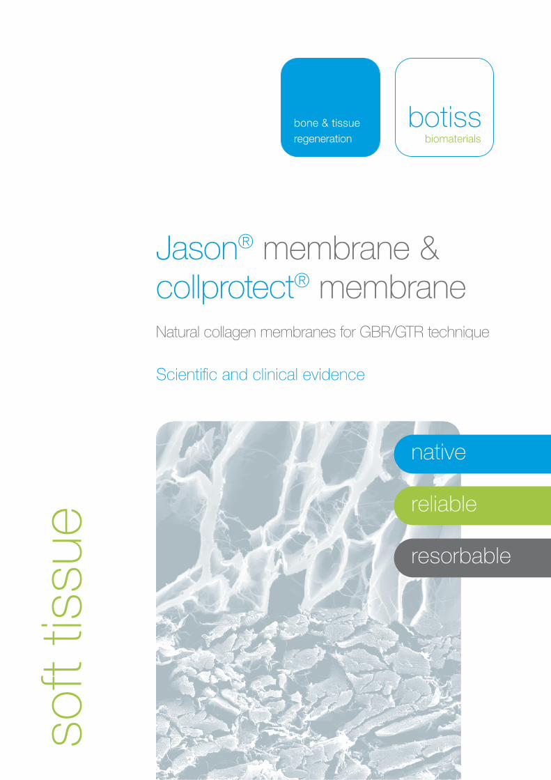
Jason® membrane &collprotect® membraneNatural collagen membranes for GBR/GTR technique
Scientific and clinical evidence
soft
tissu
e
nativ
zuverlässig
resorbierbar
native
reliable
resorbable
2
RegenerationC
ontrolled DegradationB
iolo
gica
l Pot
entia
l
High Quality Learning
* Coming soon
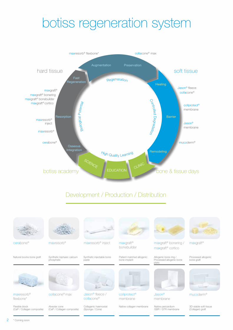
Development / Production / Distribution
Augmentation
mucoderm®
collprotect® membrane
Jason® membrane
Jason® fleece
collacone®......
collacone®..max
hard tissue
cerabone®
maxresorb®
maxresorb®
inject
maxgraft® bonebuildermaxgraft® cortico
maxgraft® boneringmaxgraft®
EDUCATION
SCIENCE CLINIC
Fast Regeneration
Preservation
OsseousIntegration Remodeling
Healing
BarrierResorption
maxresorb® flexbone*
soft tissue
botiss academy bone & tissue days
cerabone®
Natural bovine bone graft
maxresorb® inject maxgraft® bonering / maxgraft® cortico
maxgraft®
bonebuilder
Patient matched allogenic bone implant
Synthetic injectable bone paste
maxresorb®
Synthetic biphasic calcium phosphate
Allogenic bone ring / Processed allogenic bone plate
maxresorb® flexbone*
Flexible block (CaP / Collagen composite)
mucoderm®
3D-stable soft tissue (Collagen) graft
Jason® membrane
Native pericardium GBR / GTR membrane
collprotect® membrane
Native collagen membrane
Jason® fleece /collacone®
collacone® max
Collagenic haemostat (Sponge / Cone)
Alveolar cone(CaP / Collagen composite)
maxgraft®
Processed allogenic bone graft
3
Collagen –a multifaceted protein
Collagen typesCollagen type I is the most abundant protein in the body, with the
largest quantitative share. It is a fibrous protein of the connective
tissue, most frequently found in the skin, bone, tendons, ligaments
and fibrous cartilage, but also in internal organs and their fibrous
membranes, for example the pericardium and the peritoneum.
Gingival connective tissue is composed of approximately 60% colla-
gen type I. Other important collagens are collagen type II, III and IV.
Collagen type II is an important component of the extracellular ma-
trix found in hyaline- and elastic cartilage, while collagen type III is
responsible for the elastic properties of blood vessels, the skin, and
the lung. Collagen type IV is the major structural element of the basal
lamina.
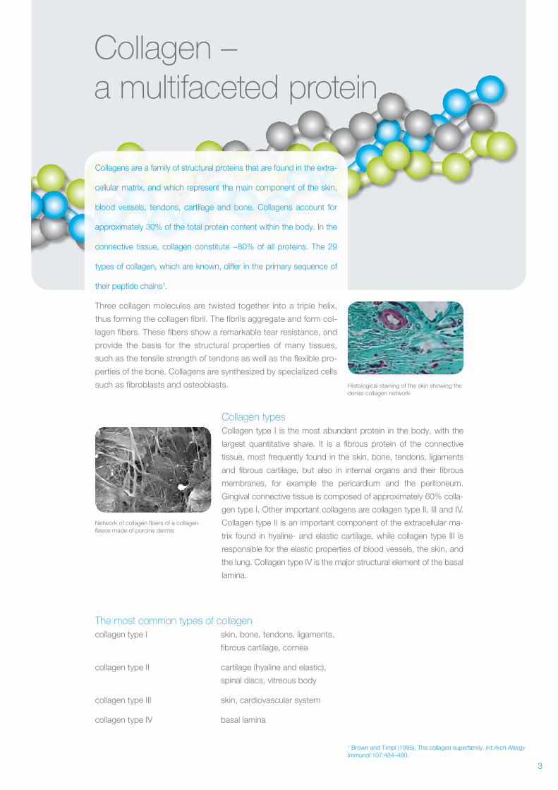
Histological staining of the skin showing the dense collagen network
Network of collagen fibers of a collagen fleece made of porcine dermis
Collagens are a family of structural proteins that are found in the extra-
cellular matrix, and which represent the main component of the skin,
blood vessels, tendons, cartilage and bone. Collagens account for
approximately 30% of the total protein content within the body. In the
connective tissue, collagen constitute ~80% of all proteins. The 29
types of collagen, which are known, differ in the primary sequence of
their peptide chains1.
Three collagen molecules are twisted together into a triple helix,
thus forming the collagen fibril. The fibrils aggregate and form col-
lagen fibers. These fibers show a remarkable tear resistance, and
provide the basis for the structural properties of many tissues,
such as the tensile strength of tendons as well as the flexible pro-
perties of the bone. Collagens are synthesized by specialized cells
such as fibroblasts and osteoblasts.
The most common types of collagencollagen type I skin, bone, tendons, ligaments,
fibrous cartilage, cornea
collagen type II cartilage (hyaline and elastic),
spinal discs, vitreous body
collagen type III skin, cardiovascular system
collagen type IV basal lamina
1 Brown and Timpl (1995). The collagen superfamily. Int Arch Allergy Immunol 107:484–490.
4
The GBR and GTR technique
Collagen membranes for the GBR and GTR technique
Collagen membranes have been used in Guided Tissue Regeneration (GTR) and Guided
Bone Regeneration (GBR) for many years. The principle of these techniques is based on
the placement of a barrier membrane for separation of slowly proliferating regenerative
cell types, such as osteoblasts and periodontal cells, from fast proliferating epithelial and
connective tissue cells, thus enabling the regeneration of lost tissue.
2 Rothamel et al. (2005). Biodegradation of differently cross-linked collagen membranes:an experimental study in the rat. Clin Oral Implants Res 16:369–378.
GTR aims at the regeneration of the periodontium. A barrier
membrane is placed between the epithelium and the tooth, to
provide space and time for regeneration of the periodontal liga-
ment. In GBR procedures, membranes are normally applied in
combination with a bone graft material. The membrane is placed
over a bony defect filled with a bone graft material. The bone graft
material prevents collapse of the membrane and serves as an
osteoconductive scaffold for ingrowth of bone and precursor cells.
The barrier membrane prevents migration of bone graft particles
into the oral cavity and ingrowth of soft tissue into the defect area,
thus enabling bony regeneration.
Guided Tissue Regeneration (GTR) Guided Bone Regeneration (GBR)
Membrane types Barrier membrane requirements
- Biocompatibility
- Tissue integration
- Cell occlusiveness
- Dimensional stability
- Easy handling
The first generation of barrier membranes was based on non-
resorbable materials e.g. cellulose acetate, titanium and expanded
polytetrafluoroethylene (ePTFE). These membranes gained satis-
fying results but had disadvantages such as the secondary surgery
required for removal, which is associated with graft site morbidity.
To avoid the limitations of the non-resorbable membranes, resorb-
able membranes were developed. Resorbable membranes are eit-
her synthetic polymers such as polyglycolides, polylactides (acidic
degradation) or animal-derived, e.g. collagen. Due to the manifold
positive natural properties of collagen, collagen membranes are
commonly the material of choice2.
5
The advantages of collagen
Advantages ofcollagen membranes
- Exceptional biocompatibility
- Support of hemostasis
- Low antigenicity
- Degradation by
specific enzymes
- Chemotactic attraction of
regenerative cells
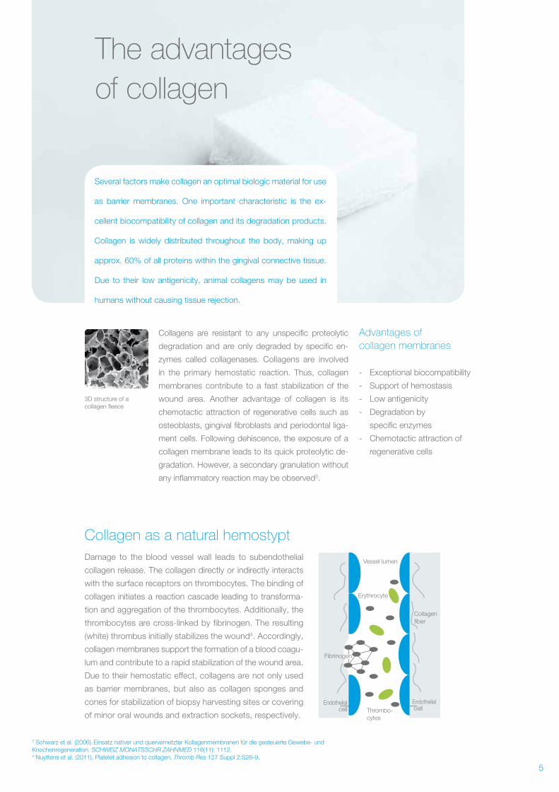
Collagen as a natural hemostypt Damage to the blood vessel wall leads to subendothelial
collagen release. The collagen directly or indirectly interacts
with the surface receptors on thrombocytes. The binding of
collagen initiates a reaction cascade leading to transforma-
tion and aggregation of the thrombocytes. Additionally, the
thrombocytes are cross- linked by fibrinogen. The resulting
(white) thrombus initially stabilizes the wound4. Accordingly,
collagen membranes support the formation of a blood coagu-
lum and contribute to a rapid stabilization of the wound area.
Due to their hemostatic effect, collagens are not only used
as barrier membranes, but also as collagen sponges and
cones for stabilization of biopsy harvesting sites or covering
of minor oral wounds and extraction sockets, respectively.
Several factors make collagen an optimal biologic material for use
as barrier membranes. One important characteristic is the ex-
cellent biocompatibility of collagen and its degradation products.
Collagen is widely distributed throughout the body, making up
approx. 60% of all proteins within the gingival connective tissue.
Due to their low antigenicity, animal collagens may be used in
humans without causing tissue rejection.
Collagens are resistant to any unspecific proteolytic
degradation and are only degraded by specific en-
zymes called collagenases. Collagens are involved
in the primary hemostatic reaction. Thus, collagen
membranes contribute to a fast stabilization of the
wound area. Another advantage of collagen is its
chemotactic attraction of regenerative cells such as
osteoblasts, gingival fibroblasts and periodontal liga-
ment cells. Following dehiscence, the exposure of a
collagen membrane leads to its quick proteolytic de-
gradation. However, a secondary granulation without
any inflammatory reaction may be observed3.
3 Schwarz et al. (2006). Einsatz nativer und quervernetzter Kollagenmembranen für die gesteuerte Gewebe- und Knochenregeneration. SCHWEIZ MONATSSCHR ZAHNMED 116(11): 1112.4 Nuyttens et al. (2011). Platelet adhesion to collagen. Thromb Res 127 Suppl 2:S26-9.
3D structure of a collagen fleece
Vessel lumen
Endothelialcell
Endothelial cell
Collagen fiber
Fibrinogen
Thrombo-cytes
Erythrocyte
6
Origin of collagen membranes
The first collagen membranes available on the market were of
bovine origin (Achilles tendon and pericardium). Nowadays, por-
cine membranes are more widely used because their usage ex-
cludes the risk of BSE transmission. Moreover, porcine collagen
exhibits a high homology to human collagen and therefore a very low
antigenicity. Due to these reasons, botiss membranes are exclusi-
vely produced from porcine collagen.
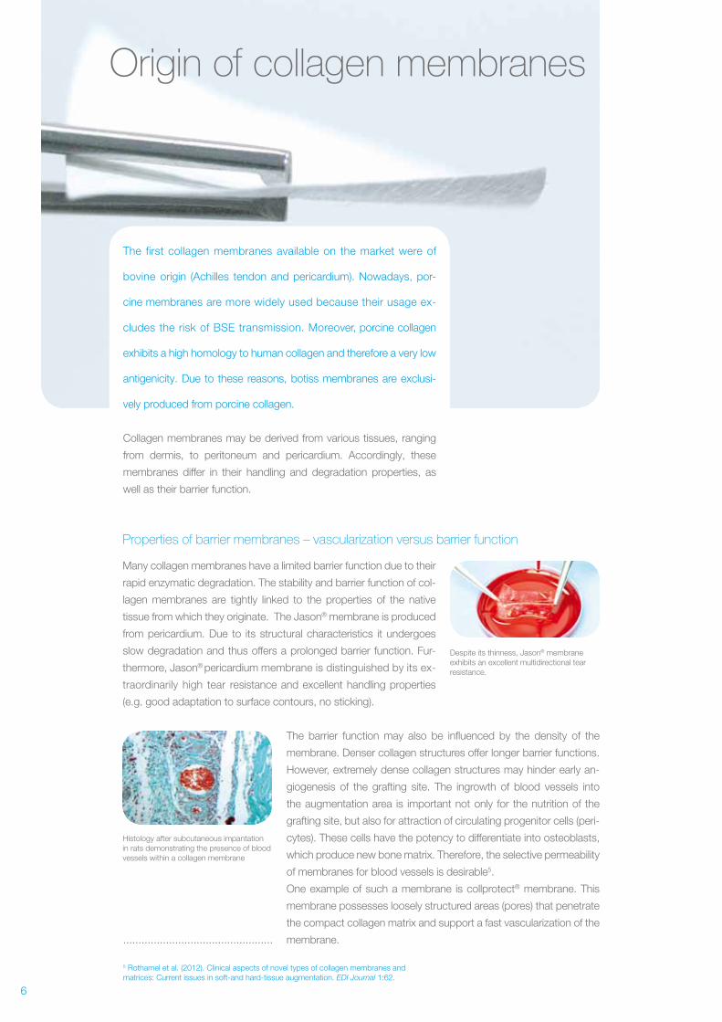
Despite its thinness, Jason® membrane exhibits an excellent multidirectional tear resistance.
Histology after subcutaneous impantation in rats demonstrating the presence of blood vessels within a collagen membrane
Collagen membranes may be derived from various tissues, ranging
from dermis, to peritoneum and pericardium. Accordingly, these
membranes differ in their handling and degradation properties, as
well as their barrier function.
Properties of barrier membranes – vascularization versus barrier function
Many collagen membranes have a limited barrier function due to their
rapid enzymatic degradation. The stability and barrier function of col-
lagen membranes are tightly linked to the properties of the native
tissue from which they originate. The Jason® membrane is produced
from pericardium. Due to its structural characteristics it undergoes
slow degradation and thus offers a prolonged barrier function. Fur-
thermore, Jason® pericardium membrane is distinguished by its ex-
traordinarily high tear resistance and excellent handling properties
(e.g. good adaptation to surface contours, no sticking).
5 Rothamel et al. (2012). Clinical aspects of novel types of collagen membranes and matrices: Current issues in soft-and hard-tissue augmentation. EDI Journal 1:62.
The barrier function may also be influenced by the density of the
membrane. Denser collagen structures offer longer barrier functions.
However, extremely dense collagen structures may hinder early an-
giogenesis of the grafting site. The ingrowth of blood vessels into
the augmentation area is important not only for the nutrition of the
grafting site, but also for attraction of circulating progenitor cells (peri-
cytes). These cells have the potency to differentiate into osteoblasts,
which produce new bone matrix. Therefore, the selective permeability
of membranes for blood vessels is desirable5.
One example of such a membrane is collprotect® membrane. This
membrane possesses loosely structured areas (pores) that penetrate
the compact collagen matrix and support a fast vascularization of the
membrane..................................................
Packaging
EO-sterilization/γ-Sterilization
Sterile product
7
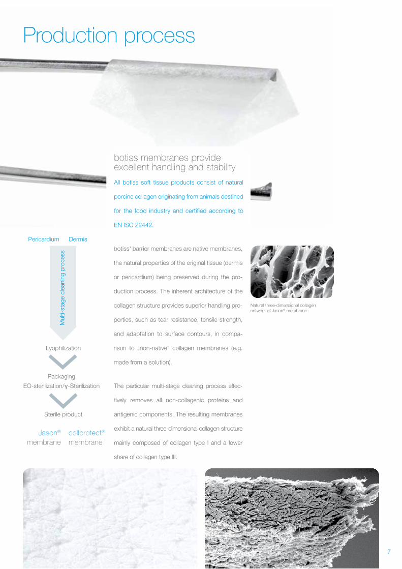
Origin of collagen membranes Production process
botiss membranes provide excellent handling and stability
All botiss soft tissue products consist of natural
porcine collagen originating from animals destined
for the food industry and certified according to
EN ISO 22442.
botiss‘ barrier membranes are native membranes,
the natural properties of the original tissue (dermis
or pericardium) being preserved during the pro-
duction process. The inherent architecture of the
collagen structure provides superior handling pro-
perties, such as tear resistance, tensile strength,
and adaptation to surface contours, in compa-
rison to „non-native“ collagen membranes (e.g.
made from a solution).
Pericardium
Lyophilization
Packaging
EO-sterilization/γ-Sterilization
Sterile product
Dermis
Jason® membrane
collprotect® membrane
Mul
ti-st
age
clea
ning
pro
cess
Natural three-dimensional collagen network of Jason® membrane
The particular multi-stage cleaning process effec-
tively removes all non- collagenic proteins and
antigenic components. The resulting membranes
exhibit a natural three-dimensional collagen structure
mainly composed of collagen type I and a lower
share of collagen type III.
8
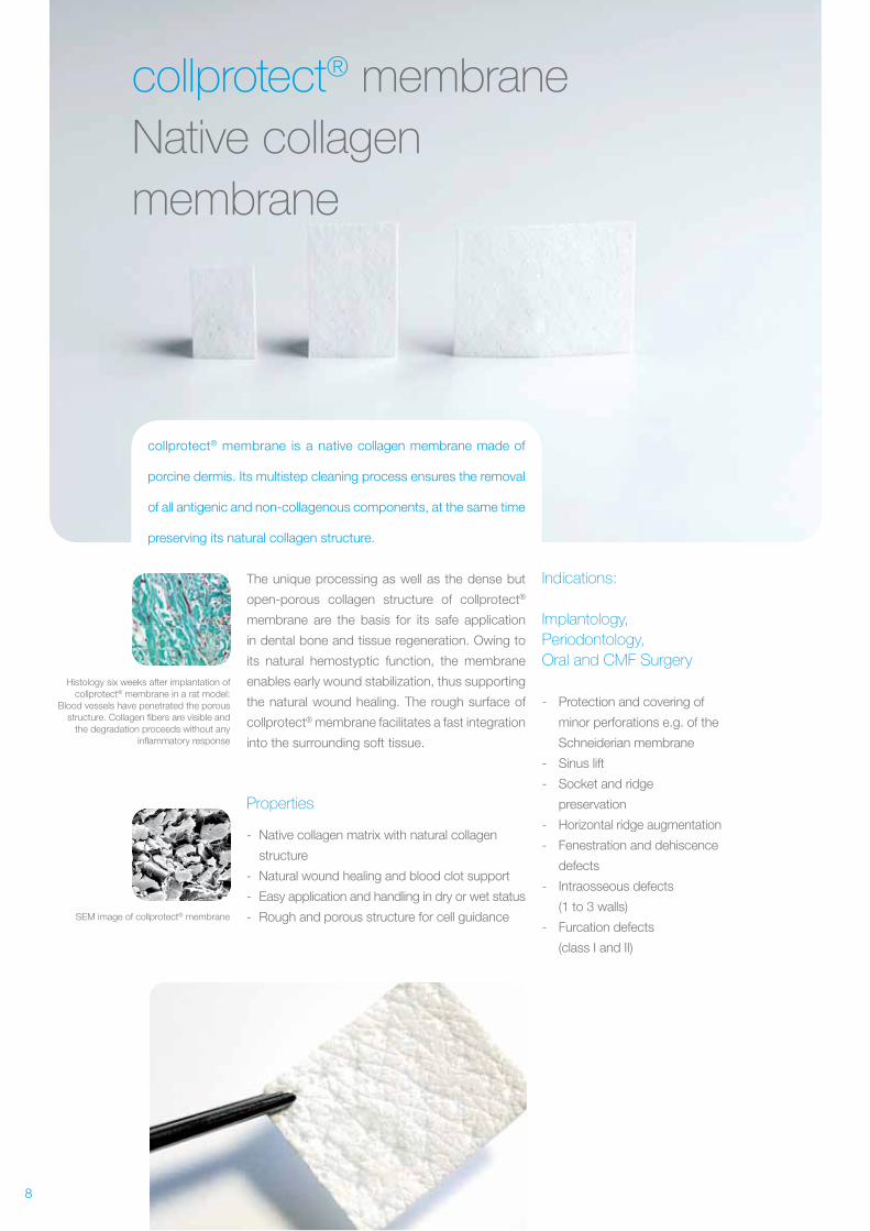
collprotect® membraneNative collagen membrane
collprotect® membrane is a native collagen membrane made of
porcine dermis. Its multistep cleaning process ensures the removal
of all antigenic and non-collagenous components, at the same time
preserving its natural collagen structure.
Histology six weeks after implantation of collprotect® membrane in a rat model:
Blood vessels have penetrated the porous structure. Collagen fibers are visible and
the degradation proceeds without any inflammatory response
SEM image of collprotect® membrane
The unique processing as well as the dense but
open-porous collagen structure of collprotect®
membrane are the basis for its safe application
in dental bone and tissue regeneration. Owing to
its natural hemostyptic function, the membrane
enables early wound stabilization, thus supporting
the natural wound healing. The rough surface of
collprotect® membrane facilitates a fast integration
into the surrounding soft tissue.
Properties
- Native collagen matrix with natural collagen
structure
- Natural wound healing and blood clot support
- Easy application and handling in dry or wet status
- Rough and porous structure for cell guidance
Indications:
Implantology, Periodontology,Oral and CMF Surgery
- Protection and covering of
minor perforations e.g. of the
Schneiderian membrane
- Sinus lift
- Socket and ridge
preservation
- Horizontal ridge augmentation
- Fenestration and dehiscence
defects
- Intraosseous defects
(1 to 3 walls)
- Furcation defects
(class I and II)
9
collprotect® membraneNative collagen membrane
SEM image of the Jason® membrane
Jason® membrane maintains the barrier
function, 56 days after subcutaneous
implantation in rats
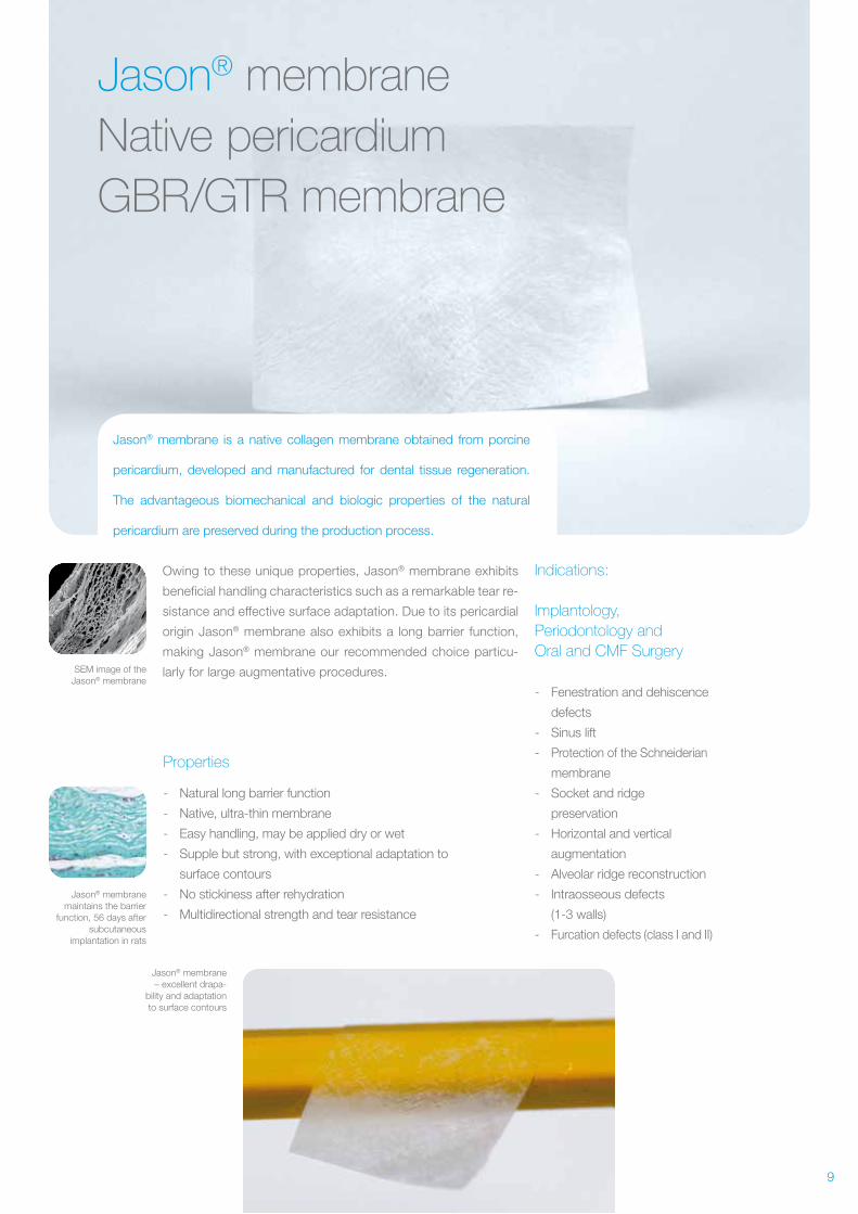
Jason® membraneNative pericardium GBR/GTR membrane
Jason® membrane is a native collagen membrane obtained from porcine
pericardium, developed and manufactured for dental tissue regeneration.
The advantageous biomechanical and biologic properties of the natural
pericardium are preserved during the production process.
Owing to these unique properties, Jason® membrane exhibits
beneficial handling characteristics such as a remarkable tear re-
sistance and effective surface adaptation. Due to its pericardial
origin Jason® membrane also exhibits a long barrier function,
making Jason® membrane our recommended choice particu-
larly for large augmentative procedures.
Properties
- Natural long barrier function
- Native, ultra-thin membrane
- Easy handling, may be applied dry or wet
- Supple but strong, with exceptional adaptation to
surface contours
- No stickiness after rehydration
- Multidirectional strength and tear resistance
Indications:
Implantology,Periodontology andOral and CMF Surgery
- Fenestration and dehiscence
defects
- Sinus lift
- Protection of the Schneiderian
membrane
- Socket and ridge
preservation
- Horizontal and vertical
augmentation
- Alveolar ridge reconstruction
- Intraosseous defects
(1- 3 walls)
- Furcation defects (class I and II)
Jason® membrane– excellent drapa-
bility and adaptation to surface contours
10
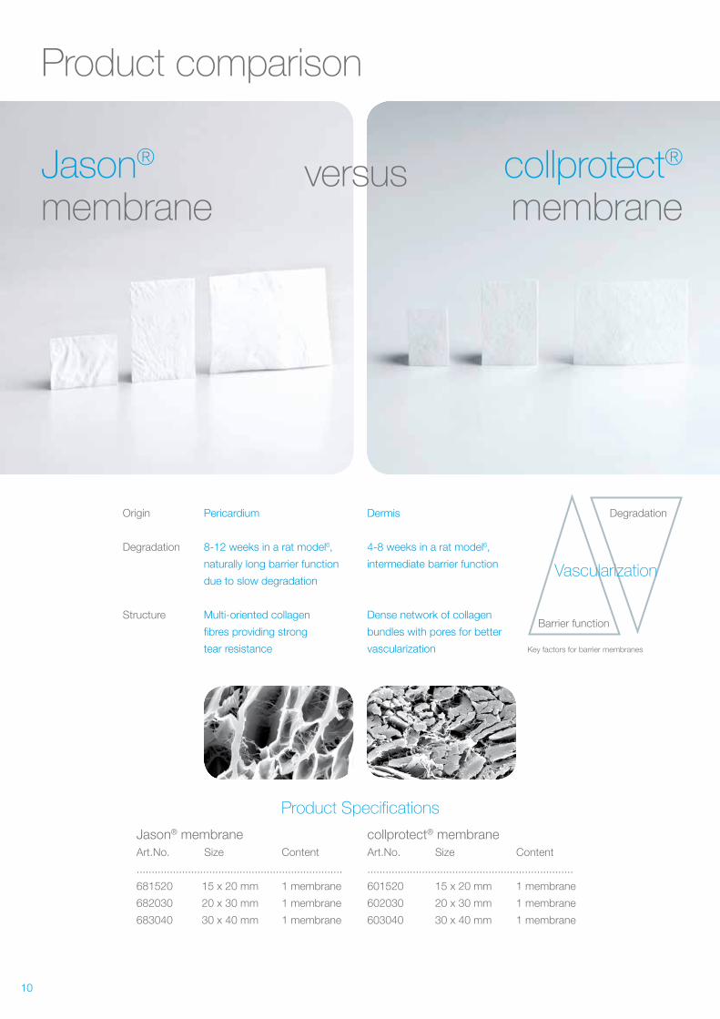
Product comparison
Jason® membrane
collprotect® membrane
versus
Origin Pericardium Dermis
Degradation 8-12 weeks in a rat model6, 4-8 weeks in a rat model6,
naturally long barrier function intermediate barrier function
due to slow degradation
Structure Multi-oriented collagen Dense network of collagen
fibres providing strong bundles with pores for better
tear resistance vascularization
Product Specifications
Jason® membrane Art.No. Size Content
....................................................................
681520 15 x 20 mm 1 membrane
682030 20 x 30 mm 1 membrane
683040 30 x 40 mm 1 membrane
collprotect® membrane Art.No. Size Content
....................................................................
601520 15 x 20 mm 1 membrane
602030 20 x 30 mm 1 membrane
603040 30 x 40 mm 1 membrane
Barrier function
Degradation
Vascularization
Key factors for barrier membranes
11
Pre-clinical testing
Incubation of the multi-layered Jason® membrane and a competetive bi-
layer membrane with osteoblast- like SaOs-2 cells showed a significantly
higher cell proliferation on the Jason® membrane after seven days.
The excellent cell attachment and proliferation on
the Jason® membrane highlights its suitability as
scaffold for osteoblast guidance which supports
of the bony regeneration of covered defects.
Resorption time and tissue integration of collagen
membranes not only depend on the animal origin,
but also differ between tissues. Tissue integra-
tion and degradation of the Jason® membrane
and the collprotect® membrane were tested by
subcutaneous implantation in rats. Jason® mem-
brane, which originates from pericardium, was
integrated within the first weeks and remained
stable for a healing period of eight to 12 weeks
(please note the different metabolic rates for
rats and humans).
The cell invasion of the dermal collagen of the
collprotect® membrane took a little longer, but
the membrane was mostly degraded within the
first four to eight weeks.
6 Rothamel et al. (2011). Biodegradation pattern of native and cross-linked porcine collagen matrices – an experimental study in rats. Poster EAO Athens, Greece. 7 Rothamel et al. (2012). Biocompatibility and Biodegradation of a Native, Porcine Pericardi-um Membrane. Results from in vitro/in vivo Examination Int J Oral Maxillofac Implants. 2012 Jan-Feb;27(1):146-54.
The Jason® membrane supports attachment and proliferation of osteoblast- like cellsIn vitro cell culture results. Dr. M. Herten, University of Münster and Prof. Dr. Dr. D. Rothamel, University of Düsseldorf7
In vivo pre- clinical testingResults from a degradation study in a rat model6,Prof. Dr. Dr. D. Rothamel, University of Düsseldorf
Only superficial cell invasion of the collpro-tect® membrane, 14 days after implantation
The collprotect® membrane prepared for subcutaneous implantation
Structural integrity of the Jason® membrane 28 days after implantation
The diagrams display degradation times of the membranes, from in vivo data obtained in an experimental rat model.
3500
3000
2500
2000
1500
1000
500
0Jason® membrane Bilayer membrane
2 hours
3 days
7 days
cells
/wel
l
400
200
0
1 2 4 128Time (weeks)
600
400
200
0
1 2 4 128Time (weeks)
Degradation of Jason® membrane
Degradation of collprotect® membrane
Thic
knes
s (µ
m)
Thic
knes
s (µ
m)
12
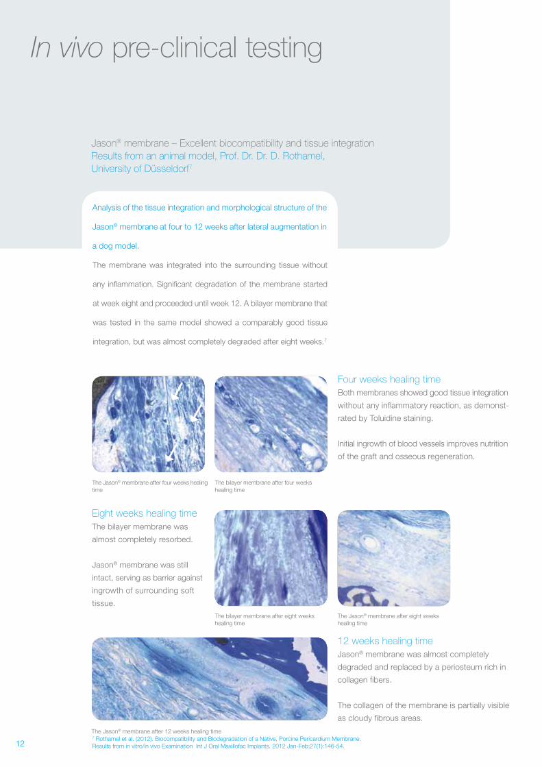
In vivo pre- clinical testing
Analysis of the tissue integration and morphological structure of the
Jason® membrane at four to 12 weeks after lateral augmentation in
a dog model.
The membrane was integrated into the surrounding tissue without
any inflammation. Significant degradation of the membrane started
at week eight and proceeded until week 12. A bilayer membrane that
was tested in the same model showed a comparably good tissue
integration, but was almost completely degraded after eight weeks.7
Jason® membrane – Excellent biocompatibility and tissue integrationResults from an animal model, Prof. Dr. Dr. D. Rothamel, University of Düsseldorf7
The Jason® membrane after four weeks healing time
The bilayer membrane after four weeks healing time
The bilayer membrane after eight weeks healing time
The Jason® membrane after eight weeks healing time
The Jason® membrane after 12 weeks healing time
Four weeks healing time Both membranes showed good tissue integration
without any inflammatory reaction, as demonst-
rated by Toluidine staining.
Initial ingrowth of blood vessels improves nutrition
of the graft and osseous regeneration.
12 weeks healing time Jason® membrane was almost completely
degraded and replaced by a periosteum rich in
collagen fibers.
The collagen of the membrane is partially visible
as cloudy fibrous areas.
Eight weeks healing time The bilayer membrane was
almost completely resorbed.
Jason® membrane was still
intact, serving as barrier against
ingrowth of surrounding soft
tissue.
7 Rothamel et al. (2012). Biocompatibility and Biodegradation of a Native, Porcine Pericardium Membrane. Results from in vitro/in vivo Examination Int J Oral Maxillofac Implants. 2012 Jan-Feb;27(1):146-54.
13
In vivo pre- clinical testing
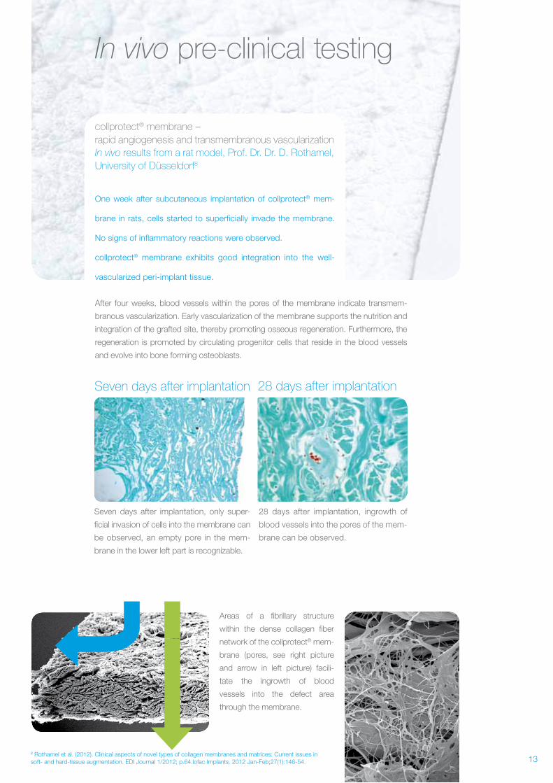
One week after subcutaneous implantation of collprotect® mem-
brane in rats, cells started to superficially invade the membrane.
No signs of inflammatory reactions were observed.
collprotect® membrane exhibits good integration into the well-
vascularized peri- implant tissue.
After four weeks, blood vessels within the pores of the membrane indicate transmem-
branous vascularization. Early vascularization of the membrane supports the nutrition and
integration of the grafted site, thereby promoting osseous regeneration. Furthermore, the
regeneration is promoted by circulating progenitor cells that reside in the blood vessels
and evolve into bone forming osteoblasts.
collprotect® membrane – rapid angiogenesis and transmembranous vascularizationIn vivo results from a rat model, Prof. Dr. Dr. D. Rothamel, University of Düsseldorf8
Areas of a fibrillary structure
within the dense collagen fiber
network of the collprotect® mem-
brane (pores, see right picture
and arrow in left picture) facili-
tate the ingrowth of blood
vessels into the defect area
through the membrane.
28 days after implantationSeven days after implantation
28 days after implantation, ingrowth of
blood vessels into the pores of the mem-
brane can be observed.
Seven days after implantation, only super-
ficial invasion of cells into the membrane can
be observed, an empty pore in the mem-
brane in the lower left part is recognizable.
8 Rothamel et al. (2012). Clinical aspects of novel types of collagen membranes and matrices: Current issues in soft- and hard-tissue augmentation. EDI Journal 1/2012; p.64.lofac Implants. 2012 Jan-Feb;27(1):146-54.
14
Clinical application of collprotect® membrane
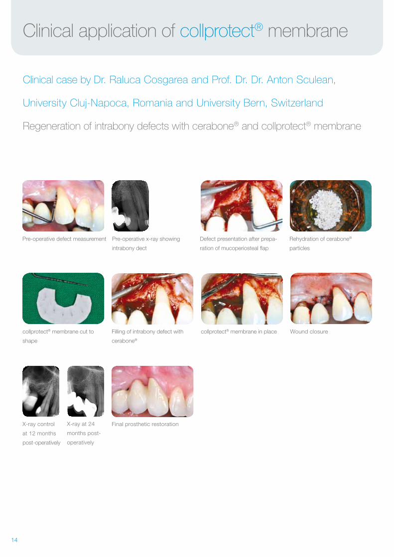
Clinical case by Dr. Raluca Cosgarea and Prof. Dr. Dr. Anton Sculean,
University Cluj-Napoca, Romania and University Bern, Switzerland
Regeneration of intrabony defects with cerabone® and collprotect® membrane
collprotect® membrane cut to
shape
Filling of intrabony defect with
cerabone®
collprotect® membrane in place
Pre-operative defect measurement Pre-operative x-ray showing
intrabony dect
Defect presentation after prepa-
ration of mucoperiosteal flap
Rehydration of cerabone®
particles
Wound closure
X-ray control
at 12 months
post-operatively
X-ray at 24
months post-
operatively
Final prosthetic restoration
16
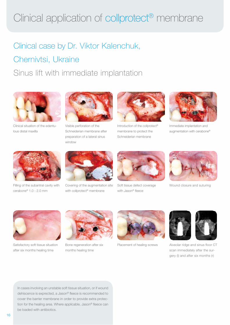
Soft tissue defect coverage
with Jason® fleece
Placement of healing screws
Covering of the augmentation site
with collprotect® membrane
Filling of the subantral cavity with
cerabone® 1.0 - 2.0 mm
Bone regeneration after six
months healing time
Satisfactory soft tissue situation
after six months healing time
Wound closure and suturing
Alveolar ridge and sinus floor CT
scan immediately after the sur-
gery (l) and after six months (r)
Visible perforation of the
Schneiderian membrane after
preparation of a lateral sinus
window
Clinical situation of the edentu-
lous distal maxilla
Introduction of the collprotect®
membrane to protect the
Schneiderian membrane
Immediate implantation and
augmentation with cerabone®
Clinical application of collprotect® membrane
Clinical case by Dr. Viktor Kalenchuk,
Chernivtsi, Ukraine
Sinus lift with immediate implantation
In cases involving an unstable soft tissue situation, or if wound
dehiscence is exprected, a Jason® fleece is recommended to
cover the barrier membrane in order to provide extra protec-
tion for the healing area. Where applicable, Jason® fleece can
be loaded with antibiotics.
17
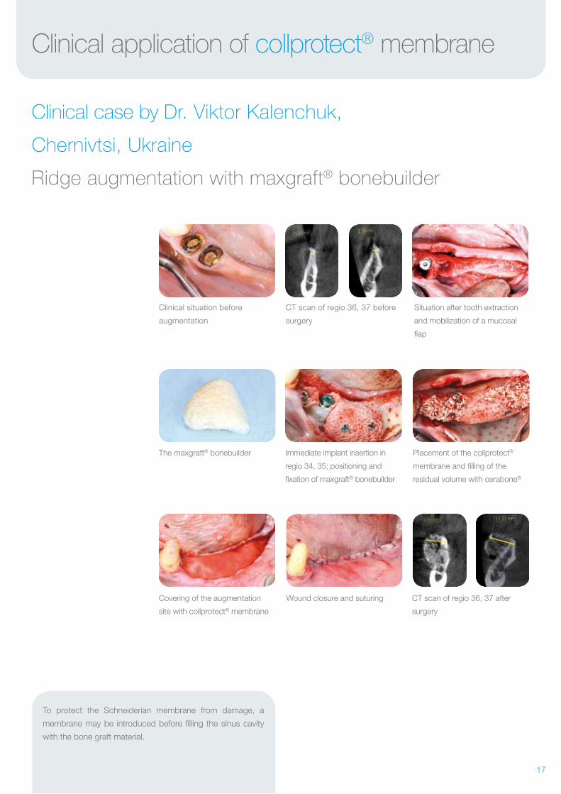
Clinical application of collprotect® membrane
Clinical case by Dr. Viktor Kalenchuk,
Chernivtsi, Ukraine
Ridge augmentation with maxgraft® bonebuilder
The maxgraft® bonebuilder
Clinical situation before
augmentation
Covering of the augmentation
site with collprotect® membrane
CT scan of regio 36, 37 before
surgery
Wound closure and suturing
Immediate implant insertion in
regio 34, 35; positioning and
fixation of maxgraft® bonebuilder
Placement of the collprotect®
membrane and filling of the
residual volume with cerabone®
Situation after tooth extraction
and mobilization of a mucosal
flap
CT scan of regio 36, 37 after
surgery
To protect the Schneiderian membrane from damage, a
membrane may be introduced before filling the sinus cavity
with the bone graft material.

18
Lateral bone defect following
root tip resection
Lateral augmentation with
maxresorb® and application of a
dry collprotect® membrane
After preparation of the implant
bed the thin vestibular wall is
visible
Complete covering of augmen-
tation site and implant with the
membrane
Insertion of implant in the
reduced bone amount
Wound closure by soft tissue
expansion without vertical
releasing incisions
Clinical application of collprotect® membrane
Clinical case by Dr. Georg Bayer,
Landsberg am Lech, Germany
Lateral augmentation
X-ray control at re- entry
CBCT image showing the redu-
ced amount of bone available in
the area of the mental foramen
Post- operative x- ray Stable keratinized gingiva after
insertion of healing abutment at
re- entry
19
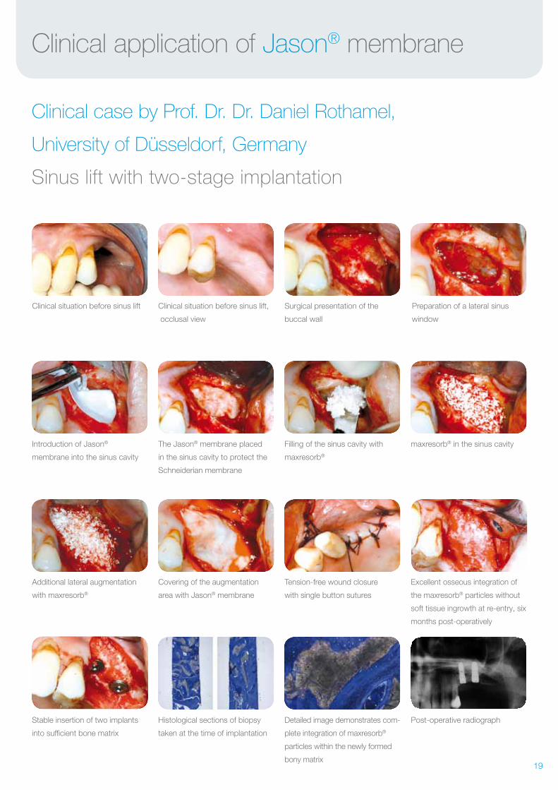
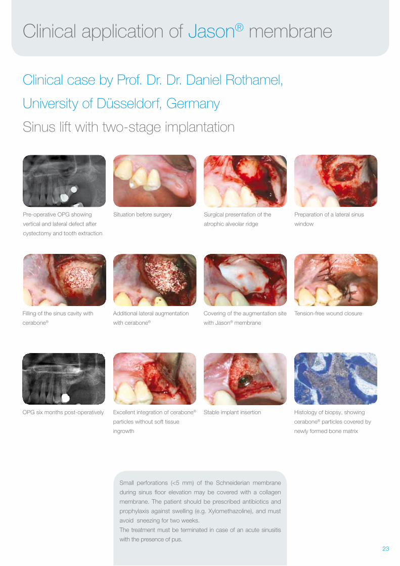
Clinical application of Jason® membrane
Clinical case by Prof. Dr. Dr. Daniel Rothamel,
University of Düsseldorf, Germany
Sinus lift with two-stage implantation
The Jason® membrane placed
in the sinus cavity to protect the
Schneiderian membrane
Introduction of Jason®
membrane into the sinus cavity
Covering of the augmentation
area with Jason® membrane
Filling of the sinus cavity with
maxresorb®
Tension-free wound closure
with single button sutures
Histological sections of biopsy
taken at the time of implantation
Stable insertion of two implants
into sufficient bone matrix
Clinical situation before sinus lift Clinical situation before sinus lift,
occlusal view
Surgical presentation of the
buccal wall
Preparation of a lateral sinus
window
maxresorb® in the sinus cavity
Excellent osseous integration of
the maxresorb® particles without
soft tissue ingrowth at re-entry, six
months post- operatively
Detailed image demonstrates com-
plete integration of maxresorb®
particles within the newly formed
bony matrix
Post-operative radiograph
Additional lateral augmentation
with maxresorb®
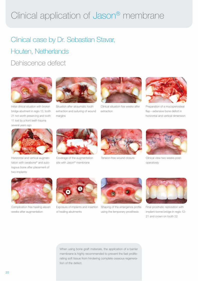
20
Horizontal and vertical augmen-
tation with cerabone® and auto-
logous bone after placement of
two implants
Clinical view two weeks post-
operatively
Preparation of a mucoperiosteal
flap - extensive bone deficit in
horizontal and vertical dimension
Tension-free wound closureCoverage of the augmentation
site with Jason® membrane
Complication free healing eleven
weeks after augmentation
Exposure of implants and insertion
of healing abutments
Shaping of the emergence profile
using the temporary prosthesis
Final prosthetic restoration with
implant-borne bridge in regio 12-
21 and crown on tooth 22
Initial clinical situation with broken
bridge abutment in regio 12, tooth
21 not worth preserving and tooth
11 lost by a front teeth trauma
several years ago
Situation after atraumatic tooth
extraction and suturing of wound
margins
Clinical situation five weeks after
extraction
Clinical application of Jason® membrane
Clinical case by Dr. Sebastian Stavar,
Houten, Netherlands
Dehiscence defect
When using bone graft materials, the application of a barrier
membrane is highly recommended to prevent the fast prolife-
rating soft tissue from hindering complete osseous regenera-
tion of the defect.
21
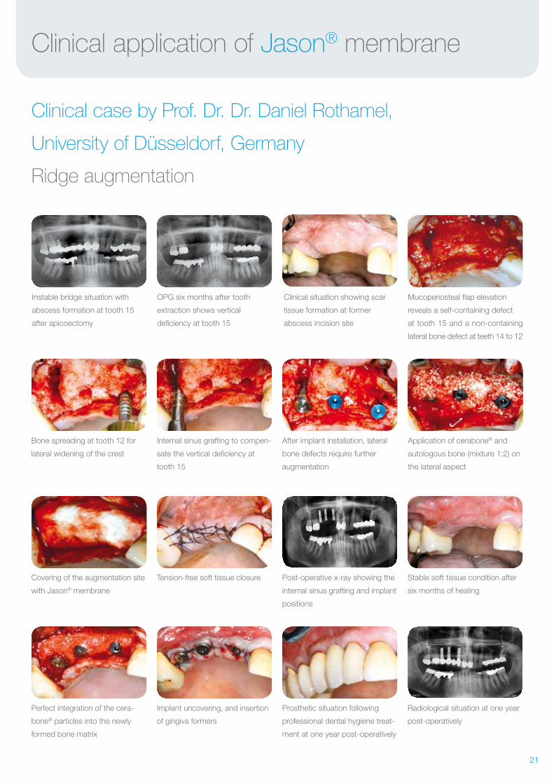
Clinical application of Jason® membrane
Clinical case by Prof. Dr. Dr. Daniel Rothamel,
University of Düsseldorf, Germany
Ridge augmentation
Bone spreading at tooth 12 for
lateral widening of the crest
Covering of the augmentation site
with Jason® membrane
Perfect integration of the cera-
bone® particles into the newly
formed bone matrix
Internal sinus grafting to compen-
sate the vertical deficiency at
tooth 15
Tension-free soft tissue closure
Implant uncovering, and insertion
of gingiva formers
Instable bridge situation with
abscess formation at tooth 15
after apicoectomy
OPG six months after tooth
extraction shows vertical
deficiency at tooth 15
Clinical situation showing scar
tissue formation at former
abscess incision site
Mucoperiosteal flap elevation
reveals a self-containing defect
at tooth 15 and a non-containing
lateral bone defect at teeth 14 to 12
After implant installation, lateral
bone defects require further
augmentation
Post-operative x-ray showing the
internal sinus grafting and implant
positions
Prosthetic situation following
professional dental hygiene treat-
ment at one year post-operatively
Stable soft tissue condition after
six months of healing
Radiological situation at one year
post-operatively
Application of cerabone® and
autologous bone (mixture 1:2) on
the lateral aspect
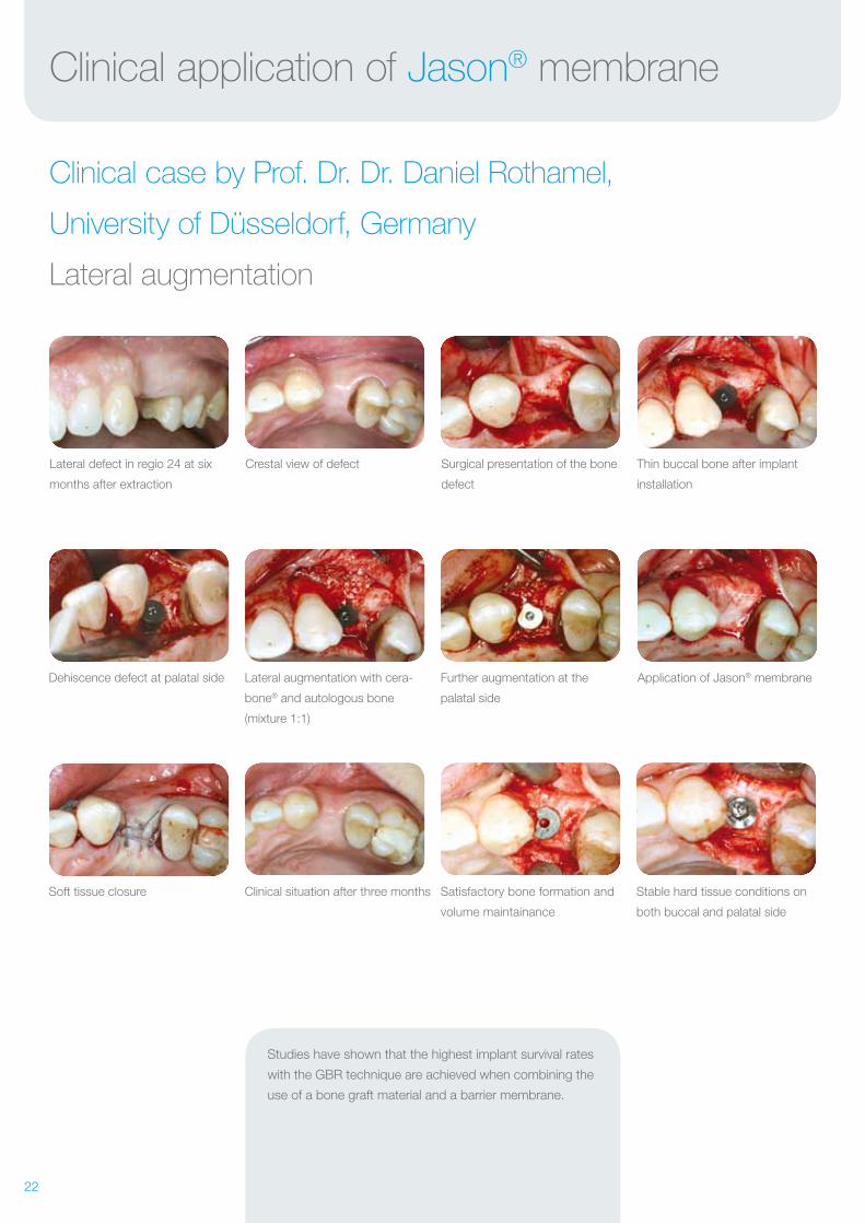
22
Further augmentation at the
palatal side
Satisfactory bone formation and
volume maintainance
Lateral augmentation with cera-
bone® and autologous bone
(mixture 1:1)
Dehiscence defect at palatal side
Clinical situation after three monthsSoft tissue closure
Application of Jason® membrane
Stable hard tissue conditions on
both buccal and palatal side
Crestal view of defectLateral defect in regio 24 at six
months after extraction
Surgical presentation of the bone
defect
Thin buccal bone after implant
installation
Clinical application of Jason® membrane
Clinical case by Prof. Dr. Dr. Daniel Rothamel,
University of Düsseldorf, Germany
Lateral augmentation
Studies have shown that the highest implant survival rates
with the GBR technique are achieved when combining the
use of a bone graft material and a barrier membrane.
23
Small perforations (<5 mm) of the Schneiderian membrane
during sinus floor elevation may be covered with a collagen
membrane. The patient should be prescribed antibiotics and
prophylaxis against swelling (e.g. Xylomethazoline), and must
avoid sneezing for two weeks.
The treatment must be terminated in case of an acute sinusitis
with the presence of pus.
Clinical application of Jason® membrane
Clinical case by Prof. Dr. Dr. Daniel Rothamel,
University of Düsseldorf, Germany
Sinus lift with two-stage implantation
Filling of the sinus cavity with
cerabone®
OPG six months post-operatively
Additional lateral augmentation
with cerabone®
Excellent integration of cerabone®
particles without soft tissue
ingrowth
Pre-operative OPG showing
vertical and lateral defect after
cystectomy and tooth extraction
Situation before surgery Surgical presentation of the
atrophic alveolar ridge
Preparation of a lateral sinus
window
Covering of the augmentation site
with Jason® membrane
Stable implant insertion Histology of biopsy, showing
cerabone® particles covered by
newly formed bone matrix
Tension-free wound closure
24
botissbiomaterials
bone & tissue regeneration
soft tissue
education
hard tissue
botiss biomaterials GmbH
Hauptstr. 28
15806 Zossen / Germany
Tel.: +49 33769 / 88 41 985
Fax: +49 33769 / 88 41 986
www.botiss.com
www.botiss-dental.com
www.facebook.com/botissdental
Innovation.
Regeneration.
Aesthetics.
Rev.: CMen-05/2017-03