jojournalurnal axin is a scaffold protein in...
TRANSCRIPT

Axin is a scaffold protein in TGF-b signaling thatpromotes degradation of Smad7 by Arkadia
Wei Liu1, Hongliang Rui1,5, Jifeng Wang2,Shuyong Lin1, Ying He2, Mingliang Chen2,Qinxi Li2, Zhiyun Ye2, Suping Zhang3,Siu Chiu Chan1, Ye-Guang Chen3,Jiahuai Han2,4 and Sheng-Cai Lin1,2,*1Department of Biochemistry, Hong Kong University of Science andTechnology, Kowloon, Hong Kong, China, 2Key Laboratory of Ministryof Education for Cell Biology and Tumor Cell Engineering, School of LifeSciences, Xiamen University, Fujian, China, 3State Key Laboratory ofBiomembrane and Membrane Biotechnology, Department of BiologicalSciences and Biotechnology, Tsinghua University, Beijing, China and4Department of Immunology, the Scripps Research Institute, La Jolla,CA, USA
TGF-b signaling involves a wide array of signaling molecules
and multiple controlling events. Scaffold proteins create a
functional proximity of signaling molecules and control the
specificity of signal transduction. While many components
involved in the TGF-b pathway have been elucidated, little is
known about how those components are coordinated by
scaffold proteins. Here, we show that Axin activates TGF-bsignaling by forming a multimeric complex consisting of
Smad7 and ubiquitin E3 ligase Arkadia. Axin depends on
Arkadia to facilitate TGF-b signaling, as their small interfer-
ing RNAs reciprocally abolished the stimulatory effect on
TGF-b signaling. Specific knockdown of Axin or Arkadia
revealed that Axin and Arkadia cooperate with each other
in promoting Smad7 ubiquitination. Pulse-chase experi-
ments further illustrated that Axin significantly decreased
the half-life of Smad7. Axin also induces nuclear export of
Smad7. Interestingly, Axin associates with Arkadia and
Smad7 independently of TGF-b signal, in contrast to its
transient association with inactive Smad3. However, coex-
pression of Wnt-1 reduced Smad7 ubiquitination by down-
regulating Axin levels, underscoring the importance of Axin
as an intrinsic regulator in TGF-b signaling.
The EMBO Journal (2006) 25, 1646–1658. doi:10.1038/
sj.emboj.7601057; Published online 6 April 2006
Subject Categories: signal transduction
Keywords: Arkadia; Axin; Smad7; TGF-b; ubiquitination
Introduction
TGF-b signaling plays a wide spectrum of roles from early
embryonic development to mature tissues in controlling
biological processes, including cell proliferation, differentia-
tion, apoptosis, and cell fate determination (Massague, 2000).
TGF-b superfamily members elicit biological responses
through a complex cascade of signaling molecules that
include receptors, Smads, and importantly the regulatory
factors that positively or negatively regulate the signaling
system (Shi and Massague, 2003).
The TGF-b superfamily can be divided into TGF-b, bonemorphogenetic proteins (BMP), and Nodal. Schematically,
much has been known about the molecular components
involved in TGF-b signaling. TGF-b ligands bind to their
receptors that are associated with a family of signal transdu-
cers called Smads (R-Smads) (Massague, 1998). TGF-b, BMP,
and Nodal receptors make use of different sets of Smads.
Upon ligand stimulation, different R-Smads bind to common
mediator Smad4 (Co-Smad) to form activated Smad complex
that is then translocated into the nucleus to regulate tran-
scription of target genes (Massague and Chen, 2000;
Massague and Wotton, 2000). The third subclass of Smads
is comprised of inhibitory Smads (I-Smads), consisting of
Smad6 and Smad7 in vertebrates. I-Smads inhibit TGF-bsignaling through binding to activated type I receptors and
competing with R-Smads for receptor interaction, and by
recruiting the receptor for degradation (Hayashi et al, 1997;
Imamura et al, 1997; Nakao et al, 1997; Hata et al, 1998).
Many ubiquitin ligases have been implicated in fine-tuning
the levels of TGF-b signaling components including receptors,
R-Smads and I-Smads. Smurf-1, an HECT type E3 ligase, was
shown to bind to and cause the ubiquitin-mediated degrada-
tion of Smad1 and Smad5 (Zhu et al, 1999; Datto and Wang,
2005; Yamashita et al, 2005). In addition, Smurfs are recruited
by inhibitory Smad6 and Smad7 to TGF-b and BMP receptors,
resulting in the degradation of these receptors (Kavsak et al,
2000; Ebisawa et al, 2001; Suzuki et al, 2002; Murakami et al,
2003). However, a recent report shows that genetic disruption
of the Smurf-1 gene does not alter the canonical Smad-
mediated TGF-b or BMP signaling, but instead enhances the
JNK MAPK cascade (Yamashita et al, 2005). Detailed analysis
has shown that ubiquitination and degradation of MEKK2,
an upstream kinase of JNK, was impaired in the Smurf1-
deficient osteoblasts. Together with the finding that Smurf1
physically interacts with MEKK2, those observations suggest
that Smurf is an E3 ligase for MEKK2 and that in Smurf�/�
mice accumulated MEKK2 elevates JNK activity, at least in
osteoblasts, leading to an age-dependent increase of bone
mass seen in the mutant mice. Most recently, Ectodermin,
another E3 ligase that possesses a RING finger on its
N-terminal region, has been shown to play a crucial role in
specification of the ectoderm by limiting the mesoderm-
inducing activity of TGF-b by targeting Smad4 to ubiquitina-
tion and degradation (Dupont et al, 2005).
Arkadia was originally identified through an insertional
mutagenesis in mice. Mice with Arkadia disrupted exhibit
abnormal formation of the mammalian organizer during
early embryogenesis (Episkopou et al, 2001; NiederlanderReceived: 21 September 2005; accepted: 1 March 2006; publishedonline: 6 April 2006
*Corresponding author. Department of Biochemistry, Hong KongUniversity of Science and Technology, Clear Water Bay, Kowloon,Hong Kong, China. Tel.: þ 852 2358 7294; Fax: þ 852 2358 1552;E-mail: [email protected] or [email protected] address: Department of Nephrology, the China-JapanFriendship Hospital, Beijing, China
The EMBO Journal (2006) 25, 1646–1658 | & 2006 European Molecular Biology Organization |All Rights Reserved 0261-4189/06
www.embojournal.org
The EMBO Journal VOL 25 | NO 8 | 2006 &2006 European Molecular Biology Organization
EMBO
THE
EMBOJOURNAL
THE
EMBOJOURNAL
1646

et al, 2001). In Arkadia mutant embryos, anterior structures
such as midbrain and forebrains are lost by mid-neurula
stages of development. Structurally, it contains a RING finger
domain in its C-terminal region that is responsible for its E3
ligase activity. Based on a genetic crossmating between
heterozygous Arkadia and Nodal mice, it was revealed that
Arkadia plays a role in Nodal signaling, and that Arkadia
depends on Nodal in the induction of nodes. It was later
shown that Arkadia enhances TGF-b signaling by physically
interacting with, and inducing polyubiquitination and sub-
sequent degradation of, the inhibitory Smad7 (Koinuma et al,
2003).
A remarkable feature in signaling transduction is that
individual pathways rely on a group of proteins referred to
as scaffolds. Major scaffolds include Axin in Wnt/b-cateninsignaling (Salahshor and Woodgett, 2005), and JIP-1 in JNK
MAP kinase signaling (Yasuda et al, 1999). These proteins are
able to bind simultaneously to several components in the
same signaling route, facilitating and augmenting specificity
during signal transduction, presumably by changing confor-
mation and providing molecular proximity towards one an-
other. In the case of Axin in the Wnt pathway, in the absence
of Wnt signal Axin interacts with APC, GSK3b, and casein
kinase Ia to promote b-catenin phosphorylation by GSK3b,which leads to the degradation of b-catenin (Zeng et al, 1997;
Hart et al, 1998; Peifer and Polakis, 2000; Liu et al, 2002).
Whether such a scaffold exists in the TGF-b signaling path-
way is unclear. SARA (Smad anchor for receptor activation)
that binds to TGF-b receptors as well as Smad2/3 has been
suggested to work as a scaffold protein to bring Smad
substrates to the receptors and thus facilitate Smad activation
(Tsukazaki et al, 1998). However, SARA is a membrane-
bound protein mainly located in early endosomes, and is
most likely confined to the perimembrane action (Hayes et al,
2002; Di Guglielmo et al, 2003). Here, we describe our finding
that Axin is a major scaffold for TGF-b signaling. Remarkably,
Axin interacts with not only Smad7 but also Arkadia. We also
show that Axin2, which has been shown to be functionally
equivalent to Axin (Chia and Costantini, 2005), also interacts
with Arkadia and Smad7. Two-step co-immunoprecipitation
experiment reveals that Arkadia, Axin, and Smad7 form a
ternary complex. Axin enhances TGF-b signaling in an
Arkadia-dependent manner. Axin sequesters Smad7 in the
cytoplasm, where Arkadia facilitates Smad7 polyubiquitina-
tion and degradation. These data all suggest the possibility
that Axin may well be a major scaffold in the TGF-b pathway,
serving to promote Smad3 phosphorylation in response to
TGF-b ligands (Furuhashi et al, 2001), and to downregulate
negative factors such as Smad7.
Results
Identification of Arkadia and Smad7 as novel
Axin-interacting proteins
In the course to identify new Axin-interacting proteins that
may cooperate or antagonize the scaffolding roles of Axin in
multiple pathways, we had previously employed a yeast two-
hybrid screen using the C-terminus of Axin as a bait, and
identified a variety of important factors (Rui et al, 2002). One
of the clones such identified (designated as AIP7 for Axin-
Interacting Protein 7) encodes an amino-acid sequence
corresponding to aa (amino acids) 83–433 of Arkadia
(Figure 1A). To test for the interaction in mammalian cells
between Axin and Arkadia in vivo, we first raised and
affinity-purified the antibody against Arkadia (for character-
ization of the Arkadia antibody, see Supplementary Figure 1).
We then carried out immunoprecipitation using the lysates
from HEK293T cells, with the newly raised anti-Arkadia and
the anti-Axin C2b antibody as described (Rui et al, 2004).
As shown in Figure 1B, Axin was readily detected in the
precipitate by anti-Arkadia, and Arkadia detected in the
immunoprecipitate of Axin, indicating that Axin indeed
interacts with Arkadia at their endogenous levels.
We also carried out a reciprocal co-immunoprecipitation
experiment using 293T cell lysates that contained ectopically
expressed FLAG-tagged Arkadia and HA-tagged Axin
(Figure 1C), and GST pulldown experiment (Figure 1D).
The results also demonstrate that Axin and Arkadia strongly
interact with each other.
Since it has been reported that the inhibitory Smad,
Smad7, is a substrate of ubiquitin ligase Arkadia (Koinuma
et al, 2003), we wondered if Axin might serve as a scaffold to
bring I-Smad to the proximity of the E3 ligase for ubiquitina-
tion, which in turn facilitates TGF-b signaling. In particular,
Smad3 has been shown to interact with Axin (Furuhashi et al,
2001). We cotransfected Axin separately with Smad3, Smad7
as well as other different Smads (indicated in Figure 2A), and
carried out co-immunoprecipitation using anti-FLAG and
anti-HA, respectively, for Smads and Axin (Figure 2A, left
and right panels). Indeed, Axin interacted with Smad7, as
strongly as with Smad3 (Figure 2A). In addition, Axin also
bound to Smad6, albeit to a lesser extent. Immuno-
precipitation using endogenous proteins in 293T cells also
indicated that Axin interacts with Smad7 (Figure 2B).
Recently, it has been shown that Axin2/Conductin is
functionally equivalent to Axin, at least as far as development
is concerned (Chia and Costantini, 2005). This raised a
critical question as to if Conductin can also facilitate TGF-bsignaling. We generated the expression plasmid and carried
out co-immunoprecipitation assay to address, first of all,
whether Conductin also interacts with Smad7 and Arkadia.
Indeed, Conductin was co-immunoprecipitated with Smad7
and Arkadia, underscoring the importance of Axin/
Conductin functional linkage to the TGF-b pathway (see
Supplementary Figure 2).
Axin, Arkadia, and Smad7 form a ternary complex
To further examine whether Axin, Arkadia, and Smad7
could form a ternary complex, we performed a two-step co-
immunoprecipitation assay (Figure 2C) (Rui et al, 2004).
As Arkadia is an E3 ubiquitin ligase, to prevent Arkadia-
mediated protein degradation, an E3-defective mutant,
ArC937A, was used in protein–protein interaction assay.
HEK293T cells were transfected with HA–Axin, Myc-Smad7,
and FLAG-ArC937A. As a control, Axin with no tag was
transfected. In the first step of immunoprecipitation, anti-
HA was used to pull down Axin, and HA peptide (Santa Cruz
Biotech.) was used to elute the complex. The eluate was then
immunoprecipitated with anti-Myc or control IgG, followed
by Western blotting to detect Arkadia. As shown in Figure 2C,
Arkadia was present in the final immunoprecipitate but not in
the control sample, indicating that Axin, Smad7, and Arkadia
are in a ternary complex. The observation that in the pre-
sence of overexpressed Axin higher levels of Myc-Smad7
Axin is a major scaffold in TGF-b signalingW Liu et al
&2006 European Molecular Biology Organization The EMBO Journal VOL 25 | NO 8 | 2006 1647

were co-precipitated with FLAG-Arkadia is also consistent
with a formation of the ternary complex, and indicates that
Axin enhances the interaction of Arkadia with Smad7
(Figure 2D).
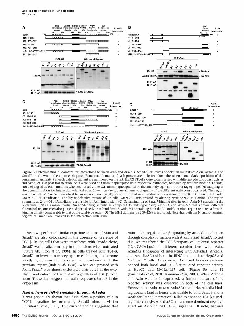
Determination of interaction regions of Axin with
Arkadia and Smad7
The above data indicated that Arkadia and Smad7 each
physically interact with Axin. We then determined the
amino-acid regions of the three proteins that are required
for their mutual interactions. Expression vectors containing
wild-type as well as their different truncation mutants are
indicated schematically (on the top of each panel, Figure 3).
When HA–Axin-M1 alone was transfected, it was not
present in the anti-FLAG (ArC937A) immunoco-precipitate
(Figure 3A, first lane); however, when cotransfected with
FLAG-ArC937A, full-length Axin was strongly co-immuno-
precipitated with Arkadia. Axin deletion mutants C1, N2, and
M1 retained their ability to form a complex with Arkadia,
albeit with lower affinity compared to full-length Axin.
However, Axin deletion mutants N1, C2, and DAr lost their
ability to interact with Arkadia, indicating that the region
around aa 507–757 in Axin is critical for Arkadia interaction.
We then cotransfected different deletion mutants of
Arkadia with full-length Axin to 293T cells to determine the
domain of Arkadia for Axin interaction. As shown in
Figure 3B, the N1, C2, and DM1 deletion mutants could not
interact with Axin, indicating that the region spanning
aa 241–404 of Arkadia is critical for Axin interaction.
To determine the mutual interaction domains between
Axin and Smad7, different constructs of Axin as indicated
(on the top of Figure 3C) were cotransfected with Smad7 into
293T cells. The N-terminal region of aa 1–183 weakly inter-
acted with Smad7; C-terminal region of aa 506–757 alone also
showed partial binding ability. Of note, as C3 and M2
exhibited similar binding affinity for Smad7, the DIX domain
of Axin (aa 757–832) is dispensable for Smad7 interaction.
These results demonstrate that Axin requires both its
N-terminal region (aa 1–183) and C-terminal region of
aa 507–757 for maximal interaction with Smad7.
1 989937 977
83 433
Arkadia WT
AIP7
GST pulldown assay
IB: FLAG FLAG-Arkadia
GST-Axin
FLAG-Arkadia
GST
Input
Ring
−++−++−++HA-Axin++−++−++−FLAG-Arkadia
IB: Axin
IB: Arkadia Arkadia
Axin
Untransfected 293T cellIP
-IgG
IP-A
r
WC
L
IP-Ig
G
IP-A
xin
pAC
T+pG
BK
T7-A
xinC
T pAC
T-AIP7+
pGB
KT7
IP-HA IP-FLAG Whole-cell lysate
IB: FLAG
IB: HA
FLAG-Arkadia
HA-Axin
pACT-AIP7+pGBKT7-AxinCT
A
B
C
D
Figure 1 Identification of Arkadia as a novel Axin-interacting protein. (A) Yeast two-hybrid screening using Axin C-terminal as a bait wasdescribed previously (Rui et al, 2002). AIP7, one of the identified clones, contains a cDNA insert corresponding to aa 83–433 of Arkadia, asdiagrammed on the top. AH109 cells cotransformed with pACT2-AIP7 and pGBKT7-AxinCT, but not the others, could grow on Ade�/Leu�/His�/Trp� medium plates. (B) Arkadia interacts with Axin at its endogenous levels. The 293T cells were treated with MG132 (10mM for 4 h)before harvest. Cell lysates were immunoprecipitated with rabbit anti-Arkadia, rabbit anti-Axin, and control rabbit IgG, respectively, followedby immunoblotting with their respective antibodies as indicated. (C) Axin and Arkadia form complex in 293T cells. FLAG-tagged Arkadia andHA-tagged Axin were transfected either alone or together into HEK293T cells. At 32 h post-transfection, cells were treated for 4 h with 10mMMG132, and were then subjected to immunoprecipitation, followed by Western blotting analysis with anti-HA or anti-FLAG as indicated.(D) GST pulldown assay. GST-Axin-fusion protein was expressed in E. coli cells and was purified as described previously (Rui et al, 2004); itwas added to the lysate of MG132-treated 293T cells ectopically expressing FLAG-Arkadia. Note that only GST-Axin but not GST could pulldown Arkadia. Experiments were repeated with essentially the same results.
Axin is a major scaffold in TGF-b signalingW Liu et al
The EMBO Journal VOL 25 | NO 8 | 2006 &2006 European Molecular Biology Organization1648

As shown in Figure 3D, among all the Smad7 mutants,
only M1 that lacks both the N- and C-terminal regions failed
to interact with Axin. Smad7 therefore appears to possess two
domains for interaction with Axin, with either N- or MH2
domains alone capable of forming complex with Axin. It is
interesting to note that both Axin and Smad7 require two
regions for their interaction.
Axin colocalizes with Arkadia and Smad7
Next, we asked if Axin is colocalized in the cell with
Arkadia and Smad7. We transfected FLAG-tagged Arkadia
into COS-7 cells alone or together with Axin. When expressed
alone, Arkadia was mainly localized in the nucleus
in the absence of TGF-b (Figure 4A), in agreement with a
previous report (Koinuma et al, 2003); when the cells
were treated with TGF-b, Arkadia appeared to translocate
into the cytoplasm and was distributed in the whole cell.
In contrast, when coexpressed with Axin, even in the
absence of TGF-b, Arkadia was translocated into the cyto-
plasm and was colocalized with Axin (Figure 4A, lower two
panels).
1st IP
2nd IP
Detection
HA-Axin orUntagged-Axin
Myc-Smad7FLAG-ArC937A
IP-HA
HA elution
IP-Myc
IB: Myc,Ar, Axin
FLAG-Smad − 1 2 3 4 5 6 7
IP-FLAG
IB: FLAG
IB: HA
LysateIB: HA
HA-Axin
HA-Axin
FLAG-Smad 3 3 6 6 7 7
HA-Axin − + − + − +
IB: FLAG
IB: HA
LysateIB: FLAG
HA-Axin
IP- HA
1st IP: IP-HA(Axin)
2nd IP: IP-Mycor IgG
Lysate HA elution
Unt
agge
dH
AU
ntag
ged
HA
Co-IP
IP: I
gGIP
: Myc
Inpu
t
HA-Axin/*Untagged Axin
Myc-Smad7
FLAG-ArC937A
FLAG-Smads
* *FLAG-Smads
IB: Axin
IB: Smad7 Smad7
Axin
Untransfected 293T cellIP
-IgG
IP-S
mad
7
WCL
*
*
FLAG-Smads
−+
+
+
+
+
−+
−FLAG-ArC937A
HA-AxinMyc-Smad7
FLAG-ArC937A
IB: Myc
IB: Myc Myc-Smad7
Lysate
IB: Arkadia
HA-Axin
IB: HA
Myc-Smad7
HA-Axin
IB: HA
IP-FLAG
IB: Axin
IB: Myc
IB: Arkadia
*
A
B
C
D
Figure 2 Axin interacts with Smad7, and Axin, Arkadia, and Smad7 form a ternary complex. (A) Identification of Smad7 as another novelAxin-interacting protein. FLAG-tagged Smads 1–7 were separately cotransfected with HA-Axin into 293Tcells. Reciprocal immunoprecipitationwith anti-FLAG and anti-HAwas carried out, followed byWestern blotting analysis. Smad3, 6, and 7 (marked by asterisks) were co-precipitatedwith Axin. (B) Endogenous Axin and Smad7 interact with each other in 293T cells. The 293T cell lysate was incubated with goat Smad7polyclonal antibody (Santa Cruz Biotech.), followed by Western blotting with rabbit AxinC2b polyclonal antibody. Three separate experimentswere carried out and similar results were obtained. (C) Two-step co-immunoprecipitation to test for ternary complex formation of Axin,Arkadia, and Smad7. The procedures of two-step co-immunoprecipitation are outlined in the left box. HEK293T cells were transfected withMyc-Smad7, FLAG-ArC937A, and HA-Axin (or untagged Axin as control, marked by asterisk). The first immunoprecipitation was performedwith anti-HA antibody. The complex was eluted by using HA peptide, followed by the second step of immunoprecipitation with anti-Myc orcontrol mouse IgG. Protein samples from each step were then subjected to Western blotting analysis separately by using anti-Axin, anti-Myc,and anti-Arkadia antibodies. The experiment was repeated with essentially the same result. (D) Axin increases the interaction affinity ofArkadia for Smad7. FLAG-ArC937A and Myc-Smad7 were cotransfected with or without Axin into 293Tcells. Immunoprecipitation was carriedout with anti-FLAG, followed by immunoblotting with anti-Myc to detect Smad7, and anti-HA to detect Axin. Increased amount of Myc-Smad7was co-immunoprecipitated with Arkadia from cells coexpressing Axin.
Axin is a major scaffold in TGF-b signalingW Liu et al
&2006 European Molecular Biology Organization The EMBO Journal VOL 25 | NO 8 | 2006 1649
Next, we performed similar experiments to see if Axin and
Smad7 are also colocalized in the absence or presence of
TGF-b. In the cells that were transfected with Smad7 alone,
Smad7 was localized mainly in the nucleus when untreated
(Figure 4B) (Itoh et al, 1998). In cells treated with TGF-b,Smad7 underwent nucleocytoplasmic shuttling to become
mostly cytoplasmically localized, in accordance with the
previous report (Itoh et al, 1998). When coexpressed with
Axin, Smad7 was almost exclusively distributed in the cyto-
plasm and colocalized with Axin regardless of TGF-b treat-
ment. These data suggest that Axin sequesters Smad7 in the
cytoplasm.
Axin enhances TGF-b signaling through Arkadia
It was previously shown that Axin plays a positive role in
TGF-b signaling by promoting Smad3 phosphorylation
(Furuhashi et al, 2001). Our current finding suggested that
Axin might regulate TGF-b signaling by an additional mean
through complex formation with Arkadia and Smad7. To test
this, we transfected the TGF-b-responsive luciferase reporter
(12�CAGA-Lux) in different combinations with Axin,
AxinDAr (incapable of interacting with Arkadia), Arkadia,
and ArkadiaDC (without the RING domain) into HepG2 and
Mv1Lu/L17 cells. As expected, Axin and Arkadia each en-
hanced both basal and TGF-b-stimulated reporter activity
in HepG2 and Mv1Lu/L17 cells (Figure 5A and B)
(Furuhashi et al, 2001; Koinuma et al, 2003). When Arkadia
and Axin were both expressed, a further increase of the
reporter activity was observed in both of the cell lines.
However, the Axin mutant AxinDAr that lacks Arkadia-bind-ing domain (and is hence also unable to bind Smad3 and is
weak for Smad7 interaction) failed to enhance TGF-b signal-
ing. Interestingly, ArkadiaDC had a strong dominant-negative
effect on Axin-induced TGF-b signaling. Of note, because
Arkadiainteraction
AxinN1: 1−506C1: 507−832N2: 1−756C2: 757−832ΔAr: 1−506/757−832M1: 507−757
89 216 353 437 506 757 8201 832 +
−++−−+
ArkadiaCAN1: 1−240N2: 1−404C1: 241−989C2: 405−989M1: 241−404ΔΔM1: 1−240/405−989
Axininteraction
+−++−+−
1 989C937A
HA-Axin
HA-Axin
Vect
or
IB: HA
IP: FLAGIB: FLAG
Lysate IB: HA
C1N1 ΔM
1
ΔM1
ΔM1
M1
N2
C2ArC
937A
IP-FLAG
Whole-cell lysateIP-FLAG
IP: FLAGIB: FLAG FLAG-Smad7
IB: HA
C3
N3 M4
Axi
n
M2
M3
Axi
n
C3
N3
M4
Axi
n
M2
M3
Axi
n
Smad7interaction
AxinN3: 1−183C3: 184−832M2: 184−756M3: 184−506M4: 1−233/507−832
++++−+
832 Smad7
ND: 1−259
MH2: 260−426
M1: 138−357
ΔM1: 1−137/358−426
1 426
Axininteraction
+
+
+
−
+
Whole-cell lysateIP-HA
IP: HAIB: HA HA-Axin
IB: FLAG
MH
2
ND
Sm
ad7
M1
Vec
tor
MH
2
ND
Sm
ad7
M1
Vec
tor
Whole-cell lysateIP-FLAG
IP: FLAGIB: FLAG FLAG-ArC937A
IB: HA
C1
N1 M1
ΔAr
ΔAr
Axi
n
N2
C2M1
C1
N1 M1
Axi
n
N2
C2
M1
RGS homologous
domain
MEKK1-interacting
domain
GSK3β-binding
site
β-Catenin-binding
site
PP2A/MEKK4
binding site
Dshhomologous
domain
89 216 353 437 506 757 8201
RGS homologous
domain
MEKK1-Interacting
domain
GSK3β-binding
site
β-Catenin-binding
site
PP2A/MEKK4-
binding site
Dshhomologous
domain
RING finger
domain937 977
MH2 domain260
A
CD
B
Figure 3 Determination of domains for interactions between Axin and Arkadia, Smad7. Structures of deletion mutants of Axin, Arkadia, andSmad7 are shown on the top of each panel. Functional domains of each protein are indicated above the schema and relative positions of theremaining fragment(s) in each deletion mutant are numbered on the left. HEK293Tcells were cotransfected with different plasmid constructs asindicated. At 36 h post-transfection, cells were lysed and immunoprecipited with respective antibodies, followed by Western blotting. Of note,none of tagged deletion mutants when expressed alone was immunoprecipitated by the antibody against the other tag epitope. (A) Mapping ofthe domain in Axin for interaction with Arkadia. Shown on the top are schematic diagrams of the different Axin constructs used. The regionaround aa 507–757 in Axin is critical for Arkadia interaction. (B) Identification of Axin-binding sites on Arkadia. The RING domain of Arkadia(aa 937–977) is indicated. The ligase-defective mutant of Arkadia, ArC937A, was created by altering cysteine 937 to alanine. The regionspanning aa 241–404 of Arkadia is responsible for Axin interaction. (C) Determination of Smad7-binding sites in Axin. Axin-N3 containing theN-terminal 183 aa showed partial Smad7-binding activity as compared to wild-type Axin; Axin-C3 and Axin-M2 that contain differentC-terminal regions each also possessed partial activity to bind Smad7. Axin-M4 containing both the N- and C-terminal region retained a Smad7-binding affinity comparable to that of the wild-type Axin. (D) The MH2 domain (aa 260–426) is indicated. Note that both the N- and C-terminalregions of Smad7 are involved in the interaction with Axin.
Axin is a major scaffold in TGF-b signalingW Liu et al
The EMBO Journal VOL 25 | NO 8 | 2006 &2006 European Molecular Biology Organization1650

Mv1Lu/L17 cells are null for TGF-b type I receptor (TbRI),a constitutively active TGF-b type I receptor (caTbRI) was
cotransfected into Mv1Lu/L17 cells instead of treating cells
with TGF-b ligand (Figure 5B).
Functional dependence between Axin and Arkadia was
further demonstrated by introducing vector-based small
interfering RNA (siRNA) of Arkadia (for knockdown effi-
ciency and specificity, see Supplementary Figure 3). The
FLAG-Arkadia
Anti-FLAG Anti-Axin Merge Hoechst
Anti-FLAG Anti-Axin Merge Hoechst
FLAG-Arkadia+TGF-β
FLAG-Arkadia+Axin
FLAG-Arkadia +Axin
+TGF-β
FLAG-Smad7
FLAG-Smad7+TGF-β
FLAG-Smad7+Axin
+TGF-β
FLAG-Smad7+Axin
A
B
Figure 4 Axin is constitutively colocalized with Arkadia or Smad7 and induces cytoplasmic translocation of Smad7. (A) Axin colocalized withArkadia. COS7 cells were transfected with FLAG-Arkadia with or without Axin. Cells treated with or without TGF-b then fixed and stained asdescribed in Materials and methods. Anti-FLAG staining for Arkadia (red), anti-Axin for Axin (green), and nuclear staining by Hoechst wereperformed. (B) Axin induces cytoplasmic translocation of Smad7 and colocalized with Smad7 in the cytoplasm. Compared to expression ofSmad7 alone, in Axin-coexpressing cells, great majority of Smad7 was seen in the cytoplasm, indicating that Axin caused translocation ofSmad7 into the cytoplasm. Most representative results from three rounds of staining are shown.
Axin is a major scaffold in TGF-b signalingW Liu et al
&2006 European Molecular Biology Organization The EMBO Journal VOL 25 | NO 8 | 2006 1651

Arkadia siRNA could attenuate Axin-stimulated TGF-b signal-
ing (Figure 5C). Conversely, siRNA against Axin (Rui et al,
2004) diminished Arkadia-induced TGF-b signaling, indicat-
ing that Axin and Arkadia depend on each other to facilitate
TGF-b signaling.
Axin enhances Arkadia-dependent Smad7
ubiquitination
It is reported that Arkadia can strongly induce polyubiquiti-
nation of Smad7 (Koinuma et al, 2003). We then examined
whether Axin facilitates the function of Arkadia in this
aspect. As expected, wild-type Arkadia, but not ArkadiaDC,could induce the polyubiquitination of Smad7 in transfected
293T cells (Figure 6A). Ubiquitination of Smad7 was en-
hanced in cells overexpressing increasing amounts of Axin
(Figure 6A and B). Axin-induced Smad7 polyubiquitination
was blocked by ArkadiaDC in a dose-dependent manner
(Figure 6C), suggesting that the latter exerts a
dominant-negative effect on the Axin-induced polyubiquiti-
nation of Smad7. To further test the cooperation between
Axin and Arkadia, vector-based siRNA of Arkadia was intro-
duced into Axin- and Smad7-transfected cells (Figure 6D).
Consistent with the above data, siRNA against Arkadia
could attenuate Axin-induced polyubiquitination of Smad7.
Similarly, when siRNA of Axin was expressed in Arkadia-
transfected cells, polyubiquitinated Smad7 was reduced
(Figure 6E). These findings suggest that Axin and Arkadia
depend on each other to promote the ubiquitination of
Smad7.
Since Axin, Arkadia, and Smad7 form a ternary complex,
to assess the functional significance of the complex forma-
tion, we transfected into 293T cells with Axin constructs
0
10
20
30
40
50
60
70
80
90
0
25
50
75
100
125 Mv1Lu/L1712× CAGA-Lux
0
5
10
15
20
25
30
35
40
45
0
5
10
15
20
25
30
+
+
ΔC ΔC ΔC ΔC+−−−Arkadia
+−−ΔAr+−Axin
HepG212× CAGA-Lux
+−+−−−pSUPER-Arkadia
−+−+−−RNAi control
++−−+−Axin
Rel
ativ
e lu
cife
rase
act
ivit
y (f
old
)
Rel
ativ
e lu
cife
rase
act
ivit
y (f
old
)
Rel
ativ
e lu
cife
rase
act
ivit
y (f
old
)
Rel
ativ
e lu
cife
rase
act
ivit
y (f
old
)
HEK29312× CAGA-Lux
−TGF-β1+TGF-β1
−TGF-β1+TGF-β1
−caTβR1+caTβR1
−TGF-β1+TGF-β1
+−+−−−pSUPER-Axin
−+−+−−RNAi control
++−−+−Arkadia
HEK29312× CAGA-Lux
+
+
+−−−Arkadia
+−−ΔAr+−Axin
A
C
B
Figure 5 Axin enhances TGF-b-dependent transcriptional activity through Arkadia. 12�CAGA-Lux reporter was cotransfected with differentAxin or Arkadia constructs into HepG2, Mv1Lu/L17, and HEK293 cells as indicated. Cells were either treated with TGF-b (A, C) orcotransfected with caTbRI (B). Variation in transfection efficiency among samples was less than 10% based on activities of cotransfected b-galactosidase. All transfections were performed in duplicate and the data are presented as means7s.d. of at least three separate experimentsafter normalizing luciferase activity from vector control to 1. (A) Axin, but not AxinDAr, can activate TGF-b-dependent gene transcription of12�CAGA-Lux reporter; the dominant-negative mutant of Arkadia, ArDC, diminished Axin-mediated activation of the reporter gene in HepG2cells. (B) Axin depends on Arkadia for stimulation of reporter transcriptional activity in Mv1Lu/L17 cells. (C) The siRNA against Arkadia, butnot control siRNA, attenuated Axin-stimulated TGF-b signaling in 293 cells (left panel). Conversely, Axin siRNA diminished the Arkadia-induced activation of TGF-b signaling (right panel).
Axin is a major scaffold in TGF-b signalingW Liu et al
The EMBO Journal VOL 25 | NO 8 | 2006 &2006 European Molecular Biology Organization1652

lacking the Arkadia-binding site (AxinDAr) or both of
Arkadia- and Smad7-binding sites (Axin-M3), and deter-
mined their contribution to the polyubiquitination of Smad7
(Figure 6F). The Axin-M4 deletion mutant that retains both
Smad7- and Arkadia-binding sites was also included for
comparison. Deletion of both Arkadia- and Smad7-binding
sites (Axin-M3) rendered Axin completely incapable of
enhancing the ubiquitination of Smad7. Axin mutants
lacking Arkadia-binding site (AxinDAr that also lacks the
C-terminal portion of Smad7 interaction site) or the
N-terminal part of the Smad7-binding site (Axin-C3)
drastically lost the function, whereas Axin-M4 induced
polyubiquitination of Smad7 as effectively as did the
wild-type Axin (Figure 6F). Specific depletion of endogenous
0
50
100Smad7 only
Smad7+Arkadia
Smad7+AxinSmad7+Arkadia+Axin
+−+−−−−HA-Arkadia−
++
−
++
+
++
+
+++−−FLAG-Smad7
−
+
−−HA-Axin
−−Myc-Ub
IB: FLAG
IB: UbIP: FLAG
(Ub)n-Smad7
FLAG-Smad7
HA-Arkadia
Lysate
IB: HA
+++
+++
−+++
−HA-Arkadia−
++FLAG-Smad7
HA-Axin
Myc-Ub
HA-Arkadia
IB: UbIP: FLAG
IB: FLAG FLAG-Smad7
Lysate
IB: Arkadia
HA-AxinIB: HA
(Ub)n-Smad7
M3
+
+
M4
+
+
−+
+
+
+
+
C3
+
+
ΔAr
+
+FLAG-Smad7
HA-Axin
Myc-Ub
(Ub)n-Smad7IB: UbIP: FLAG
IB: FLAG FLAG-Smad7
Lysate
IB: HA
+++−HA-Axin+−−−pSUPER-Arkadia−+−−RNAi control
++
++
++
++FLAG-Smad7
Myc-Ub
(Ub)n-Smad7IB: UbIP: FLAG
IB: FLAG FLAG-Smad7
Lysate
IB: HA HA-Axin
+++−HA-Arkadia+−−−pSUPER-Axin−+−−RNAi control
++
++
++
++FLAG-Smad7
Myc-Ub
(Ub)n-Smad7IB: UbIP: FLAG
IB: FLAG FLAG-Smad7
Lysate
IB: Arkadia HA-Arkadia
HA-Axin/Axin deletions
−7.552.50Chase time (h)
++ + +FLAG-Smad7
+Axin
IP: FLAG
35S-Smad7
+Arkadia
+Axin+Arkadia
35S
-Fla
g-S
mad
7(p
erce
nta
ge
of
tim
e 0
h)
+
++
−−HA-Arkadia Δ Δ C−
++
+
++
+
++
+
++FLAG-Smad7
HA-Axin
Myc-Ub
HA-ArkadiaΔC
IB: UbIP: FLAG
IB: FLAG FLAG-Smad7
Lysate
IB: HA
HA-Axin
(Ub)n-Smad7
IB: HA
+Vector
(Ub)n-Smad7IB: Ub
IB: Smad7 Smad7Lysate
IB: Tubilin Tubilin
++++−MG132
−+−
−−−−pSUPER-Arkadia−−+−RNAi control
IgGSmad7IP
− − + −pSUPER-Axin
IP: Smad7/IgG
2.5 5 7.5Time (h)
A
D
G H
E F
B C
Figure 6 Axin promotes Arkadia-induced ubiquitin-dependent degradation of Smad7. (A) Axin induces ubiquitination of Smad7. The 293Tcells were transfected as indicated, and treated with 10mM of MG132 for 4 h before cell lysis. Lysates from cells were subjected to anti-FLAGimmunoprecipitation followed by anti-ubiquitin immunoblotting. Polyubiquitination species of Smad7, (Myc-ubiquitin)n–FLAG-Smad7, areindicated in the top panel. (B) Axin promotes Arkadia-induced polyubiquitination of Smad7 in a dose-dependent manner. Increasing amounts(0, 0.5, and 2mg) of HA-Axin were cotransfected with 2mg of Arkadia into 293Tcells. Smad7 ubiquitination was determined as described above.(C) Axin-induced polyubiquitination of Smad7 is negatively regulated by ArkadiaDC in a dose-dependent manner. (D) Arkadia siRNAdiminished polyubiquitination of Smad7 induced by Axin. The 293T cells were transfected with Axin with or without pSUPER-Arkadiaas indicated to assess the effect of siRNA against Arkadia on Smad7 ubiquitination. (E) Axin siRNA attenuated Arkadia-induced polyubiquiti-nation of Smad7. pSUPER-Axin (4mg) was transfected into 293Tcells. (F) Axin scaffolds Arkadia and Smad7 to enhance the polyubiquitinationof Smad7. The 293T cells were transfected with FLAG-Smad7, Myc-ubiquitin, and wild-type or different Axin deletion mutants as indicated.(G) Specific knockdown of Axin or Arkadia reduced polyubiquitinated Smad7 in 293T cells. HEK293T cells were transfected with pSUPERvectors expressing control siRNA, Axin siRNA, or Arkadia siRNA as indicated. MG132 (10mM) was added to the cells at 32 h post-transfection,and were incubated for additional 4 h. Cell lysates were then subjected to immunoprecipitation with anti-Smad7 or control IgG, followed byWestern blotting with anti-ubiquitin. (H) Axin facilitates Arkadia to accelerate ubiquitin-dependent degradation of Smad7. The 293Tcells weretransfected with FLAG-Smad7 with or without wild-type Arkadia or wild-type Axin, or both of them. [35S]methionine- and cysteine-labeled celllysates were immunoprecipitated by anti-FLAG antibody followed by autoradiography after separation on SDS–PAGE. The autoradiographic signalswere quantified and the values plotted relative to 0-h values. All of the above experiments were repeated with essentially the same results.
Axin is a major scaffold in TGF-b signalingW Liu et al
&2006 European Molecular Biology Organization The EMBO Journal VOL 25 | NO 8 | 2006 1653

Axin or Arkadia by their respective siRNA significantly
reduced the levels of polyubiquitinated Smad7 in 293T cells
(Figure 6G). These observations indicate that Axin scaffolds
Arkadia and Smad7 to facilitate Arkadia-induced polyubiqui-
tination of Smad7.
To further ascertain the augmentative effect of Axin on
Arkadia-induced proteasome-dependent degradation of
Smad7, we analyzed turnover of Smad7 by pulse-chase
experiments (Figure 6H). As expected, Smad7 was rapidly
degraded in the presence of Arkadia or Axin alone. Moreover,
it is found that Axin could significantly enhance Arkadia-
induced degradation of Smad7. The results thus demonstrate
that Axin accelerates turnover of Smad7 by recruiting an E3
ubiquitin ligase, Arkadia.
Axin associates with Arkadia and Smad7 independently
of TGF-b signaling
In the case of Smad3 interaction with Axin, it has been shown
that Smad3 becomes disassociated upon stimulation by TGF-
b signal. Our results obtained from the cell staining experi-
ments, however, raised the possibility that Axin may be
colocalized with Smad7 and Arkadia before and after TGF-btreatment. To verify this, we carried out interaction assays
by co-immunoprecipitation between Axin and separately
Arkadia, Smad7, along with Smad3, Smad3D407E, and
Smurf1, after cotransfection with or without caTbRI as in-
dicated (Figure 7A). Indeed, Arkadia and Smad7 bind to Axin
with the same affinities regardless of coexpression of caTbRI.Smurf1 did not interact with Axin. In the absence of TGF-b
Figure 7 Axin constitutively associates with Arkadia and Smad7. (A) HA-Axin together with the various plasmids indicated were cotransfectedwith or without caTbRI into 293Tcells. After 4 h treatment with MG132 (10mM), cells were lysed and lysates were immunoprecipitated by anti-FLAG, followed by Western blotting using anti-HA and rabbit anti-FLAG. (B) Wnt-1 attenuated both basal and Axin-induced Smad7ubiquitination. HEK293T cells were transfected with 0.5mg of FLAG-Smad7, Myc-ubiquitin with or without 1.5 mg of HA-Axin or/and Wnt-1as indicated. Cells were treated with MG132 (10mM) for 4 h before lysed. Cell lysates were subjected to immunoprecipitation with anti-FLAGantibody and polyubiquitination of Smad7 was detected with anti-ubiquitin antibody. Wnt-1 in total cell lysates was probed with anti-Wnt-1.Similar results were obtained in three separate experiments. (C) Effects of Wnt-1 on the TGF-b-dependent transcriptional activity. 12�CAGA-Lux was transfected into HEK293 cells with or without Axin, caTbRI, or/and Wnt-1 as indicated. Wnt-1 could reduce the reporter genetranscription in the presence or absence of transfected Axin. (D) Model of scaffolding roles of Axin in TGF-b signaling. Axin forms a complexwith Smad3, Arkadia, and Smad7, of which Smad3 has physical contact with the type I receptor. Upon ligand stimulation, Axin promotesSmad3 phosphorylation; phosphorylated Smad3 dissociates from the Axin complex, and then combines with Smad4 to activate transcription inthe nucleus. In addition, Axin acts as a scaffold to facilitate Arkadia-mediated polyubiquitination of Smad7 regardless of TGF-b signaling,leading to Smad7 degradation. Implied here is also that Axin can sequester Smad7 in the cytoplasm.
Axin is a major scaffold in TGF-b signalingW Liu et al
The EMBO Journal VOL 25 | NO 8 | 2006 &2006 European Molecular Biology Organization1654

signaling, Smad3 strongly interacted with Axin; however,
when coexpressed with caTbRI, Smad3 dissociated
from the Axin complex. In contrast, the Smad3 mutant
D407E that is not phosphorylated upon TGF-b stimulation
(Goto et al, 1998) remained associated with Axin in the
presence of TGF-b signaling, in agreement with the
previous report that the mutant D407E is constitutively
associated with Axin. These data have thus demonstrated
that whereas only unstimulated Smad3 but not stimulated
Smad3 interacts with Axin, Arkadia and Smad7 are constitu-
tively associated with Axin. Consistently, caTbRI did not
have any effect on Axin and Arkadia-induced polyubiquitina-
tion of Smad7, as determined by both ubiquitination and
pulse-chase labeling experiments (Supplementary Figure 4A
and B).
However, it is possible that Wnt proteins may exert an
effect on Axin-mediated Smad7 ubiquitination as the
stability and function of Axin have been shown to be
regulated by the Wnt proteins. We therefore coexpressed
mouse Wnt-1 with Smad7 in the presence or absence of
overexpressed Axin and determined the levels of Smad7
ubiquitination in 293T cells. As shown in Figure 7B, Wnt-1
significantly reduced Smad7 ubiquitination in the cell. In
addition, Wnt-1 also attenuated Axin-induced Smad7 ubiqui-
tination, in agreement with its ability to downregulate the
levels of Axin (Yamamoto et al, 1999), further underscoring
the importance of Axin in the induction of Smad7 degrada-
tion. In parallel, we assayed for the effect of Wnt-1 on the
transcriptional activity of the 12�CAGA-Lux as shown in
Figure 7C. Wnt-1 could reduce the TGF-b-dependent tran-
scriptional activity of the reporter gene in the presence or
absence of overexpressed Axin. The results were reproduci-
ble in three separate experiments.
Discussion
TGF-b signaling has been an area of intensive studies, owing
to its plethoric biological roles in development and cellular
homeostasis. Although many signaling molecules have been
elucidated, how these intracellular signaling components are
coordinated remains largely obscure. Here, we have uncov-
ered a novel aspect of TGF-b signaling regulation in that
promotion of R-Smad activation and I-Smad downregulation
are coordinated by the same scaffolding molecule Axin that
has been demonstrated to play a central and scaffolding role
in the Wnt and JNK MAP kinase pathways (Luo and Lin,
2004; Salahshor and Woodgett, 2005). We present a simpli-
fied summary of the roles of Axin in the regulation of TGF-bsignaling in Figure 7D. Despite the observation that Axin
forms a complex with Arkadia and Smad7 independently of
TGF-b signaling, Wnt-1, a Wnt ligand, appears to play an
inhibitory role in Axin-stimulated TGF-b signaling by down-
regulating Axin levels in the cell. We have thus provided new
mechanistic insights into the notion that Axin is an integrator
for the Wnt and TGF-b signaling pathways (Attisano and
Labbe, 2004).
Axin acts as a scaffold for multiple components in the
TGF-b superfamily signaling pathway
Abundant evidence exists to suggest that Wnt signaling
interacts or crosstalks with TGF-b signaling (Labbe et al,
2000; Nishita et al, 2000; Edlund et al, 2005). In this study,
we have demonstrated that Axin, a central scaffold in the Wnt
pathway, interacts with the E3 ubiquitin ligase Arkadia that
has been linked to TGF-b signaling. The interaction has been
confirmed by multiple means, a yeast two-hybrid interaction
assay, co-immunoprecipitation of endogenous proteins, and
subcellular colocalization by immunofluorescent staining.
This unexpected finding prompted us to check whether
Axin would also bind to Smads as they have been shown to
be Arkadia substrates. Indeed, Axin also interacts with
Smad7; moreover, ternary complex assay by two-step co-
immunoprecipitation has revealed that Axin simultaneously
interacts with Arkadia and Smad7. Interestingly, the C-term-
inal region of Axin participates in the interaction with
both Arkadia and Smad7, suggesting that Axin can provide
proximity of Smad7 to its E3 ligase Arkadia. As for the
structural requirements of Smad7 for Axin interaction,
both the N-terminus region and the carboxyl region of MH2
are both needed. This may imply that Axin may also
modulate Smad7 functions in pathways other than TGF-bsignaling, as the MH2 domain alone is responsible for recep-
tor interaction, formation of homomeric and heteromeric
Smad complexes.
Axin as an activator of TGF-b by downregulating
I-Smads through sequestration and polyubiquitination-
mediated degradation
Smad3 in its inactive state binds to Axin. When phosphory-
lated in response to TGF-b treatment, it dissociates from
the Axin complex (Furuhashi et al, 2001), suggesting that
Axin may not be directly involved in Smad3-dependent
transcriptional activation of target genes. In contrast, Axin
seems to be constitutively associated with Arkadia, and
Smad7 unless degraded. Even in the absence of TGF-btreatment, coexpression of Axin itself can translocate
Smad7 from the nucleus to the cytoplasm as revealed by
immunostaining. Co-immunoprecipitation experiment also
showed that complex formation between Axin and
Arkadia or Smad7 did not change after stimulation by the
constitutively active type I receptor. Moreover, TGF-b signal
did not seem to further increase Axin-induced Smad7 poly-
ubiquitination and degradation (Supplementary Figure 4). All
these observations indicate that Axin is closely associated
with Arkadia and Smad7, and acts to constantly decrease
cellular levels of the I-Smad(s) in the presence or absence of
TGF-b.The nucleocytoplasmic translocation of I-Smad induced by
Axin may be achieved by two possible means. One is that
Axin serves as a carrier to mediate nuclear export, and the
other is that Axin simply traps I-Smad in the cytoplasm to
prevent nuclear entry of Smad7. The first possibility is
supported by the fact that Axin is also a nuclear protein,
albeit mostly cytoplasmic (Cong and Varmus, 2004; Wiechens
et al, 2004), and by our previous observation that it is able to
undergo nucleocytoplasmic shuttling upon stimulation by
signals such as UV (Rui et al, 2004). However, Smad7 was
still present in the cytoplasm when coexpressed with an Axin
mutant that did not exhibit any discernible nuclear localiza-
tion after its NLS sequences were deleted (data not shown).
Therefore, it remains unclear how Axin induces Smad7
nuclear export.
It is demonstrated here that Axin facilitates TGF-b signal-
ing in an Arkadia-dependent manner. First of all, AxinDAr
Axin is a major scaffold in TGF-b signalingW Liu et al
&2006 European Molecular Biology Organization The EMBO Journal VOL 25 | NO 8 | 2006 1655

that is deleted of the Arkadia-interacting domain fails to
activate TGF-b signaling. Second, when Arkadia expression
is knocked down by siRNA, the Axin-induced activation of
TGF-b signaling is drastically diminished. Moreover, E3
ligase-deficient mutant ArkadiaDC also greatly attenuated
Axin activation of TGF-b signaling. All these observations
point to a critical role of Arkadia in the biological function of
Axin. Furthermore, the observation that Arkadia always
stimulates TGF-b signaling to a greater extent than Axin is
also consistent with the above notion. To test if the stimula-
tory role of Arkadia is indeed exerted by its E3 ligase activity,
a series of ubiquitination assays as well as half-life determi-
nation of Smad7 were carried. Indeed, Axin strongly pro-
motes polyubiquitination of Smad7 and cooperates with
Arkadia to reduce stability of Smad7.
A general role of Axin in cytostasis?
TGF-b plays a crucial role in maintaining proliferative homeo-
stasis (Massague, 1998). In light of all the existing evidence,
it is fascinating to speculate that Axin, a known tumor
suppressor (Polakis, 2000; Hsu et al, 2001), exerts a dual
role in a constitutive manner to prevent escape of cells from
cytostasis by acting to downregulate oncogenic b-catenin, inone way, and to enforce antiproliferative function induced by
TGF-b. As far as Wnt signaling is concerned, Axin in un-
stimulated cells provides an architectural platform for mole-
cular assembly containing APC, GSK-3b, casein kinases, and
b-catenin to enable phosphorylation of b-catenin, leading to
ubiquitination and degradation. In the case of TGF-b signal-
ing, besides a role in facilitating Smad3 phosphorylation
upon TGF-b treatment (Furuhashi et al, 2001), Axin promotes
ubiquitination and degradation of Smads that play an inhibi-
tory role in this pathway, again leading to enhancement of
antiproliferative mode. Equally interesting is that Axin
directly stimulates p53 function through augmenting its phos-
phorylation by HIPK-2, leading to cell apoptosis (Rui et al,
2004). Of note, Axin-mediated JNK MAP kinase activation
seems to also play a role in the induction of apoptosis by Axin
(Neo et al, 2000). These lines of evidence all point to a
general role of Axin in downregulating cellular proliferation.
This conjecture raises another interesting question as to
whether Axin has evolved as a major tumor suppressor, in
that when one aspect of antiproliferative function is lost,
it can still manage to utilize another avenue to prevent cell
from escape from cytostasis. Certainly, much awaits further
investigation.
Materials and methods
Plasmid constructionsFull-length cDNA encoding mouse Arkadia was cloned throughfusing the EST clone with HA- or FLAG-tag using a PCR-basedapproach. Arkadia point mutation C937A and Smurf1C698A, andvarious deletion mutants were generated as described previously(Rui et al, 2004). The constructs of caTbRI, Smad1–7, Smurf1, andSmad7 mutants were as described previously (Chen et al, 1998).The expression plasmid for mouse Wnt-1 was purchased fromUpstate Biotech. (Lake Placid, NY).
Cell culture and transient transfectionHEK293T, HEK293, and HepG2 were obtained from ATCC (Mana-ssas, VA). All of the three cell lines and Mv1Lu/L17 cells werecultured in Dulbecco’s modified Eagle’s medium (DMEM) supple-mented with 10% fetal bovine serum, 2mM L-glutamine, andantibiotics, and maintained at 371C, 5% CO2, in a humidified
incubator. Transfections were performed in 60-mm dishes usingcalcium phosphate precipitation.
Preparation of antibodiesMouse anti-HA (F-7), anti-Myc (9E10), anti-ubiquitin (P4D1), andgoat anti-Smad7 (N-19) were purchased from Santa Cruz Biotech.Mouse anti-FLAG (M2) was purchased from Sigma. The polyclonalantibody against Axin was described previously (Rui et al, 2004).Arkadia GST-fusion protein-containing amino-acid sequence of aa1–132 was produced in BL21 Escherichia coli cells and was injectedinto rabbits following purification using glutathione–agarose beads.The Arkadia antibody was purified and characterized as describedpreviously (Rui et al, 2004). Rabbit polyclonal Wnt-1 antibody waspurchased from Zymed (Cat. no.: 36-5800).
Co-immunoprecipitation and Western blottingCells were transfected with various plasmids and incubated for 36 hbefore analysis. For inhibition of proteasomal degradation, cellswere treated with 10mM MG132 (Sigma) for 4 h. Cell lysatepreparation, immunoprecipitation, and Western blotting wereperformed as described previously (Zhang et al, 1999). Essentiallythe same results were obtained in at least three separateexperiments for each interaction assay.
GST pulldown assayThe GST-Axin (full length) was prepared as described previously(Rui et al, 2004). Approximately 4mg of GST-fusion protein boundto agarose beads were added to each total lysate from 293T cells,and incubated for 3 h with gentle rotation. The beads were washedthree times with cell lysis buffer, and the proteins were eluted with2� SDS sample buffer. Protein samples were analyzed by Westernblotting.
Immunofluorescent stainingCOS7 cells grown on coverslips in six-well plates were transfectedwith expression plasmids as indicated. After 30h, immunofluor-escent staining was carried out as described previously (Rui et al,2004). To determine the effects of TGF-b, cells were washed withserum-free medium and treated with TGF-b (10 ng/ml, in serum-free medium) for 8 h before fixation. Experiments were repeated formultiple times.
Transcription reporter assayHEK293, HepG2, and Mv1Lu/L17 cells were transfected with 0.1mgof 12�CAGA-Lux, and 2 mg each of Axin and Arkadia constructs,with or without 0.5 mg of caTbRI or/and 0.5 mg Wnt-1 as indicated.At 36 h post-transfection, luciferase activity was measured andpresented as described previously (Rui et al, 2004). HepG2 andHEK293 cells were washed with serum-free medium and incubatedfor additional 12 h in serum-free medium supplemented with TGF-b1 (5 ng/ml) before analysis. All transfections were performed induplicate and the data are presented as means7s.d. of at least threeseparate experiments after normalizing luciferase activity fromvector control to 1.
Two-step co-immunoprecipitationTwo-step co-immunoprecipitation was performed essentially ac-cording to the procedures described previously (Rui et al, 2004).Briefly, 293T cells were transfected with HA–Axin, Myc-Smad7 andFLAG-ArC937A. For the control of first immunoprecipitation, theAxin lacking the HA-tag was used. At 36 h after transfection, thecells were lysed with lysis buffer (Zhang et al, 1999), sonicatedbriefly, and centrifuged. The supernatant was incubated with anti-HA antibody bound to protein A/G–agarose beads for 3 h at 41C.The beads were washed with lysis buffer three times, and the HA–Axin protein complex was eluted with 300 ml of lysis buffercontaining 250mM NaCl and HA peptide for 3 h at 41C. Secondimmunoprecipitation was performed using 150ml of eluate from thefirst immunoprecipitation and 350ml of lysis buffer containing464mM NaCl and 10ml of anti-Myc antibody or control IgG followedby the addition of protein A/G–agarose beads.
Metabolic labeling and pulse-chase analysisHEK293T cells transfected with plasmids indicated were treatedwith 100 mCi/ml [35S]methionine and cysteine (Amersham) inmethionine- and cysteine-free DMEM (Amersham), and were
Axin is a major scaffold in TGF-b signalingW Liu et al
The EMBO Journal VOL 25 | NO 8 | 2006 &2006 European Molecular Biology Organization1656

incubated for 1 h at 371C, then washed with PBS twice and chasedin DMEM supplemented with 10% FBS, 2mM methionine, and0.5mM cysteine for the time periods indicated. Cells were lysedwith RIPA buffer and subjected to immunoprecipitation followedby SDS–PAGE. The gels were fixed, dried, and examined byautoradiography.
Supplementary dataSupplementary data are available at The EMBO Journal Online.
Acknowledgements
We thank our colleagues for technical assistance and Drs JiaweiWu and Zhenguo Wu for critical comments. This research wassupported by grants from Research Grant Council of Hong Kong(HKUST6127/04M; HKUST6416/05M), National Outstanding YoungInvestigator Award (No.30125012; 30125021), and grants from theNational Science Foundation of China (Nos. 90208015 and30370306), as a grant from Fujian Council of Science andTechnology (2002F002).
References
Attisano L, Labbe E (2004) TGFbeta and Wnt pathway cross-talk.Cancer Metast Rev 23: 53–61
Chen YG, Hata A, Lo RS, Wotton D, Shi Y, Pavletich N, Massague J(1998) Determinants of specificity in TGF-beta signal transduc-tion. Genes Dev 12: 2144–2152
Chia IV, Costantini F (2005) Mouse axin and axin2/conductinproteins are functionally equivalent in vivo. Mol Cell Biol 25:4371–4376
Cong F, Varmus H (2004) Nuclear-cytoplasmic shuttling of Axinregulates subcellular localization of beta-catenin. Proc Natl AcadSci USA 101: 2882–2887
Datto M, Wang XF (2005) Ubiquitin-mediated degradation amechanism for fine-tuning TGF-beta signaling. Cell 121: 2–4
Di Guglielmo GM, Le Roy C, Goodfellow AF, Wrana JL (2003)Distinct endocytic pathways regulate TGF-beta receptor signallingand turnover. Nat Cell Biol 5: 410–421
Dupont S, Zacchigna L, Cordenonsi M, Soligo S, Adorno M,Rugge M, Piccolo S (2005) Germ-layer specification and controlof cell growth by Ectodermin, a Smad4 ubiquitin ligase. Cell 121:87–99
Ebisawa T, Fukuchi M, Murakami G, Chiba T, Tanaka K, Imamura T,Miyazono K (2001) Smurf1 interacts with transforming growthfactor-beta type I receptor through Smad7 and induces receptordegradation. J Biol Chem 276: 12477–12480
Edlund S, Lee SY, Grimsby S, Zhang S, Aspenstrom P, Heldin CH,Landstrom M (2005) Interaction between Smad7 and beta-cate-nin: importance for transforming growth factor beta-inducedapoptosis. Mol Cell Biol 25: 1475–1488
Episkopou V, Arkell R, Timmons PM, Walsh JJ, Andrew RL, Swan D(2001) Induction of the mammalian node requires Arkadia func-tion in the extraembryonic lineages. Nature 410: 825–830
Furuhashi M, Yagi K, Yamamoto H, Furukawa Y, Shimada S,Nakamura Y, Kikuchi A, Miyazono K, Kato M (2001) Axinfacilitates Smad3 activation in the transforming growth factorbeta signaling pathway. Mol Cell Biol 21: 5132–5141
Goto D, Yagi K, Inoue H, Iwamoto I, Kawabata M, Miyazono K, KatoM (1998) A single missense mutant of Smad3 inhibits activationof both Smad2 and Smad3, and has a dominant negative effect onTGF-beta signals. FEBS Lett 430: 201–204
Hart MJ, de los Santos R, Albert IN, Rubinfeld B, Polakis P (1998)Downregulation of beta-catenin by human Axin and its associa-tion with the APC tumor suppressor, beta-catenin and GSK3 beta.Curr Biol 8: 573–581
Hata A, Lagna G, Massague J, Hemmati-Brivanlou A (1998) Smad6inhibits BMP/Smad1 signaling by specifically competing with theSmad4 tumor suppressor. Genes Dev 12: 186–197
Hayashi H, Abdollah S, Qiu Y, Cai J, Xu YY, Grinnell BW, RichardsonMA, Topper JN, Gimbrone Jr MA, Wrana JL, Falb D (1997) TheMAD-related protein Smad7 associates with the TGFbeta receptorand functions as an antagonist of TGFbeta signaling. Cell 89:1165–1173
Hayes S, Chawla A, Corvera S (2002) TGF beta receptor internaliza-tion into EEA1-enriched early endosomes: role in signaling toSmad2. J Cell Biol 158: 1239–1249
Hsu W, Shakya R, Costantini F (2001) Impaired mammary glandand lymphoid development caused by inducible expression ofAxin in transgenic mice. J Cell Biol 155: 1055–1064
Imamura T, Takase M, Nishihara A, Oeda E, Hanai J, Kawabata M,Miyazono K (1997) Smad6 inhibits signalling by the TGF-betasuperfamily. Nature 389: 622–626
Itoh S, Landstrom M, Hermansson A, Itoh F, Heldin CH, HeldinNE, ten Dijke P (1998) Transforming growth factor beta1
induces nuclear export of inhibitory Smad7. J Biol Chem 273:29195–29201
Kavsak P, Rasmussen RK, Causing CG, Bonni S, Zhu H, ThomsenGH, Wrana JL (2000) Smad7 binds to Smurf2 to form an E3ubiquitin ligase that targets the TGF beta receptor for degrada-tion. Mol Cell 6: 1365–1375
Koinuma D, Shinozaki M, Komuro A, Goto K, Saitoh M, Hanyu A,Ebina M, Nukiwa T, Miyazawa K, Imamura T, Miyazono K (2003)Arkadia amplifies TGF-beta superfamily signalling throughdegradation of Smad7. EMBO J 22: 6458–6470
Labbe E, Letamendia A, Attisano L (2000) Association of Smadswith lymphoid enhancer binding factor 1/T cell-specific factormediates cooperative signaling by the transforming growthfactor-beta and wnt pathways. Proc Natl Acad Sci USA 97:8358–8363
Liu C, Li Y, Semenov M, Han C, Baeg GH, Tan Y, Zhang Z, Lin X, HeX (2002) Control of beta-catenin phosphorylation/degradation bya dual-kinase mechanism. Cell 108: 837–847
Luo W, Lin SC (2004) Axin: a master scaffold for multiple signalingpathways. Neurosignals 13: 99–113
Massague J (1998) TGF-beta signal transduction. Annu Rev Biochem67: 753–791
Massague J (2000) How cells read TGF-beta signals. Nat Rev MolCell Biol 1: 169–178
Massague J, Chen YG (2000) Controlling TGF-beta signaling. GenesDev 14: 627–644
Massague J, Wotton D (2000) Transcriptional control by the TGF-beta/Smad signaling system. EMBO J 19: 1745–1754
Murakami G, Watabe T, Takaoka K, Miyazono K, Imamura T (2003)Cooperative inhibition of bone morphogenetic protein signaling bySmurf1 and inhibitory Smads. Mol Biol Cell 14: 2809–2817
Nakao A, Afrakhte M, Moren A, Nakayama T, Christian JL, HeuchelR, Itoh S, Kawabata M, Heldin NE, Heldin CH, ten Dijke P (1997)Identification of Smad7, a TGFbeta-inducible antagonist of TGF-beta signalling. Nature 389: 631–635
Neo SY, Zhang Y, Yaw LP, Li P, Lin SC (2000) Axin-inducedapoptosis depends on the extent of its JNK activation and itsability to down-regulate beta-catenin levels. Biochem Biophys ResCommun 272: 144–150
Niederlander C, Walsh JJ, Episkopou V, Jones CM (2001) Arkadiaenhances nodal-related signalling to induce mesendoderm.Nature 410: 830–834
Nishita M, Hashimoto MK, Ogata S, Laurent MN, Ueno N, ShibuyaH, Cho KW (2000) Interaction between Wnt and TGF-betasignalling pathways during formation of Spemann’s organizer.Nature 403: 781–785
Peifer M, Polakis P (2000) Wnt signaling in oncogenesis andembryogenesis—a look outside the nucleus. Science 287: 1606–1609
Polakis P (2000) Wnt signaling and cancer. Genes Dev 14:1837–1851
Rui HL, Fan E, Zhou HM, Xu Z, Zhang Y, Lin SC (2002) SUMO-1modification of the C-terminal KVEKVD of Axin is required forJNK activation but has no effect on Wnt signaling. J Biol Chem277: 42981–42986
Rui Y, Xu Z, Lin S, Li Q, Rui H, Luo W, Zhou HM, Cheung PY, Wu Z,Ye Z, Li P, Han J, Lin SC (2004) Axin stimulates p53 functions byactivation of HIPK2 kinase through multimeric complex forma-tion. EMBO J 23: 4583–4594
Salahshor S, Woodgett JR (2005) The links between axin andcarcinogenesis. J Clin Pathol 58: 225–236
Shi Y, Massague J (2003) Mechanisms of TGF-beta signaling fromcell membrane to the nucleus. Cell 113: 685–700
Axin is a major scaffold in TGF-b signalingW Liu et al
&2006 European Molecular Biology Organization The EMBO Journal VOL 25 | NO 8 | 2006 1657

Suzuki C, Murakami G, Fukuchi M, Shimanuki T, Shikauchi Y,Imamura T, Miyazono K (2002) Smurf1 regulates the inhibitoryactivity of Smad7 by targeting Smad7 to the plasma membrane.J Biol Chem 277: 39919–39925
Tsukazaki T, Chiang TA, Davison AF, Attisano L, Wrana JL (1998)SARA, a FYVE domain protein that recruits Smad2 to the TGFbetareceptor. Cell 95: 779–791
Wiechens N, Heinle K, Englmeier L, Schohl A, Fagotto F (2004)Nucleo-cytoplasmic shuttling of Axin, a negative regulator of theWnt-beta-catenin pathway. J Biol Chem 279: 5263–5267
Yamamoto H, Kishida S, Kishida M, Ikeda S, Takada S, Kikuchi A(1999) Phosphorylation of axin, a Wnt signal negative regulator,by glycogen synthase kinase-3beta regulates its stability. J BiolChem 274: 10681–10684
Yamashita M, Ying SX, Zhang GM, Li C, Cheng SY, Deng CX, ZhangYE (2005) Ubiquitin ligase Smurf1 controls osteoblast activity and
bone homeostasis by targeting MEKK2 for degradation. Cell 121:101–113
Yasuda J, Whitmarsh AJ, Cavanagh J, Sharma M, Davis RJ (1999)The JIP group of mitogen-activated protein kinase scaffold pro-teins. Mol Cell Biol 19: 7245–7254
Zeng L, Fagotto F, Zhang T, Hsu W, Vasicek TJ, Perry III WL, Lee JJ,Tilghman SM, Gumbiner BM, Costantini F (1997) The mousefused locus encodes Axin, an inhibitor of the Wnt signaling path-way that regulates embryonic axis formation. Cell 90: 181–192
Zhang Y, Neo SY, Wang X, Han J, Lin SC (1999) Axin forms acomplex with MEKK1 and activates c-Jun NH(2)-terminal kinase/stress-activated protein kinase through domains distinct fromWnt signaling. J Biol Chem 274: 35247–35254
Zhu H, Kavsak P, Abdollah S, Wrana JL, Thomsen GH (1999) ASMAD ubiquitin ligase targets the BMP pathway and affectsembryonic pattern formation. Nature 400: 687–693
Axin is a major scaffold in TGF-b signalingW Liu et al
The EMBO Journal VOL 25 | NO 8 | 2006 &2006 European Molecular Biology Organization1658