jonas anderud on guided bone regeneration using …on guided bone regeneration using ceramic...
TRANSCRIPT

JONAS ANDERUDON GUIDED BONE REGENERATION USING CERAMIC MEMBRANES
JON
AS
AN
DE
RU
D
MA
LM
Ö U
NIV
ER
SIT
Y 2
01
6O
N G
UID
ED
BO
NE
RE
GE
NE
RA
TIO
N U
SIN
G C
ER
AM
IC M
EM
BR
AN
ES
DO
CT
OR
AL
DIS
SE
RTA
TIO
N IN
OD
ON
TO
LOG
Y


O N G U I D E D B O N E R E G E N E R A T I O N U S I N G C E R A M I C M E M B R A N E S

Malmö UniversityFaculty of Odontology Doctoral Dissertation 2016
© Jonas Anderud 2016
ISBN 978-91-7104-676-5 (print)
ISBN 978-91-7104-677-2 (pdf)
Holmbergs, Malmö 2016

JONAS ANDERUD ON GUIDED BONE REGENERATION USING CERAMIC MEMBRANES
Malmö University, 2016Faculty of Odontology
Department of Prosthodontics

This publication is available in electronic format at:http://dspace.mah.se/handle/2043/20132

To Malin, August and Linn
To my Mother and Father

This thesis is number 48 in a series of investigations on implants, hard tissue, and the locomotor apparatus originating from the Department of Biomaterials, University of Gothenburg and the Department of Prosthodontics/Material Sciences, Malmö University, Sweden.
1. Anders R Eriksson DDS, 1984. Heat-induced Bone Tissue Injury. An in vivo investigation of heat tolerance of bone tissue and temperature rise in the drilling of cortical bone. Thesis defended 21.2.1984. External examiner: Docent K-G. Thorngren.
2. Magnus Jacobsson MD, 1985. On Bone Behaviour after Irradiation. Thesis defended 29.4.1985. External examiner: Docent A. Nathanson.
3. Fredric Buch MD, 1985. On Electrical Stimulation of Bone Tissue. Thesis defended 28.5.1985. External examiner: Docent T. Ejsing-Jörgensen.
4. Peter Kälebo MD, 1987. On Experimental Bone Regeneration in Titanium Implants. A quantitative microradiographic and histologic investigation using the Bone Harvest Chamber. Thesis defended 1.10.1987. External examiner: Docent N. Egund.
5. Lars Carlsson MD, 1989. On the Development of a new Concept for Orthopaedic Implant Fixation. Thesis defended 2.12.1989. External examiner: Docent L-Å Broström.
6. Tord Röstlund MD, 1990. On the Development of a New Arthroplasty. Thesis defended 19.1.1990. External examiner: Docent Å. Carlsson.
7. Carina Johansson Res Tech, 1991. On Tissue Reaction to Metal Implants. Thesis defended 12.4.1991. External examiner: Professor K. Nilner.
8. Lars Sennerby DDS, 1991. On the Bone Tissue Response to Titanium Implants. Thesis defended 24.9.1991. External examiner: Dr J.E. Davies.

7
9. Per Morberg MD, 1991. On Bone Tissue Reactions to Acrylic Cement. Thesis defended 19.12.1991. External examiner: Docent K. Obrant.
10. Ulla Myhr PT, 1994. On factors of Importance for Sitting in Children with Cerebral Palsy. Thesis defended 15.4.1994. External examiner: Docent K. Harms-Ringdahl.
11. Magnus Gottlander MD, 1994. On Hard Tissue Reactions to Hydroxyapatite- Coated Titanium Implants. Thesis defended 25.11.1994. External examiner: Docent P. Aspenberg.
12. Edward Ebramzadeh MScEng, 1995. On Factors Affecting Long-Term Outcome of Total Hip Replacements. Thesis defended 6.2.1995. External examiner: Docent L. Linder.
13. Patricia Campbell BA, 1995. On Aseptic Loosening in Total Hip Replacement: the Role of UHMWPE Wear Particles. Thesis defended 7.2.1995. External examiner: Professor D. Howie.
14. Ann Wennerberg, DDS, 1996. On Surface Roughness and Implant Incorporation. Thesis defended 19.4.1996. External examiner: Professor P-O. Glantz.
15. Neil Meredith BDS MSc FDS RCSm, 1997. On the Clinical Measurement of Implant Stability Osseointegration. Thesis defended 3.6.1997. External examiner: Professor J. Brunski.
16. Lars Rasmusson DDS, 1998. On Implant Integration in Membrane-Induced and Grafter Bone. Thesis defended 4.12.1998. External examiner: Professor R. Haanaes.
17. Thay Q Lee MSc, 1999. On the Biomechanics of the Patellfemoral Joint and Patellar Resurfacing in Total Knee Arthroplasty. Thesis defended 19.4.1999. External examiner: Docent G. Nemeth.
18. Anna Karin Lundgren DDS, 1999. On Factors Influencing Guided Regeneration and Augmentation of Intramembraneous Bone. Thesis defended 7.5.1999. External examiner: Professor B. Klinge.
8
19. Carl-Johan Ivanoff DDS, 1999. On Surgical and Implant Related Factors Influencing Integration and Function of Titanium Implants. Experimental and Clinical Aspects. Thesis defended 12.5.1999. External examiner: Professor B. Rosenquist.
20. Bertil Friberg DDS MDS, 1999. On Bone Quality and Implant Stability Measurements. Thesis defended 12.11.1999. External examiner: Docent P. Åstrand.
21. Åse Allansdotter Johansson MD, 1999. On Implant Integration in Irradiated Bone. An Experimental Study of the Effects of Hyperbaric Oxygeneration and Delayed Implant Placement. Thesis defended 8.12.1999. External examiner: Docent K. Arvidsson-Fyrberg.
22. Börje Svensson FFS, 2000. On Costochondral Grafts Replacing Mandibular Condyles in Juvenile Chronic Arthritis. A Clinical, Histologic and Experimental Study. Thesis defended 22.5.2000. External examiner: Professor Ch. Lindqvist.
23. Warren Macdonald BEng, MPhil, 2000. On Component Integration on Total Hip Arthroplasties: Pre-Clinical Evaluations. Thesis defended 1.9.2000. External examiner: Dr A.J.C. Lee.
24. Magne Røkkum MD, 2001. On Late Complications with HA Coated Hip Arthroplasties. Thesis defended 12.10.2001. External examiner: Professor P. Benum.
25. Carin Hallgren Höstner DDS, 2001. On the Bone Response to Different Implant Textures. A 3D analysis of roughness, wavelength and surface pattern of experimental implants. Thesis defended 19.11.2001. External examiner: Professor S. Lundgren.

9
26. Young-Taeg Sul DDS, 2002. On the Bone Response to Oxidised Titanium Implants: The role of microporous structure and chemical composition of the surface oxide in enhanced osseointegration. Thesis defended 7.6.2002. External examiner: Professor J.E. Ellingsen.
27. Victoria Franke Stenport DDS, 2002. On Growth Factors and Titanium Implant Integration in Bone. Thesis defended 11.6.2002. External examiner: Associate Professor E. Solheim.
28. Mikael Sundfeldt MD, 2002. On the Aetiology of Aseptic Loosening in Joint Arthroplasties and Routes to Improved cemented Fixation. Thesis defended 14.6.2002. External examiner: Professor N. Dahlen.
29. Christer Slotte CCS, 2003. On Surgical Techniques to Increase Bone Density and Volume. Studies in Rat and Rabbit. Thesis defended 13.6.2003. External examiner: Professor C.H.F. Hämmerle.
30. Anna Arvidsson MSc, 2003. On Surface Mediated Interactions Related to Chemomechanical Caries Removal. Effects on surrounding tissues and materials. Thesis defended 28.11.2003. External examiner: Professor P. Tengvall.
31. Pia Bolind DDS, 2004. On 606 retrieved oral and cranio-facial implants. An analysis of consequently received human specimens. Thesis defended 17.12.2004. External examiner: Professor A. Piattelli.
32. Patricia Miranda Burgos DDS, 2006. On the influence of micro- and macroscopic surface modifications on bone integration of titanium implants. Thesis defended 1.9.2006. External examiner: Professor A. Piattelli.
33. Jonas P. Becktor DDS, 2006. On factors influencing the outcome of various techniques using endosseous implants for reconstruction of the atrophic edentulous and partially dentate maxilla. Thesis defended 17.11.2006. External examiner: Professor K.F. Moos.

10
34. Anna Göransson DDS, 2006. On Possibly Bioactive CP Titanium Surfaces. Thesis defended 8.12.2006. External examiner: Professor B. Melsen.
35. Andreas Thor DDS, 2006. On platelet-rich plasma in reconstructive dental implant surgery. Thesis defended 8.12.2006. External examiner: Professor E.M. Pinholt.
36. Luiz Meirelles DDS MSc, 2007. On Nano Size Structures for Enhanced Early Bone Formation. Thesis defended 13.6.2007. External examiner: Professor Lyndon F. Cooper.
37. Pär-Olov Östman DDS, 2007. On various protocols for direct loading of implant- supported fixed prostheses. Thesis defended 21.12.2007. External examiner: Professor B. Klinge.
38. Kerstin Fischer DDS, 2008. On immediate/early loading of implant supported prostheses in the maxilla. Thesis defended 8.2.2008. External examiner: Professor K. Arvidsson Fyrberg.
39. Alf Eliasson 2008. On the role of number of fixtures, surgical technique and timing of loading. Thesis defended 23.5.2008. External examiner: Professor K. Arvidsson Fyrberg.
40. Victoria Fröjd DDS, 2010. On Ca2+ incorporation and nanoporosity of titanium surfaces and the effect on implant performance. Thesis defended 26.11.2010. External examiner: Professor J.E. Ellingsen.
41. Lory Melin Svanborg DDS, 2011. On the importance of nanometer structures for implant incorporation in bone tissue. Thesis defended 01.06.2011. External examiner: Associate professor C. Dahlin.
42. Byung-Soo Kang MSc, 2011. On the bone tissue response to surface chemistry modifications of titanium implants. Thesis defended 30.09.2011. External examiner: Professor J. Pan.
43. Kostas Bougas DDS, 2012. On the influence of biochemical coating on implant bone incorporation. Thesis defended 12.12.2012. External examiner: Professor T. Berglundh.
11
44. Arne Mordenfeld DDS, 2013. On tissue reaction to and adsorption of bone substitutes. Thesis defended 29.5.2013. External examiner: Professor C. Dahlin.
45. Ramesh Chowdhary DDS, 2014. On efficacy of implant thread design for bone stimulation. Thesis defended 21.05.2014. External examiner: Professor Flemming Isidor.
46. Anders Halldin MSc, 2015. On a biomechanical approach to analysis of stability and load bearing capacity of oral implants. Thesis defended 28.05.2015. External examiner: Professor J. Brunski.
47. Francesca Cecchinato MSc, 2015. On magnesium-modified titanium coatings and magnesium alloys for oral and orthopaedic applications: in vitro investigation. Thesis defended 20.11.2015. External examiner: Professor C. Stanford.
48. Jonas Anderud DDS, 2016. On guided bone regeneration using ceramic membranes. Thesis to be defended 27.05.2016. External examiner: Professor S. Lundgren
12
THESIS AT A GLANCE
THES
IS A
T A
GLA
NC
E
Stud
y O
bjec
tive
s M
etho
ds
Illu
stra
tion
s M
ain
find
ings
/con
clus
ions
(I
) G
uide
d bo
ne a
ugm
enta
tion
us
ing
a ce
ram
ic s
pace
-m
aint
aini
ng d
evic
e
To
eval
uate
3-d
imen
sion
ally
w
heth
er v
erti
cal b
one
augm
enta
tion
can
be
achi
eved
us
ing
a hy
drox
yapa
tite
sp
ace-
mai
ntai
ning
dev
ice
in a
ra
bbit
cal
vari
a m
odel
48 h
ollo
w d
omes
in 4
di
ffer
ent
desi
gns
wer
e pl
aced
su
bper
iost
eally
on
rabb
it
calv
aria
. The
res
ults
wer
e an
alyz
ed 3
-dim
ensi
onal
ly
usin
g m
icro
-CT
.
A la
rger
pro
duct
ion
of b
one
volu
me
whe
n us
ing
an
occl
usiv
e, d
ense
hy
drox
yapa
tite
sp
acem
aint
aini
ng
devi
ce w
ith
a ro
ugh
inne
r su
rfac
e.
(II)
G
uide
d bo
ne a
ugm
enta
tion
us
ing
cera
mic
spa
ce-
mai
ntai
ning
dev
ices
: the
im
pact
of
chem
istr
y
To
eval
uate
whi
ch o
f m
icro
poro
us h
ydro
xyap
atit
e an
d zi
rcon
ia
spac
emai
ntai
ning
dev
ices
w
ere
mos
t su
itab
le f
or
guid
ed b
one
gene
rati
on.
24 1
2 Z
irco
nia
+12
HA
µ w
ere
plac
ed o
n ra
bbit
ca
lvar
ia. T
he r
esul
ts w
ere
anal
ysed
usi
ng li
ght
mic
rosc
opy
and
SEM
.
The
eff
ect
of t
he
mic
ropo
rous
str
uctu
re o
f hy
drox
yapa
tite
see
ms
to
faci
litat
e fo
r th
e bo
ne c
ells
to
adhe
re t
o th
e m
ater
ial a
nd
that
zir
coni
a en
hanc
e a
slig
htly
larg
er v
olum
e of
ne
wly
for
med
bon
e.
(III
) T
he im
pact
of
surf
ace
roug
hnes
s an
d pe
rmea
bilit
y in
hyd
roxy
apat
ite
bone
re
gene
rati
on m
embr
anes
To
eval
uate
the
impa
ct o
f su
rfac
e ro
ughn
ess
and
perm
eabi
lity
of d
ense
hy
drox
yapa
tite
spa
ce
mai
ntai
ning
dev
ices
.
36 h
ydro
xyap
atit
e sp
ace
mai
ntai
ning
dev
ices
wer
e pl
aced
on
rabb
it c
alva
ria.
T
he r
esul
ts w
ere
anal
ysed
us
ing
light
mic
rosc
opy
and
SEM
.
A m
oder
atel
y ro
ugh
inne
r su
rfac
e of
a c
eram
ic
mem
bran
e al
ong
wit
h a
non-
perm
eabl
e de
vice
pro
duce
s m
ore
bone
tha
n a
smoo
th in
ner
surf
ace.
(I
V)
Gui
ded
bone
reg
ener
atio
n us
ing
indi
vidu
aliz
ed c
eram
ic
shee
ts
To
desc
ribe
a n
ew m
etho
d of
gu
ided
bon
e re
gene
rati
on
usin
g in
divi
dual
ized
cer
amic
m
embr
anes
.
3 pa
tien
ts h
ad G
BR
tr
eatm
ent
wit
h in
divi
dual
ized
cer
amic
m
embr
anes
. Aft
er h
ealin
g of
7
mon
ths
the
mem
bran
es
wer
e re
mov
ed a
nd t
he r
esul
t an
alys
ed.
Cer
amic
indi
vidu
aliz
ed
mem
bran
es c
an r
egen
erat
e la
rge
volu
mes
of
bone
in
vert
ical
and
hor
izon
tal
dire
ctio
ns w
ith
good
bi
olog
ical
acc
epta
nce
of t
he
mat
eria
l.

13
ABSTRACT
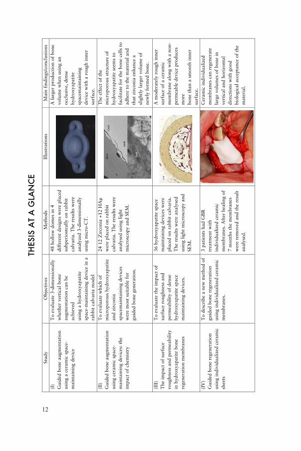
Regeneration of bone in the oral and maxillofacial region can be achieved with different techniques such as autologous bone grafts, bone substitutes and guided bone regeneration. Guided bone regeneration is defined as creating a space between the bone and its surrounding tissues, using a barrier that allows new bone to migrate into the space while preventing other cell types from interfering. The barrier material should be biocompatible, have suitable occlusive properties and be able to maintain the created space for bone regeneration. A wide range of different materials has been used.
The general aim was to evaluate a novel method of guided bone regeneration using designed ceramic space maintaining devices on animals and humans.
An experimental rabbit model was used in studies I, II and III. 60 different domes shaped as halfspheres were fixed with titanium screws to the skull bone of 30 rabbits. The domes had 5 different characteristics; 1) Dense hydroxyapatite with a moderately rough inner surface (HA rough), 2) Dense hydroxyapatite with a smooth inner surface (HA smooth), 3) Microporous hydroxyapatite with a moderately rough inner surface (HA μ), 4) Dense hydroxyapatite with a moderately rough inner surface and macroscopic holes (HA holes) and 5) Zirconia with a moderately rough inner surface (Zirconia). The domes were left to heal for 12 weeks before the animals were euthanized and the results were analysed with histomorphology and micro-CT.

14
The results revealed that Zirconia with a moderately rough inner surface produced the largest amount of newly formed bone although the results were difficult to interpret as the Zirconia domes were difficult to X-ray because of the very dense nature of the material.
In study IV, 3 patients had bone regeneration treatment with a Zirconia barrier. Patient 1 had posterior maxillary bone deficiency in a transverse direction. Patient 2 had anterior maxillary bone deficiency in a vertical and transverse direction. Patient 3 had posterior mandibular bone deficiency in a vertical direction. Based on individual digital models, Zirconia membranes corresponding to the amount of bone intended to be regenerated were designed and manufactured. The Zirconia membranes, were attached to the underlying bone with titanium screws and covered with the periosteum and mucosa. After a mean healing time of 7 months the membranes were removed and dental implants were installed.
The results showed new bone regeneration corresponding to the design of the space maintained. None of the patients had any major complications aside from normal postoperative discomfort. According to evaluation with CBCT all patients gained new bone in the desired and preoperatively planned region.

15
LIST OF PAPERS
I: Guided bone augmentation using a ceramic space-maintaining device.Anderud J, Jimbo R, Abrahamsson P, Isaksson SG, Adolfsson E, Malmström J, Kozai Y, Hallmer F, Wennerberg AOral Surg Oral Med Oral Pathol Oral Radiol. 2014 Nov;118(5):532-8
II: Guided bone augmentation using ceramic space-maintaining devices: the impact of chemistry.Anderud J, Jimbo R, Abrahamsson P, Isaksson S, Adolfsson E, Malmström J, Naito Y, Wennerberg AClin Cosmet Investig Dent 2015 Mar 12;7:45-53.
III: The impact of surface roughness and permeability in hydroxyapatite bone regeneration membranes.Anderud J, Abrahamsson P, Jimbo R, Adolfsson E, Malmström J, Wennerberg AClin Oral Implants Res. 2015 Oct 15. doi: 10.1111/clr.12717. (Epub ahead of print)
IV: Guided Bone Regeneration using using Individualized Ceramic Sheets.Malmström J, Anderud J, Abrahamsson P, Wälivaara DÅ, Isaksson SG, Adolfsson ESubmitted to International Journal of Oral and Maxillofacial Surgery 2015


TABLE OF CONTENTS
THESIS AT A GLANCE ................................................... 12
ABSTRACT .................................................................. 13
LIST OF PAPERS ........................................................... 15
INTRODUCTION .......................................................... 19Clinical problem ...................................................................19Bone ...................................................................................19Current methods of bone augmentation ...................................21
Autogenic grafts ..............................................................21Allogenic grafts ...............................................................21Xenogenic grafts ..............................................................22Alloplastic grafts ..............................................................22Other methods of augmentation .........................................22
Guided bone regeneration ....................................................24Experimental vertical guided bone regeneration .......................27Ceramics and Bio-ceramics ....................................................31
HYPOTHESIS ............................................................... 34
AIMS ......................................................................... 35
MATERIALS AND METHODS ........................................... 36Preoperative investigations ....................................................36Paper I-III .............................................................................37
Surface topography .........................................................39Animals and anaesthesia and surgery ................................39Preparation of samples .....................................................40Three-dimensional trabecular bone analysis study 1 .............41Histological analyses study 2 and 3 ...................................42Statistics .........................................................................42

Paper IV ..............................................................................43Surgery ..........................................................................44Measuring of gained bone volume and soft tissue topographic ....................................................................45Sample preparation .........................................................46
RESULTS ..................................................................... 47Surface measurements ...........................................................47Three dimensional results from micro-CT investigation ................47Histological results ...............................................................52Paper IV ..............................................................................57
Clinical outcome ..............................................................57Materials ........................................................................58Radiologic evaluation .......................................................59Histological outcomes .......................................................60
DISCUSSION ............................................................... 61Surface roughness ................................................................61Material porosity ..................................................................63Permeability .........................................................................64Zirconia ..............................................................................65Individualized membranes in a clinical situation .......................66
CONCLUSIONS ........................................................... 72
POPULÄRVETENSKAPLIG SAMMANFATTNING .................. 73
ACKNOWLEDGEMENTS ............................................... 75
REFERENCES ............................................................... 77
PAPERS I – IV ............................................................... 89

19
INTRODUCTION
Clinical problemAccording to Wolff’s law (Wolff, 1892) the bone in a healthy human being or animal will adapt to the loads under which it is placed. After loss of teeth the alveolar processes are no longer exposed to chewing forces. This may result in loss of dentoalveolar bone. Surgery, infection and other traumatic loss of alveolar bone can alter the contour and thereby the volume of the alveolar process. Large, critical sized bony defects will not regenerate to full volume spontaneously.
Treatment with dental implants is a common and well-documented method of supporting dental prosthesis. Dental implant treatment is considered to have a good long-term prognosis. To allow successful implantation, the jawbone needs sufficient quality and volume. Low quality or volume of bone can jeopardize the outcome of such treatment. Interference of anatomical structures such as the inferior alveolar nerve or the maxillary sinuses reduces the possibility to install dental implants.
If the bone volume is insufficient in volume it can be augmented by various methods. Several methods and techniques to increase bone volume and restore to the original shape of the bone are at hand. The gold standard is still considered to be transplantation of autogenous cortical bone as blocks or as particlutated bone.
BoneThe definition of bone is “a hard form of connective tissue composed of osteocytes and calcified collagenous intracellular substance arranged in thin plates” (Mosby, 2012). Its role is to support and
20
protect other tissues and organs, to be a reservoir for minerals and be involved in mineral homeostasis. The marrow of bone hosts the production of blood cells. In adults there are two types of bone tissue; compact, cortical bone and trabecular (cancellous, spongy) bone. Compact bone forms the outer layer and represents nearly 80% of the mass of the skeleton. It consists of several passages for diffusion of blood vessels and molecules. The blood vessels interconnect through perforated canals with vessels on the surface outside the bone where the periosteum is present. Trabecular bone is less dense than compact bone. It is built of plates and bars of bone along irregular cavities that contain red bone marrow. The trabeculae seem to be arranged randomly but are still able to provide maximum strength as they follow the lines of stress. There are two types of cells that contribute to bone homeostasis; osteoblasts and osteoclasts. These cells are important in the remodelling and repair of bone tissue. Osteoblasts are bone-forming cells that form osteoid by depositing extracellular matrix (collagen). The osteoid becomes mineralised by calcium withdrawn from blood. Some of the osteoblasts differentiate by entrapment into osteocytes. Osteoblasts can be found on the outer surface of bone and bone cavities. Osteoclasts are devoted to bone resorption as bone are involved in a continuous cycle of resorption and apposition. This process is called “bone turnover”.
The periosteum is a structure composed of 2 layers. The outer “fibrous” layer consists of fibroblasts, collagen and elastin along with a nerve and micro- vascular network. These components provide mechanical stability to the periosteum. The inner “cambium layer” is densly populated with cells that influence bone formation and bone repair (Allen et al., 2004). These features provide the periosteum with regenerative capacity. Because of its high vascularity, numerous endothelial pericytes can be found within the periosteum (Diaz-Flores et al., 1992). Pericytes are cells in physical contact with capillary endothelial cells. These cells can differentiate into several cell types, including osteoblasts (Reilly et al., 1998).
Bone fracture healing is initially characterized by an acute inflammatory response that includes blood clot formation, inflammatory cell migration and granular tissue formation. The inflammatory phase is believed to stimulate cell migration and proliferation of mesenchymal cells. Following the inflammation

21
period, mesenchymal cells aggregate at the repair site and differentiate into chondroblasts and osteoblasts. A collagen matrix is formed and mineralized. These events result in soft callus formation that fuses the 2 fracture margins together. With time, the soft callus will continue to ossify and woven bone will form. Eventually the healing bone will restore its original shape and structure (Colnot et al., 2012, Dimitriou et al., 2011).
The systematic recruitment of cells during bone repair is mainly dependent on skeletal progenitors from the bone marrow (Taguchi et al., 2005). These cells are brought to the injured site via blood vessels. As the systemic recruitment of skeletal progenitors during bone repair appears to be minimal, the recruitment within the local environment predominates. These skeletal progenitors may come from the bone marrow within the injured bone, from the surrounding periosteum and from soft tissues close to the bone (Einhorn, 1995). The source of cells and route of delivery are still being investigated experimentally (Colnot, 2011).
Current methods of bone augmentationAutogenic graftsAutogenous bone can be transplanted from intraoral approaches as well as from extra-oral approaches such as the iliac crest. Autogenous bone has proved to be both osteoinductive (contains living cells and proteins needed for new bone formation and healing) and osteoconductive (can act as a scaffold for bone forming cells and blood vessels). A disadvantage of autogenous bone grafting is morbidity of the donor site. This means often that a second wound needs to be made which can be uncomfortable for the patient. Prolonged surgical time and graft resorption are other bone grafting disadvantages.
Allogenic graftsAllogenic bone graft is bone harvested from another individual of the same species. Different allografts have been evaluated such as; Mineralized, demineralized, frozen or freeze-dried bone. Allografts are considered to be osteoconductive but most likely have less osteoinductive properties due to the absences of living cells. Allogenic bone grafts have routinely been used in orthopaedic surgery. Gapski

22
et al showed in 2006 that the use of allografts in sinuslift procedures can be successful (Gapski et al., 2006).
Xenogenic graftsXenogenic bone grafts originate from other species. Examples include bovine or porcine sources. To obtain immunological safety, necessary to prevent rejection of the graft, the proteins of this bone have been removed by different procedures. Hereby the osteoinductive effect is removed and the graft can only act as an osteoconductive scaffold (Calvo Guirado et al., 2013). The most frequently used and evaluated xenogenic bone graft is deproteinized bovine bone (Esposito et al., 2006).
Alloplastic graftsAlloplastic bone grafting materials are synthetically produced in many different compositions such as calcium-based ceramics, calcium- sulphates and bioactive glass. Different biological and mechanical characteristics have been reported such as dissolving rate and speed. In order to combine different properties, different materials are mixed together. For example Bone Ceramic® (Straumann, Basel, Switzerland), that consists of 60% hydroxyapatite and 40% β-tricaliumphosphate. The aim is hereby to let the more soluble β-tricaliumphosphate dissolve to give space to bone forming cells and blood vessels and let the harder hydroxyapatite act as a osteoconductive scaffold.
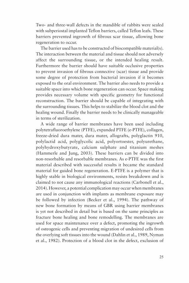
Other methods of augmentationIf the transplanted bone is particulated it needs to be stabilised by a mesh or membrane to stay in place. Titanium mesh has been used to stabilize various kinds of bone transplants. (Rasia-dal Polo et al., 2014).
This method has been described as successful both in horizontal and in vertical direction of new bone formation (Corinaldesi et al., 2009). However disadvantages have also been described such as penetration of the oral mucosa covering the titanium mesh with an unfavourable aesthetic result (von Arx et al., 1996). Between 0-50% of the meshes inserted have shown complications in the form of mucosal penetration. However von Arx states that this complication doesn’t necessarily jeopardize the functional outcome of the treatment.

23
Splitting the alveolar crest is another method where the maxillary alveolar crest is too narrow but has the height necessary for implant installation.
Hereby the alveolar crest is divided with instruments to allow implants to be installed between the buccal and palatal bone plates. This method is difficult to perform and has not been well evaluated in long term outcomes (Garcez-Filho et al., 2015) although favourable short term results (6 months) have been reported (Simion et al., 1992).
When the width of the maxilla is sufficient for implant installation but the height is not, the floor of the maxillary sinus along with the Schnederian membrane can be elevated, so called sinuslift. Hereby
Figure 2. Installaton of dental implants using split crest method
Figure 1. Guided bone regeneration using titanium mesh and autologous bone

24
the membrane is lifted with either the implant itself or as a two-stage procedure with a bone graft. The clinical effectiveness of sinuslift procedures is well described (Johansson et al., 2013, Johansson et al., 2010) with good survival rates of the inserted implants. According to the literature the most common complication with sinuslift procedures is perforation of the sinus membrane (Shlomi et al., 2004) which will adversely affect the implant survival rate.
To avoid damage to the inferior alveolar nerve in the mandible, short implants have been suggested in the premolar and molar region (Slotte et al., 2012). This seems to be a promising technique with similar survival as longer implants as long as the implants are connected with a dental bridge. The prosthodontic constructions on these implants may have an unfavourable ratio of height to implant length. This has not been shown to affect the survival or success rate, but it might cause unfavourable hygienic problem for the patient. Another method to avoid damage to the inferior alveolar nerve is nerve transposition. Using this technique the bone covering the inferior alveolar nerve is removed and the nerved is transferred laterally allowing insertion of standard dental implants without disturbing the nerve. Success rates varying from 76-100% have been reported (Khojasteh et al., 2015). However permanent dysfunction of the nerve or even mandibular fracture, although very rare, might occur postoperatively. Therefore this technique is rarely used today.
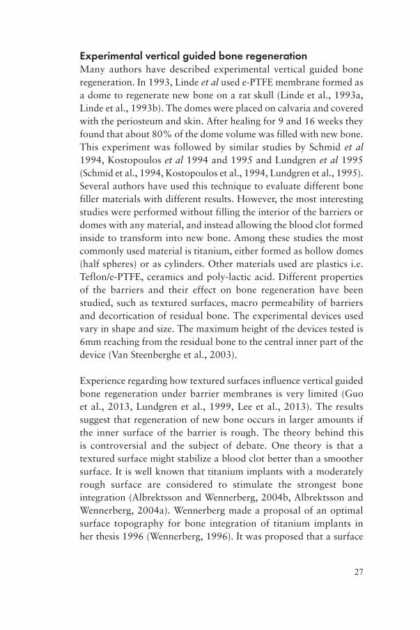
Guided bone regenerationGuided Bone Regeneration (GBR) is defined as creating a space between the bone surface and surrounding soft tissues, using a barrier that allows new bone to migrate into the space while preventing other cell types from interfering. Murray et al described the principle of physically sealing off an anatomical site for improved healing of bone in 1957 (Murray et al., 1957), followed by Hurley in 1959 who described when cell-occlusive membranes were used for spinal fusions (Hurley et al., 1959). In this publication it was stated that three things were necessary for regeneration of bone; 1) the presence of a blood clot, 2) preserved osteoblasts, and 3) contact with living tissue. Hurley and co-workers protected the blood clot with a plastic cage and the interior of the cage became filled with bone. Kahnberg evaluated this concept experimentally in 1979 (Kahnberg, 1979).
25
Two- and three-wall defects in the mandible of rabbits were sealed with subperiostal implanted Teflon barriers, called Teflon leafs. These barriers prevented ingrowth of fibrous scar tissue, allowing bone regeneration to occur.
The barrier used has to be constructed of biocompatible material(s). The interaction between the material and tissue should not adversely affect the surrounding tissue, or the intended healing result. Furthermore the barrier should have suitable occlusive properties to prevent invasion of fibrous connective (scar) tissue and provide some degree of protection from bacterial invasion if it becomes exposed to the oral environment. The barrier also needs to provide a suitable space into which bone regeneration can occur. Space making provides necessary volume with specific geometry for functional reconstruction. The barrier should be capable of integrating with the surrounding tissues. This helps to stabilize the blood clot and the healing wound. Finally the barrier needs to be clinically manageable in terms of sterilization.
A wide range of barrier membranes have been used including polytetrafluoroethylene (PTFE), expanded PTFE (e-PTFE), collagen, freeze-dried dura mater, dura mater, allografts, polyglactin 910, polylactid acid, polyglycolic acid, polyortoester, polyurethane, polyhydroxybutyrate, calcium sulphate and titanium meshes (Hammerle and Jung, 2003). These barriers can be divided into non-resorbable and resorbable membranes. As e-PTFE was the first material described with successful results it became the standard material for guided bone regeneration. E-PTFE is a polymer that is highly stable in biological environments, resists breakdown and is claimed to not cause any immunological reactions (Carbonell et al., 2014). However, a potential complication may occur when membranes are used in conjunction with implants as membrane exposure may be followed by infection (Becker et al., 1994). The pathway of new bone formation by means of GBR using barrier membranes is yet not described in detail but is based on the same principles as fracture bone healing and bone remodelling. The membranes are used for space maintenance over a defect, promoting the ingrowth of osteogenic cells and preventing migration of undesired cells from the overlying soft tissues into the wound (Dahlin et al., 1989, Nyman et al., 1982). Protection of a blood clot in the defect, exclusion of

26
gingival connective tissue and provision of a secluded space into which osteogenic cells from the bone can migrate are essential for a successful outcome. The sequence of bone healing in GBR is not only affected by invasion of non-osteogenic tissue, but even more so by the defect size and morphology. To be able to stabilize and thereby protect the blood clot, titanium reinforced e-PTFE membranes are available on the market. The titanium threads inserted in the membrane add more rigidity to the material that will further stabilize the blood clot and maintain the space created.
Figure 3. Guided bone regeneration using e-PTFE membrane
High-density polytetrafluoroethylene (hd-PTFE) membranes offer an interesting alternative to e-PTFE (Bartee, 1995, Bartee and Carr, 1995). Hd-PTFE is made of 100% PTFE, which is non-porous, non-expanded and non-permeable (Marouf and El-Guindi, 2000). The thickness of commercial available membranes ranges from 0,13-0,25 mm. Different sizes and textures are available. As the hd-PTFE membranes are totally occlusive they show minimal degree of infection and inflammation if exposed to the environment in the oral cavity (Krauser, 1996).
27
Experimental vertical guided bone regenerationMany authors have described experimental vertical guided bone regeneration. In 1993, Linde et al used e-PTFE membrane formed as a dome to regenerate new bone on a rat skull (Linde et al., 1993a, Linde et al., 1993b). The domes were placed on calvaria and covered with the periosteum and skin. After healing for 9 and 16 weeks they found that about 80% of the dome volume was filled with new bone. This experiment was followed by similar studies by Schmid et al 1994, Kostopoulos et al 1994 and 1995 and Lundgren et al 1995 (Schmid et al., 1994, Kostopoulos et al., 1994, Lundgren et al., 1995). Several authors have used this technique to evaluate different bone filler materials with different results. However, the most interesting studies were performed without filling the interior of the barriers or domes with any material, and instead allowing the blood clot formed inside to transform into new bone. Among these studies the most commonly used material is titanium, either formed as hollow domes (half spheres) or as cylinders. Other materials used are plastics i.e. Teflon/e-PTFE, ceramics and poly-lactic acid. Different properties of the barriers and their effect on bone regeneration have been studied, such as textured surfaces, macro permeability of barriers and decortication of residual bone. The experimental devices used vary in shape and size. The maximum height of the devices tested is 6mm reaching from the residual bone to the central inner part of the device (Van Steenberghe et al., 2003).
Experience regarding how textured surfaces influence vertical guided bone regeneration under barrier membranes is very limited (Guo et al., 2013, Lundgren et al., 1999, Lee et al., 2013). The results suggest that regeneration of new bone occurs in larger amounts if the inner surface of the barrier is rough. The theory behind this is controversial and the subject of debate. One theory is that a textured surface might stabilize a blood clot better than a smoother surface. It is well known that titanium implants with a moderately rough surface are considered to stimulate the strongest bone integration (Albrektsson and Wennerberg, 2004b, Albrektsson and Wennerberg, 2004a). Wennerberg made a proposal of an optimal surface topography for bone integration of titanium implants in her thesis 1996 (Wennerberg, 1996). It was proposed that a surface

28
with an average height deviation of 1,5 μm, an average wavelength of about 11,1 μm and a developed area ratio of 50% was optimal for integration. Lundgren et al experimented with surface roughness of barrier walls in 1999 (Lundgren et al., 1999). The experimental devices were made of titanium. Two different surfaces were tested. One machined used as control with a surface that was considered smooth and one grit blasted with titanium dioxide particles that were considered moderately rough. The test was performed on the skull of rabbits and the healing period was 12 weeks. In all of the devices, bone regeneration of various degrees had occurred and no significant differences in the morphological appearance or the amount of new bone could be seen. However, the mineralized bone did reach a higher level above the surface of the residual bone adjacent to the grit blasted titanium walls. Another attempt to evaluate different surfaces in GBR using titanium barriers were made by Guo et al 2013 (Guo et al., 2013). They used a machined surface to compare to a surface modified in an electrolytic solution that contained 3,5% glycophosphate disodium salt pentahydrate and 1,2% calcium acetate monohydrate by microarc oxidation (MAO). The devices were fixed to the calvaria of rabbits and left to heal for 4 weeks. No attempts to analyse the surface roughness of the devices were made. The results revealed that there were more bone produced under the MAO treated barriers and that this surface possessed osteoconductive potential. Lee et al tested rough surface hydrophobic titanium (SLA) domes and modified hydrophilic titanium (SLActive) domes in a comparison study (Lee et al., 2013). The study aimed to reveal any differences in bone regeneration according to GBR in rats with uncontrolled diabetes, controlled diabetes or healthy non-diabetic animals. Domes of each kind were place on the skull of rats and left to heal for 1 or 6 weeks. After 1 week there were no differences detected comparing SLA and SLActive domes among the different animal groups. After 6 weeks no significant differences could be shown between the 2 surface modifications but a trend towards larger bone production could be seen for the SLActive surface compared to the SLA surface.
In summary it might be suggested that a moderately rough, hydrophilic inner surface might enhance guided bone regeneration, although investigated on titanium surfaces.
29
The permeability of the barrier has been evaluated with different results. Some authors claim a better bone growth using a totally occlusive barrier compared to a barrier with permeability and some authors claim the opposite. Lundgren et al studied 7 different perforation sizes, 0, 10, 25, 50, 75, 100 and 300 μm, experimentally on the skulls of rats (Lundgren et al., 1998). The membranes were made of polyester. The outer skull bone plate was penetrated with 4 small holes, and the healing periods were 4, 8 and 12 weeks. The authors concluded that a totally occlusive barrier permitted bone regeneration just as well as the permeable barriers at 12 weeks. However, the perforated membranes showed a faster rate of bone regeneration. Yamada et al presented a paper where the occlusiveness of titanium caps was evaluated (Yamada et al., 2003). They compared 2 different titanium caps, one that was totally occlusive and one with several macroscopic holes. The caps were placed on rabbit calvaria and examined after 4 and 12 weeks of healing. No significant difference could be seen after 4 weeks but at 12 week there was significantly more bone in the caps that were totally occlusive. The authors concluded that an occlusive titanium barrier is preferable to a permeable one. Schmid et al stated 1994 that membrane permeability is unnecessary for guided bone regeneration (Schmid et al., 1994). Test cylinders made of titanium were attached to the calvaria of rabbits. Half of the cylinders were totally occlusive (in titanium and half of them had an open top that were covered with an e-PTFE membrane. After 8 months of healing it was concluded that membrane permeability is not needed for GBR. Another evaluation of the influence of permeability of barrier membranes was performed on rats by Zellin & Linde (Zellin and Linde, 1996). Three different e-PTFE membranes with different porosities were evaluated over 6, 12, 18 and 26 weeks of healing time. The results showed that the greatest amount of new bone achievable was obtained using the two most permeable membranes among the three investigated (Internodal distance of 20-25 and 100 μm) after 6 weeks and after 12 weeks there were no differences between the 3 tested membranes. Mardas et al compared a cell-permeable Teflon capsule to a cell-occlusive Teflon capsule both packed with demineralized bone matrix (Mardas et al., 2003). The healing periods were set at 30, 60 and 120 days. The results showed
30
similar amounts of bone formed in the two different capsules suggesting that permeability is unnecessary for bone formation with GBR. Another interesting experiment was performed by Ikeno et al (Ikeno et al., 2013). Four titanium cylinders were attached to the skulls of rabbits and filled with autologous iliac bone. The cylinders were randomly either uncovered or covered with; 1) titanium mesh, 2) e-PTFE membrane or 3) titanium plate. After 8 weeks the results were evaluated revieling that the titanium plate had the best result in terms of volume of new bone followed by the e-PTFE memrane, the titanium mesh and last the uncovered control.
In summary, the above-presented findings suggest that a certain degree of occlusiveness is crucial to GBR.
Decortication or perforation of the underlying cortical bone is stated to enhance the new bone formation under a barrier membrane. It is stated that the initial surgical trauma will start the bone healing cascade with blood clot formation, inflammation and so on. Rompen et al showed in 1999 that creating a wound, using an occlusive titanium barrier and filling it with peripheral blood on rat skull bone will stimulate the bone formation within the barrier chamber (Rompen et al., 1999). Majzoub et al tested occlusive titanium barriers on rabbit calvaria comparing cortical perforations to non-perforated bone (Majzoub et al., 1999). Healing periods were 10, 21, 42 and 60 days. The devices used were shaped as domes. The results showed that intramarrow penetration accelerated the rate of osseous neogenesis and increased bone fill and density inside the space created. These results were later confirmed by Min et al (Min et al., 2007) and Lee et al (Lee et al., 2014) also using dome shaped titanium barriers. Using titanium cylinders on rabbit calvaria with a healing period of 3 months. Lundgren et al showed the opposite results (Lundgren et al., 2000). They removed the whole outer cortex of the bone on one cylinder and left it intact on the other. After a healing time of 3 months no statistically differences could be seen. The conclusion was that decortication doesn’t result in more bone formation. Similar results have been presented by Slotte & Lundgren also using a cylinder shaped barrier (Slotte and Lundgren, 2002).

31
Ceramics and Bio-ceramicsThe word ceramic originates from the Greek word keramikos, which means “of pottery” or “for pottery”. Ceramic pottery several thousand years old has been found in archaeological excavations. In general, ceramics are inorganic materials with a combination of ionic and covalent bonds. They are composed of metal, non-metal or metalloid atoms and their crystallinity ranges from highly oriented to semi-crystalline and often completely amorphous. Ceramics are good thermal and electrical insulators due to the ionic and covalent bonds. Ceramics have, in general, a high melting temperature, high hardness, poor conductivity, high elastic moduli, chemical resistance and low ductility with known exceptions. The porosity of ceramic materials is defined as the void fraction and is a measure of the void (i.e. “empty”) spaces in a material. Porosity is the ratio of void volume to its total volume in %.
Bio-ceramics are ceramics used as the medical applications to restore lost function and aesthetics. Furthermore, its surrounding tissues must accept the material. The medical needs in an increasingly aging population have driven the further refinement of bio-ceramics. As a class of biomaterials, ceramics are generally used to repair or replace skeletal hard connective tissues damaged by disease or trauma. Their success depends on achieving stable attachment to these tissues. The mechanism of tissue attachment is related to the response at the material-tissue interface. Alumina, Zirconia, calcium phosphates and certain glasses and glass-ceramics are examples of bioceramics.
Aluminium oxide (Al2O3) dental implants osseointegrated well but were withdrawn from the market because of poor survival rate due to implant fracture. Zirconia (ZrO2) was and is being used as material for dental implants. Zirconium oxide (ZrO2) is considered to be a biostable ceramic. This means that the material undergoes very little chemical change during long-term exposure to in vivo environment. The material was introduced as an alternative to alumina for femoral ball heads in total hip replacements in the 1980s, for its higher fracture toughness, due to a transformation toughening mechanism operating in its microstructure (Piconi and Maccauro, 1999, Piconi et al., 1999). When a crack propagates, the adjacent grains transform

32
in structure, producing a 3-4% increase in volume, which creates a compressive stress of field that tends to close the crack.
Usually, biostable ceramics with very smooth surfaces form a fibrous layer approximately 1μm thick according to Hench 1998 (Hench, 1998). The thickness of the fibrous layer depends on several parameters such as the shape, size, surface morphology, porosity and composition of the implant as well as the type of the surrounding tissues. Studies have shown that coating titanium dental implants with zirconia favoured bone apposition compared to titanium dental implants with no coating (Sollazzo et al., 2008). When bone to implant contact of Zirconia dental implants was compared with bone to implant contact of titanium and alumina dental implants no significant differences were found (Dubruille et al., 1999). As mentioned before it is known that a certain degree of surface roughness is beneficial in bone apposition to the dental implant surface (Albrektsson and Wennerberg, 2004b, Albrektsson and Wennerberg, 2004a). Since shorter healing times are practiced more often in implant dentistry, smooth surfaced Zirconia dental implants appears to be a disadvantage (Ozkurt and Kazazoglu, 2011). Tests of biocompability in vitro have shown that laser-modified Zirconia exhibits better adhesion to osteoblasts than non treated surfaces due to better wettability characteristics (Kohal et al., 2002). Liagre et al claim that Zirconia does not provoke any inflammatory pathway (Liagre et al., 2002). Tests of biocompability have been conducted in vivo as well, where it was found that if Zirconia was implanted in the soft tissue it became encapsulated by a thin layer of fibrous tissue (Ichikawa et al., 1992). Overall the soft tissue response to Zirconia seems to be comparable to the soft tissue response of titanium (van Brakel et al., 2012).
Hydroxyapatite (HA) is a naturally occurring calcium-phosphate mineral with a Ca/P ratio of 1,67. The formula is Ca5(PO4)3(OH) but is usually written Ca10(PO4)6(OH)2. The hydroxyl ion can be replaced with other ions like fluoride, chloride or carbonate, which is a unique characteristic of the apatite structure. HA usually crystallises in hexagonal patterns. Incorporation of different ions and vacancies in the crystal structure affords HA different properties in terms of crystallinity, thermal stability and solubility. It has also been

33
found to form the inorganic components of hard tissues, such as bone and tooth substances, in man and animals. These materials are of great interest because they are widely applied as biomaterials in bone fillers, bone engineering scaffolds, bioactive coatings, soft tissue repairs and drug loading/delivering systems. A great advantage of HA is its ability to form apatite bone-like layer on its surface to which bone bonds in vivo and in vitro conditions (Oyane et al., 2003).
Hydroxyapatite has been used as a coating material on dental implants (Xuereb et al., 2015). It has been reported that hydroxyapatite coating of dental titanium implants shows enhanced bone bonding and accelerated new bone formation (Lin et al., 2009). Studies have shown that titanium dental implants coated with hydroxyapatite have better bone-to-implant contact and increased removal torque values when compared to uncoated control implants (Svanborg et al., 2011). This suggests that hydroxyapatite is a biomaterial with high acceptance of the surrounding tissues. For the past three decades several hydroxyapatite products have been presented as fillers for bony defects. It has been manufactured in a variety of forms including granules and porous blocks. Although hydroxyapatite accounts for nearly 70% of the mineral content of bone, the hydroxyapatite in the body exists in a substituted form. Carbonate, silicates and other ions may replace the hydroxyl or phosphate groups of the apatite structure. This is why researchers have tried to produce alginate- (Chae et al., 2013), strontium- (Landi et al., 2007), carbonate- and magnesium- (TenHuisen and Brown, 1997) substituted synthetic hydroxyapatite to produce hydroxyapatite that more closely resembles the mineral content of native bone in order to enhance bioactivity and osteoconduction.

34
HYPOTHESIS
• Hydroxyapatite is suitable as a biomaterial for creating a space between the bone cortex and the periosteum to enhance new bone regeneration.
• Zirconia is suitable as a biomaterial for creating a space between the bone cortex and the periosteum to enhance new bone regeneration.
• A moderately rough inner surface of a ceramic space maintaining device will enhance the quantity and quality of newly formed bone.
• A porous ceramic material of a space maintaining device will enhance the quantity and quality of newly formed bone.
• A permeable ceramic space maintaining device will enhance the quantity and quality of newly formed bone.
35
AIMS
The general aim was to evaluate a novel method of guided bone regeneration using designed ceramic space maintaining devices on animals and humans.
The specific aims were;
• To evaluate hydroxyapatite and Zirconia ceramics as a barrier for guided bone regeneration related to different factors to optimize the outcome.
• To evaluate the effect of increased inner surface roughness on guided bone regeneration.
• To evaluate the effect of increased material porosity on guided bone regeneration.
• To evaluate the effect of increased permeability on guided bone regeneration.
• To evaluate the bone response of a ceramic barrier on a material-tissue interface level.
• To evaluate the bone response of a ceramic space maintaining device on a macroscopic level.

36
MATERIALS AND METHODS
Preoperative investigationsAll ceramic components, paper I-IV, were made of the same batch of hydroxyapatite powder (Plasma Biotal Limited, Derbyshire, United Kingdom) and Zirconia powder (TZ3YSBE, Tosoh Corporation, Tokyo, Japan). At the start of the trials, there was no previously reported method of how to manufacture rough or smooth surfaces of ceramic materials. A series of surface roughness measurements on discs of dense hydroxyapatite, micro porous hydroxyapatite and zirconia were done in order to obtain similar surface characteristics on different materials. The discs were 10mm in diameter. Altogether, 34 different kinds of discs were produced. Three of each variety were produced, i.e. a total of 102 discs. Each disc was characterized using an interferometer (MicroXam; ADE Phase Shift Technology, Inc., Tucson, Arizona, USA). Each disc was measured at 5 positions (one at the centre and four at the outer radius). The parametric calculation was performed after form errors and waviness were removed with a 50 μm X 50 μm Gaussian filter. The following 3-D parameters were selected: Sa (μm) = the arithmetic average height deviation from a mean plane and Sdr (%) = the developed surface ratio. From the results different techniques were chosen to manufacture the surfaces of the devices used in study I-IV.

37
Paper I-III60 hollow domes were manufactured.
They were shaped as half spheres with an inner diameter of 6 mm and an outer diameter of 8 mm. Twelve of them were made of dense hydroxyapatite with a rough inner surface and a mean Sa of 1,1 μm (HA rough). Twelve were manufactured with the same properties as HA rough but had a smooth inner surface (HA smooth) with a mean Sa of 0,3 μm. Twelve were made of microporous hydroxyapatite with a rough inner surface with a mean Sa of 1,1 μm (HA μ). Twelve were manufactured with the same properties as HA rough including the rough inner surface but with macroscopic holes on top (HA holes). Twelve were made of Zirconia with similar properties as HA rough (Zirconia)
Figure 4. The different space maintaining devices used in study I-III
38
Table 1. The different space maintaining devices used in paper I - III
Material Inner surface Occlusiveness Material porosity
HA rough
Hydroxyapatite Moderately rough (Sa 1,1 ± 0,1 µm)(Sdr 66,0 ± 1,4%)
None No
HA smooth
Hydroxyapatite Smooth (Sa 0,3 ± 0,1 µm)(Sdr 16,0 ± 0,9%)
None No
HA µ Microporous hydroxyapatie
Moderately rough (Sa 1,1 ± 0,2 µm)
(Sdr85,3%± 1,6%)
None 50%
HA holes
Hydroxyapatite Moderately rough (Sa 1,1 ± 0,1 µm)(Sdr 66,0 ± 1,4%)
Macroscopic No
Zirconia Zirconia Moderately rough (Sa 1,1 ± 0,1 µm)(Sdr 29,8 ± 1,9%)
None No
The hydroxyapatite components were produced from a hydroxyapatite powder (Plasma Biotal Limited, Derbyshire, United Kingdom), which was further processed with water and a dispersant (polycyclic acid) to prepare a ceramic suspension by ball milling. The suspension was sprayed into liquid nitrogen (freeze granulation) and the frozen granules were freeze-dried. Ceramic green bodies of hydroxyapatite and Zirconia (TZ3YSBE, Tosoh Corporation, Tokyo, Japan) were prepared by isostatic compaction of the hydroxyapatite and zirconia granules at a pressure of 300MPa.
Based on the CAD model of the components, a five axis CNC-machine (Dechel-Maho, DMU 60, Deckel Maho, Pfronten, Germany) was used to form the green body into the desired macroscopic shape. To ensure that all components, despite different characteristics had the same sample size even when different sintering temperature were used during the fabrication process, the sintering shrinkage was measured at different sintering temperatures and different values were used to compensate for the enlargement of the CAD model depending on the fabrication process.
The smooth surface of the dense hydroxyapatite was obtained by sintering the machined components at 1250°C for 2 hours. The

39
dense and rough surface was obtained by presintering the machined material at 900°C, blasting with 110μm alumina particles at 1 bar followed by sintering at 1250°C for 2 hours. The rough micro porous surface was produced by presintering the machined material at 900°C for 2 hours, blasted with 110μm alumina particles at 1 bar and an additional heat treatment at 900°C. The surface on Zirconia was produced by presintering at 900°C. The inner surface was blasted in the presintered state with 110μm alumina particles at 1 bar, and sintered at 1450°C for 2 hours. Some Alumina particles could be detected on the blasted HA surfaces (HA rough, HA holes, HA μ). An image analysis from SEM was made and it was estimated that 0,2% of the μ HA surface was covered with Alumina particles and 0,5% of the Ha rough- and HA holes surfaces were covered with Alumina particles. No Alumina particles could be seen on the Zirconia surface.
Surface topographyThe topography of the inner surfaces of the spacemaintaining devices was characterized using an interferometer (MicroXam; ADE Phase Shift Technology, Inc., Tucson, AZ). Three devices per group were randomly selected for analysis. Each device was measured at 5 positions (one at the most inner top and four at the inner radius areas). The parametric calculation was performed after form errors and waviness were removed with a 50 μm X 50 μm Gaussian filter. The following 3-D parameters were selected: Sa (μm) = the arithmetic average height deviation from a mean plane and Sdr (%) = the developed surface ratio.
Animals and anaesthesia and surgeryThe study was approved by the Malmö/Lund regional animal ethics committee (approval no. M 314-10). Thirty lop-eared rabbits of mixed sex with a mean body weight of 4,15 kg were used in study I-III. Two implants were inserted on each rabbit’s calvaria according to a randomized scheme. Before surgery, the rabbits’ skulls were shaved and disinfected with chlorhexidine (5mg/ml, Pharmacia AB, Stockholm, Sweden). The animals were anaesthetized by intramuscular injection of a mixture of 0.15 ml kg-1 medetomidine (1 mg ml-1 Dormitor; Orion Pharma, Sollentuna, Sweden) and 0.35 ml kg-1 ketamine hydrochloride (50 mg ml-1 Ketalar; Pfizer AB,

40
Sollentuna, Sweden). Lidocaine hydrochloride (Xylocaine 10mg/ml; AstraZeneca AB, Södertälje, Sweden) was administered as local anaesthetic at each insertion site at a dose of 0,5 ml. Sterile conditions were maintained during surgical procedure. An incision through the skin and periosteum was made along the midsagittal line on top of the skull measuring approximately 2 cm. The periosteum was carefully resected from the bone. A trephine bur was used to make one slit in the cortical bone on each side of the midline to a depth of 0,5mm. The area was constantly irrigated with saline solution. Two space maintaining devices were fitted to the bone slits and fixated with 2 titanium screws each (De Puy Synthes 04.503.203 Ø1.5 mm 3mm self drilling, De Puy Synthes, Zuchvil, Switzerland). The periosteum was then closed in position using Vicryl 4-0 continuous stitching before the skin was closed in the same fashion. Postoperatively, buprenorphine hydrochloride (0.5 ml Temgesic; Reckitt Benckiser, Slough, UK) was administered as an analgesic for 3 days. No antibiotics were used.
Figure 5. One ceramic space maintaining device attached to the skull of a rabbit
Preparation of samplesAt twelve weeks postoperatively, the rabbits were euthanised with an overdose (60 mg ml 1) of sodium pentobarbital (Apoteksbolaget AB, Stockholm, Sweden). Samples of the space maintaining devices including the attached underlying bone were retrieved and placed in 4% formaldehyde for 24 h, after which they were placed in 70% ethanol. All samples were processed for undecalcified ground

41
sectioning according to the technique described by Donath (Donath, 1985) In brief, after a series of dehydrations and infiltrations in resin, the samples were embedded in light-curing resin (Technovit 7200 VLC; Heraeus Kulzer Wehrheim, Germany). At this stage, 48 of the domes were analyzed with micro CT. The embedded specimens were divided into two identical blocks. Thereafter, one central cut and ground section from one of the two blocks was prepared from each sample by using Exakt sawing and grinding equipment. The sections were ground to a final thickness of approximately 25 μm and stained with toluidine blue. It was extremely difficult to make the sample any thinner because the ceramic peeled off if ground further. The second block was prepared for scanning electron microscopy (SEM). The blocks were ground and polished with a fine-grain abrasive paper (SiC-Paper, grit 4000, Struers A/S, Denmark) that contained particles of 9 μm, 3 μm and 1 μm. They were then coated with a thin conductive carbon layer with a thickness of approximately 1000Å by vacuum evaporation (AGAR SEM Carbon Coater, Germany). These specimens were examined with SEM (JEOL JXA-8600; JEOL, Tokyo, Japan). The interface between the ceramic material and newly formed bone was evaluated.
Three-dimensional trabecular bone analysis study 1A micro CT equipped with a micro focus X-ray tube (focus size 868 mm, MCT-100MF; Hitachi Medical Corporation, Tokyo, Japan) produced a 3D image of each spacemaintaining device from 201 image slices. The tube voltage, tube current, magnification and voxel size were 70 kV, 100 μA, x 6, and 21.0 x 21.0 x 21.0 μm, respectively. Trabecular structure analysis software (TRI/3D BON; Ratoc System Engineering Co. Ltd, Tokyo, Japan) calculated the 3D trabecular structure parameters from the image, information from 110 slices from the very top of the space maintaining device and down. From binary images, the trabecular structure parameters were measured, including tissue volume (TV), bone volume (BV), bone surface per bone volume (BS/ BV), and trabecular thickness (Tb-Th). The star volume of marrow space (V*m.space) and the star volume of trabeculae (V*tr) were measured by star volume analysis. The trabecular connectivity was measured using node-strut analysis described by Garrahan et al. (Garrahan et al., 1990). Nodes (Nd;

42
connective point of three or more trabeculae), termini (Tm; terminal of trabeculae) and cortex (Ct; connective point of trabeculae and cortical bone) was identified by node-strut analysis, and the parameter of the number (n) of Nd per tissue volume (nNd/TV), the number of Tm per tissue volume (nTm/TV), the number of Ct per tissue volume (nCt/TV), and the total strut length per tissue volume (TSL/TV) was obtained by node-strut analysis.
Histological analyses study 2 and 3Histological evaluations were performed using light microscopy (Eclipse ME600; Nikon, Japan). The material surface on the inside of the domes was photographed using 40 x magnifications. The pictures were stitched together using Adobe Photoshop Elements (Adobe Systems, San Jose, USA) creating a panoramic image. The bone to material contact was measured in percentage using the free software ImageJ (National Institutes of Health, Bethesda, Maryland, USA). First the total surface was measured from where the newly formed bone began and ended. Secondly the amount of bone in contact with the material surface was measured and calculated and this figure was divided from the total surface giving the percentage of bone in contact with the surface.
The bone volume was measured as percentage of new bone formed related to the total volume using the same method as described above with the magnification of the objective x10. No stitching of images was needed. Only the volume of newly formed bone was measured.
Samples with more than 15 measuring points were measured twice as a control of the measuring technique. If the measures differed more than 1% the samples were measured a third time and an average of the measurements were accepted as the true result. This had to be done with approximately one third (18) of the samples.
StatisticsDifferences in the mean values between the control group (n = 12) and the three experimental groups (n = 12/group) were calculated for each parameter to obtain the median percentages. The means for each parameter were compared using Mann-Whitney U test. Values of p>0.05 were considered statistically significant. IBM SPSS Statistics was used for all analyses.
43
Paper IVThree patients, two female and one male, volunteered to be included and were consented for surgery. They all had inadequate bone volumes for optimal installation of implants and required bone augmentation prior to implant surgery.
Patient 1 had posterior maxillary bone deficiency in the transverse direction.
Patient 2 had anterior maxillary bone deficiency in the vertical and transverse direction.
Patient 3 had posterior mandibular bone deficiency in the vertical direction.
All patients were examined preoperatively with one panoramic image and a cone-beam computed tomography, volume 60x60, 360 degrees, (3D Accuitomo 170, J Morita, Tokyo, Japan) through the area of interest.
The CT images of each patient were used to build a plastic model of the jaw. On this model a wax was moulded to represent the bone that was supposed to be regenerated. This was covered with a sheet of wax that was formed to represent a membrane or barrier. Based on this wax-model, a five axis CNC-machine (Dechel-Maho, DMU 60, Deckel Maho, Pfronten, Germany) was used to mill a cold isostatically pressed and presintered green body of Zirconia (TZ3YSBE, Tosoh Corporation, Tokyo, Japan) to the desired macroscopic shape.
Figure 6. Manufacturing process of an individualized ceramic membrane

44
The size of the machined membrane was enlarged in order to compensate for the sintering shrinkage performed at 1450°C. The inner surface was blasted in the presintered state with 110μm alumina particles at 1 bar, corresponding to the manufacturing method of the space maintaining devices used in study II. The outer surface was characterized with the same interferometer method used in studies I-III. The inner surfaces were impossible to measure as the interferometer collided with the flanks of the material. After manufacturing the devices were cleaned in ultrasonic bath of 70% alcohol for 15 min followed by a brief heating to 1200 °C. They were again cleaned in an ultrasonic bath for 3x15min using 90% alcohol. Thereafter they were put in an autoclave (Getinge Quadro Avanti, Getinge, Sweden) and sterilized using a standard program (134 °C/3min).
SurgeryThe intraoral area was initially cleaned with Chlorhexidine 1mg/ml (Hexident®, Meda AB, Solna, Sweden). Local anaesthetic, 20 mg/ml Lidocaine + 12,5 μg/ml epinephrine (Xylocain Dental Adrenalin, Dentsply Limited, Surrey, Great Britain) was administered. A mucoperiosal flap of full thickness was raised using crestal incision and vertical releasing incisions on the buccal aspect of the surgical site. The cortical plate of the defect was perforated with a small roundbur to provoke bleeding. Fixation of the individualized ceramic membranes where done with Synthes® (De Puy Synthes, Johnson & Johnson, New Brunswick, New Jersey, USA) fracture screws. No adaptation was needed and membranes were stable using only 1 screw. Membranes were cleaned with 3% hydrogen peroxide prior to soft tissue closure.
In patient 3, bone chips were harvested from the mandibular ramus on the left side with a Safescraper Twist (Safescraper®, META, Reggio Emilia, Italy) and placed between the underlying bone and the ceramic barrier. The membrane on the right side was placed without any bone chips.
After fixation of the ceramic barriers, the mucoperiosteal flaps were coronally repositioned with significant apical undermining. Split-thickness periosteal release incisions were completed, to aid primary tension-free closure using Vicryl ® 4-0 (Ethicon Inc., Johnson & Johnson, New Brunswick, New Jersey, USA). All patients received antibiotic (phenoxymethylpenicillin) 1g three times per day for 1 week postoperatively.

45
After a mean of 7 months of healing, the augmented sites were reopened using the similar surgical technique. The ceramic barriers and fixations screws were removed. After that installation of dental implants followed. Before the implants were installed the patient were again examined with one panoramic image and a cone-beam computed tomography, volume 60x60, 360 degrees, (3D Accuitomo 170, J Morita) over the area of interest. Samples of the newly grown bone and covering soft tissues were taken and sent for histological preparation and analysis.
Measuring of gained bone volume and soft tissue topographicData from pre- and postoperative cone-beam-tomography investi-gations were analysed in 3D software (SimPlant Pro, Materialise Dental, Belgium). The amount of gained bone volume in 3 dimensions postoperatively was compared to the same area from the preoperative investigation by an overlay function.
The intra oral soft-tissue profile of the alveolar defect in patient 2 was documented using a three-dimensional metering equipment
Figure 7. The individualized ceramic membrane attached to mandible on patient 3

46
(PRIMOS® optical 3D (Primos 5.7 and Primos Body 6.6), GFMess-technik Germany). The measurements were performed at two different occasions: 1) preoperatively and 2) after removal of the membrane. The difference was measured in a 90 degree cut from the alveolar crest.
Sample preparationSamples of the regenerated bone were retrieved using a trephine bur and placed in 4% formaldehyde for 24 h, after which they were placed in 70% ethanol. All samples were processed for undecalcified ground sectioning according to the same technique used in paper I-III. The sections were ground to a final thickness of approximately 40 μm and stained with toluidine blue. Histological evaluations were performed using a light microscope (Eclipse ME600; Nikon, Japan).
Samples of the soft tissue found between the ceramic membranes and the newly formed bone were retrieved and placed in 4% formaldehyde. The samples were processed for soft tissue sectioning. After processing, the tissues were embedded in paraffin and serial sections 3μm thick were prepared and stained with Hematoxylin-Eosin and van Gieson. Histological evaluations were performed using a light microscope (Leica DMD 108, Leica microsystems, Wetzlar, Germany).

47
RESULTS
Surface measurementsThe interferometry measurements of the inner surface of the space maintainers used in paper I – III revealed statistically significant differences in roughness parameters between the ceramic space maintaining devices considered smooth (HA smooth) and the other four groups that were considered rough (HA rough, HA μ, HA holes, Zirconia). Only a small variation within the samples was found for the Sa values. Based on the average Sa value of 0.3 μm for HA smooth and Sa value of 1,1 μm for the other groups, the space maintainers can be categorized as smooth and moderately rough. The microporous hydroxyapatite surface had an Sdr of 85,3% while the Zirconia surface had an Sdr of 29,8%. For exact measured values see table 1.
Three dimensional results from micro-CT investigationAll hydroxyapatite space maintaining devices (HA rough, HA smooth, HA μ and HA holes) were possible to examine with micro-CT. The space maintaining devices made of Zirconia were too dense. The 3 dimensional trabecular bone microarchitecture on the 4 different hydroxyapatite ceramic space maintaining devices is described in table 2 a-c. The bone volume (BV) values for each hydroxyapatite space maintaining device are presented in diagram 1.
The differences in BV between HA rough and HA smooth were not significant. Nonetheless, HA rough produced a slightly larger median bone volume, BV of 18,09 mm3 compared to 16,22 mm3 for HA smooth (p=0,133). The median number of trabecular connective points was also higher for HA rough with 1072 (max 1407/min 488) compared to 868 (max 1440/ min 413) for HA smooth (p=0,356).

48
The values of BV, nNd/TV, nTm/TV, and nCt/TV were higher for HA rough than HA μ with a significant difference (p < 0,05). BV was 18,09 mm3 for HA rough compared to 11,67 mm3 for HA μ (p=0,011). Tb-Th was lower in HA rough than HA μ again with significant difference. These findings suggest that a larger porosity of a ceramic space maintaining device significantly decrease the volume of new bone produced and the connectivity of the internal new trabeculae produced.
When comparing HA rough and HA holes, dense hydroxyapatite with a rough inner surface and macroscopic holes, Tb-Th was higher in HA holes than HA rough (p=0,001) and nNd/TV was higher in HA rough than HA holes (p=0,050) with significant difference. Similar to the findings for HA μ where there was a larger bone volume BV produced by the control HA rough than by HA holes with 18,09 mm3 compared to 15,86 mm3 as median values, without significant difference (p=0,204). These findings suggest that a higher permeability of a ceramic spacemaintaining device is negative in terms of produced bone volume although it seems to promote thicker trabeculae.
0
5
10
15
20
25
30
Three dimensional bone volume
HA rough
HA smooth
HA micro
HA holes
Diagram 1. Showing median, max and min of three dimensional bone volume paper I
Figure 8. Three dimensional reconstruction of micro-CT investigation

49
Tabl
e 2
a. T
hree
dim
ensi
onal
trab
ecul
ar b
one
struc
ture
par
amet
ers
com
paris
on b
etw
een
HA
roug
h an
d H
A s
moo
th
HA
rou
gh (c
ontr
ol)
HA
sm
ooth
Med
ian
max
min
Med
ian
max
m
in
P-va
lue
Trab
ecul
ar s
truc
ture
BV(m
m3 )
18,0
927
,58
13,2
916
,22
21,8
28,
230,
133
Tb-T
h(µ
m)
69,3
081
,60
58,4
465
,63
79,0
554
,51
0,38
6
Star
vol
ume
anal
ysis
V*m
spa
ce(m
m3 )
1,32
2,51
0,89
1,39
2,57
1,14
0,45
3
V*tr
(mm
3 )0,
164,
270,
020,
162,
900,
021,
000
Nod
e-st
rut a
naly
sis
nNd/
TV(1
mm
-3 )10
7214
0748
886
814
4041
30,
356
nTm
/TV
(1 m
m-3 )
733
1016
486
775
938
677
0,29
9
nCt/
TV(1
mm
-3 )14
1425
8486
713
7721
6760
50,
341

50
Tabl
e 2
b. T
hree
dim
ensi
onal
trab
ecul
ar b
one
struc
ture
par
amet
ers
com
paris
on b
etw
een
HA
roug
h an
d H
A µ
HA
rou
gh (c
ontr
ol)
HA
µ
Med
ian
max
min
Med
ian
max
m
inP-
valu
e
Trab
ecul
ar s
truc
ture
BV(m
m3 )
18,0
9*27
,58
13,2
911
,67
28,4
17,
210,
011
Tb-T
h(µ
m)
69,3
0*81
,60
58,4
479
,36
94,8
358
,27
0,05
0
Star
vol
ume
anal
ysis
V*m
spa
ce(m
m3 )
1,32
2,51
0,89
1,74
3,59
0,53
0,16
6
V*tr
(mm
3 )0,
164,
270,
020,
093,
040,
010,
273
Nod
e-st
rut a
naly
sis
nNd/
TV(1
mm
-3 )10
72*
1407
488
448
1307
161
0,00
1
nTm
/TV
(1 m
m-3 )
733*
1016
486
546
898
129
0,03
3
nCt/
TV(1
mm
-3 )14
14*
2584
867
668
1706
221
0,00
1

51
Tabl
e 2
c. T
hree
dim
ensi
onal
trab
ecul
ar b
one
struc
ture
par
amet
ers
com
paris
on b
etw
een
HA
roug
h an
d H
A h
oles
HA
rou
gh (c
ontr
ol)
HA
hol
es
Med
ian
max
min
Med
ian
max
m
inP-
valu
e
Trab
ecul
ar s
truc
ture
BV(m
m3 )
18,0
927
,58
13,2
915
,86
25,2
611
,04
0,20
4
Tb-T
h(µ
m)
69,3
0*81
,60
58,4
477
,63
121,
6170
,64
0,00
0
Star
vol
ume
anal
ysis
V*m
spa
ce(m
m3 )
1,32
2,51
0,89
1,66
2,59
1,03
0,24
8
V*tr
(mm
3 )0,
164,
270,
020,
181,
930,
040,
908
Nod
e-st
rut a
naly
sis
nNd/
TV(1
mm
-3 )10
72*
1407
488
708
1255
425
0,05
0
nTm
/TV
(1 m
m-3 )
733
1016
486
683
1086
447
0,27
3
nCt/
TV(1
mm
-3 )14
1425
8486
714
9325
2295
60,
773

52
Histological results Cross-sections from the histological investigation showed newly formed bone, residual bone and the ceramic space maintaining device. The newly formed bone consisted of mineralized trabecular bone and large marrow spaces with a fat rich content. Newly formed bone was seen in close contact with the ceramic material of all barriers. In the cross-sections of HA holes, soft tissue ingrowth through the perforations was observed. The morphological results from light microscopy are represented in figure 9.
Figure 9. Examples of histological results from study I-III
Two different morphologies of bone to material contact were observed. At the surface of the microporous hydroxyapatite a close contact between material and new bone was seen. At the surface of Zirconia, intermediate zones of about 10 μm widths from the material without bone contact could be seen. The bone adjacent to these zones had the same micro morphology as the inner surface of the Zirconia wall. This fact presumably indicates that the bone had grown in contact with the material but had been torn away from the Zirconia in the grinding process. There was no presence of a soft tissue lining between the Zirconia wall and the new bone. When observing the morphology of bone growth one could notice that the bone climbs the walls of the microporous hydroxyapatite very well, whereas the Zirconia dome predominantly seems to be invaded by bone from the residual tissues.
The histological results of the different ceramic space maintaining devices are described in diagram 2 and 3.

53
0
20
40
60
80
100
120
Bone to material contact
HA rough
HA smooth
HA micro
HA holes
Zirconia
Diagram 2. Showing median, max and min of bone to material contact in study I-III
Diagram 3. Showing median, max and min of bone volume in paper I-III
13,5
10,9 11,6 12,1
16
Bone volume HA rough HA smooth HA micro
HA holes Zirconia
13,5
10,9 11,6 12,1
16
Bone volume HA rough HA smooth HA micro
HA holes Zirconia

54
Tabl
e 3.
Tw
o di
men
sion
al h
isto
logi
cal b
one
struc
ture
s. C
ompa
rison
bet
wee
n H
A ro
ugh
(con
trol)
and
HA
sm
ooth
, HA
µ, H
A h
oles
, Zirc
onia
.
Bone
to m
ater
ial c
onta
ct (%
)Bo
ne v
olum
e (%
)
Med
ian
max
min
P-va
lue
Med
ian
max
m
in
P-va
lue
HA
roug
h80
,585
,768
,1(C
ontro
l)13
,5
20,0
11,4
(Con
trol)
HA
sm
ooth
84,1
94,7
67,4
0,08
310
,917
,19,
50,
004
HA
µ88
,1
96,7
76,3
0,00
111
,616
,76,
60,
13
HA
hol
es38
,451
,922
,0<
0,00
112
,115
,26,
90,
14
Zirc
onia
61,8
76,7
25,3
0,00
116
,0
18,9
12,2
0,09

55
The differences between HA rough and HA smooth were very small. The moderately rough surfaced hydroxyapatite produced a lesser median bone volume of 80,5 % (max 85,7/min 68,1) compared to 84,1% (max 94,7/min 67,4) for HA smooth (p=0,083). The median percentage of bone to material contact was higher for HA rough with 13,5% (max 20,0/min 11,4) compared to 10,9% (max 17,1/ min 9,5) for HA smooth with significance (p=0,004).
The differences between HA rough and HA holes regarding bone to material contact were statistically significant. The rougher surfaced occlusive hydroxyapatite produced a larger median bone to material contact of 80,5 % (max 85,7/min 68,1) compared to 34,8% (max 51,9/min 22,0) for hydroxyapatite with macroscopic holes (p<0,0001). The median percentage of bone volume was higher for HA rough with 13,5% (max 20,0/min 11,4) compared to 12,1% (max 15,2/ min 6,9) for HA holes. This difference was not significant (p=0,14). These findings suggest that an occlusive design produces more bone to material contact than a ceramic space maintaining device with macroscopic holes.
When comparing Zirconia with HA rough the difference in bone-to-material contact was significant. Zirconia produced a smaller median bone-to-material contact of 61,8% (max 76,7/min 25,3) compared to 80,5 % (max 85,7/min 68,1) for HA rough (p<0,001). The median percentage of bone volume was lower for HA rough; 13,5% (max 20,0/min 11,4) compared to Zirconia 16,0% (max 18,9/min 12,2) without statistical significance (p=0,09).
The differences between HA rough and HA μ in terms of bone-to-material contact were statistically significant. HA rough produced a lesser bone-to-material contact of 80,5 % (max 85,7/min 68,1) compared to 88,1% (max 96,7/min 76,3) for HA μ (p=0,001). No significant differences could be shown when comparing the bone volume between HA rough and HA μ.
Differences between the samples could be observed in the SEM investigation. There was ingrowth of newly formed bone in the micropores of the moderately rough surface represented by group 1 and 3. The smoother surface represented by group 2 showed a space between the newly formed bone and the surface of the dome in the SEM. Close contact without intervening soft tissue was seen between
56
bone and the inner surface of the microporous hydroxyapatite using scanning electron microscopy. Presence of bone inside the micropores was demonstrated as well (see Figure 10b). A non-union area at the immediate interface between Zirconia and bone was detected by SEM. This could not be detected at the interface between microporous hydroxyapatite. Cracks in the material were detected in the microporous hydroxyapatite but not in the Zirconia.
Figure 10b. SEM investigation of HA micro and Zirconia
Figure 10a. Results from SEM investigation of HA rough and HA smooth

57
Paper IVClinical outcomeNone of the three patients had any postoperative complications from the installation of ceramic barriers or the implant surgery. All surgical sites healed without signs of infection or penetration of the barriers. Only minor postoperative initial discomforts such as slight swelling and soreness were reported by the patients. At implant installation it was observed that the mucoperiosteal flap was very easy to dissect from the surface of the ceramic membrane, without damaging the tissues. The membrane itself was easily removed after the fixation-screws had been removed. No attachment of the surrounding tissues to the ceramic barrier could be detected. Beneath the ceramic membrane a layer of soft tissue (approximately 0,5 – 1mm thick) could be identified. The structure contained capillary blood vessels and resembled macroscopically and microscopically of periosteum. The newly formed bone was observed to have normal density and texture within its location.
Figur 11. After removal of the individualized ceramic membrane on patient 3.
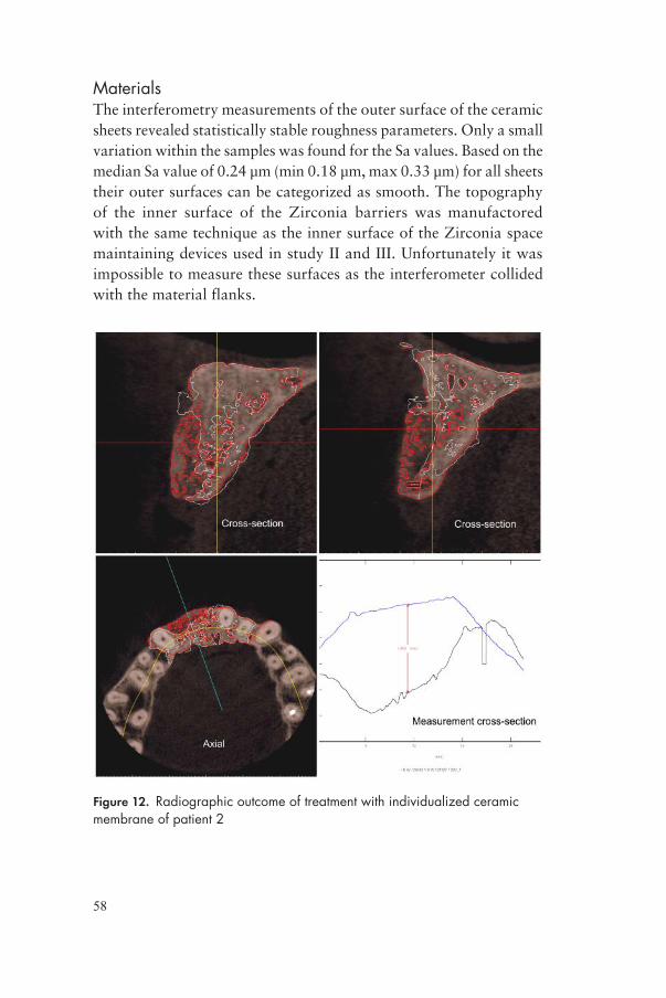
58
MaterialsThe interferometry measurements of the outer surface of the ceramic sheets revealed statistically stable roughness parameters. Only a small variation within the samples was found for the Sa values. Based on the median Sa value of 0.24 μm (min 0.18 μm, max 0.33 μm) for all sheets their outer surfaces can be categorized as smooth. The topography of the inner surface of the Zirconia barriers was manufactored with the same technique as the inner surface of the Zirconia space maintaining devices used in study II and III. Unfortunately it was impossible to measure these surfaces as the interferometer collided with the material flanks.
Figure 12. Radiographic outcome of treatment with individualized ceramic membrane of patient 2
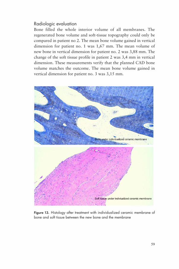
59
Radiologic evaluationBone filled the whole interior volume of all membranes. The regenerated bone volume and soft-tissue topography could only be compared in patient no 2. The mean bone volume gained in vertical dimension for patient no. 1 was 1,67 mm. The mean volume of new bone in vertical dimension for patient no. 2 was 3,88 mm. The change of the soft tissue profile in patient 2 was 3,4 mm in vertical dimension. These measurements verify that the planned CAD bone volume matches the outcome. The mean bone volume gained in vertical dimension for patient no. 3 was 3,15 mm.
Figure 13. Histology after treatment with individualized ceramic membrane of bone and soft tissue between the new bone and the membrane

60
Histological outcomesThe bone biopsies on all 3 patients revealed newly formed trabecular bone with lamellar structure including a few osteocyte lacunaes. Normal osteoblastic and osteoclastic activity could be observed together with a mellow mature stroma and dilated vessels with thin walls. This was considered to be the picture of normal trabecular bone.
The microscopy of the soft tissue biopsies shows connective tissue with absense of inflammatory response. Bone trabeculae could be seen on the edges and because of their localization between the newly formed bone and the ceramic membrane this was considered to be newly formed periosteum.

61
DISCUSSION
This thesis has demonstrated that a ceramic barrier permits new bone formation and osteoconduction within the space created. Furthermore, an individualized ceramic barrier offers the opportunity to design the regenerated bone in any shape needed. The key elements of bone healing and bone regeneration include; a) osteogenic progenitor cells, b) osteoconductive matrices’, c) osteoinductive growth factors and d) a sufficient blood supply (Drosse et al., 2008). So far, no single technique fulfils all these requirements at the same time. The general aim of this thesis was to evaluate a novel method of guided bone regeneration using designed ceramic space maintaining devices on animals and humans. In the initial three studies, experimental space maintaining devices made of dense hydroxyapatite; microporous hydroxyapatite and zirconia were evaluated on a rabbit calvaria model. The design of these space maintaining devices had four purposes: to evaluate the effect of surface roughness, porosity, permeability and the effect of chemistry on GBR.
Surface roughnessThe results of study I-III revealed newly formed trabecular bone with mineralization. The moderately rough inner surface of the dense hydroxyapatite space maintaining device showed slightly larger volumes of newly formed bone than its equivalent with a smooth surface in the micro-CT and histological investigations. These findings indicate that a moderately rough surface probably increased the osteoconductivity in the area. One reason for this might be that the blood clot forming inside the space maintaining device

62
stabilizes more easily on a rough surface than on a smooth one. This may induce early recruitment of osteogenic cells, which will enhance the healing and regeneration of bone (Yang et al., 2015). From the results of the SEM investigation it can be speculated that the newly formed bone possessed a stronger mechanical bond to the hydroxyapatite devices with a rough inner surface. Cracks that could be seen on the rough surface were formed during the manufacturing process. The cracks often form micropores were new bone grow undisrupted. This was observed at the moderately rough surface of the microporous hydroxyapatite space maintaining device but not at the moderately rough surface of the device made of Zirconia. As hydroxyapatite accounts for nearly 70% of the mineral content of bone (Robinson, 1979) it can be speculated if there is also a chemical bond between the bone and the hydroxyapatite. The influence of surface topography on bone regeneration has been described by Wennerberg and Albrektsson (Wennerberg and Albrektsson, 2010, Wennerberg and Albrektsson, 2009). They reported that a surface roughness in a certain range will facilitate rapid and enhanced bone regeneration. Although this was reported on titanium surfaces, it is believed that the effect of surface topography distinguishes itself from surface chemistry. Therefore the prominent osteoconductivity on hydroxyapatite surfaces seen in the animal experiments in this thesis is believed to be the effect of surface topography. On the contrary, when looking at the results of bone to material contact histologically, there were higher values for the smooth hydroxyapatite surface compared to the dense hydroxyapatite rough surface. The microporous had the highest value of bone to material contact. The reason for this is not yet fully understood, as the results were actually the opposite in the investigation with micro-CT. Previous studies when comparing micro-CT with histomorphometrical measurements have shown correlation of approximately 16% error (Marechal et al., 2005). Traditional histological analysis of undecalcified tissues involves time consuming processes such as embedding in methylmetacrylate, sawing, grinding and analysing. Limiting factors are the destructive nature of the procedure as some material might get lost and the fact that the morphology presented is analysed from a two-dimensional cut not representing the full volume (Kochi et al., 2010). This might explain the reason for the different results. As mentioned in the

63
introduction some authors have evaluated the impact of surface roughness on GBR (Lundgren et al., 1999, Lee et al., 2013, Guo et al., 2013). Their results suggest that the regeneration of new bone occurs in larger amounts and with faster kinetic if the inner surface of the barrier is moderately rough and hydrophilic. These findings are supported by the results in this thesis.
Material porosityThe microporous hydroxyapatite space maintaining device was compared to the dense hydroxyapatite device with micro-CT and to Zirconia with histomorphometry. Microporous materials are characterized by pore sizes in the micrometre range (Malmström, 2007). In vivo experiments have indicated that micro porosity within hydroxyapatite scaffolds can accelerate bone regeneration (Hing et al., 2005). The histomorphological results of bone to material contact of the microporous hydroxyapatite space maintaining devices indicate a positive effect of the microporous structure towards new bone regeneration. Pores in biomaterial scaffolds are considered necessary to allow influx of osteogenic cells as well as vascularization (Kuboki et al., 1998). Additionally, a porous surface is considered to allow mechanical interlocking between the biomaterial and the surrounding bone at the interface (Story et al., 1998). As the mechanical stability between regenerated bone and the space maintaining devices was not tested this cannot be confirmed. A general idea of how micro porosity enhances bone regeneration is firstly that the established connections between micropores improves transportation and circulation of proteins, thereby facilitating ingrowth of blood vessels and osteogenic cells (Eggli et al., 1988). Secondly, through this combination of enhanced angiogenesis and cell adhesion, the sensitivity of osteogenic cells appears to be altered (Annaz et al., 2004, Hing, 2004). Micropores appear to make the material osteoinductive by triggering the relevant cells to differentiate into an osteogenic population (Habibovic et al., 2005). As the microporous hydroxyapatite showed thicker trabecular than the dense hydroxyapatite under micro-CT investigation this might be confirmed in this thesis.

64
PermeabilityThe permeability of membranes used for guided bone regeneration has been intensely discussed. As expected, new bone formed inside the hydroxyapatite space maintaining devices with macroscopic holes (HA holes). The theory behind the design of a barrier with macroscopic holes was increased permeability. The holes would allow the periosteum to provide the residual bone with osteogenic proteins and thereby promote new bone formation. Gutta et al claimed in 2009 that a barrier with macropores facilitates greater bone regeneration compared to barriers with micropores (Gutta et al., 2009). The results presented in this thesis showed the opposite in histomorphometry and micro-CT investigations. Schmid et al evaluated the involvement of periosteum for bone formation in a study on rabbit calvaria (Schmid et al., 1994). Their results suggested that membranes do not necessarily have to be permeable in guided bone regeneration. The authors compared a totally occlusive titanium cylinder with an open titanium cylinder covered with e-PTFE membrane. The healing period of this study was 8 months, which is considered to be a very long time for evaluations of new bone growth on the rabbit calvaria (Black et al., 2015). Differences in the kinetics of bone regeneration using membranes with different pore size have also been described by Lundgren et al and Zellin & Linde (Zellin and Linde, 1996, Lundgren et al., 1998). Lundgren et al evaluated 6 different perforation sizes of polyester material and compared them to a control that was totally occlusive towards the periosteum. The healing periods were 4, 8 and 12 weeks before histological analysis. The results showed that the devices with perforated barriers showed faster bone regeneration than the occlusive barrier. Although, after 12 weeks of healing no significant differences in the amount of newly formed mineralized bone could be seen. Zellin & Linde conducted a similar experiment in 1996. They evaluated 3 different e-PTFE membranes with different internodal distances between the pores (<8, 20-25 and 100 μm) for healing periods of 6, 12 18 and 26 weeks. Their results suggested that the membranes with the smallest internodal distance were less efficient than the others in that the bone regeneration was delayed. The total amount of new bone was already reached after 6 weeks by the two most permeable membranes. After 12 weeks there were no differences of new bone regeneration among the used membranes.

65
These findings indicate that there is a relationship between the pore size of barriers used for GBR and the initial stage of bone regeneration. However, this cannot be confirmed by the experiments in this thesis. Yamada et al performed a study on rabbit calvaria using titanium domes comparing a totally occlusive dome to a dome with macroscopic holes (Yamada et al., 2003). The healing periods were 4 and 12 weeks. The titanium devices were shaped as a dome with an inner height of 4 mm. The device with holes had 13 holes of 1,5mm in diameter. At the 12-week evaluation their result suggested that a totally occlusive barrier produced significantly more bone than the permeable one, which is confirmed by the results presented in this thesis.
ZirconiaThe space maintaining devices made of Zirconia have a different chemistry compared to the devices made of hydroxyapatite. Unfortunately it was not possible to obtain any data from the Zirconia devices in the micro-CT investigation, as Zirconia is too dense for the X-ray beams to penetrate. The space maintaining devices made of Zirconia produced the largest amount of new bone overall but bone-to-material contact was low. As Zirconia is a very dense material one might suspect that the amount of micro movements in the material and the contact of the material to the underlying bone is minimal. The fact that the space maintaining devices of Zirconia were hard made them possible to attach very firmly to the bone with the titanium screws. This may be one explanation to the theory of minimal micro movements. When comparing the bone-to-material contact of the Zirconia space maintaining device against the other experimental barriers, the Zirconia devices did not perform very well. One of the reasons might be the fact that the moderately rough surface of the Zirconia barrier differed in Sdr compared to the other barriers with a lower value. Due to the hardness of the material, higher values were not achievable with the tested manufacturing procedures. As the bone-to-material contact was low on Zirconia compared to the hydroxyapatite space maintaining devices it could be speculated that the Zirconia device would be easier to remove from the newly formed bone. This would be benificial in clinical situations. Zirconia devices would probably not integrate into the
66
newly formed bone and run the risk that unwanted materials could disturb the implant installation procedure. Zirconia is successfully used as material for dental implants (Cionca et al., 2015). However, long term clinical follow-up studies are still lacking. It has also shown favourable biological reactions; with no adverse response of osteoblasts and osteoblast-like cells in cell culture studies (Kohal et al., 2008). A common failure pattern for Zirconia implant loss has been described as suddenly increased mobility (Cionca et al., 2015). Analysis of retrieved implants by scanning electron microscopy showed complete absence of any bone. The reason suggested was a sudden mechanical breakdown of the interface between implant and bone. A non-infectious process resulting in bone resorption has been described as “aseptic loosening” by orthopaedic literature (Allain et al., 1999). Defined as a multifactorial aetiology induced by wear debris modulating the expression of proteins responsible for bone resorption (Gehrke et al., 2003). It is less likely this was the scenario using Zirconia space maintaining devices since they were not exposed to any wear that could form debris of the material.
Individualized membranes in a clinical situationDue to the results in the 3 animal studies using ceramic space maintaining devices of hydroxyapatite, microporous hydroxyapatite and Zirconia with different characteristics it was decided to test an individualized Zirconia barrier on 3 patients. The Zirconia space maintaining devices had performed well in the preclinical studies with high volume of regenerated bone, as they were occlusive, ridgid and were probably easy to remove after the healing period. The main advantages of using a ridgid ceramic individualized ceramic membrane for GBR are; a) the possibility to design the shape of the regenerated bone, b) the reduced time of surgery, and c) the possibility of augmenting large volumes of bone.
Bone block transplants have a tendency of partly resorbing during the healing process (Chiapasco et al., 2007). In a clinical study, Cordaro et al reported decrease of bone-volume in the lateral and vertical direction of 23,5% and 42% respectively (Cordaro et al., 2002). It has been suggested to maintain the space and protect the bone grafts with different membranes such as titanium meshes, PTFE or collagen. There is, however, controvery regarding the necessity of covering onlay grafts with a membrane (Dongieux et al., 1998).

67
Dongieux et al reported that there were no significant differences in bone volume or histological outcome for collagen membranes, e-PTFE membranes or no coverage of onlay bone graft healing in a rabbit model. Chiapasco et al suggested that narrow ridges should be augmented with bone blocks without the use of a membranes because it would increase the risk of wound dehiscence and membrane exposure, resulting in a high risk of infection (Chiapasco et al., 1999). Titanium mesh needs to be removed before implant placement and has a risk of penetrating the soft tissue mucosa (Roccuzzo et al., 2004, von Arx et al., 1996). When this occurs, the titanium mesh is more difficult to remove and can cause severe trauma to the mucosa resulting in unfavourable aesthetic outcome. Hereby it was suggested that a layer of collagen, covering the autologous bone graft would maintain the created space longer and thereby the resorption rate would decrease (Kim et al., 2009). A major drawback of collagen membranes is their rapid biodegrading which can result in poor resistance to collapse which allows undesirable cells to enter the wound area (Tatakis and Trombelli, 1999). Donos et al stated that resorption could not be predicted without the use of membrane coverage using e-PTFE membranes in combination with onlay mandibular bone grafts (Donos et al., 2002). In paper IV one of the patients had particlutated bone transplanted to fill the space created inside the ceramic membrane. As the results indicated there was no difference compared to the sites that were left empty. This indicates that a ridgid membrane able to maintain the space created does not need any bone grafts for regenerating new bone. On the other hand if a membrane is more flexible the need for supporting it with a scaffold such as transplanted bone will be higher.
The surgical time to install the ceramic membranes was believed to be reduced compared to a bone grafting procedure or a mouldable membrane that needs to be adapted to the surgical site. The membranes were excellently fitted and there was no need of shaping during surgery. Customized, patient- and site-specific forms of ceramic materials are possible to manufacture through a combination of imaging and ceramic fabrication techniques. The designing of a three dimensional outcome will be predictable. The reduced surgical time comparing to autologous bone grafting is obvious. Another big advantage of this technique is the lack of morbidity from the donor site, in the grafting procedure (Sasso et al., 2005). During surgery,

68
sterility of the transplanted bone must be protected. Prolonged exposure or heat will affect bone cells and osteogenic proteins of the bone and needs to be avoided (Urist, 1980).
Compared to the single use of PTFE membranes large volume of bone could be regenerated in a predictable manner using the customized ceramic membranes. The term “critical size defect” is defined as a defect that will not heal without intervention (Spicer et al., 2012). By classical definition, a critical size defect is the smallest size tissue defect that will not completely heal over the natural lifetime of an animal. The defects critical dimensions differ depending on animal model, the location of the defects direction and shape, i.e. vertical or horizontal (Mardas et al., 2014). When discussing vertical or horizontal defects in terms of defects in the jaw bone one must remember that it is most likely that bone needs to augmented outside the skeletal envelope. No definition of critical size defects outside the skeletal borders exists. Therefore it is the clinical goal of restoring function and aesthetics that needs to be defined rather than expressing the need for new bone as a critical size defect. The size of the defect that is possible to regenerate using the presented technique is yet not known. When using e-PTFE membranes in combination with particlutated bone grafts to maintain the space created the results seems promising independent of volume regenerated (Urban et al., 2009).
An interesting finding was that a new periosteum was formed between the regenerated bone and the ceramic membrane independent of size and shape of the membrane. The histological investigation showed no adverse reactions or inflammation. The periosteum covers the external surfaces of most bones to serve as a transitional region between cortical bone and the overlying soft tissues (Allen et al., 2004). The periosteum contains a microvascular network and progenitor cells, osteoblast and fibroblasts to support the remodulation of cortical bone. The absence of periosteum resulted in 75% reduction of osteoclast numbers on bone graft reported by Zhang et al (Zhang et al., 2008). The authors propose that the development of a flexible cellular layer would serve as an osteogenic and angiogenic “periosteum” to fit around bone or bone substitutes for enhancement of bone repair and regeneration. The authors suggests three important components must be present; a) Live osteogenic cells, capable of producing new bone, b) osteoinductive factors and c) an osteoconductive scaffold
69
maintaining the space. The ceramic membranes along with cortical perforations used in paper IV provided all this. As the regenerated bone inside the ceramic membranes were totally secluded from the overlying natural periosteum it could be speculated that the need of a new periosteum was essential. Similar results have been reported using hd-PTFE membranes (Carbonell et al., 2014), although only 2 authors support the use of hd-PTFE membranes alone for ridge augmentation particularly in the vertical direction (Bartee, 1998, Krauser, 1996).
The need for soft tissue coverage is essential. As the ceramic membrane is completely occlusive, secluding the regenerating area from the outer world it is most likely also occlusive against bacterial invasion. Bartee, Krauser, and Barber have shown that an occlusive hd-PTFE membrane could be left exposed during bone regeneration without causing any infections (Bartee, 1995, Krauser, 1996, Barber et al., 2007). Bartee covered fresh extraction sockets with an hd-PTFE membrane and left the socket to heal for 21 days before removing the membrane. No signs of infection at the healing site could be detected during healing. The fact that the ceramic membrane very likely is occlusive to bacterial infection gives it a major advantage over e-PTFE membranes which need full and passive mucosal coverage (Wang and Boyapati, 2006). It could be questioned if a ceramic membrane exposed to the oral cavity could be left in place without risk of infection. As the ceramic membrane used in paper IV were approximately 0,7mm thick it was difficult to receive full mucosal coverage. Significant undermining of the mucoperiost flaps had to be performed. If undermining of a raised mucoperiosteal flap would not be enough to receive tension free closure there is a need to use a different flap-technique or to expand the soft tissues preoperatively. If there is too much tension after the closure of a wound the covering soft tissue might necrotize.
Osteoinduction was investigated by Urist in 1965 (Urist, 1965). Osteoinduction means recruitment of immature cells and stimulation of these cells to differentiate into osteogenic cells capable of regenerating new bone tissue. In fracture healing this happens spontaneously as there is a trauma to the bone. When bone is regenerated in terms of GBR the need of surgical trauma to promote osteoinduction has been discussed. A surgical trauma can be induced during reflection of the mucoperiosteal flap, by drilling and scraping

70
the bone. Slotte & Lundgren reported no differences in new bone regeneration comparing procedures with or without perforations in a rabbit study observed 3 months postoperatively (Slotte and Lundgren, 2002). Favourable results of bone penetration have also been reported demonstrating an acceleration of bone neogenesis (Majzoub et al., 1999). Rabbits were studied during healing periods of 10, 21, 42 and 60 days comparing mechanically perforated calvaria bone under titanium domes to unperforated calvaria bone under similar titanium domes. A mean area of 29% newly formed bone was demonstrated at the experimental sites compared to 9% at the control sites. In study IV we chose to mechanically perforate the underlying bone on all three patients in order to cause a surgical trauma.
Different techniques of measuring changes in the overlying soft tissues of augmented bone areas have been described. For instance tattoos and adjacent teeth can be used as reference points (Uijlenbroek et al., 2011). Measuring techniques like that are time consuming and have less precision. In one of the patients (patient 2) soft tissue alterations were measured with PRIMOS® optical 3D. This method is highly precise, is non-invasive and time efficient (Walivaara et al., 2007). As the PRIMOS® device only can operate in the anterior part of the oral cavity it was only possible to measure one of the patients and no statistically differences could be calculated. It is believed that this is a more exact method of soft tissue analysis compared to other methods. It is a predictable and repeatable method and has been used to evaluate external soft tissue alterations after orthognatic surgery (Soncul and Bamber, 2004).
An attempt to evaluate the three dimensional bone changes using overlay function of CBCT images was made. Tomographic imaging gives the opportunity to evaluate bone structures in a more realistic way than conventional two-dimensional images as the images can be reconstructed to three-dimensional models. The resolution of CBCT images can detect fine detailed structures and morphologies (nominal 0,076- to 0,125-mm voxel resolution) (Pharoah, 2014). By image fusion, two imaging data sets can be integrated and compared. This technique is mostly used when CBCT volumes are fused with extraoral facial (photographic) or intraoral (impression) optical data. Hereby it is also possible for rapid prototyping of life-sized, dimensionally accurate models of anatomical structures. By superimposing the original CBCT images with the images at the

71
end of the healing period the result of the GBR treatment can be evaluated quantitatively (Weissheimer et al., 2015). Since study IV only included three patients no statistics could be calculated. However changes in the bone morphology could be measured at a transverse or an axial cut of the image volume representing new bone growth, (see figure 12). In the future there might be a method of simulating the desired bone shape in an atrophied jaw and by rapid prototyping one could manufacture an individualized ceramic membrane with excellent fit using DICOM data.
Although the results in paper IV point in a promising direction some limitations and questions arises. The fit of the ceramic membranes used was excellent. Shaping of the Zirconia can only be made to a limited extent, as the material is too dense. Any adjustments have to be performed on the recipient jaw bone to be able to fit the membrane. This of course would be disadvantageous, as that bone might be needed in the implant installation procedure later on. Limitation of soft tissues covering the ceramic membrane could be a problem as well, although the membrane is occlusive and should theoretically resist bacterial invasion. In the second stage surgery when dental implants are supposed to be installed, soft tissues are essential for a successful outcome (Herford et al., 2015). Intraoral soft tissue expansion techniques could be used prior to membrane installation in order to obtain tension-free soft tissue coverage (Abrahamsson et al., 2012). An osmotic soft tissue expander is hereby installed under the periosteum and left to expand for 2 weeks, raising the keratinized mucosa from the underlying bone prior to bone augmentation. None of the patients presented in paper IV wore any provisional prosthesis during the healing period. The aim of this was to avoid repeated trauma to the soft tissues during healing. The ceramic membrane is ridgid and the risk of micro movements due to chewing was considered minor.
The main advantage of using a ridgid individualized ceramic membrane for GBR is the ability to design the regenerated bone in any shape. A short surgical time of exposed bone and an excellent acceptance of the soft tissues are also advantageous of this method. Overall it is believed to be a simple and effective surgical GBR technique to be used in complicated grafting areas in conjunction with dental implant installation.
72
CONCLUSIONS
• Ceramic space maintaining devices regardless of the choice of material (hydroxyapatite and Zirconia), inner surface roughness, microporosity or permeability, permit new bone formation and osteoconduction within the space created.
• A ceramic barrier with a moderately rough inner surface regenerated a larger volume of new bone compared to a ceramic barrier with a smooth inner surface with similar characteristics.
• A micro porous hydroxyapatite barrier regenerated less volume of new bone compared to a dense hydroxyapatite barrier with similar characteristics.
• An occlusive hydroxyapatite barrier regenerated a larger volume of new bone compared to a permeable hydroxyapatite barrier with similar characteristics.
• A barrier made of Zirconia regenerated a larger volume of new bone compared to a barrier made of hydroxyapatite with similar characteristics.
• The limit of vertical bone regeneration is not clarified.
• An individualized ceramic barrier make it possible to design the regenerated bone in any shape needed with a predictable result and no need for transplanted bone.
73
POPULÄRVETENSKAPLIG SAMMANFATTNING
Att få nya tänder med hjälp av tandimplantat är idag en väl etablerad teknik med god prognos. För att kunna operera in implantat i käkbenet måste käkbenet ha tillräcklig volym och kvalite. När benet inte har tillräcklig volym kan det byggas upp till exempel genom att transplantera ben från någon annat ställe. “Guided Bone Regeneration” eller “Styrd ben läkning” är en teknik för ben-uppbyggnad där ett membran skapar ett tomt utrymme ovanför det befintliga benet. Mellan membranet och det befintliga benet skapas ett blodkoagel som senare omvandlas till nytt ben. De membraner som idag finns tillgängliga på marknaden är mjuka vilket begränsar användningsmöjligheterna.
Det övergripande målet med denna avhandling var att introducera och utvärdera en ny metod för styrd ben läkning genom att använda ett membran av keramiskt material.
I de 3 inledande djurstudierna användes kanin som försöksdjur. Härvid utvärderades hur en rå yta påverkar ben läkningen. Vidare utvärderades hur en porös keram påverkar ben läkningen, hur en keram med hål i påverkar ben läkningen samt hur olika keramiska material (hydroxyapatit och Zirconia) påverkar ben läkningen. Fem olika sorters kupor (12 st av varje sort) fästes på skallbenet på kaniner och täcktes med benhinnan och huden. Dessa fick läka i 12 veckor innan kaninerna avlivades och därefter analyserades resultatet. Resultatet mättes med röntgen och vävnadsprover. Slutsatsen blev att en keramisk barriär av Zirconia med en rå inneryta utan hål var den bästa lösningen.

74
Efter djurförsöken gjordes behandlingar på 3 stycken patienter med keramiska membraner. Patient 1 hade för smalt käkben i överkäkens sidoparti, patient 2 hade för smalt och för lågt ben i överkäksfronten och patient 3 hade för lågt ben i underkäkens båda sidor. Membranen var anpassade för varje individ och passade perfekt. Operationen för att fästa membranen tog kort tid och patienterna hade inga komplikationer under läkningen. Efter 7 månader avlägsnades membranen och implantat kunde installeras komplikationsfritt och efter ytterligare en läk period kunde patienterna få nya tänder ovanpå implantaten. Benläkningen utvärderades med datortomografi röntgen, vävnadsprover och tredimensionellt fotografi. Resultatet visade att ”styrd ben läkning med hjälp av keramiska membraner är förutsägbart med gott resultat. Slutsatsen blev att individuellt anpassade keramiska membran ger operatören möjlighet att designa formen på det nybildade benet med ett förutsägbart resultat utan att behöva transplantera ben.
75
ACKNOWLEDGEMENTS
This project has been an amazing experience for me and would not have been possible without a lot of helping hands along the way. I would like to express my thanks to all of you, and especially:
Ann Wennerberg, my main supervisor for sharing your knowledge of the scientific process and for always being available for questions and discussions.
Sten Isaksson, my co-supervisor for inspiration and for a neverending source of new ideas. Thank you also for guiding me through my dental studies, sending me to the Royal London Hospital in 2006 and guiding specialist training.
Peter Abrahamsson, my co-supervisor for inspiration and for making it possible to do research at Specialisttandvården in Halmstad. Thank you also for the time you take discussing scientific work and life in general.
Johan Malmström, my co-author for endless discussions about ceramic materials and how to raise kids.
Ryo Jimbo, my co-author for help with a lot of practical issues and co-authorship. Thank you for showing me a piece of Japan.
Erik Adolfsson, my co-author for technical expertise concerning ceramic materials, for manufacturing the ceramic components and for answering a lot of necessary and unnecessary questions.

76
Silvia Galli, for helping me urgently with the animal samples before the trip to Japan.
Lars-Åke Johansson, for interesting discussions about this research project and ideas along the way. Thank you for lending me that book.
Dan-Åke Wälivaara, my co-writer and for helping me with the photos.
Tomas Albreksson, for inspirational discussions and review of texts.
Anders Holmen, for help with computers, computer programs and statistical calculations.
Rishi Bandari, my English colleague and friend for helping out with the English grammar of the final thesis.
Chris Reynolds, my American cousin for helping out with the language review of the final thesis. Thank you also for reading this work critacally and reviewing the facts. Best regards to all your family.
Yusuke Kozai, my co-author for helping out with the micro-CT investigations.
Yoshihito Naito, my co-author for helping me with sawing and grinding.
Fredrik Hallmer, my co-author for a helping hand during animal surgery and for the good company during dental training and all courses.

77
REFERENCES
ABRAHAMSSON, P., WALIVAARA, D. A., ISAKSSON, S. & ANDERSSON, G. 2012. Periosteal expansion before local bone reconstruction using a new technique for measuring soft tissue profile stability: a clinical study. J Oral Maxillofac Surg, 70, e521-30.
ALBREKTSSON, T. & WENNERBERG, A. 2004a. Oral implant surfaces: Part 1--review focusing on topographic and chemical properties of different surfaces and in vivo responses to them. Int J Prosthodont, 17, 536-43.
ALBREKTSSON, T. & WENNERBERG, A. 2004b. Oral implant surfaces: Part 2--review focusing on clinical knowledge of different surfaces. Int J Prosthodont, 17, 544-64.
ALLAIN, J., LE MOUEL, S., GOUTALLIER, D. & VOISIN, M. C. 1999. Poor eight-year survival of cemented zirconia-polyethylene total hip replacements. J Bone Joint Surg Br, 81, 835-42.
ALLEN, M. R., HOCK, J. M. & BURR, D. B. 2004. Periosteum: biology, regulation, and response to osteoporosis therapies. Bone, 35, 1003-12.
ANNAZ, B., HING, K. A., KAYSER, M., BUCKLAND, T. & DI SILVIO, L. 2004. An ultrastructural study of cellular response to variation in porosity in phase-pure hydroxyapatite. J Microsc, 216, 97-109.
BARBER, H. D., LIGNELLI, J., SMITH, B. M. & BARTEE, B. K. 2007. Using a dense PTFE membrane without primary closure to achieve bone and tissue regeneration. J Oral Maxillofac Surg, 65, 748-52.
BARTEE, B. K. 1995. The use of high-density polytetrafluoroethylene membrane to treat osseous defects: clinical reports. Implant Dent, 4, 21-6.
BARTEE, B. K. 1998. Evaluation of a new polytetrafluoroethylene guided tissue regeneration membrane in healing extraction sites. Compend Contin Educ Dent, 19, 1256-8, 1260, 1262-4.
BARTEE, B. K. & CARR, J. A. 1995. Evaluation of a high-density polytetrafluoroethylene (n-PTFE) membrane as a barrier material to facilitate guided bone regeneration in the rat mandible. J Oral Implantol, 21, 88-95.

78
BECKER, W., DAHLIN, C., BECKER, B. E., LEKHOLM, U., VAN STEENBERGHE, D., HIGUCHI, K. & KULTJE, C. 1994. The use of e-PTFE barrier membranes for bone promotion around titanium implants placed into extraction sockets: a prospective multicenter study. Int J Oral Maxillofac Implants, 9, 31-40.
BLACK, C. R., GORIAINOV, V., GIBBS, D., KANCZLER, J., TARE, R. S. & OREFFO, R. O. 2015. Bone Tissue Engineering. Curr Mol Biol Rep, 1, 132-140.
CALVO GUIRADO, J. L., RAMIREZ FERNANDEZ, M. P., NEGRI, B., DELGADO RUIZ, R. A., MATE SANCHEZ DE-VAL, J. E. & GOMEZ-MORENO, G. 2013. Experimental model of bone response to collagenized xenografts of porcine origin (OsteoBiol(R) mp3): a radiological and histomorphometric study. Clin Implant Dent Relat Res, 15, 143-51.
CARBONELL, J. M., MARTIN, I. S., SANTOS, A., PUJOL, A., SANZ-MOLINER, J. D. & NART, J. 2014. High-density polytetrafluoroethylene membranes in guided bone and tissue regeneration procedures: a literature review. Int J Oral Maxillofac Surg, 43, 75-84.
CHAE, T., YANG, H., LEUNG, V., KO, F. & TROCZYNSKI, T. 2013. Novel biomimetic hydroxyapatite/alginate nanocomposite fibrous scaffolds for bone tissue regeneration. J Mater Sci Mater Med, 24, 1885-94.
CHIAPASCO, M., ABATI, S., ROMEO, E. & VOGEL, G. 1999. Clinical outcome of autogenous bone blocks or guided bone regeneration with e-PTFE membranes for the reconstruction of narrow edentulous ridges. Clin Oral Implants Res, 10, 278-88.
CHIAPASCO, M., ZANIBONI, M. & RIMONDINI, L. 2007. Autogenous onlay bone grafts vs. alveolar distraction osteogenesis for the correction of vertically deficient edentulous ridges: a 2-4-year prospective study on humans. Clin Oral Implants Res, 18, 432-40.
CIONCA, N., MULLER, N. & MOMBELLI, A. 2015. Two-piece zirconia implants supporting all-ceramic crowns: a prospective clinical study. Clin Oral Implants Res, 26, 413-8.
COLNOT, C. 2011. Cell sources for bone tissue engineering: insights from basic science. Tissue Eng Part B Rev, 17, 449-57.
COLNOT, C., ZHANG, X. & KNOTHE TATE, M. L. 2012. Current insights on the regenerative potential of the periosteum: molecular, cellular, and endogenous engineering approaches. J Orthop Res, 30, 1869-78.
CORDARO, L., AMADE, D. S. & CORDARO, M. 2002. Clinical results of alveolar ridge augmentation with mandibular block bone grafts in partially edentulous patients prior to implant placement. Clin Oral Implants Res, 13, 103-11.
79
CORINALDESI, G., PIERI, F., SAPIGNI, L. & MARCHETTI, C. 2009. Evaluation of survival and success rates of dental implants placed at the time of or after alveolar ridge augmentation with an autogenous mandibular bone graft and titanium mesh: a 3- to 8-year retrospective study. Int J Oral Maxillofac Implants, 24, 1119-28.
DAHLIN, C., SENNERBY, L., LEKHOLM, U., LINDE, A. & NYMAN, S. 1989. Generation of new bone around titanium implants using a membrane technique: an experimental study in rabbits. Int J Oral Maxillofac Implants, 4, 19-25.
DIAZ-FLORES, L., GUTIERREZ, R. & VARELA, H. 1992. Behavior of postcapillary venule pericytes during postnatal angiogenesis. J Morphol, 213, 33-45.
DIMITRIOU, R., JONES, E., MCGONAGLE, D. & GIANNOUDIS, P. V. 2011. Bone regeneration: current concepts and future directions. BMC Med, 9, 66.
DONATH, K. 1985. The diagnostic value of the new method for the study of undecalcified bones and teeth with attached soft tissue (Sage-Schliff (sawing and grinding) technique). Pathol Res Pract, 179, 631-3.
DONGIEUX, J. W., BLOCK, M. S., MORRIS, G., GARDINER, D. & DEAN, K. 1998. The effect of different membranes on onlay bone graft success in the dog mandible. Oral Surg Oral Med Oral Pathol Oral Radiol Endod, 86, 145-51.
DONOS, N., KOSTOPOULOS, L. & KARRING, T. 2002. Alveolar ridge augmentation by combining autogenous mandibular bone grafts and non-resorbable membranes. Clin Oral Implants Res, 13, 185-91.
DROSSE, I., VOLKMER, E., CAPANNA, R., DE BIASE, P., MUTSCHLER, W. & SCHIEKER, M. 2008. Tissue engineering for bone defect healing: an update on a multi-component approach. Injury, 39 Suppl 2, S9-20.
DUBRUILLE, J. H., VIGUIER, E., LE NAOUR, G., DUBRUILLE, M. T., AURIOL, M. & LE CHARPENTIER, Y. 1999. Evaluation of combinations of titanium, zirconia, and alumina implants with 2 bone fillers in the dog. Int J Oral Maxillofac Implants, 14, 271-7.
EGGLI, P. S., MULLER, W. & SCHENK, R. K. 1988. Porous hydroxyapatite and tricalcium phosphate cylinders with two different pore size ranges implanted in the cancellous bone of rabbits. A comparative histomorphometric and histologic study of bony ingrowth and implant substitution. Clin Orthop Relat Res, 127-38.
EINHORN, T. A. 1995. Enhancement of fracture-healing. J Bone Joint Surg Am, 77, 940-56.
80
ESPOSITO, M., GRUSOVIN, M. G., COULTHARD, P. & WORTHINGTON, H. V. 2006. The efficacy of various bone augmentation procedures for dental implants: a Cochrane systematic review of randomized controlled clinical trials. Int J Oral Maxillofac Implants, 21, 696-710.
GAPSKI, R., NEIVA, R., OH, T. J. & WANG, H. L. 2006. Histologic analyses of human mineralized bone grafting material in sinus elevation procedures: a case series. Int J Periodontics Restorative Dent, 26, 59-69.
GARCEZ-FILHO, J., TOLENTINO, L., SUKEKAVA, F., SEABRA, M., CESAR-NETO, J. B. & ARAUJO, M. G. 2015. Long-term outcomes from implants installed by using split-crest technique in posterior maxillae: 10 years of follow-up. Clin Oral Implants Res, 26, 326-31.
GARRAHAN, N. J., CROUCHER, P. I. & COMPSTON, J. E. 1990. A computerised technique for the quantitative assessment of resorption cavities in trabecular bone. Bone, 11, 241-5.
GEHRKE, T., SERS, C., MORAWIETZ, L., FERNAHL, G., NEIDEL, J., FROMMELT, L. & KRENN, V. 2003. Receptor activator of nuclear factor kappaB ligand is expressed in resident and inflammatory cells in aseptic and septic prosthesis loosening. Scand J Rheumatol, 32, 287-94.
GUO, Z., ZHOU, L., RONG, M., DING, J., ZHU, A., LI, S. & LU, H. 2013. Bone augmentation in a titanium cap with a porous surface modified by microarc oxidation. Int J Oral Maxillofac Implants, 28, 767-73.
GUTTA, R., BAKER, R. A., BARTOLUCCI, A. A. & LOUIS, P. J. 2009. Barrier membranes used for ridge augmentation: is there an optimal pore size? J Oral Maxillofac Surg, 67, 1218-25.
HABIBOVIC, P., YUAN, H., VAN DER VALK, C. M., MEIJER, G., VAN BLITTERSWIJK, C. A. & DE GROOT, K. 2005. 3D microenvironment as essential element for osteoinduction by biomaterials. Biomaterials, 26, 3565-75.
HAMMERLE, C. H. & JUNG, R. E. 2003. Bone augmentation by means of barrier membranes. Periodontol 2000, 33, 36-53.
HENCH, L. L. 1998. Biomaterials: a forecast for the future. Biomaterials, 19, 1419-23.
HERFORD, A. S., TANDON, R., PIVETTI, L. & CICCIU, M. 2015. Pedicled lingual flap to provide keratinized tissue regeneration over dental implants: a description of the technique and a case report. J Oral Implantol, 41, 196-9.
HING, K. A. 2004. Bone repair in the twenty-first century: biology, chemistry or engineering? Philos Trans A Math Phys Eng Sci, 362, 2821-50.
HING, K. A., ANNAZ, B., SAEED, S., REVELL, P. A. & BUCKLAND, T. 2005. Microporosity enhances bioactivity of synthetic bone graft substitutes. J Mater Sci Mater Med, 16, 467-75.

81
HURLEY, L. A., STINCHFIELD, F. E., BASSETT, A. L. & LYON, W. H. 1959. The role of soft tissues in osteogenesis. An experimental study of canine spine fusions. J Bone Joint Surg Am, 41-A, 1243-54.
ICHIKAWA, Y., AKAGAWA, Y., NIKAI, H. & TSURU, H. 1992. Tissue compatibility and stability of a new zirconia ceramic in vivo. J Prosthet Dent, 68, 322-6.
IKENO, M., HIBI, H., KINOSHITA, K., HATTORI, H. & UEDA, M. 2013. Effects of the permeability of shields with autologous bone grafts on bone augmentation. Int J Oral Maxillofac Implants, 28, e386-92.
JOHANSSON, L. A., ISAKSSON, S., BRYINGTON, M. & DAHLIN, C. 2013. Evaluation of bone regeneration after three different lateral sinus elevation procedures using micro-computed tomography of retrieved experimental implants and surrounding bone: a clinical, prospective, and randomized study. Int J Oral Maxillofac Implants, 28, 579-86.
JOHANSSON, L. A., ISAKSSON, S., LINDH, C., BECKTOR, J. P. & SENNERBY, L. 2010. Maxillary sinus floor augmentation and simultaneous implant placement using locally harvested autogenous bone chips and bone debris: a prospective clinical study. J Oral Maxillofac Surg, 68, 837-44.
KAHNBERG, K. E. 1979. Restoration of mandibular jaw defects in the rabbit by subperiosteally implanted Teflon mantle leaf. Int J Oral Surg, 8, 449-56.
KHOJASTEH, A., HASSANI, A., MOTAMEDIAN, S. R., SAADAT, S. & ALIKHASI, M. 2015. Cortical Bone Augmentation Versus Nerve Lateralization for Treatment of Atrophic Posterior Mandible: A Retrospective Study and Review of Literature. Clin Implant Dent Relat Res.
KIM, S. H., KIM, D. Y., KIM, K. H., KU, Y., RHYU, I. C. & LEE, Y. M. 2009. The efficacy of a double-layer collagen membrane technique for overlaying block grafts in a rabbit calvarium model. Clin Oral Implants Res, 20, 1124-32.
KOCHI, G., SATO, S., EBIHARA, H., HIRANO, J., ARAI, Y. & ITO, K. 2010. A comparative study of microfocus CT and histomorphometry in the evaluation of bone augmentation in rat calvarium. J Oral Sci, 52, 203-11.
KOHAL, R. J., ATT, W., BACHLE, M. & BUTZ, F. 2008. Ceramic abutments and ceramic oral implants. An update. Periodontol 2000, 47, 224-43.
KOHAL, R. J., PAPAVASILIOU, G., KAMPOSIORA, P., TRIPODAKIS, A. & STRUB, J. R. 2002. Three-dimensional computerized stress analysis of commercially pure titanium and yttrium-partially stabilized zirconia implants. Int J Prosthodont, 15, 189-94.
KOSTOPOULOS, L., KARRING, T. & URAGUCHI, R. 1994. Formation of jawbone tuberosities by guided tissue regeneration. An experimental study in the rat. Clin Oral Implants Res, 5, 245-53.

82
KRAUSER, J. T. 1996. High-density PTFE membranes: uses with root-form implants. Dent Implantol Update, 7, 65-9.
KUBOKI, Y., TAKITA, H., KOBAYASHI, D., TSURUGA, E., INOUE, M., MURATA, M., NAGAI, N., DOHI, Y. & OHGUSHI, H. 1998. BMP-induced osteogenesis on the surface of hydroxyapatite with geometrically feasible and nonfeasible structures: topology of osteogenesis. J Biomed Mater Res, 39, 190-9.
LANDI, E., TAMPIERI, A., CELOTTI, G., SPRIO, S., SANDRI, M. & LOGROSCINO, G. 2007. Sr-substituted hydroxyapatites for osteoporotic bone replacement. Acta Biomater, 3, 961-9.
LEE, S. B., RETZEPI, M., PETRIE, A., HAKIMI, A. R., SCHWARZ, F. & DONOS, N. 2013. The effect of diabetes on bone formation following application of the GBR principle with the use of titanium domes. Clin Oral Implants Res, 24, 28-35.
LEE, S. H., LIM, P. & YOON, H. J. 2014. The influence of cortical perforation on guided bone regeneration using synthetic bone substitutes: a study of rabbit cranial defects. Int J Oral Maxillofac Implants, 29, 464-71.
LIAGRE, B., MOALIC, S., VERGNE, P., CHARISSOUX, J. L., BERNACHE-ASSOLLANT, D. & BENEYTOUT, J. L. 2002. Effects of alumina and zirconium dioxide particles on arachidonic acid metabolism and proinflammatory interleukin production in osteoarthritis and rheumatoid synovial cells. J Bone Joint Surg Br, 84, 920-30.
LIN, A., WANG, C. J., KELLY, J., GUBBI, P. & NISHIMURA, I. 2009. The role of titanium implant surface modification with hydroxyapatite nanoparticles in progressive early bone-implant fixation in vivo. Int J Oral Maxillofac Implants, 24, 808-16.
LINDE, A., ALBERIUS, P., DAHLIN, C., BJURSTAM, K. & SUNDIN, Y. 1993a. Osteopromotion: a soft-tissue exclusion principle using a membrane for bone healing and bone neogenesis. J Periodontol, 64, 1116-28.
LINDE, A., THOREN, C., DAHLIN, C. & SANDBERG, E. 1993b. Creation of new bone by an osteopromotive membrane technique: an experimental study in rats. J Oral Maxillofac Surg, 51, 892-7.
LUNDGREN, A., LUNDGREN, D. & TAYLOR, A. 1998. Influence of barrier occlusiveness on guided bone augmentation. An experimental study in the rat. Clin Oral Implants Res, 9, 251-60.
LUNDGREN, A. K., LUNDGREN, D., HAMMERLE, C. H., NYMAN, S. & SENNERBY, L. 2000. Influence of decortication of the donor bone on guided bone augmentation. An experimental study in the rabbit skull bone. Clin Oral Implants Res, 11, 99-106.

83
LUNDGREN, A. K., LUNDGREN, D., WENNERBERG, A., HAMMERLE, C. H. & NYMAN, S. 1999. Influence of surface roughness of barrier walls on guided bone augmentation: experimental study in rabbits. Clin Implant Dent Relat Res, 1, 41-8.
LUNDGREN, D., LUNDGREN, A. K., SENNERBY, L. & NYMAN, S. 1995. Augmentation of intramembraneous bone beyond the skeletal envelope using an occlusive titanium barrier. An experimental study in the rabbit. Clin Oral Implants Res, 6, 67-72.
MAJZOUB, Z., BERENGO, M., GIARDINO, R., ALDINI, N. N. & CORDIOLI, G. 1999. Role of intramarrow penetration in osseous repair: a pilot study in the rabbit calvaria. J Periodontol, 70, 1501-10.
MALMSTRÖM, J. 2007. On bone regeneration in porous bioceramics : studies in humans and rabbits using free form fabricated scaffolds. Diss (sammanfattning) Göteborg Göteborgs universitet, 2007, Institute of Clinical Sciences, Department of Biomaterials, Göteborg University,.
MARDAS, N., DEREKA, X., DONOS, N. & DARD, M. 2014. Experimental model for bone regeneration in oral and cranio-maxillo-facial surgery. J Invest Surg, 27, 32-49.
MARDAS, N., KOSTOPOULOS, L., STAVROPOULOS, A. & KARRING, T. 2003. Evaluation of a cell-permeable barrier for guided tissue regeneration combined with demineralized bone matrix. Clin Oral Implants Res, 14, 812-8.
MARECHAL, M., LUYTEN, F., NIJS, J., POSTNOV, A., SCHEPERS, E. & VAN STEENBERGHE, D. 2005. Histomorphometry and micro-computed tomography of bone augmentation under a titanium membrane. Clin Oral Implants Res, 16, 708-14.
MAROUF, H. A. & EL-GUINDI, H. M. 2000. Efficacy of high-density versus semipermeable PTFE membranes in an elderly experimental model. Oral Surg Oral Med Oral Pathol Oral Radiol Endod, 89, 164-70.
MIN, S., SATO, S., MURAI, M., OKUNO, K., FUJISAKI, Y., YAMADA, Y. & ITO, K. 2007. Effects of marrow penetration on bone augmentation within a titanium cap in rabbit calvarium. J Periodontol, 78, 1978-84.
MOSBY 2012. Mosby’s Medical Dictionary, Elevier.
MURRAY, G., HOLDEN, R. & ROSCHLAU, W. 1957. Experimental and clinical study of new growth of bone in a cavity. Am J Surg, 93, 385-7.
NYMAN, S., GOTTLOW, J., KARRING, T. & LINDHE, J. 1982. The regenerative potential of the periodontal ligament. An experimental study in the monkey. J Clin Periodontol, 9, 257-65.
OYANE, A., KAWASHITA, M., NAKANISHI, K., KOKUBO, T., MINODA, M., MIYAMOTO, T. & NAKAMURA, T. 2003. Bonelike apatite formation on ethylene-vinyl alcohol copolymer modified with silane coupling agent and calcium silicate solutions. Biomaterials, 24, 1729-35.
84
OZKURT, Z. & KAZAZOGLU, E. 2011. Zirconia dental implants: a literature review. J Oral Implantol, 37, 367-76.
PHAROAH, S. C. W. A. M. J. 2014. Oral Radiology, Principles and Interpretation, Elevier.
PICONI, C., LABANTI, M., MAGNANI, G., CAPORALE, M., MACCAURO, G. & MAGLIOCCHETTI, G. 1999. Analysis of a failed alumina THR ball head. Biomaterials, 20, 1637-46.
PICONI, C. & MACCAURO, G. 1999. Zirconia as a ceramic biomaterial. Biomaterials, 20, 1-25.
RASIA-DAL POLO, M., POLI, P. P., RANCITELLI, D., BERETTA, M. & MAIORANA, C. 2014. Alveolar ridge reconstruction with titanium meshes: a systematic review of the literature. Med Oral Patol Oral Cir Bucal, 19, e639-46.
REILLY, T. M., SELDES, R., LUCHETTI, W. & BRIGHTON, C. T. 1998. Similarities in the phenotypic expression of pericytes and bone cells. Clin Orthop Relat Res, 95-103.
ROBINSON, R. A. 1979. Bone tissue: composition and function. Johns Hopkins Med J, 145, 10-24.
ROCCUZZO, M., RAMIERI, G., SPADA, M. C., BIANCHI, S. D. & BERRONE, S. 2004. Vertical alveolar ridge augmentation by means of a titanium mesh and autogenous bone grafts. Clin Oral Implants Res, 15, 73-81.
ROMPEN, E. H., BIEWER, R., VANHEUSDEN, A., ZAHEDI, S. & NUSGENS, B. 1999. The influence of cortical perforations and of space filling with peripheral blood on the kinetics of guided bone generation. A comparative histometric study in the rat. Clin Oral Implants Res, 10, 85-94.
SASSO, R. C., LEHUEC, J. C., SHAFFREY, C. & SPINE INTERBODY RESEARCH, G. 2005. Iliac crest bone graft donor site pain after anterior lumbar interbody fusion: a prospective patient satisfaction outcome assessment. J Spinal Disord Tech, 18 Suppl, S77-81.
SCHMID, J., HAMMERLE, C. H., OLAH, A. J. & LANG, N. P. 1994. Membrane permeability is unnecessary for guided generation of new bone. An experimental study in the rabbit. Clin Oral Implants Res, 5, 125-30.
SHLOMI, B., HOROWITZ, I., KAHN, A., DOBRIYAN, A. & CHAUSHU, G. 2004. The effect of sinus membrane perforation and repair with Lambone on the outcome of maxillary sinus floor augmentation: a radiographic assessment. Int J Oral Maxillofac Implants, 19, 559-62.
SIMION, M., BALDONI, M. & ZAFFE, D. 1992. Jawbone enlargement using immediate implant placement associated with a split-crest technique and guided tissue regeneration. Int J Periodontics Restorative Dent, 12, 462-73.

85
SLOTTE, C., GRONNINGSAETER, A., HALMOY, A. M., OHRNELL, L. O., STROH, G., ISAKSSON, S., JOHANSSON, L. A., MORDENFELD, A., EKLUND, J. & EMBRING, J. 2012. Four-millimeter implants supporting fixed partial dental prostheses in the severely resorbed posterior mandible: two-year results. Clin Implant Dent Relat Res, 14 Suppl 1, e46-58.
SLOTTE, C. & LUNDGREN, D. 2002. Impact of cortical perforations of contiguous donor bone in a guided bone augmentation procedure: an experimental study in the rabbit skull. Clin Implant Dent Relat Res, 4, 1-10.
SOLLAZZO, V., PEZZETTI, F., SCARANO, A., PIATTELLI, A., BIGNOZZI, C. A., MASSARI, L., BRUNELLI, G. & CARINCI, F. 2008. Zirconium oxide coating improves implant osseointegration in vivo. Dent Mater, 24, 357-61.
SONCUL, M. & BAMBER, M. A. 2004. Evaluation of facial soft tissue changes with optical surface scan after surgical correction of Class III deformities. J Oral Maxillofac Surg, 62, 1331-40.
SPICER, P. P., KRETLOW, J. D., YOUNG, S., JANSEN, J. A., KASPER, F. K. & MIKOS, A. G. 2012. Evaluation of bone regeneration using the rat critical size calvarial defect. Nat Protoc, 7, 1918-29.
STORY, B. J., WAGNER, W. R., GAISSER, D. M., COOK, S. D. & RUST-DAWICKI, A. M. 1998. In vivo performance of a modified CSTi dental implant coating. Int J Oral Maxillofac Implants, 13, 749-57.
SVANBORG, L. M., HOFFMAN, M., ANDERSSON, M., CURRIE, F., KJELLIN, P. & WENNERBERG, A. 2011. The effect of hydroxyapatite nanocrystals on early bone formation surrounding dental implants. Int J Oral Maxillofac Surg, 40, 308-15.
TAGUCHI, K., OGAWA, R., MIGITA, M., HANAWA, H., ITO, H. & ORIMO, H. 2005. The role of bone marrow-derived cells in bone fracture repair in a green fluorescent protein chimeric mouse model. Biochem Biophys Res Commun, 331, 31-6.
TATAKIS, D. N. & TROMBELLI, L. 1999. Adverse effects associated with a bioabsorbable guided tissue regeneration device in the treatment of human gingival recession defects. A clinicopathologic case report. J Periodontol, 70, 542-7.
TENHUISEN, K. S. & BROWN, P. W. 1997. Effects of magnesium on the formation of calcium-deficient hydroxyapatite from CaHPO4.2H2O and Ca4(PO4)2O. J Biomed Mater Res, 36, 306-14.
UIJLENBROEK, H. J., LIU, Y., HE, J. F., VISSCHER, C., VAN WAAS, M. A. & WISMEYER, D. 2011. Expanding soft tissue with Osmed tissue expanders in the goat maxilla. Clin Oral Implants Res, 22, 121-8.

86
URBAN, I. A., JOVANOVIC, S. A. & LOZADA, J. L. 2009. Vertical ridge augmentation using guided bone regeneration (GBR) in three clinical scenarios prior to implant placement: a retrospective study of 35 patients 12 to 72 months after loading. Int J Oral Maxillofac Implants, 24, 502-10.
URIST, M. R. 1965. Bone: formation by autoinduction. Science, 150, 893-9.
URIST, M. R. 1980. Fundamental and Clinical Bone Physiology, Philadelphia, USA, Lippincott Williams & Wilkins.
VAN BRAKEL, R., MEIJER, G. J., VERHOEVEN, J. W., JANSEN, J., DE PUTTER, C. & CUNE, M. S. 2012. Soft tissue response to zirconia and titanium implant abutments: an in vivo within-subject comparison. J Clin Periodontol, 39, 995-1001.
VAN STEENBERGHE, D., JOHANSSON, C., QUIRYNEN, M., MOLLY, L., ALBREKTSSON, T. & NAERT, I. 2003. Bone augmentation by means of a stiff occlusive titanium barrier. Clin Oral Implants Res, 14, 63-71.
VON ARX, T., HARDT, N. & WALLKAMM, B. 1996. The TIME technique: a new method for localized alveolar ridge augmentation prior to placement of dental implants. Int J Oral Maxillofac Implants, 11, 387-94.
WALIVAARA, D. A., ISAKSSON, S. & ROSEN, S. 2007. Description of a technique for evaluation of three-dimensional shape alterations in soft tissue after intra oral bone reconstruction. Br J Oral Maxillofac Surg, 45, 382-6.
WANG, H. L. & BOYAPATI, L. 2006. “PASS” principles for predictable bone regeneration. Implant Dent, 15, 8-17.
WEISSHEIMER, A., MENEZES, L. M., KOERICH, L., PHAM, J. & CEVIDANES, L. H. 2015. Fast three-dimensional superimposition of cone beam computed tomography for orthopaedics and orthognathic surgery evaluation. Int J Oral Maxillofac Surg, 44, 1188-96.
WENNERBERG, A. 1996. On surface roughness and implant incorporation. Diss (sammanfattning) Göteborg Univ.
WENNERBERG, A. & ALBREKTSSON, T. 2009. Effects of titanium surface topography on bone integration: a systematic review. Clin Oral Implants Res, 20 Suppl 4, 172-84.
WENNERBERG, A. & ALBREKTSSON, T. 2010. On implant surfaces: a review of current knowledge and opinions. Int J Oral Maxillofac Implants, 25, 63-74.
WOLFF, J. 1892. Das Gesetz Der Transformation Der Knochen. Berlin.
XUEREB, M., CAMILLERI, J. & ATTARD, N. J. 2015. Systematic review of current dental implant coating materials and novel coating techniques. Int J Prosthodont, 28, 51-9.

87
YAMADA, Y., NANBA, K. & ITO, K. 2003. Effects of occlusiveness of a titanium cap on bone generation beyond the skeletal envelope in the rabbit calvarium. Clin Oral Implants Res, 14, 455-63.
YANG, J., ZHOU, Y., WEI, F. & XIAO, Y. 2015. Blood clot formed on rough titanium surface induces early cell recruitment. Clinical Oral Implants Research [Online]. Available: http://dx.doi.org/10.1111/clr.12672.
ZELLIN, G. & LINDE, A. 1996. Effects of different osteopromotive membrane porosities on experimental bone neogenesis in rats. Biomaterials, 17, 695-702.
ZHANG, X., AWAD, H. A., O’KEEFE, R. J., GULDBERG, R. E. & SCHWARZ, E. M. 2008. A perspective: engineering periosteum for structural bone graft healing. Clin Orthop Relat Res, 466, 1777-87.