jose carreras international leukaemia foundation · relapsed/refractory leukemia, and marrow...
TRANSCRIPT
1
JOSE CARRERAS INTERNATIONAL LEUKEMIA FOUNDATION
FORM 1 Jaebok Choi, PhD
Project Title Epigenetic modulation of GvHD and GvL by In Vivo Administration of
Azacitidine
Applicant Name Jaebok Choi
Address 660 South Euclid Avenue
Campus Box 8007
St. Louis, MO 63110-1010, USA
e-mail [email protected] Telephone (314)362-9335 Fax (314)362-9333
Sponsor Name John F. DiPersio, MD PhD
Address 660 South Euclid Avenue
Campus Box 8007
St. Louis, MO 63110-1010, USA
e-mail [email protected] Telephone (314)362-9337 Fax (314)362-9333
Institutional Financial Officer Name Joe Gindhart
Address 700 Rosedale Avenue
Campus Box 1034
St. Louis, MO 63122-1408
e-mail [email protected] Telephone (314)935-7089 Fax (314)935-4309
Project will involve:
[X] Biohazards
[_] Human Subjects [_] Adults [_] Children [_] Fetal Material
[X] Laboratory Animals

2
Form 2 Choi, Jaebok
Abstract (limit 300 words)
Regulatory T cells (Tregs) contribute to the maintenance of self-tolerance and mitigate graft-versus-host
disease (GvHD), a major complication of allogeneic bone marrow transplantation (BMT), while preserving
the beneficial graft-versus-leukemia (GvL) effect2. The locus of Foxp3, the master regulator of Tregs, is
methylated and silenced in conventional T cells and unmethylated and expressed only in Tregs3-5
. We have
recently reported that the hypomethylating agent azacitidine (AzaC) induces Foxp3 expression and increases
Tregs in vivo, mitigating GvHD without abrogating GvL in mouse models6. However, the mechanisms
underlying the AzaC-mediated regulatory effects are largely unknown. Thus, in this proposal, we will
determine whether AzaC affects trafficking and expansion of donor T cells and leukemia cells in vivo using
bioluminescence imaging (BLI) and whether the regulatory effects is solely mediated by AzaC-induced
Tregs (azacTs) (Aim 1). Since AzaC also induces FOXP3 expression in human T cells,6 we hypothesize that
AzaC will induce similar immunomodulatory effects in human xenograft GvHD/leukemia models. Using
NOD/SCID/γcnull
mice transplanted with human T cells and G2 human B cell-derived acute lymphoblastic
leukemia cells, we will determine the effects of AzaC on xenogeneic GvHD and GvL (Aim 2). Our recent
report and preliminary data identified three candidate genes that were consistently overexpressed in Tregs
and azacTs and might be critical for AzaC-induced suppressor function6. We will overexpress these genes in
non-Tregs and perform viral-based RNAi in Tregs and azacTs, followed by functional readouts in vitro
(MLR) and in vivo (allogeneic BMT) to identify one or more of these genes that contribute to the observed
suppressive effects (Aim 3). Our studies will provide key insights into the molecular mechanisms underlying
the suppressor function of Tregs and azacTs and the foundation for future human clinical trials aimed at
mitigating GvHD without abrogating GvL using AzaC-mediated epigenetic therapy, a simple alternative to
Treg therapy.

3
Fig 1. Effect of AzaC on FOXP3 expression. Foxp3-ires-GFP KI mice
1 used.
Form 3 Choi, Jaebok
A. Specific Aims: We have recently reported that the DNA hypomethylating agent AzaC induces Foxp3
expression, thereby increasing Tregs in vivo, and mitigates GvHD without abrogating GvL in a murine
allogeneic transplant model6. Possible hypotheses for this immunomodulatory effect of AzaC are: 1) AzaC
inhibits trafficking of alloreactive T cells to GvHD target organs, 2) AzaC modulates trafficking of leukemia,
3) AzaC directly suppress the proliferation of alloreactive T cells (i.e. Tregs are not necessary for prevention
of GvHD), and 4) AzaC-induced Tregs are the active and direct regulator of GvHD and GvL. In this
proposal, we will investigate these hypotheses to elucidate the cellular mechanisms underlying the AzaC-
mediated regulation of GvHD and GvL (Aim1). We will also determine whether AzaC induces similar
immunomodulatory effects in human T cells (Aim 2). Finally, we will identify the molecular mechanisms
underlying the suppressor function of AzaC-induced Tregs (azacTs) (Aim 3).
Aim 1. Determine the efficacy of AzaC to mitigate GvHD while maintaining GvL.
1) Determine effect of AzaC on trafficking and expansion of donor T cells in vivo using BLI.
2) Determine effect of AzaC on leukemia trafficking and expansion in vivo using BLI and two different
GvL models; luciferase-expressing murine A20 lymphoid leukemia cells and acute promyelocytic
leukemia (APL) cells.
3) Determine whether AzaC-mediated prevention of GvHD requires Tregs using Foxp3DTR
KI mice.
Aim 2. Determine the role of AzaC on GvHD and GvL in a xenograft GvHD/leukemia model.
1) Determine effect of AzaC on xenogeneic GvHD using a NOD/SCID/γcnull
(NSG) mouse model.
2) Determine effect of AzaC on GvL using NSG mice engrafted with luciferase-expressing G2 human B
cell-derived acute lymphoblastic leukemia (ALL) cells.
Aim 3. Determine the molecular mechanisms underlying the suppressor function of Tregs and azacTs.
1) Determine whether the expression of the three candidate genes is regulated via DNA methylation
using bisulfite sequencing.
2) Determine the roles of three candidate genes in Tregs and azacTs by retrovirus-mediated
overexpression and viral-based RNAi.
3) Validate the differential expression of these candidate genes in human Tregs and azacTs.
B. Background and Significance: Allogeneic BMT is the only curative treatment for patients with
relapsed/refractory leukemia, and marrow failure states such as myelodysplasia and aplastic anemia.
However, allogeneic BMT is complicated by GvHD which is mediated by alloreactive donor T cells and can
be life threatening especially in recipients of unrelated or HLA-mismatched stem cell products. These same
alloreactive donor T cells can mediate GvL. Managing the threat of GvHD while maximizing the beneficial
GvL effect would broaden the scope and usefulness of allogeneic BMT. In animal models Tregs have been
shown to prevent GvHD by suppressing alloreactive donor T cells without sacrificing GvL, thereby
providing a promising treatment option2. However, several limitations have prevented the routine clinical use
of Tregs7-10
: 1) the low circulating numbers of Tregs in peripheral blood, 2) loss of suppressor activity
following in vitro expansion and 3) the lack of Treg-specific surface markers necessary to purify in vitro
expanded Tregs. We have recently reported that in vivo AzaC treatment of mice transplanted with allogeneic
T cells increases donor Tregs in vivo, thereby mitigating GvHD while maintaining GvL6. These data suggest
that epigenetic immunomodulation using AzaC may be a simple alternative to cellular Treg therapy. Our
study, if successful, will provide insights into cellular (Aim 1) and molecular (Aim 3) mechanisms
underlying AzaC-mediated regulation of GvHD and GvL and the foundation for future clinical trials aimed
at mitigating GvHD and overcoming HLA barriers while maintaining a potent GvL effect in humans
undergoing allogeneic BMT (Aim 2).
B.1. Immunomodulatory effect of AzaC. FOXP3 is a forkhead box transcription factor exclusively
expressed in CD4+CD25+ Tregs11-13
. It has been shown that mutations in Foxp3 lead to autoimmune
diseases due to the loss of functional Tregs and that forced expression of Foxp3 in non-Tregs, such as
CD4+CD25- T cells, results in conversion of these T cells to functional Tregs11-16
. This suggests that Foxp3
is necessary and sufficient for the development of Tregs. Interestingly,
the Foxp3 locus in both humans and mice is hypomethylated in Tregs
but are heavily methylated in CD4+CD25- T cells3-5
. These findings led
us to examine whether the FDA-approved hypomethylating agent,
AzaC, could be used to enhance the expression of Foxp3 via epigenetic
modulation and convert CD4+CD25- alloreactive T cells into functional
Tregs in an effort to mitigate GvHD after allogeneic BMT. We have

4
Fig 2. Effect of AzaC on GvHD and GvL. a. *p<0.0001. Y axis (b) indicates photon flux in log scale measured over the entire body of each mouse (BLI). Figure from ref. 6.
Fig 3. Relative mRNA expression of three candidate genes in MoFlo sorted cells.
recently reported that AzaC markedly induced FOXP3 expression6 (Fig. 1) and that these AzaC-induced
Tregs (azacTs) were able to function as suppressors in vitro6. More
importantly, in vivo AzaC treatment of mice transplanted with allogeneic
conventional pan T cells (Tconv) (B6 to Balb/c) resulted in the absence of
GvHD-related symptoms and significantly improved survival compared to
mice that received PBS after transplant (Fig. 2a)6. In addition, while AzaC
treatment had no effect on the growth of luciferase-expressing (for BLI6,17
)
murine A20 leukemia cells (A20+PBS-Tconv vs. A20+AzaC-Tconv), the
AzaC group (A20+AzaC+Tconv) had a significantly lower leukemic
burden and percentage of mice with leukemia than the control group
(A20+AzaC-Tconv) (Fig. 2b) and a significantly improved survival rate
than the PBS group (A20+PBS+Tconv)6. These data strongly suggest that
AzaC preferentially suppresses GvHD while maintaining GvL6. In this
proposal, we will test four hypotheses mentioned above in Section A to
elucidate the cellular mechanisms underlying the AzaC-mediated
regulation of GvHD and GvL. We will also test the hypothesis that AzaC
induces similar immunomodulatory effects in human T cells using
xenograft GvHD/leukemia models.
B.2. Molecular mechanisms of the suppressor function of Tregs and azacTs. We found that the
suppressor function of azacTs was independent of granzymes A and B but partially dependent on perforin 16.
In addition, their suppressor function was cell-contact dependent6. Most importantly, the suppressor
function of azacTs was Foxp3 independent, based on the observations that azacTs generated from Foxp3 -/-
T cells were as suppressive as azacTs from wild-type littermate controls in vitro and that in vivo
administration of AzaC significantly prolonged survival of Foxp3 -/- mice by suppressing autoimmunity6.
Considering the global hypomethylating effect of AzaC, it is likely that AzaC not only upregulates the
expression of Foxp3 but also other genes critical for suppressor function and that these genes are
regulated in a similar fashion to Foxp3, via DNA methylation. Our genome-wide RNA profiling analyses
identified three genes in addition to Foxp3 (84 fold) consistently upregulated in naïve Tregs (nTregs), anti-
CD3/CD28 bead-activated Tregs (aTregs), and azacTs compared to PBS-
treated T cells (pbsTs)6. 2700079J08Rik (11 fold) may encode a short non-
coding RNA with no known function. Tmem176a (9 fold) has recently been
shown to be highly expressed in a model of allograft tolerance and Ms4a4c
(7 fold) has no known function but its homolog Ms4a4b is critically
involved in Th1 function and/or differentiation18-20
. We validated that these
three genes were overexpressed in nTregs, expanded Tregs (expTregs) and
azacTs compared to pbsTs using real-time RT-PCR (Fig. 3). We
hypothesize that overexpression of one or more of these candidate genes is
responsible for the suppressor function of Tregs and azacTs.
C. Materials and Methods:
C.1. Aim 1. Determine the efficacy of AzaC to mitigate GvHD while maintaining GvL.
C.1.1. Determine effect of AzaC on trafficking and expansion of donor T cells using BLI. Allogeneic
BMT will be performed as follows6,17
. The recipient mice (Balb/c; H-2d) will be conditioned with lethal
irradiation (925 cGy) (day -1) and injected with T cell depleted bone marrow (TCD BM) (5x10⁶ cells)
obtained from donor mice (B6, CD45.2+; H-2b) (day 0). We will transduce Tconv with a murine retrovirus
containing the click beetle red luciferase/GFP fusion (CBRluc/gfp) gene and inject mice with these
transduced Tconv (2x10⁶) (day 11). Mice will be subcutaneously injected with either PBS or AzaC (2 mg/kg,
day 15, 17, 19 and 21) and BLI will be performed immediately prior to the first dose of AzaC, three days
after the last injection of AzaC, then weekly until the last day of experiment (day 52) to compare the
trafficking and expansion of the donor Tconv in collaboration with Dr. David Piwnica-Worms at Molecular
Imaging Center at WUSM.
C.1.2. Determine effect of AzaC on myeloid and lymphoid leukemia trafficking and expansion.
Allogeneic BMT will be performed as follows. The recipient mice (Balb/c; H-2d) will be conditioned (925
cGy) (day -1) and injected with TCD BM (5x10⁶ cells) (B6, CD45.2+; H-2b) along with 1x10
4 A20-
CBRluc/gfp leukemia cells or murine APL-CBRluc/gfp cells21
(day 0; both H-2b), followed by the injection
of Tconv (2x10⁶) (day 11) and AzaC (C.1.1). BLI will be performed one day prior to donor T cell infusion,
immediately prior to the first dose of AzaC, three days after the last injection of AzaC, then weekly until the
Choi, Jaebok

5
Fig 4. Effect of AzaC on human FOXP3.
last day of experiment (day 52) to compare the trafficking and expansion of the leukemia cells.
C.1.3 Determine whether AzaC-mediated prevention of GvHD requires Tregs using Foxp3DTR
KI mice.
Recently, Perez-Simon and colleagues proposed that AzaC might directly suppress the proliferation of
alloreactive T cells and thus GvHD, based on in vitro cell culture data
22. In contrast, our data demonstrated
that AzaC prevents GvHD by increasing donor T cell-derived FOXP3+ Tregs6. To address the requirement
for Tregs in the AzaC-mediated inhibition of GvHD, we obtained Foxp3DTR
KI mice that express the human
diphtheria toxin receptor (DTR) under the control of the Foxp3 promoter23
. BMT and AzaC treatment will be
performed as described in C.1.1 with one modification in which 2x10⁶ T cells obtained from Foxp3
DTR mice
(CD45.2+) will be injected (day 11) in place of transduced Tconv. Diphtheria toxin (50 μg/kg body weight;
Sigma) will be injected intraperitoneally after each AzaC treatment (days 16, 18, 20, and 22) to deplete
FOXP3+ Tregs. We hypothesize that in vivo depletion of FOXP3+ Tregs after administration of diphtheria
toxin will lead to the loss of alloreactive T cell suppression and an exacerbation of lethal GvHD. If true, the
data would support our belief that AzaC prevents GvHD by increasing the peripheral conversion of
CD4+CD25
- alloreactive T cells into functionally suppressive FOXP3
+ Tregs. If our hypothesis is incorrect
and AzaC continues to prevent GvHD in the absence of Tregs, the data would support the theory proposed
by the Perez-Simon group22
that AzaC directly suppresses the proliferation of alloreactive T cells.
C.1.4. GvHD assessment. Throughout this proposal, mice will be weighed and monitored for survival and
signs of GvHD as describe by Cooke et al.24
. All surviving animals will be bled four days after the last
treatment of AzaC to determine complete blood counts and to assess donor chimerism and engraftment using
flow cytometry. Mice will be sacrificed at the last day of experiment (day 100 and day 50 for xenograft
models below section C.2) or at the time of >20% weight loss from GvHD and peripheral blood and
splenocytes will be analyzed using flow cytometry to determine donor chimerism and engraftment. Portions
of colon, kidney, liver, lung, small intestine, skin, and spleen will be saved for assessment of donor T cell
subset infiltration and GvHD by histology, immunohistochemistry, and fluorescence microscopy.
C.1.5. Statistics. Throughout this proposal, all animal studies will be statistically analyzed using Kaplan-
Meier survival curves and log rank test to determine statistical significance between groups. 10 mice each
group will be used in multiple independent experiments. Dr. William Shannon at Washington University
School of Medicine (WUSM) will be our statistics consultant.
C.2. Aim 2. Determine the role of AzaC on GvHD and GvL in a xenograft GvHD/leukemia model.
C.2.1. Determine effect of AzaC on xenogeneic GvHD using a NOD/SCID/γcnull
(NSG) mouse model.
The effect of AzaC on human T cells in vitro is comparable to that in murine T cells (Fig. 4). Our lab has
developed a unique and informative xenotransplant model to assess the GvHD potential of human T cells in
immunodeficient mice25
. NSG immunodeficient mice will be conditioned with sublethal irradiation (250
cGy). Naïve human T cells isolated from human PBMCs using the AutoMACS will be administered one day
after sublethal irradiation by retro-orbital injection (3x10⁶ cells), followed by AzaC injection. Various doses
of AzaC (0.1, 0.5, 1 or 5 mg/kg) will be injected subcutaneously every other day into the mice at various
time points (starting at day 5, 7, or 10 post T cell infusion; up to 4 doses). GvHD assessment will be
performed as described above (C.1.4) to determine optimal timing and dosing of AzaC treatment (C.1.5).
C.2.2. Determine effect of AzaC on GvL using NSG mice engrafted with luciferase-expressing G2
human B cell-derived ALL cells. Methods to evaluate GvL will be identical to the GvHD studies described
above with the following modifications. Luciferase-expressing G2 cells were generated in our lab by
transducing the G2 human pre-B ALL cell line26,27
with retrovirus containing the CBRluc/gfp gene. NSG
mice will be injected with 1x10⁶ G2-CBRluc/gfp leukemia cells on day -15. These mice will be sublethally
irradiated (250 cGy; no effect on G2 expansion in vivo) on day -1, followed by the injection of human T cells
(3x10⁶) on day 0, followed by AzaC treatment as determined in C.2.1. GvL assessment will be performed
using flow cytometry of peripheral blood and BLI at day 4 post human T cell infusion, and then weekly until
day 46 post human T cell infusion.
C.3. Aim 3. Determine the molecular mechanisms underlying
the suppressor function of Tregs and azacTs.
C.3.1. Determine whether the expression of the three candidate
genes is regulated via DNA methylation using bisulfite
sequencing. Using the CpG Island Searcher28
, we identified
putative CpG islands in their upstream regulatory sequences.
Therefore, we will perform bisulfite sequencing to further confirm that these three genes are regulated by
DNA methylation. First, we will isolate/generate nTregs, GFP+ expanded Tregs, GFP- expanded Tregs,
GFP+ azacTs, GFP- azacTs, naïve CD4+CD25- T cells, and pbsTs from Foxp3-ires-GFP KI mice. Next,
Choi, Jaebok

6
genomic DNA isolation and bisulfite conversion will be performed with the ZR genomic DNA II kit and EZ
DNA Methylation-Gold (Zymo Research). Nested PCR primers for each CpG target have been designed
using MethPrimer29
. PCR products will be cloned using a TOPO TA cloning kit (Invitrogen). Plasmid DNA
from at least 15 colonies will be sequenced (see Section D).
C.3.2. Determine the roles of three candidate genes in Tregs and azacTs. Since antibodies against these
three candidate genes are not commercially available, our study will be focused on their roles in the
suppressor function of Tregs and azacTs.
C.3.2.1. Retrovirus-mediated over expression of three candidate genes in non-Tregs. We will first
overexpress these genes via retroviral transduction in CD4+CD25- T cells. The retroviral construct will
include GFP as a cell-sorting marker. GFP+ cells will be sorted using the MoFlo cell sorter. Next, we will
perform mixed lymphocyte reaction (MLR) to determine if the overexpression of any of these candidate
genes can convert non-Tregs into suppressive Treg-like cells. nTregs and GFP+ transductants with empty
vector will be used as positive and negative controls, respectively. To further validate the role of each
candidate gene we will also overexpress them in bulk CD4+ T cells from Foxp3 -/- mice to determine if
forced expression can rescue the loss of suppressor function seen in Foxp3 -/- T cells using MLR. If one of
these candidate genes is indeed sufficient for the generation and suppressor function of Tregs and azacTs in
vitro, we will attempt to validate their suppressor function in vivo by performing allogeneic BMT. BMT will
be performed as follows. 5x10⁶ TCD BM (B6, CD45.2+) and 5x10
5 naive Tconv (B6, CD45.1+) along with
5x10⁵ non-Tregs overexpressing one of these three candidate genes will be injected into lethally irradiated
(925cGy) recipient mice (Balb/c; H-2d) on day 0. nTregs and GFP+ transductants with empty vector will
function as positive and negative controls respectively. All surviving animals will be bled at days 30 and 60
post BMT to determine complete blood counts, donor chimerism and engraftment using flow cytometry.
Mice will be bled and sacrificed at the last day of experiment (day 100 post BMT) and peripheral blood,
splenocytes and other GvHD target organs will be analyzed as described above (C.1.4).
C.3.2.2. viral-based RNAi-mediated down regulation of three candidate genes in Tregs and azacTs. To
determine whether these genes are necessary for the suppressor function of Tregs and azacTs, we will knock
down the expression of these genes in Tregs and azacTs using viral-based RNAi (Section D). Gene knock
down will be confirmed by quantitative RT-PCR. Their suppressor function will be determined by
performing MLR in which these cells will function as suppressors, CFSE-labeled CD4+CD25- Teff as
responders (both from B6), and irradiated whole splenocytes from Balb/c as stimulators. We will next
validate their suppressor function in vivo by performing allogeneic BMT as described above (C.3.2.1). To
test whether the viral-based RNAi-transduced Tregs and azacTs lose their suppressor properties, we will
inject them on day 0 in a 1:1 ratio with Tconv and both survival and GvHD will be assessed (C.1.4 and
C.3.2.1). Positive (Tregs and azacTs transduced with scramble shRNA) and negative (pbsTs transduced
either with scramble shRNA or with the RNAi construct) controls will be tested. More than one candidate
genes may be necessary to induce suppressor function. Therefore, we will also overexpress combinations of
genes using IRES or 2A peptide base retroviral vectors30
currently in use in our lab.
C.3.3. Validate the differential expression of the three candidate genes in human Tregs and azacTs. We
will examine mRNA expression of the three candidate genes in human Tregs (naïve and anti-CD3/CD28
bead-activated) and azacTs using real-time RT-PCR (QuantiTect RT-PCR kit (Qiagen)). Since two of the
candidate genes, Tmem176a (human ortholog is TMEM176A) and Ms4a4c (a.k.a. Ms4a9 and the human
ortholog is MS4A10), are predicted to produce transmembrane proteins, they may provide new cell surface
markers for the identification and purification of nTregs or in vitro expanded Tregs and azacTs. Successful
results will lead us to examine the function of these genes in human Tregs and AzaC-induced Tregs as
described in mouse models. However, this is outside the scope for this proposal.
D. Institutional Environment and Support. Besides the support provided by Drs. Piwnica-Worms and
Shannon for BLI and Biostatistics, respectively, our lab has closely interacted with Drs. Tim Ley (Foxp3-
ires-GFP KI) and Chyi-Song Hsieh (Foxp3 -/- and Foxp3DTR
). The resources and services provided include
Siteman Cancer Center Core Facilities (High Speed Cell Sorter Core; MoFlo and FACScan, Biostatistics
Core, Clinical Trials Core, Good Manufacturing Practice (GMP) Facility, and Multiplexed Gene Analysis
Core) as well as ICTS cores including the Research Design and Biostatistics and Biomedical Informatics. All
viral-based RNAi constructs will be purchased at the Genome Center at WUSM. DNA sequencing will be
performed at the Genome Center (WUSM). Our lab (AutoMACS) is juxtaposed to many of the Siteman
Cancer Center Core facilities and all other members of the Section of Stem Biology in which the faculty
have had a long track record of mentoring and training future independent scientists and physician-
investigators who will enhance the success in the project described in this proposal.
Choi, Jaebok

7
E. Literature Cited:
1. Haribhai D, Lin W, Relland LM, Truong N, Williams CB, Chatila TA. Regulatory T cells
dynamically control the primary immune response to foreign antigen. J Immunol. 2007;178:2961-
2972.
2. Edinger M, Hoffmann P, Ermann J, et al. CD4+CD25+ regulatory T cells preserve graft-
versus-tumor activity while inhibiting graft-versus-host disease after bone marrow transplantation.
Nat Med. 2003;9:1144-1150.
3. Floess S, Freyer J, Siewert C, et al. Epigenetic control of the foxp3 locus in regulatory T
cells. PLoS Biol. 2007;5:e38.
4. Baron U, Floess S, Wieczorek G, et al. DNA demethylation in the human FOXP3 locus
discriminates regulatory T cells from activated FOXP3(+) conventional T cells. Eur J Immunol.
2007;37:2378-2389.
5. Kim HP, Leonard WJ. CREB/ATF-dependent T cell receptor-induced FoxP3 gene
expression: a role for DNA methylation. J Exp Med. 2007;204:1543-1551.
6. Choi J, Ritchey J, Prior JL, et al. In vivo administration of hypomethylating agents mitigate
graft-versus-host disease without sacrificing graft-versus-leukemia. Blood. 2010;116:129-139.
7. Yamazaki S, Patel M, Harper A, et al. Effective expansion of alloantigen-specific Foxp3+
CD25+ CD4+ regulatory T cells by dendritic cells during the mixed leukocyte reaction. Proc Natl
Acad Sci U S A. 2006;103:2758-2763.
8. Allan SE, Broady R, Gregori S, et al. CD4+ T-regulatory cells: toward therapy for human
diseases. Immunol Rev. 2008;223:391-421.
9. De Rosa V, Procaccini C, Cali G, et al. A key role of leptin in the control of regulatory T
cell proliferation. Immunity. 2007;26:241-255.
10. Karakhanova S, Munder M, Schneider M, Bonyhadi M, Ho AD, Goerner M. Highly
efficient expansion of human CD4+CD25+ regulatory T cells for cellular immunotherapy in
patients with graft-versus-host disease. J Immunother. 2006;29:336-349.
11. Hori S, Nomura T, Sakaguchi S. Control of regulatory T cell development by the
transcription factor Foxp3. Science. 2003;299:1057-1061.
12. Khattri R, Cox T, Yasayko SA, Ramsdell F. An essential role for Scurfin in CD4+CD25+ T
regulatory cells. Nat Immunol. 2003;4:337-342.
13. Fontenot JD, Gavin MA, Rudensky AY. Foxp3 programs the development and function of
CD4+CD25+ regulatory T cells. Nat Immunol. 2003;4:330-336.
14. Bennett CL, Brunkow ME, Ramsdell F, et al. A rare polyadenylation signal mutation of the
FOXP3 gene (AAUAAA-->AAUGAA) leads to the IPEX syndrome. Immunogenetics.
2001;53:435-439.
15. Bennett CL, Christie J, Ramsdell F, et al. The immune dysregulation, polyendocrinopathy,
enteropathy, X-linked syndrome (IPEX) is caused by mutations of FOXP3. Nat Genet. 2001;27:20-
21.
16. Chae WJ, Henegariu O, Lee SK, Bothwell AL. The mutant leucine-zipper domain impairs
both dimerization and suppressive function of Foxp3 in T cells. Proc Natl Acad Sci U S A.
2006;103:9631-9636.
17. Rettig MP, Ritchey JK, Prior JL, Haug JS, Piwnica-Worms D, DiPersio JF. Kinetics of in
vivo elimination of suicide gene-expressing T cells affects engraftment, graft-versus-host disease,
and graft-versus-leukemia after allogeneic bone marrow transplantation. J Immunol.
2004;173:3620-3630.
18. Condamine T, Le Texier L, Howie D, et al. Tmem176B and Tmem176A are associated with
the immature state of dendritic cells. J Leukoc Biol. 2010.
19. Xu H, Williams MS, Spain LM. Patterns of expression, membrane localization, and effects
of ectopic expression suggest a function for MS4a4B, a CD20 homolog in Th1 T cells. Blood.
2006;107:2400-2408.
20. Howie D, Nolan KF, Daley S, et al. MS4A4B is a GITR-associated membrane adapter,
expressed by regulatory T cells, which modulates T cell activation. J Immunol. 2009;183:4197-
Choi, Jaebok

8
4204.
21. Westervelt P, Lane AA, Pollock JL, et al. High-penetrance mouse model of acute
promyelocytic leukemia with very low levels of PML-RARalpha expression. Blood.
2003;102:1857-1865.
22. Sanchez-Abarca LI, Gutierrez-Cosio S, Santamaria C, et al. Immunomodulatory effect of 5-
azacytidine (5-azaC): potential role in the transplantation setting. Blood. 2010;115:107-121.
23. Kim JM, Rasmussen JP, Rudensky AY. Regulatory T cells prevent catastrophic
autoimmunity throughout the lifespan of mice. Nat Immunol. 2007;8:191-197.
24. Cooke KR, Kobzik L, Martin TR, et al. An experimental model of idiopathic pneumonia
syndrome after bone marrow transplantation: I. The roles of minor H antigens and endotoxin. Blood.
1996;88:3230-3239.
25. Nervi B, Rettig MP, Ritchey JK, et al. Factors affecting human T cell engraftment,
trafficking, and associated xenogeneic graft-vs-host disease in NOD/SCID beta2mnull mice. Exp
Hematol. 2007;35:1823-1838.
26. Kamel-Reid S, Dick JE, Greaves A, et al. Differential kinetics of engraftment and induction
of CD10 on human pre-B leukemia cell lines in immune deficient scid mice. Leukemia. 1992;6:8-
17.
27. Freedman MH, Grunberger T, Correa P, Axelrad AA, Dube ID, Cohen A. Autocrine and
paracrine growth control by granulocyte-monocyte colony-stimulating factor of acute lymphoblastic
leukemia cells. Blood. 1993;81:3068-3075.
28. Takai D, Jones PA. Comprehensive analysis of CpG islands in human chromosomes 21 and
22. Proc Natl Acad Sci U S A. 2002;99:3740-3745.
29. Li LC, Dahiya R. MethPrimer: designing primers for methylation PCRs. Bioinformatics.
2002;18:1427-1431.
30. Szymczak AL, Workman CJ, Wang Y, et al. Correction of multi-gene deficiency in vivo
using a single 'self-cleaving' 2A peptide-based retroviral vector. Nat Biotechnol. 2004;22:589-594.
Choi, Jaebok

9
Form 4 Choi, Jaebok
BUDGET
See the next pages for Budget Justifications.
Identification Year 1 Year 2 Year 3 Total
Salary Choi, Jaebok
15,450 15,914 16,391 47,755
Fringes Choi, Jaebok
3,795 4,055 4,344 12,194
Direct Costs
Research Supplies
Mouse Cage Costs
Other Expenses
15,370
6,661
5,020
14,646
6,661
5,020
13,880
6,661
5,020
43,896
19,983
15,060
Indirect Costs (8%)
3,704 3,704 3,704 11,112
Year Totals
50,000 50,000 50,000 150,000
Check Payable To: Washington University
Check Mailed to the Attention of:
Joe Gindhart, Director
Washington University
Sponsored Projects Accounting
Campus Box 1034
700 Rosedale Avenue
St. Louis, MO 63112-1408
10
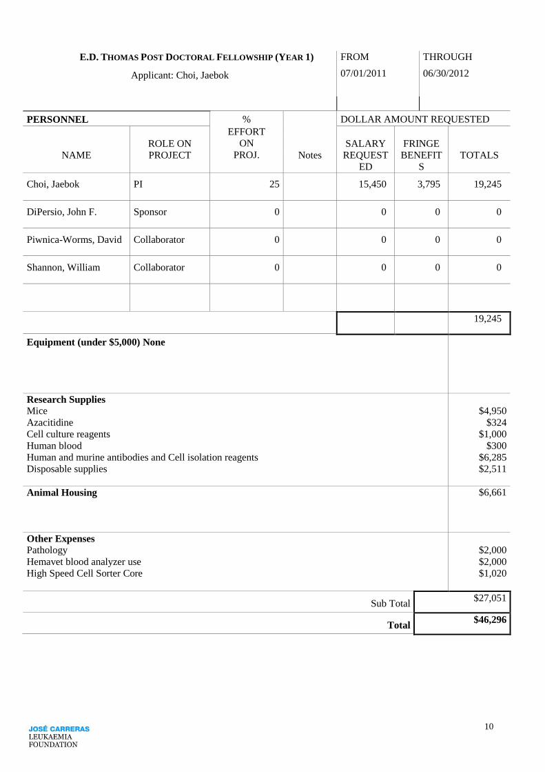
E.D. THOMAS POST DOCTORAL FELLOWSHIP (YEAR 1) FROM THROUGH
Applicant: Choi, Jaebok 07/01/2011 06/30/2012
PERSONNEL %
EFFORT
ON
PROJ.
DOLLAR AMOUNT REQUESTED
ROLE ON SALARY FRINGE
NAME PROJECT Notes REQUEST
ED
BENEFIT
S
TOTALS
Choi, Jaebok PI 25 15,450 3,795 19,245
DiPersio, John F. Sponsor 0 0 0 0
Piwnica-Worms, David Collaborator 0 0 0 0
Shannon, William Collaborator 0 0 0 0
19,245
Equipment (under $5,000) None
Research Supplies Mice
Azacitidine
Cell culture reagents
Human blood
Human and murine antibodies and Cell isolation reagents
Disposable supplies
$4,950
$324
$1,000
$300
$6,285
$2,511
Animal Housing $6,661
Other Expenses Pathology
Hemavet blood analyzer use
High Speed Cell Sorter Core
$2,000
$2,000
$1,020
Sub Total $27,051
Total $46,296

11
Applicant: Choi, Jaebok
Budget Justification for Direct Costs (Year 1)
Research Supplies
Mouse acquisition: The budget for mice is based on the anticipated purchase of 25 mice, Balb/c (transplant
recipients) and C57BL/6 (5.2+) (bone marrow donors), per month for 12 months ($16.50/mouse).
NOD/SCID/γcnull
(transplant recipients for a xenogeneic GvHD model), C57BL/6 (5.1+) (T cell donors), and
Foxp3 KO mice will be bred in house.
Total cost for mouse acquisition per year $4,950.
Azacitidine ($32.40/vial X 10).
Total cost for azacitidine per year $324.
Tissue culture reagents: T cells are isolated and cultured for transduction and expansion. Supplies include
media: Dulbecco Minimum Essential Medium (DMEM), Fetal Calf Serum, interleukin-2, and anti-
CD3/CD28 antibody coated beads.
Total cost for tissue culture per year $1,000.
Human blood for isolation peripheral blood mononuclear cells.
Total cost for human blood per year $300.
Antibodies and cell isolation reagents: Reagents for AutoMACS sorting including MACS columns,
magnetic beads and fluorochrome conjugated antibodies. Human Pan T isolation kits ($685 X 3), mouse pan
T isolation kits ($550 X 3), and mouse bone marrow isolation kit ($610 X 3), and antibodies (CD4, CD8 and
CD25: $250/antibody).
Total cost for antibodies and cell isolation reagents per year $6,285.
Disposable supplies: 6-well plates, 24-well plates, 70 um filter, pipettes (5, 10, and 25 mL), pipette tips (10,
200, 1000 ul), polypropylene tubes (15 and 50 mL), 12x75 FACS tubes, blood collection tubes and
hematocrit tubes.
Total cost for disposable supplies per year $2,511.
Animal housing NOD/SCID/γc
null, CD57BL/6 (5.1+), Foxp3-ires-GFP KI, and Foxp3 KO mice will be bred in house.
Housing is also required for the purchased mice such as Balb/c and CD57BL/6 (5.2+). Minimum of 25
cages housed daily @ $0.73/day X 365 days.
Total cost for animal housing per year $6,661.
Other Expenses
Pathology on murine tissue to evaluate GvHD and engraftment.
Total cost for pathology per year $2,000.
Hemavet for white blood cell counts.
4.00/sample X 500 samples.
Total cost for hemavet per year $2,000.
High Speed Cell Sorting: used to isolate various cell types 12 hours at $85/hour.
Total cost for high speed cell sorting per year $1020.

12
E.D. THOMAS POST DOCTORAL FELLOWSHIP (YEAR 2) FROM THROUGH
Applicant: Choi, Jaebok 07/01/2012 06/30/2013
PERSONNEL %
EFFORT
ON
PROJ.
DOLLAR AMOUNT REQUESTED
ROLE ON SALARY FRINGE
NAME PROJECT Notes REQUEST
ED
BENEFIT
S
TOTALS
Choi, Jaebok PI 25 15,914 4,055 19,969
DiPersio, John F. Sponsor 0 0 0 0
Piwnica-Worms, David Collaborator 0 0 0 0
Shannon, William Collaborator 0 0 0 0
19,969
Equipment (under $5,000) None
Research Supplies Mice
Azacitidine
Cell culture reagents
Human blood
Human and murine antibodies and Cell isolation reagents
Disposable supplies
$4,950
$324
$1,000
$300
$6,285
$1,787
Animal Housing $6,661
Other Expenses Pathology
Hemavet blood analyzer use
High Speed Cell Sorter
$2,000
$2,000
$1,020
Sub Total $26,327
Total $46,296

13
Applicant: Choi, Jaebok
Budget Justification for Direct Costs (Year 2)
Research Supplies
Mouse acquisition: The budget for mice is based on the anticipated purchase of 25 mice, Balb/c (transplant
recipients) and C57BL/6 (5.2+) (bone marrow donors), per month for 12 months ($16.50/mouse).
NOD/SCID/γcnull
(transplant recipients for a xenogeneic GvHD model), C57BL/6 (5.1+) (T cell donors),
Foxp3-ires-GFP KI, and Foxp3 KO mice will be bred in house.
Total cost for mouse acquisition per year $4,950.
Azacitidine ($32.40/vial X 10).
Total cost for azacitidine per year $324.
Tissue culture reagents: T cells are isolated and cultured for transduction and expansion. Supplies include
media: Dulbecco Minimum Essential Medium (DMEM), Fetal Calf Serum, interleukin-2, and anti-
CD3/CD28 antibody coated beads.
Total cost for tissue culture per year $1,000.
Human blood for isolation peripheral blood mononuclear cells.
Total cost for human blood per year $300.
Antibodies and cell isolation reagents: Reagents for AutoMACS sorting including MACS columns,
magnetic beads and fluorochrome conjugated antibodies. Human Pan T isolation kits ($685 X 3), mouse pan
T isolation kits ($550 X 3), and mouse bone marrow isolation kit ($610 X 3), and antibodies (CD4, CD8 and
CD25: $250/antibody).
Total cost for antibodies and cell isolation reagents per year $6,285.
Disposable supplies: 6-well plates, 24-well plates, 70 um filter, pipettes (5, 10, and 25 mL), pipette tips (10,
200, 1000 ul), polypropylene tubes (15 and 50 mL), 12x75 FACS tubes, blood collection tubes and
hematocrit tubes.
Total cost for disposable supplies per year $1,787.
Animal housing
NOD/SCID/γcnull
, CD57BL/6 (5.1+), Foxp3-ires-GFP KI, and Foxp3 KO mice will be bred in house.
Housing is also required for the purchased mice such as Balb/c and CD57BL/6 (5.2+). Minimum of 25
cages housed daily @ $0.73/day X 365 days.
Total cost for animal housing per year $6,661.
Other Expenses
Pathology on murine tissue to evaluate GvHD and engraftment.
Total cost for pathology per year $2,000.
Hemavet for white blood cell counts.
4.00/sample X 500 samples.
Total cost for hemavet per year $2,000.
High Speed Cell Sorting: used to isolate various cell types 12 hours at $85/hour.
Total cost for high speed cell sorting per year $1020.

14
E.D. THOMAS POST DOCTORAL FELLOWSHIP (YEAR 3) FROM THROUGH
Applicant: Choi, Jaebok 07/01/2013 06/30/2014
PERSONNEL %
EFFORT
ON
PROJ.
DOLLAR AMOUNT REQUESTED
ROLE ON SALARY FRINGE
NAME PROJECT Notes REQUEST
ED
BENEFIT
S
TOTALS
Choi, Jaebok PI 25 16,391 4,344 20,735
DiPersio, John F. Sponsor 0 0 0 0
Piwnica-Worms, David Collaborator 0 0 0 0
Shannon, William Collaborator 0 0 0 0
20,735
Equipment (under $5,000) None
Research Supplies Mice
Azacitidine
Cell culture reagents
Human blood
Human and murine antibodies and Cell isolation reagents
Disposable supplies
$4,950
$324
$1,000
$300
$6,285
$1,021
Animal housing
$6,661
Other Expenses Pathology
Hemavet blood analyzer use
High Speed Cell Sorter
$2,000
$2,000
$1,020
Sub Total $25,561
Total $46,296

15
Applicant: Choi, Jaebok
Budget Justification for Direct Costs (Year 3)
Research Supplies
Mouse acquisition: The budget for mice is based on the anticipated purchase of 25 mice, Balb/c (transplant
recipients) and C57BL/6 (5.2+) (bone marrow donors), per month for 12 months ($16.50/mouse).
NOD/SCID/γcnull
(transplant recipients for a xenogeneic GvHD model), C57BL/6 (5.1+) (T cell donors),
Foxp3-ires-GFP KI, and Foxp3 KO mice will be bred in house.
Total cost for mouse acquisition per year $4,950.
Azacitidine ($32.40/vial X 10).
Total cost for azacitidine per year $324.
Tissue culture reagents: T cells are isolated and cultured for transduction and expansion. Supplies include
media: Dulbecco Minimum Essential Medium (DMEM), Fetal Calf Serum, interleukin-2, and anti-
CD3/CD28 antibody coated beads.
Total cost for tissue culture per year $1,000.
Human blood for isolation peripheral blood mononuclear cells.
Total cost for human blood per year $300.
Antibodies and cell isolation reagents: Reagents for AutoMACS sorting including MACS columns,
magnetic beads and fluorochrome conjugated antibodies. Human Pan T isolation kits ($685 X 3), mouse pan
T isolation kits ($550 X 3), and mouse bone marrow isolation kit ($610 X 3), and antibodies (CD4, CD8 and
CD25: $250/antibody).
Total cost for antibodies and cell isolation reagents per year $6,285.
Disposable supplies: 6-well plates, 24-well plates, 70 um filter, pipettes (5, 10, and 25 mL), pipette tips (10,
200, 1000 ul), polypropylene tubes (15 and 50 mL), 12x75 FACS tubes, blood collection tubes and
hematocrit tubes.
Total cost for disposable supplies per year $1,021.
Animal housing
NOD/SCID/γcnull
, CD57BL/6 (5.1+), Foxp3-ires-GFP KI, and Foxp3 KO mice will be bred in house.
Housing is also required for the purchased mice such as Balb/c and CD57BL/6 (5.2+). Minimum of 25
cages housed daily @ $0.73/day X 365 days.
Total cost for animal housing per year $6,661.
Other Expenses
Pathology on murine tissue to evaluate GvHD and engraftment.
Total cost for pathology per year $2,000.
Hemavet for white blood cell counts.
4.00/sample X 500 samples.
Total cost for hemavet per year $2,000.
High Speed Cell Sorting: used to isolate various cell types 12 hours at $85/hour.
Total cost for high speed cell sorting per year $1020.

16
Form 5 Choi, Jaebok
Brief list of equipment and space available for the project. Limit 1 page.
A. Equipment.
The laboratory consists of two fume hoods, two tissue culture hoods, CO2 incubators, analytical balances,
spectrophotometer, -20ºC freezer, 2 -80ºC freezers, large double door refrigerators, complete electrophoresis
capacity (11 power supplies, 16 gel units, both horizontal and vertical), multiple centrifuges, ELISA plate
reader, a Coulter counter, state-of-the-art Becton Dickinson FACScan and Beckman Coulter Gallios. We
share the use of 2 ultracentrifuges, 4 preparative centrifuges, speed vac, autoclave, 37ºC warm room, 200 sq
ft. cold room and a dark room, with the members of the Division.
We interact extensively with other members of the Department of Medicine, Pathology, Biochemistry, and
Genetics at Washington University, who provide frequent consultation and advice regarding our work. We
also have access to community DNA oligonucleotide production and sequencing, amino acid sequencing,
and peptide production.
B. Space and Facilities.
1. Laboratory:
Dr. DiPersio’s laboratory is on the sixth floor of the Southwest Tower, Room 626. It consists of 1200 sq. ft.
of bench and desk space. I occupy two benches in the DiPersio lab and has adjacent desk space.
2. Animal:
The East McDonnell Transgenic Barrier Facility will house all of the animals used in this study. This facility
is managed by the Division of Comparative Medicine at Washington University Medical School, and meets
all local, state, and federal government guidelines for animal care.
3. Computer:
Dell Precision 670 and Phaser 8860 color printer.
4. Other:
The Siteman Cancer Center Bioinformatics Core, High Speed Cell Sorter Core (MoFlo, Dako Cytomation,
Inc.) and Small Animal Imaging Core are all available for our use.

17
Form 6 Choi, Jaebok
List the name, address phone number and Fax number, if available for the two persons you have requested to
supply letters of recommendation in addition to your sponsor.
Name Timothy J. Ley
Address 660 South Euclid Avenue
Campus Box 8007
St. Louis, MO 63110-1010, USA
e-mail [email protected] Telephone (314)362-9337 Fax (314)362-9333
Name David Piwnica-Worms
Address 510 South Kingshighway Blvd.
Campus Box 8225
St. Louis, MO 63110-1010, USA
e-mail [email protected] Telephone (314)362-9359 Fax (314)362-0152

18
Form 7 Choi, Jaebok
List all current and pending support for the applicant.
Current Molecular Imaging Center Pilot Research Project 2010 Awards (PI: Jaebok Choi) 02/01/10-12/31/10
Title: Epigenetic Control of GvHD and GvL using the Hypomethylating Agent Azacitidine.
$25,000 total
Translational Oncology Group (PI: Jaebok Choi) 07/01/10-06/30/11
Title: Effect of AzaC on Trafficking of AzaC-induced Tregs and Leukemia in a Murine Allogeneic
Transplant Model.
$15,000/year
Siteman Cancer Center Research Development Awards (PI: Jaebok Choi) 07/01/10-06/30/11
Title: Effect of AzaC on GvHD and GvL in Xenotransplant Models
$20,000/year
Pending N/A

Choi, Jaebok
1
Jaebok Choi, PhD
Name : Jaebok Choi (DOB: 01/13/1972)
Citizenship: South Korea (and Permanent Resident of USA)
Position Title: Research Instructor
Institution: Washington University School of Medicine
A. EDUCATION and TRAINING
03/1990 - 08/1996 B.S. (Genetic Engineering), Kyungpook National University,
Daegu, Korea (03/1991 ~ 10/1993: mandatory military service
in Korea)
01/1999 - 05/2001 M.A. (Biology), University of Nebraska at Omaha, Omaha,
NE 68182
Advisor: Bruce A. Chase, Ph.D.
Thesis: The mind-meld Gene Encoding a Drosophila ADAM (a
disintegrin and metalloprotease) Protein.
Teaching Assistant, Department of Biology, University of
Nebraska at Omaha, Omaha, NE
Instructed undergraduate laboratory courses in introductory biology,
microbiology and molecular biology.
08/2001 – 06/2006 Ph.D. (Biochemistry and Molecular Biology), Baylor College
of Medicine, Houston, TX 77030
Advisor: Anna P. Newman, Ph.D.
Thesis: Genetic Study of Cell Fusion Factors in C. elegans.
07/2006 – 06/2009 Postdoctoral Research Associate (Division of Oncology),
Washington University School of Medicine, St. Louis, MO
63110
Advisor: John F. DiPersio, M.D., Ph.D.
Epigenetic Regulation of Graft-versus-Host Disease (GvHD) and
Graft-versus-Leukemia (GvL) using Inhibitors of DNA Methylation.
07/2009 – Present Research Instructor (Division of Oncology), Washington
University School of Medicine, St. Louis, MO 63110
Mentor: John F. DiPersio, M.D., Ph.D.
In Vivo Administration of Hypomethylating Agents to Mitigate
GvHD While Preserving GvL.

Choi, Jaebok
2
B. RESEARCH AND PROFESSIONAL EXPERIENCE
MEMBERSHIP IN PROFESSIONAL SOCIETIES
06/26/2009 - present Research Associate Member, Siteman Cancer Center.
01/01/2010 - present Active Member, American Society of Hematology.
RESEARCH GRANTS/GIFTS
Completed
Bryan Thomas Campbell Foundation (PI: Jaebok Choi)
07/01/09-06/30/10
Title: Epigenetic Regulation of GvHD and GvL using Inhibitors of DNA Methylation.
$50,000/year
Active
Molecular Imaging Center Pilot Research Project 2010 Awards (PI: Jaebok Choi)
02/01/10-12/31/10
Title: Epigenetic Control of GvHD and GvL using the Hypomethylating Agent Azacitidine.
$25,000 total
Translational Oncology Group (PI: Jaebok Choi)
07/01/10-06/30/11
Title: Effect of AzaC on Trafficking of AzaC-induced Tregs and Leukemia in a Murine
Allogeneic Transplant Model.
$15,000/year
Siteman Cancer Center Research Development Awards (PI: Jaebok Choi)
07/01/10-06/30/11
Title: Effect of AzaC on GvHD and GvL in Xenotransplant Models.
$20,000/year
AWARDS
ASBMT Best Abstract Awards for Basic Science Research. Epigenetic
modulation of Foxp3: Generation of CD4+CD25- T cells with a
suppressor phenotype. 2009 international BMT Tandem Meeting. Tampa,
FL, February 11-15, 2009.
Keystone Symposia Scholarships/Awards. Epigenetic Therapy of GvHD after
Allogeneic Bone Marrow Transplantation. Keystone Symposia:
Developmental Origins and Epigenesis in Human Health and
Disease. Singapore, April 26-30, 2010.
C. BIBLIOGRAPHY
PUBLICATIONS
Choi J and Newman AP, “A two-promoter system of gene expression in C.
elegans” Deelopmental Biology 296:537-544, 2006.

Choi, Jaebok
3
Choi J, Richards KL, Cinar HN and Newman AP, “N-ethylmaleimide sensitive
factor is required for fusion of the C. elegans uterine anchor cell” Developmental
Biology 297:87-102, 2006.
Sapir A*, Choi J* (*equal contribution), Leikina E, Avinoam O, Valansi C,
Chernomordik LV, Newman AP, Podbilewicz B., “AFF-1, a FOS-1-regulated
fusogen, mediates fusion of the anchor cell in C. elegans.” Developmental Cell
12:683-698, 2007.
Choi J, Ritchey J, Prior J, Holt M, Shannon WD, Deych E, Piwnica-Worms
DR, DiPersio JF., “In vivo administration of hypomethylating agents mitigates
GvHD without sacrificing GvL” Blood 116:129-139, 2010.
ORAL PRESENTATIONS
Epigenetic modulation of Foxp3: Generation of CD4+CD25- T cells with a
suppressor phenotype. 2009 international BMT Tandem Meeting. Tampa,
FL, February 11-15, 2009.
Generation of Treg-like cells from CD4+CD25- T cells occurs via both Foxp3
dependent and independent pathways. 50th
International Annual Meeting
of American Society of Hematology. San Francisco, CA, December 6-9,
2008.
Generation of Treg-like cells from CD4+CD25- T cells via epigenetic
modification using a demethylating agent decitabine. 49th
International
Annual Meeting of American Society of Hematology. Atlanta, GA,
December 8-11, 2007.
Genetic Study of Cell Fusion Factors. Baylor College of Medicine Biochemistry
Department Annual Retreat. Galveston, TX, August 26-27, 2004.
Genetic Study of Cell Fusion Factors. Baylor College of Medicine Biochemistry
Department Annual Retreat. Columbia Lakes, TX, November 6-7, 2003.
POSTER PRESENTATIONS
Epigenetic control of GvHD and GvL using the hypomethylating agent
Azacitidine. 51th
International Annual Meeting of American Society of
Hematology. New Orleans, LA, December 5-8, 2009.
Mark A. Schroeder, Jonathan Ricks, Jaebok Choi, Julie K. Ritchey, Matthew
Holt, Brian Dieckgraefe, John F. DiPersio. Pegylated recombinant murine
GM-CSF is a potent mobilize of murine bone marrow progenitors,
synergizes with BIO5192 and Plerixafor (AMD3100), and skews
mobilized cells to a tolerogenic phenotype. 51th
International Annual
Meeting of American Society of Hematology. New Orleans, LA,
December 5-8, 2009.
Suppression of GvHD by Treg-like cells generated from non-Treg cells via
epigenetic modulation: Foxp3 might not be necessary. 2009 Keystone
Symposia: Regulatory T cells. Keystone, CO, March 1-6, 2009

Choi, Jaebok
4
Generation of Treg-like cells from CD4+CD25- T cells via epigenetic
modification using a demethylating agent decitabine. 2008 BMT Tandem
Meeting. San Diego, CA, February 13-17, 2008.
Generation of Treg-like cells from CD4+CD25- T cells via epigenetic
modification using a demethylating agent decitabine. 2008 Hematopoietic
Development and Malignancy Retreat, St. Louis, MO, June 2008.
Epigenetic and Genetic Approaches to Convert CD4+CD25- T cells into
CD4+CD25+ Treg-like T cells. 2007 Hematopoietic Development and
Malignancy Retreat, St. Louis, MO, June 2007.
Genetic Study of Cell Fusion Factors. Baylor College of Medicine Graduate
student symposium, Houston, TX, October 24, 2003.
Genetic Study of Cell Fusion Factors. 14th
international C. elegans conference,
Los Angeles, CA, June 29 – July 3, 2003.
Characterization of the mind-meld gene in Drosophila. 42nd
Annual Drosophila
Research Conference, Washington DC, March 21-25, 2001.
CURRICULUM VITAE
Personal Information Name John F. DiPersio
Date of Birth July 4, 1952
Citizenship USA
Education
1973 Williams College, BA Biology
1980 University of Rochester, MD, PhD, Microbiology
1980-81 Intern, Parkland Memorial Hosp, The University of Texas Southwestern Medical Center, Dallas, TX
1981-83 Resident, Parkland Memorial Hosp, Dallas, TX
1983-84 Chief Resident, Parkland Memorial Hosp, Dallas, TX
1984-87 Fellow, Division of Hematology/Oncology, UCLA School of Medicine
Professional Experience 2000 – Present Chief, Division of Oncology, Washington University School of Medicine, St. Louis, MO
2000 – 2006 Director, Section of BMT and Leukemia, Washington University School of Medicine, St. Louis, MO
2000 – Present Deputy Director, Siteman Cancer Center, Washington University School of Medicine, St. Louis, MO
2000 – 2003 Acting Director, Medical Oncology, Washington University School of Medicine, St. Louis, MO
1997 – Present Professor of Medicine, Pediatrics and Pathology/Immunology, Washington University School of Medicine, St.
Louis, MO
1994 – 2000 Chief, Division of Bone Marrow Transplantation and Stem Cell Biology, Washington University School of Medicine,
St. Louis, MO
1994 – 1997 Associate Professor of Medicine, Pediatrics and Pathology, Washington
University School of Medicine, St. Louis, MO
1990 – 1994 Assistant Professor of Medicine, Hematology Unit, University of Rochester School of Medicine, Rochester, New
York
1990 – 1994 Director, Bone Marrow Transplant Program, Strong Memorial Hospital, Rochester, NY
1990 – 1994 Assistant Professor of Oncology, University of Rochester School of Medicine and Dentistry, Rochester, NY
1988 – 1990 Assistant Professor of Medicine, Division of Hematology-Oncology, UCLA School of Medicine, Los Angeles, CA
1987 – 1988 Instructor of Medicine, Division of Hematology-Oncology, UCLA School of Medicine, Los Angeles, CA
1984 – 1987 Fellow, Division of Hematology-Oncology, UCLA School of Medicine, Los Angeles, CA
1983 – 1984 Chief Resident, Internal Medicine, Parkland Memorial Hospital, University of Texas, Southwestern Medical Center
at Dallas, TX
1981 – 1983 Resident, Internal Medicine, Parkland Memorial Hospital, Dallas, TX
1980 – 1981 Intern (Straight Medicine), Parkland Memorial Hospital, Dallas, TX
Selected Honors & Awards 1973 B.A., Magna Cum Laude, Honors in Biology, Williams College
1980 Alpha Omega Alpha, University of Rochester
1986 Special Fellow, Leukemia Society of America
1989 Junior Faculty Research Award, American Cancer Society
1992 Bauman Award - Teacher of the Year, Internal Medicine, University of Rochester
1993 Member, Editorial Board Journal of Experimental Hematology
1995 Member, Nominating Committee, International Society for Experimental Hematology
1996 Elected, American Society of Clinical Investigation (ASCI)
1996 Member, American Society of Clinical Investigation
1997 Chairman, Nominating Committee, International Society for Experimental Hematology (ISEH)
1997 Member, Stem Cell Evaluation Committee, International Society for Hematotherapy and Graft Engineering (ISHAGE)
1997 Councilor, International Society of Experimental Hematology
1997 Recipient, Lewis T. and Rosalind B. Apple Chair in Oncology
1998 Member, Barnard Free Skin and Cancer Board of Directors
1998 Guest Editor (3 yrs), Blood Journal
1999 – 2006 Member, External Advisory Panel, M.D. Anderson, (MDACC) PPG-AML
1999 Deputy Director, Siteman Cancer Center, Washington University School of Medicine
2000 – 2006 Leukemia and Lymphoma Society of America, Career Development Award
Study Section Member
2000 Chairman, NHLBI Consensus Conference on Allogeneic Transplantation for Non-Malignant Diseases Bethesda, MD
2000 ISHAGE Meeting, Quebec Canada. First Prize, Scientific Merit: “Control of GvHD using genetically modified T cells”
2001 Reviewer, University of Minnesota NCI-Designated Comprehensive Cancer Center Grant (NCF)
2002 – 2006 Member, Board of Directors IBMTR/ABMTR
2002 Member, ASH Committee on Educational Affairs, Washington D.C.
2003 – 2006 Member, Board of Directors, American Society of Biology and Marrow Transplant.
2003 Chairman, American Society of Biology and Marrow Transplant Council on Education and
Standards
2003 NHLBI P01 Study Section Member University of Illinois, Chicago
2004 Member, Board of Directors, Barnard Free Skin and Cancer Hospital.
Applicant: Choi, Jaebok

2003 – 2006 Study Section Member, American Society of Hematology Faculty and Fellow Scholar
Career Development Award
2004 External Advisor, Ohio State University P01 “Retrovirus Models of Lymphocyte Transformation and Disease”
2004 – 2009 NCI (NIH) Permanent Member CONC Study Section Member
2005 Member, Medical Advisory Board, the Bone Marrow Foundation
2005 Chairman, Study Section RFA HL-04-017 (NHLBI) Specialized Centers for Cell Based Therapy (SCCT). Annapolis, MD
2005 NIH/CSR Study Section ZRG1 - Ad Hoc Reviewer
2006 – 2009 Member, Clinical Oncology Study Section, Center for Scientific Review
2006 Member, K12 Advisory Committee
2006 – 2009 Mentor, K12 Awardee
2006 NIH (ZRG)/CSR – Gene Therapy and Inborn Errors – Special Emphasis Review Panel
2006 Member, ASH Committee on Gene and Cell Therapy
2007 Member, Editorial Board of Clinical and Translational Science
2007 Member, Editorial Board of Clinical Medicine: Oncology
2007 Lecturer, “Great Teachers Series of Medical Grand Rounds at the NIH Clinical Center in Bethesda, MD
2008 2008-12 Member ASH Scientific Committee on Hematopoietic Cytokines and Factors
2008 – 2010 Alternate Member, Scientific and Medical Research Funding Working Group of the California Institute of
Regenerative Medicine
2008 Member, ASH Scientific Committee on Hematopoietic Cytokines and Factors
2009 Member, AACR
2010 Member, College of CSR Reviewers
2010 Recipient, Virginia E. and Samuel J. Golman Endowed Professorship in Medicine
2010 Member, External Advisory Board, University of Texas Health Science Center at San Antonio, the Cancer Therapy and
Research Center
2010 Chairman, NIH NIAID ZAI1 MFH-1 P01 Special Emphasis Panel
Selected peer-reviewed publications (selected from 196):
1. Rettig MP, Ritchey JK, Meyerrose TE, Haug JS, DiPersio JF: Transduction and selection of human T cells with novel
CD34/thymidine kinase chimeric suicide genes for the treatment of graft-versus-host disease. Mol Ther 8:29-41, 2003.
2. Rettig MP, Ritchey JK, Prior JL, Haug JS, Piwnica-Worms D, DiPersio JF: Kinetics of in vivo elimination of suicide gene-
expressing T cells affects engraftment, graft-versus-host disease, and graft-versus-leukemia after allogeneic bone marrow
transplantation. J.Immunol, 173:3620-3630, 2004.
3. Devine SM, Brown RA, Mathews V, Trinkaus K, Khoury H, Adkins D, Vij R, Sempek D, Graubert T, Tomasson M,
Goodnough LT, DiPersio JF: Reduced risk of acute GVHD following mobilization of HLA-identical sibling donors with GM-
CSF alone. Bone Marrow Transpl 36:531-538, 2005.
4. Nervi B, Rettig MP, Ritchey JK, Wang HL, Bauer G, Walker J, Bonyhadi ML, Berenson RJ, Prior JL, Piwnica-Worms D, Nolta
JA, DiPersio JF. Factors affecting human T cell engraftment, trafficking and associated xenogeneic graft-vs. host disease in
NOD/SCID beta2m (null) mice. Exp Hematol 35:1823-1838, 2007.
5. Pusic, I., Jiang, S. Y., Landua, S., Uy, G. L., Rettig, M. P., Cashen, A. F., Westervelt, P., Vij, R., Abboud, C. N., Stockerl-
Goldstein, K. E., Sempek, D. S., Smith, A. L., and DiPersio, J. F. Impact of mobilization and remobilization strategies on
achieving sufficient stem cell yields for autologous transplantation. Biol Blood Marrow Transpl 14:1045-56; 2008.
6. Nervi, B., Pablo Ramirez, P., Rettig, M.P., Uy, G.L., Holt, M.S., Ritchey, Prior, J.L., Piwnica-Worms, D., Bridger, G Ley, T.J.,
and DiPersio, J.F. Chemosensitization of AML following mobilization by the CXCR4 antagonist AMD3100. Blood, 2009 Jun
11;113(24):6206-14 Epub 2008 Dec 2. PMCID: PMC2699239
7. Ley TJ, Mardis ER, Ding L, Fulton B, McLellan MD, Chen K, Dooling D, Dunford-Shore BH, McGrath S, Hickenbotham M,
Cook L, Abbott R, Larson DE, Koboldt DC, Pohl C, Smith S, Hawkins A, Abbott S, Locke D, Hillier LW, Miner T, Fulton L,
Magrini V, Wylie T, Glasscock J, Conyers J, Sander N, Shi X, Osborne JR, Minx P, Gordon D, Chinwalla A, Zhao Y, Ries RE,
Payton JE, Westervelt P, Tomasson MH, Watson M, Baty J, Ivanovich J, Heath S, Shannon WD, Nagarajan R, Walter MJ, Link
DC, Graubert TA, DiPersio JF, Wilson RK. DNA sequencing of a cytogenetically normal acute myeloid leukemia genome.
Nature. 2008 Nov 6;456(7218):66-72.. PMCID: PMC2303574.
8. DiPersio, J. F., Stadtmauer, E. A., Nademanee, A., Micallef, I. N., Stiff, P. J., Kaufman, J. L., Maziarz, R.T., Hosing, C.,
Fruehauf, S., Horwitz, M., Cooper, D., Bridger, G., Calandra, G. Plerixafor and G-CSF versus placebo and G-CSF to mobilize
hematopoietic stem cells for autologous stem cell transplantation in patients with multiple myeloma. Blood, 2009 Jun
4;113(23) 5720-6 Epub 2009 Apr 10. PMCID: PMC Journal – In Process.
9. Ramirez,P., Rettig, M., Uy
, G.L., Deych, E., Holt, M.S., Ritchey, J.K., and DiPersio, J.F. BIO5192, a small molecule
inhibitor of VLA-4, mobilizes hematopoietic stem and progenitor cells. Blood, 2009 (Epub ahead of print). PMCID:
PMC2727418
10. Mardis ER, Ding L, Dooling DJ, Larson DE, McLellan MD, Chen K, Koboldt DC, Fulton RS, Delehaunty KD, McGrath SD,
Fulton LA, Locke DP, Magrini VJ, Abbott RM, Vickery TL, Reed JS, Robinson JS, Wylie T, Smith SM, Carmichael L,
Eldred JM, Harris CC, Walker J, Peck JB, Du F, Dukes AF, Sanderson GE, Brummett AM, Clark E, McMichael JF, Meyer
RJ, Schindler JK, Pohl CS, Wallis JW, Shi X, Lin L, Schmidt H, Tang Y, Haipek C, Wiechert ME, Ivy JV, Kalicki J, Elliott
G, Ries RE, Payton JE, Westervelt P, Tomasson MH, Watson MA, Baty J, Heath S, Shannon WD, Nagarajan R, Link DC,
Walter MJ, Graubert TA, DiPersio JF, Wilson RK, Ley TJ. Reucrring mutations found by sequencing an acute myeloid
leukemia genome. N Engl J Med. 2009 Aug 5.[Epub ahead of print). PMCID: PMC Journal – In Process
11. Choi J, Ritchey J, Prior J, Holt M, Shannon WD, Deych E, Piwnica-Worms DR, DiPersio JF., In vivo administration of
hypomethylating agents mitigate graft-versus-host disease without sacrificing graft-versus-leukemia. Blood 116:129-139,
2010.
Applicant: Choi, Jaebok

doi:10.1182/blood-2009-12-257253 Prepublished online Apr 27, 2010;2010 116: 129-139
Piwnica-Worms and John F. DiPersio Jaebok Choi, Julie Ritchey, Julie L. Prior, Matthew Holt, William D. Shannon, Elena Deych, David R.
graft-versus-host disease without sacrificing graft-versus-leukemiaIn vivo administration of hypomethylating agents mitigate
http://bloodjournal.hematologylibrary.org/cgi/content/full/116/1/129Updated information and services can be found at:
(1542 articles)Transplantation � collections: BloodArticles on similar topics may be found in the following
http://bloodjournal.hematologylibrary.org/misc/rights.dtl#repub_requestsInformation about reproducing this article in parts or in its entirety may be found online at:
http://bloodjournal.hematologylibrary.org/misc/rights.dtl#reprintsInformation about ordering reprints may be found online at:
http://bloodjournal.hematologylibrary.org/subscriptions/index.dtlInformation about subscriptions and ASH membership may be found online at:
. Hematology; all rights reservedCopyright 2007 by The American Society of 200, Washington DC 20036.semimonthly by the American Society of Hematology, 1900 M St, NW, Suite Blood (print ISSN 0006-4971, online ISSN 1528-0020), is published
only.For personal use at WASHINGTON UNIV SCH MEDICINE on October 12, 2010. www.bloodjournal.orgFrom
TRANSPLANTATION
In vivo administration of hypomethylating agents mitigate graft-versus-hostdisease without sacrificing graft-versus-leukemiaJaebok Choi,1 Julie Ritchey,1 Julie L. Prior,2 Matthew Holt,1 William D. Shannon,3 Elena Deych,3 David R. Piwnica-Worms,2
and John F. DiPersio1
1Division of Oncology, Department of Medicine, 2Molecular Imaging Center, Mallinckrodt Institute of Radiology and Department of Developmental Biology, and3Division of General Medical Sciences, Department of Medicine, Washington University, St Louis, MO
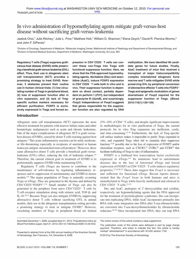
Regulatory T cells (Tregs) suppress graft-versus-host disease (GVHD) while preserv-ing a beneficial graft-versus-leukemia (GVL)effect. Thus, their use in allogeneic stemcell transplantation (SCT) provides apromising strategy to treat GVHD. How-ever, 3 obstacles prevent their routineuse in human clinical trials: (1) low circu-lating number of Tregs in peripheral blood,(2) loss of suppressor function after invitro expansion, and (3) lack of Treg-specific surface markers necessary forefficient purification. FOXP3 is exclu-sively expressed in Tregs and forced ex-
pression in CD4�CD25� T cells can con-vert these non-Tregs into Tregs withfunctional suppressor function. Here, weshow that the FDA-approved hypomethy-lating agents, decitabine (Dec) and azaci-tidine (AzaC), induce FOXP3 expressionin CD4�CD25� T cells both in vitro and invivo. Their suppressor function is depen-dent on direct contact, partially depen-dent on perforin 1 (Prf1), but independentof granzyme B (GzmB), and surprisingly,Foxp3. Independence of Foxp3 suggeststhat genes responsible for the suppres-sor function are also regulated by DNA
methylation. We have identified 48 candi-date genes for future studies. Finally,AzaC treatment of mice that received atransplant of major histocompatibilitycomplex mismatched allogeneic bonemarrow and T cells mitigates GVHD whilepreserving GVL by peripheral conversionof alloreactive effector T cells into FOXP3�
Tregs and epigenetic modulation of genesdownstream of Foxp3 required for thesuppressor function of Tregs. (Blood.2010;116(1):129-139)
Introduction
Allogeneic stem cell transplantation (SCT) represents the mosteffective treatment for patients with marrow failure states and otherhematologic malignancies such as acute and chronic leukemias.One of the major complications of allogeneic SCT is graft-versus-host disease (GVHD), caused by donor T cells reacting against hostantigens.1 This acute inflammatory reaction can be mild, moderate,or life-threatening especially in recipients of unrelated or humanleukocyte antigen–mismatched stem cell products.2 However, thesesame alloreactive donor T cells provide a beneficial graft-versus-leukemia (GVL) effect, reducing the risk of leukemia relapse.3,4
Therefore, the current clinical goal in treatment of GVHD is topreferentially suppress GVHD while maintaining GVL.
Regulatory T cells (Tregs) are known to contribute to themaintenance of self-tolerance by regulating inflammatory re-sponses and to suppression of autoimmunity and GVHD in mousemodels.5-9 The major population of Tregs is naturally occurringTregs or nTregs. They are generated in the thymus and defined byCD4�CD25�FOXP3�.5-8 Small number of Tregs can also begenerated in the periphery from naive CD4�CD25� T cells byT cell–receptor stimulation along with retinoic acid, TGF-�, andIL-10.10,11 Because Tregs can also mitigate GVHD by suppressingalloreactive donor T cells without sacrificing GVL in animalmodels, their use in the allogeneic transplantation setting providesa promising strategy to treat or mitigate GVHD.9 However,circulating numbers of Tregs in peripheral blood are limited
(5%–10% of CD4� T cells), and despite significant improvementsin methodologies for in vitro purification of Tregs, the currentprotocols for in vitro Treg expansion are inefficient, costly,and time-consuming.12-15 Furthermore, the lack of Treg-specificcell surface markers makes it impossible to purify Tregs expanded invitro, and expanded Tregs often fail to maintain their suppressorfunction,13,16 possibly due to the loss of expression of FOXP3 and/orchemokine receptors, such as CXCR3,17 CCR6,18 and CCR819 thatfacilitate trafficking of Tregs to sites of inflammation.
FOXP3 is a forkhead box transcription factor exclusivelyexpressed in nTregs.5-8 Its mutations lead to autoimmunediseases due to the loss of functional nTregs and forcedexpression of FOXP3 in CD4�CD25� T cells induces regulatoryproperties.5,7,8,20-22 These data suggest that Foxp3 is necessaryand sufficient for functional nTregs. Recent reports demon-strated that the Foxp3 locus in both humans and mice isunmethylated in Tregs while heavily methylated and silenced inCD4�CD25� T cells.23-25
Dec and AzaC, analogues of 2�-deoxycytidine and cytidine,respectively, are hypomethylating agents that the FDA approvedfor the treatment of myelodysplastic syndromes. Dec can incorpo-rate into replicating DNA, while AzaC incorporates primarily intoRNA with some integration into DNA after 5-aza-ribonucleotidesare converted into 5-aza-deoxyribonucleotides by ribonucleotidereductase.26-29 Once incorporated into DNA, they can trap DNA
Submitted December 1, 2009; accepted April 21, 2010. Prepublished online asBlood First Edition paper; April 27, 2010; DOI 10.1182/blood-2009-12-257253.
Presented in abstract form at the 50th annual meeting of the American Societyof Hematology, San Francisco, CA, December 8, 2008.
The online version of this article contains a data supplement.
The publication costs of this article were defrayed in part by page chargepayment. Therefore, and solely to indicate this fact, this article is herebymarked ‘‘advertisement’’ in accordance with 18 USC section 1734.
© 2010 by The American Society of Hematology
129BLOOD, 8 JULY 2010 � VOLUME 116, NUMBER 1
only.For personal use at WASHINGTON UNIV SCH MEDICINE on October 12, 2010. www.bloodjournal.orgFrom

methyltransferase 1 (DNMT1),30 thereby inhibiting DNAmethylation.27
Based on these reports, we hypothesized that Dec and AzaCcould be used to induce the expression of FOXP3 in CD4�CD25�
T cells via epigenetic modification and convert these non-Tregsinto Tregs. In this study, we report that these drugs induce theexpression of Foxp3 in activated CD4�CD25� T cells generatingfunctional Tregs with suppressor properties. We further demon-strate that in vivo treatment of mice with AzaC after allogeneicSCT dramatically mitigates GVHD while preserving GVL at leastin part by increasing the peripheral conversion of CD4�CD25�
alloreactive T effector cells (Teffs) into functionally suppressiveFOXP3� Tregs. In addition, the suppressor function of theseAzaC-induced Tregs is independent of Foxp3, suggesting thatAzaC modifies the expression of not only Foxp3 but also othergenes that are necessary for Treg suppressor function. Thus, ourstudy suggests that epigenetic modulation of events distal to Foxp3is also a critical mechanism by which in vivo administration ofAzaC controls GVHD. Our study provides a solid foundation for apharmacologic therapy to limit GVHD without sacrificing GVL.
Methods
Mice
Balb/c (H-2Kd, CD45.2�) and C57BL/6 (B6; H-2Kb, CD45.2�) mice wereobtained from Taconic Farms. Congenic B6 mice expressing the CD45.1gene were purchased from The Jackson Laboratory. Animal care andeuthanasia were approved by the Washington University School of Medi-cine Animal Studies Committee. Six- to 12-week-old mice were usedexcept for Foxp3 KO experiments in which 3-week-old Foxp3 KO (B6background) mice5 and littermate control mice were used.
Cell culture
Human peripheral blood mononuclear cells (PBMCs) were harvested byficoll gradient centrifugation. CD4�CD25� T cells and CD4�CD25�
T cells were isolated from the human PBMCs using Miltenyi microbeadsand AutoMACS (Miltenyi Biotec).31 The isolated CD4�CD25� T cellswere activated for 2 to 3 days in the presence of anti-CD3/CD28 beads(bead:cell � 1:1; Invitrogen) and Stemline T-cell expansion medium (Sigma-Aldrich) supplemented with L-glutamine (4mM), penicillin (100 U/mL),streptomycin (100 �g/mL), and human recombinant IL-2 (hIL-2; 50 U/mL).The activated T cells were incubated in the presence of Dec (0.1-10�M;Sigma-Aldrich) or phosphate-buffered saline (PBS) for an additional 1 to4 days. Mouse CD4�CD25�T cells and CD4�CD25� T cells were isolatedfrom mouse spleens using Miltenyi microbeads and AutoMACS. Theisolated CD4�CD25� T cells were activated for 2 days (2 or 4 days forFoxp3 KO and littermate controls) in the presence of beads (bead:cell � 1:1) and Dulbecco modified Eagle medium supplemented with 10% FCS,L-glutamine (2mM), penicillin (100 U/mL), streptomycin (100 �g/mL),MEM nonessential amino acids (1�), sodium pyruvate (1mM), HEPESbuffer (20mM), 2-mercaptoethanol (50�M) and hIL-2 (10 U/mL), calledXcyte media. The activated T cells were incubated in the presence of Dec(0.1-10�M), AzaC (0.5-4�M), or PBS for an additional 2 days.
Real-time RT-PCR
Human CD4�CD25� T cells that were activated with beads for 3 days(defined as day 0) and incubated in the presence of Dec or PBS foradditional 1 to 4 days (day 1 through day 4) as described in the previousparagraph. Total RNA was isolated from these cells every day (day 0through day 4) using the RNeasy Plus Mini Kit (QIAGEN). The 7300 RealTime PCR system (Applied Biosystems), QuantiTect Primer Assaysprimers (GAPDH as an internal control), and QuantiTect SYBR GreenRT-PCR kits (QIAGEN) were used.
RNA profiling analysis
Total RNA was isolated from the following cells (all B6, CD45.2�) usingTRIzol Reagent (Invitrogen): CD4�CD25� naive Tregs, CD4�CD25�
Tregs that were incubated in Xcyte media (100 U/mL hIL-2) for 4 days inthe presence of the beads (bead:cell � 1:1), PBS-treated T cells (pbsTs),and Dec-induced Tregs (dcTs). Target preparation, gene chip hybridization,and analysis were performed by the Washington University Siteman CancerCenter Gene Chip Facility. Labeled target was made from non-amplifiedtotal RNA and was hybridized to Mouse Genome 430 2.0 array (Affymetrix).
Flow cytometric analysis
The following antibodies were used. For human T cells: CD4-APC,CD25-FITC (BD Pharmingen), and FOXP3-PE (clone PCH101; eBio-science), for mouse T cells: CD4-FITC, CD25-PE (BD Pharmingen),Foxp3-PE and Foxp3-PECy5 (clone FJK-16s; eBioscience), and for SCT:H-2Kb-FITC, CD4-PE, CD3-PECy7, B220-APC and CD45.2-biotin/streptavidin APC-Cy7 (BD Pharmingen). All cells were analyzed on aFACScan cytometer (BD Biosciences).
CFSE-based proliferation assays and MLR
CD4�CD25� T cells (B6, CD45.1) were labeled with carboxyfluoresceindiacetate, succinimidyl ester (CFSE) at a final concentration of 300nM. TheCFSE-labeled cells were incubated in 200 �L of Xcyte media withsuppressors, such as CD4�CD25� nTregs, dcTs, azacTs, or pbsTs, (all B6,CD45.2), and �-irradiated (20 Gy) splenocytes/antigen presenting cells(APCs; Balb/c, CD45.2) for mixed lymphocyte reaction (MLR) or beads forproliferation assays. 1:1:1 � Teff:bead/APC:suppressor ratio was usedunless otherwise indicated. DcTs and azacTs are extensively washed withPBS (2�) before coculture with T effectors, thus T effectors are notexposed to any detectable levels of decitabine or azacitidine.
SCT
SCT was performed as previously described31 with the following modifica-tions. Total-body irradiation (TBI; 900 cGy) using a Mark I cesiumirradiator (J. L. Shepherd and Associates) was used to condition recipientmice (Balb/c). B6 mice (CD45.1 or CD45.2) T cell–depleted (TCD) bonemarrow (BM; 5 � 106 cells) was used as a stem cell source. To induceGVHD, 5 � 105 B6 conventional CD4�/CD8� T cells (Tconv; CD45.1)were infused along with donor (B6) TCD BM. To test suppressor functionof dcTs, 5 � 105 B6 CD4�CD25� nTregs (CD45.2; for control), pbsTs ordcTs (CD45.2) generated from CD4�CD25� T cells were injected withTCD BM and Tconv. In some cases, 10 � 106 pbsTs or dcTs (CD45.2)generated from Tconv were given in the place of Tconv and pbsTs or dcTsthat were derived from CD4�CD25� T cells. For delayed donor lympho-cyte infusions, 2 � 106 or 10 � 106 Tconv were given on day 11 after SCT.For examination of GVL effect, 1 � 104 A20-luc/egfp leukemic cells weregiven along with TCD BM. Animals losing 20% of their starting bodyweights were killed.
BLI
GVL effect assessment and bioluminescence imaging (BLI) of animalswere done as previously described.31,32 Briefly, mice were injected intra-peritoneally with 150 �g/g D-luciferin (Biosynth) in PBS and imaged10 minutes later. Imaging was done using a charge-coupled device camera(IVIS 100; Caliper Corporation; exposure time, 1-60 seconds; binning, 8;field of view, 15; f/stop 1, open filter) at the Molecular Imaging Center(Washington University). Mice were anesthetized using isoflourane (2.5%vaporized in O2). For analysis, total photon flux (photons per second) wasmeasured from a fixed region of interest over the entire abdomen and thoraxusing Living Image 2.50 and IgorPro software (Wavemetrics). BLI wasperformed 1 day before donor T-cell infusion and the first injection of AzaCand 3 days after the last injection of AzaC, then once every week untilday 52 after SCT.
130 CHOI et al BLOOD, 8 JULY 2010 � VOLUME 116, NUMBER 1 only.For personal use at WASHINGTON UNIV SCH MEDICINE on October 12, 2010. www.bloodjournal.orgFrom

Administration of Dec and AzaC
AzaC and Dec were prepared in cold PBS and were injected 4 timessubcutaneously or intraperitoneally every other day starting on day 4after donor lymphocyte infusion (day 15 after SCT). Foxp3 KO micewere also injected 4 times subcutaneously every other day starting at16 to 26 days of age.
Statistical analysis
The significance of differences in survival of treatment groups wasanalyzed using the log-rank test. For proliferation assays and MLR,unpaired t test was used. P values less than .05 were considered significant.For RNA profiling analyses, the probes were identified using GA-Mantelmethod.33 This method uses genetic algorithm strategy to select probes thatoptimize the Mantel correlation among the samples. GA-Mantel was run100 times and the probes that appeared in at least 30 of 100 solutions werepicked. All analyses were performed using SAS Version 9.1 (SAS Institute).
Results
The effect of hypomethylating agents on FOXP3 expression inhuman and mouse CD4�CD25�FOXP3� T cells
Because the hypomethylating effect of Dec and AzaC can onlyoccur in cells actively undergoing cellular division,29 we stimulatedhuman CD4�CD25� T cells using anti-CD3/CD28 antibody-coated beads and hIL-2 (50 U/mL) before treatment with variousconcentrations of Dec and AzaC. Approximately 60% of Dec-treated CD4�CD25� T cells (dcTs) and only approximately 10% ofpbsTs expressed FOXP3 (Figure 1A). Maximum FOXP3 expres-sion was achieved after exposure to 5 to 10�M Dec for
4 days. However, cell counts significantly declined when T cellswere exposed to concentrations of Dec in excess of 5�M (cellcounts were 69.7% 14.1% of the 1�M Dec cell counts [n � 4]).Treating activated CD4�CD25� T cells or conventional T cells(Tconv; isolated using Miltenyi pan T-cell isolation kit) with 1�MDec for 4 days optimized both cell expansion and FOXP3expression. Real-time reverse transcription–polymerase chain reac-tion (RT-PCR) demonstrated levels of mRNA for FOXP3 that werecomparable to those seen in bead-activated nTregs (10- to 12-foldincrease above baseline; Figure 1B). Dec also induced FOXP3expression in approximately 80% of bead-activated murineCD4�CD25� T cells (Figure 1A). Consistent with these results,both Dec and AzaC markedly increased GFP expression inactivated CD4�CD25� T cells obtained from Foxp3-ires-GFPknock-in (KI) mice34 (Figure 1C). To determine the duration ofFOXP3 expression in dcTs, we stained for intracellular FOXP3daily for 7 days and found that approximately 50% of dcTsremained FOXP3� at day 7 (Figure 1D). These results suggest thatboth hypomethylating agents induce both FOXP3 mRNA andprotein expression in CD4�CD25� non-Tregs.
Suppressor function of hypomethylating agent-induced Tregsin vitro
We examined whether dcTs and AzaC-induced Tregs (azacTs) aresuppressive in proliferation assays in which anti-CD3/CD28 beadswere used as stimulators (Figure 2, supplemental Figures 1-2,available on the Blood Web site; see the Supplemental Materialslink at the top of the online article). Both dcTs and azacTs were ableto function as suppressors, while pbsTs had no effect (Figure 2A,
Figure 1. Effect of Dec on FOXP3 expression in anti-CD3/CD28 antibody coated bead-activated CD4�CD25� T cells. (A) Both human and mouse CD4�CD25� T cellsexpress FOXP3 after Dec treatment in the presence of anti-CD3/CD28 antibody coated beads. (B) Real-time RT-PCR for human FOXP3 mRNA was performed in triplicate atvarious times after Dec treatment of activated T cells. Levels of mRNA for FOXP3 increase each day and are comparable to that seen in bead-activated Tregs (brown) by day 4.(C) CD4�CD25� T cells obtained from Foxp3-ires-GFP KI mice confirm induction of FOXP3 expression after Dec and AzaC treatment (indirectly measured as GFPexpression). (D) DcT FOXP3 expression (blue) persists for at least 7 days, suggesting prolonged and stable expression of FOXP3 in vitro. Treg (red) is shown for comparison.A pool of 2 independent experiments.
HYPOMETHYLATING AGENTS AND GVHD 131BLOOD, 8 JULY 2010 � VOLUME 116, NUMBER 1 only.For personal use at WASHINGTON UNIV SCH MEDICINE on October 12, 2010. www.bloodjournal.orgFrom
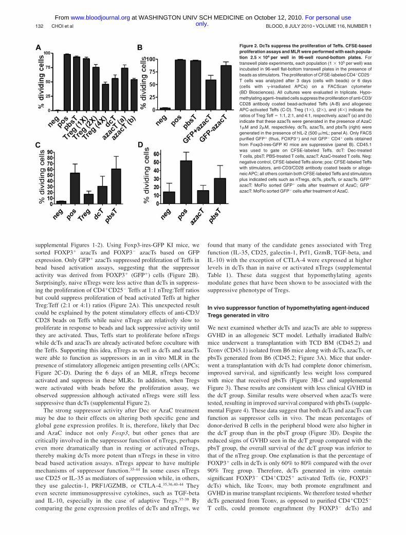
supplemental Figures 1-2). Using Foxp3-ires-GFP KI mice, wesorted FOXP3� azacTs and FOXP3� azacTs based on GFPexpression. Only GFP� azacTs suppressed proliferation of Teffs inbead based activation assays, suggesting that the suppressoractivity was derived from FOXP3� (GFP�) cells (Figure 2B).Surprisingly, naive nTregs were less active than dcTs in suppress-ing the proliferation of CD4�CD25� Teffs at 1:1 nTreg:Teff ratiosbut could suppress proliferation of bead activated Teffs at higherTreg:Teff (2:1 or 4:1) ratios (Figure 2A). This unexpected resultcould be explained by the potent stimulatory effects of anti-CD3/CD28 beads on Teffs while naive nTregs are relatively slow toproliferate in response to beads and lack suppressive activity untilthey are activated. Thus, Teffs start to proliferate before nTregswhile dcTs and azacTs are already activated before coculture withthe Teffs. Supporting this idea, nTregs as well as dcTs and azacTswere able to function as suppressors in an in vitro MLR in thepresence of stimulatory allogeneic antigen presenting cells (APCs;Figure 2C-D). During the 6 days of an MLR, nTregs becomeactivated and suppress in these MLRs. In addition, when Tregswere activated with beads before the proliferation assay, weobserved suppression although activated nTregs were still lesssuppressive than dcTs (supplemental Figure 2).
The strong suppressor activity after Dec or AzaC treatmentmay be due to their effects on altering both specific gene andglobal gene expression profiles. It is, therefore, likely that Decand AzaC induce not only Foxp3, but other genes that arecritically involved in the suppressor function of nTregs, perhapseven more dramatically than in resting or activated nTregs,thereby making dcTs more potent than nTregs in these in vitrobead based activation assays. nTregs appear to have multiplemechanisms of suppressor function.35-44 In some cases nTregsuse CD25 or IL-35 as mediators of suppression while, in others,they use galectin-1, PRF1/GZMB, or CTLA-4.35,36,40-44 Theyeven secrete immunosuppressive cytokines, such as TGF-betaand IL-10, especially in the case of adaptive Tregs.37-39 Bycomparing the gene expression profiles of dcTs and nTregs, we
found that many of the candidate genes associated with Tregfunction (IL-35, CD25, galectin-1, Prf1, GzmB, TGF-beta, andIL-10) with the exception of CTLA-4 were expressed at higherlevels in dcTs than in naive or activated nTregs (supplementalTable 1). These data suggest that hypomethylating agentsmodulate genes that have been shown to be associated with thesuppressive phenotype of Tregs.
In vivo suppressor function of hypomethylating agent-inducedTregs generated in vitro
We next examined whether dcTs and azacTs are able to suppressGVHD in an allogeneic SCT model. Lethally irradiated Balb/cmice underwent a transplantation with TCD BM (CD45.2) andTconv (CD45.1) isolated from B6 mice along with dcTs, azacTs, orpbsTs generated from B6 (CD45.2; Figure 3A). Mice that under-went a transplantation with dcTs had complete donor chimerism,improved survival, and significantly less weight loss comparedwith mice that received pbsTs (Figure 3B-C and supplementalFigure 3). These results are consistent with less clinical GVHD inthe dcT group. Similar results were observed when azacTs weretested, resulting in improved survival compared with pbsTs (supple-mental Figure 4). These data suggest that both dcTs and azacTs canfunction as suppressor cells in vivo. The mean percentages ofdonor-derived B cells in the peripheral blood were also higher inthe dcT group than in the pbsT group (Figure 3D). Despite thereduced signs of GVHD seen in the dcT group compared with thepbsT group, the overall survival of the dcT group was inferior tothat of the nTreg group. One explanation is that the percentage ofFOXP3� cells in dcTs is only 60% to 80% compared with the over90% Treg group. Therefore, dcTs generated in vitro containsignificant FOXP3� CD4�CD25� activated Teffs (ie, FOXP3�
dcTs) which, like Tconv, may both promote engraftment andGVHD in murine transplant recipients. We therefore tested whetherdcTs generated from Tconv, as opposed to purified CD4�CD25�
T cells, could promote engraftment (by FOXP3� dcTs) and
Figure 2. DcTs suppress the proliferation of Teffs. CFSE-basedproliferation assays and MLR were performed with each popula-tion 2.5 � 104 per well in 96-well round-bottom plates. Fortranswell plate experiments, each population (1 � 105 per well) wasincubated in 96-well flat-bottom transwell plates in the presence ofbeads as stimulators. The proliferation of CFSE-labeled CD4�CD25�
T cells was analyzed after 3 days (cells with beads) or 6 days(cells with �-irradiated APCs) on a FACScan cytometer(BD Biosciences). All cultures were evaluated in triplicate. Hypo-methylating agent–treated cells suppress the proliferation of anti-CD3/CD28 antibody coated bead-activated Teffs (A-B) and allogeneicAPC-activated Teffs (C-D). Treg (1�), (2�), and (4�) indicate theratios of Treg:Teff � 1:1, 2:1, and 4:1, respectively. azacT (a) and (b)indicate that these azacTs were generated in the presence of AzaC1�M and 2�M, respectivley. dcTs, azacTs, and pbsTs (right) weregenerated in the presence of hIL-2 (500 �/mL; panel A). Only FACSpurified GFP� (thus, FOXP3�) and not GFP� CD4� cells obtainedfrom Foxp3-ires-GFP KI mice are suppressive (panel B). CD45.1was used to gate on CFSE-labeled Teffs. dcT: Dec-treatedT cells, pbsT: PBS-treated T cells, azacT: AzaC-treated T cells. Neg:negative control, CFSE-labeled Teffs alone; pos: CFSE-labeled Teffswith stimulators, anti-CD3/CD28 antibody coated beads or alloge-neic APC; all others contain both CFSE-labeled Teffs and stimulatorsplus indicated cells such as nTregs, dcTs, pbsTs, or azacTs. GFP�
azacT: MoFlo sorted GFP� cells after treatment of AzaC; GFP�
azacT: MoFlo sorted GFP� cells after treatment of AzaC.
132 CHOI et al BLOOD, 8 JULY 2010 � VOLUME 116, NUMBER 1 only.For personal use at WASHINGTON UNIV SCH MEDICINE on October 12, 2010. www.bloodjournal.orgFrom
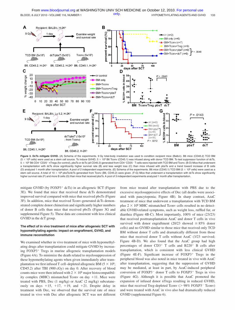
mitigate GVHD (by FOXP3� dcTs) in an allogeneic SCT (Figure3E). We found that mice that received these dcTs demonstratedimproved survival compared with mice that received pbsTs (Figure3F). In addition, mice that received Tconv-generated dcTs demon-strated complete donor chimerism and significantly higher numbersof donor B cells than mice that received pbsTs (Figure 3G andsupplemental Figure 5). These data are consistent with less clinicalGVHD in the dcT group.
The effect of in vivo treatment of mice after allogeneic SCT withhypomethylating agents: impact on engraftment, GVHD, andimmune reconstitution
We examined whether in vivo treatment of mice with hypomethyl-ating drugs after transplantation could mitigate GVHD by increas-ing FOXP3� Tregs in murine allogeneic transplantation models(Figure 4A). To minimize the death related to myelosuppression ofthese hypomethylating agents when given immediately after trans-plantation we first infused T cell–depleted allogeneic BM (5 � 106;CD45.2) after TBI (900 cGy) on day 0. After recovery of bloodcounts mice were then infused with 2 � 106 major histocompatibil-ity complex (MHC) mismatched Tconv on day �11. Mice weretreated with PBS, Dec (1 mg/kg) or AzaC (2 mg/kg) subcutane-ously on days �15, �17, �19, and �21. Despite delay intreatment with Dec, we observed that the survival rate of micetreated in vivo with Dec after allogeneic SCT was not different
from mice treated after transplantation with PBS due to theexcessive myelosuppressive effects of Dec (all deaths were associ-ated with pancytopenia; Figure 4B). In sharp contrast, AzaCtreatment of mice that underwent a transplantation with TCD BMplus 2 � 106 MHC mismatched Tconv cells resulted in no detect-able GVHD-related symptoms, such as weight loss, ruffled fur, ordiarrhea (Figure 4B-C). Most importantly, 100% of mice (23/23)that received posttransplantation AzaC and donor T cells in vivosurvived with donor engraftment (20/23 showed � 85% donorcells) and no GVHD similar to those mice that received only TCDBM without donor T cells and dramatically different from thosemice that received donor T cells without AzaC (3/23 survived;Figure 4B-D). We also found that the AzaC group had highpercentages of donor CD3� T cells and B220� B cells aftertransplantation, which is consistent with less clinical GVHD(Figure 4E-F). Significant increase of FOXP3� Tregs in theperipheral blood was also noted in mice treated in vivo with AzaCafter transplantation, suggesting that the suppression of GVHDmay be mediated, at least in part, by AzaC-induced peripheralconversion of FOXP3� donor T cells to FOXP3� Tregs in vivo(Figure 4G). Although it is possible that AzaC promoted theexpansion of infused donor nTregs resulting in reduced GVHD,mice that received Treg-depleted Tconv ( 98% FOXP3� Tconv)and were treated with AzaC in vivo also had dramatically reducedGVHD (supplemental Figure 6).
Figure 3. DcTs mitigate GVHD. (A) Schema of the experiments. 9 Gy total-body irradiation was used to condition recipient mice (Balb/c). B6 mice (CD45.2) TCD BM(5 � 106 cells) were used as a stem cell source. To induce GVHD, 5 � 105 B6 Tconv (CD45.1) was infused along with donor TCD BM. To test suppressor function of dcTs,5 � 105 B6 CD4�CD25� nTregs (for control), pbsTs or dcTs (all CD45.2) generated from CD4�CD25� T cells were injected with TCD BM and Tconv. (B-D) Mice that underwenta transplantation with dcTs show significantly higher survival rate (B) and less weight loss (C) than mice infused with pbsTs and a trend toward increase of B cells(D) analyzed 1 month after transplantation. A pool of 2 independent experiments. (E) Schema of the experiments. B6 mice (CD45.1) TCD BM (5 � 106 cells) were used as astem cell source. A total of 10 � 106 pbsTs/dcTs generated from Tconv (B6, CD45.2) were given. (F-G) Mice that underwent a transplantation with dcTs show significantlyhigher survival rate (F) and more B cells (G) than mice that received pbsTs. A pool of 3 independent experiments analyzed 1 month after transplantation.
HYPOMETHYLATING AGENTS AND GVHD 133BLOOD, 8 JULY 2010 � VOLUME 116, NUMBER 1 only.For personal use at WASHINGTON UNIV SCH MEDICINE on October 12, 2010. www.bloodjournal.orgFrom

Recently, Perez-Simon and colleagues proposed that AzaCmight directly suppress the proliferation of Tconv and thus GVHDbased on in vitro cell culture data in which more than 99% of Tconvwere blocked at G0/G1 phase after CD3/CD28 bead activation in
the presence of AzaC.45 To determine the impact of AzaC in vivo,Tconv were first labeled with CFSE and then infused on day �11after transplantation. Mice were treated in vivo with either PBS,or AzaC as described above. Peripheral blood was obtained on
Figure 5. AzaC increase FOXP3� Tregs that may inhibit the proliferation of allogeneic Tconv in vivo. In vivo AzaC treatment (blue) attenuates the proliferation ofallogeneic Tconv compared with the PBS control (red, left panel; peripheral blood on day 19 after SCT, gated on CD45.1� donor T cells). This inhibited proliferation of Tconv islikely to be mediated by FOXP3� Tregs induced by AzaC (middle and right; splenocytes on day 19 after SCT, gated on CD45.1� donor T cells). One representative from eachgroup with identical results is shown. AzaC (n � 4), PBS (n � 2).
Figure 4. AzaC treatment of mice that underwent a transplantation with delayed allogeneic T cells mitigates GVHD. (A) Schema of the experiments. B6 mice (CD45.2) TCD BM(5 � 106 cells) were used as a stem cell source. To induce GVHD, 2 � 106 Tconv (B6, CD45.1) were given on day 11 after SCT followed by the treatment withAzaC/Dec/PBS (every otherday; 4 doses) starting on day 15 after SCT. (B-F) Mice treated withAzaC (2 mg/kg) show significantly higher survival rate (B), less weight loss (C), and more B cells (E) and T cells (F) withdonor engraftment (D) than mice infused with pbsTs. (G) AzaC group also show increased Treg population in peripheral blood. T rec: T cells from recipient; T BM: T cells from donor BM;T donT: T cells from donor T cells; B BM: B cells from donor BM. (D-G)Analyzed 1 month after transplantation.Apool of 4 independent experiments.
134 CHOI et al BLOOD, 8 JULY 2010 � VOLUME 116, NUMBER 1 only.For personal use at WASHINGTON UNIV SCH MEDICINE on October 12, 2010. www.bloodjournal.orgFrom

day �19 and FACS gated on donor Tconv (H-2Kb; CD45.1) wasperformed. Surprisingly, we observed that Tconv proliferatedrobustly in vivo after AzaC treatment (Figure 5). Moreover, all ofthe FOXP3� Tregs were derived from donor Tconv that haddivided the most in vivo (Figure 5). These results are consistentwith the critical role of cellular proliferation on the biologicactivities of both Dec and AzaC. Therefore, the modest reduction ofproliferation of donor Tconv in transplant recipients treated withAzaC could be explained by a direct effect on Tconv proliferationor more likely on the peripheral conversion of FOXP3� Tconv toFOXP3� Tregs. These in vivo generated “azacTs” could thensuppress the proliferation of Tconv, thus limiting clinical GVHDwhile maintaining a potent GVL effect (see next paragraph).
The effect of in vivo treatment of mice after allogeneic SCT withhypomethylating agents: impact on GVL
It has been shown that nTregs preferentially suppress GVHD whilemaintaining GVL. Thus, we examined whether AzaC injectionafter allogeneic SCT results in similar effects. Using a well-established GVL model involving luciferase-expressing murineA20 leukemia cells, we tested the clearance of leukemia cells after
allogeneic SCT (PBS vs AzaC treatment after transplantation invivo) using in vivo bioluminescent imaging (BLI)31 (Figure 6A).We tested 2 different doses of Tconv: 2 � 106 and 10 � 106. Whileboth doses preserved GVL activity after AzaC treatment,10 � 106 Tconv induced a more robust GVHD only in those micetreated with PBS (supplemental Figure 7). Thus, we used10 � 106 Tconv for all the following GVL experiments. WhileAzaC treatment had no effect on the growth of A20 leukemia cellsin vivo (A20�PBS-Tconv vs A20�AzaC�Tconv; Figure 6B-C), theAzaC group (A20�AzaC�Tconv) had a significantly lower leuke-mic burden and percentage of mice with leukemia than the controlgroup (A20�AzaC�Tconv) and comparable to the PBS group and asignificantly better survival rate than the PBS group (A20�PBS�Tconv),suggesting that AzaC treatment of mice after allogeneic SCTpreferentially suppresses GVHD while maintaining GVL (Figure6B-C, supplemental Figures 7-8).
The mechanisms of hypomethylating agent-mediated inductionof Tregs
Two appealing models of how Tregs suppress the proliferation ofTeffs are (1) a cell-contact (or close proximity) dependent
Figure 6. AzaC treatment of mice that underwent a transplantation with delayed allogeneic T cells mitigates GVHD while preserving GVL. (A) Schema of theexperiments. B6 mice (CD45.2) TCD BM (5 � 106 cells) were used as a stem cell source. To induce GVHD, 10 � 106 Tconv (B6, CD45.1) were given on day 11 after SCTfollowed by the treatment with AzaC or PBS (every other day; 4 doses) starting on day 15 after SCT. For examination of GVL effect, 1 � 104 A20-luc/egfp leukemic cells weregiven along with TCD BM. (B-C) Mice treated with AzaC show significantly higher survival rate (B) and lower leukemic burden (C). Y axis in top panels indicates photon flux(photons/sec) in log scale measured from the dorsal and the ventral view with a region of interest drawn over the entire body of each mouse. Actual images of 1 representativemouse from each group are shown in bottom panels (scale: photons/sec/cm2/sr). A pool of 3 independent experiments.
HYPOMETHYLATING AGENTS AND GVHD 135BLOOD, 8 JULY 2010 � VOLUME 116, NUMBER 1 only.For personal use at WASHINGTON UNIV SCH MEDICINE on October 12, 2010. www.bloodjournal.orgFrom

mechanism via perforin 1 (PRF1) and granzyme B (GZMB), IL-35,cell surface-bound TGF-� or Galectin-1, and CD25, and (2) acell-contact independent mechanism mediated by secreted immuno-suppressive cytokines, such as TGF-� and IL-10.35-44 To determinewhether or not dcTs function in a cell-contact dependent fashion,we performed proliferation assays using coculture (direct contact)and transwell plates (cell contact-independent). As shown insupplemental Figure 9, coculture of dcTs and Teffs was necessaryfor suppression, suggesting that dcTs function in a cell-contact orclose proximity dependent manner. Next, we determined whethersuppressor function of dcTs requires PRF1 and GZMB using dcTsgenerated from Prf1 and GzmB deficient (KO) mice.46,47 DcTs fromPrf1 KO and GzmB KO mice were highly suppressive, suggestingthat these proteins are not necessary for the suppressor function ofdcTs, although PRF1 might be partially required (Figure 7A).
Because Dec and AzaC treatment leads to global hypomethyla-tion, it raised the question of whether dcTs and azacTs function in aFOXP3-dependent manner. We observed that the dcTs from theFoxp3 KO mice5 (Foxp3�/Y) were as suppressive as dcTs from theFoxp3 wild-type (WT) littermate controls (Foxp3�/Y) and WT B6mice (Figure 7B). We also observed similar results with azacTsgenerated from Foxp3 KO mice (Figure 7C). These results suggest
that the suppressor function of dcTs and azacTs does not requireFOXP3 expression and that the treatment with Dec and AzaCinduces the expression of not only FOXP3, but also modulates theexpression of other candidate genes that are necessary for thesuppressor function of azacTs and dcTs. These genes may beindependent or downstream targets of FOXP3 and regulated, likeFoxp3, by DNA methylation. Therefore, we performed genome-wide RNA profiling analyses using non-amplified total RNAs in aneffort to identify candidate genes that are regulated by hypomethy-lating agents and potentially mediate the observed suppressivefunction of nTregs, dcTs, and azacTs. Using GA-Mantel analysis,33
we identified 49 probes/genes that were expressed in a similarfashion within the 3 Treg groups (naive nTregs, activated nTregs,and dcTs) but differently in the pbsT control group (Figure 7D andsupplemental Table 2). As we expected, Foxp3 was one of thesegenes. Among these 48 candidate genes, 46 were up-regulated and2 down-regulated in the Treg groups.
Effect of AzaC on Foxp3 KO mice
Because the suppressor function of these hypomethylating agent-induced Tregs is independent of FOXP3 and since AzaC injection
Figure 7. Mechanism of hypomethylating agent-induced generation of Tregs. (A) Perforin (Prf1) is partially required for the suppressor function of dcT. DcTs from both WTand granzyme B (GzmB)–deficient mice were equally suppressive while those from Prf1-deficient mice were significantly less suppressive. A pool of 2 independentexperiments. (B-C) The impact of dcTs (B) and azacTs (C) from WT, Foxp3 heterozygote (Foxp3�/Y) and Foxp3 deficient (Foxp3�/Y) mice on Teffs were compared inproliferation assays. They are equally suppressive regardless of their origins. Neg: negative control, CFSE-labeled Teffs alone; pos: CFSE-labeled Teffs with stimulators,anti-CD3/CD28 antibody coated beads or allogeneic APC; all others contain both CFSE-labeled Teffs and stimulators plus indicated cells. (D) GA-Mantel33 analysis was run100 times. Forty-nine probes appeared in at least 30 of 100 solutions (Mantel correlation � 0.97). Foxp3 is the most frequently selected probe. Heat map shows these49 probes that separate the 3 Treg groups from the pbsT group. nnTreg: naive nTregs (n � 2), anTreg: activated nTregs (n � 3), dcT (n � 3), pbsT (n � 3). (E) AzaC treatmentprolongs survival of Foxp3 deficient mice. A treatment of 2 mg/kg AzaC was given every other day (4 doses) starting at 16 to 26 days of age. A pool of 4 independentexperiments. (F) Shown are 41-day-old Foxp3 KO mice treated with PBS (left) and AzaC (right); 25% (n � 8) had WT-looking ears.
136 CHOI et al BLOOD, 8 JULY 2010 � VOLUME 116, NUMBER 1 only.For personal use at WASHINGTON UNIV SCH MEDICINE on October 12, 2010. www.bloodjournal.orgFrom

in vivo postallogeneic SCT mitigates GVHD by increasing Tregs,we hypothesized that injection of AzaC might suppress autoimmu-ity and increase survival of Foxp3 KO mice by convertingauto-reactive T cells into Foxp3-independent Tregs. We observedthat injections of AzaC shortly after birth (days 16-26) significantlyimproved survival of these mice compared with Foxp3 KO miceinjected with PBS control (Figure 7E). In addition, AzaC sup-pressed autoimmunity, based on the observation that the AzaCgroup had normal looking ears while the PBS group had small,scaly, and thick ears, which is one of the features of Foxp3 KOmice (Figure 7F).
Discussion
In this report, we describe a novel and simple approach to limitGVHD while maintaining GVL by generating Tregs from alloreac-tive donor T cells in vivo using AzaC. This approach may help toovercome the current obstacles for the routine use of nTregs in thehuman clinical trials including limiting numbers of nTregs inperipheral blood, loss of suppressor properties after in vitroexpansion, and lack of cell surface markers necessary for efficientaffinity purification.5,7-9,20-22
The Foxp3-independence of dcTs and azacTs suggests thatFoxp3 and the genes responsible for the suppressor function ofTregs are probably regulated by the same mechanism, that is, DNAmethylation. Thus, although Dec and AzaC can directly induceFOXP3 expression, they can also modulate the expression ofFOXP3 target genes required for the suppressor function of Tregs.This suggests that these genes as well as Foxp3 remain hypomethy-lated in Tregs, but methylated in non-Tregs. A recent reportdemonstrates that Foxp3 is not required for the development ofnTregs, but for the maintenance of nTreg suppressor properties bymaintaining the expression of Foxp3 target genes.48 This observa-tion suggests that Foxp3 might be involved in maintaining hypom-ethylation status of these Foxp3 target genes or is essential for theincreased or continued expression of these candidate genes. Thesedata may suggest why AzaC treatment of Foxp3 KO mice did notpermanently rescue them in the absence of continued expression ofFoxp3. In an elegant system in which the Foxp3 gene has beenreplaced with GFP, the same group showed that GFP� (FOXP3�)cells contain gene expression profiles similar to nTregs.48 Togetherwith our data this observation strongly suggests that there is acommon regulatory mechanism that controls the DNA methylationstatus of both Foxp3 and its target genes.
In an effort to identify these target genes, we found 48 candidategenes that are differentially expressed in the Treg groups compar-ing to the pbsT group. Since their functions in Tregs are currentlyunknown (with the exception of Foxp3), it will be of interest todetermine whether one or more of these genes are responsible forthe suppressor function of Tregs and if their expression can bemodulated by hypomethylating agents in CD4� cells from both WTand Foxp3 KO mice. Finally, it would be of interest to determinewhether any of these gene products are expressed on the cellsurface and could potentially be used for purification of nTregs, exvivo–expanded nTregs, or hypomethylating agent–induced Tregs.
Although the suppressor function of dcTs and azacTs areFoxp3-independent, FOXP3 expression is currently the only surro-gate marker of the hypomethylation status of those genes critical inthe generation of Tregs. As shown in Figure 2B, only GFP� azacTswere suppressive. It is possible that GFP� azacTs might benonproliferating cells limiting the ability of AzaC to reduce the
promoter methylation status of critical Treg-dependent genes inthese cells since AzaC can incorporate only into replicatingDNA.26-29 In addition to the direct hypomethylation of target genesby Dec and AzaC, it is also possible that Dec and AzaC might altermiRNA expression, thus altering expression of Foxp3 target genesand their suppressor functions.49 In addition, Dec and AzaC mighthave other unknown functions. It has been recently suggested thatAzaC may have indirect effects on gene expression via DNAdamage/stress response signaling pathways.50,51
It is possible that AzaC inhibits activation and proliferation ofalloreactive donor T cells, thereby decreasing GVHD as proposedby Perez-Simon and colleagues based on their in vitro experi-ments.45 According to our CFSE-based in vivo experiments (Figure5) and GVL experiments (Figure 6), however, it is not likely thatAzaC simply eliminates or blocks the proliferation of allogeneicTconv. Rather, it is more likely that AzaC treatment suppressesGVHD while maintaining a potent GVL effect by peripheralconversion of donor T cells into FOXP3� Tregs. Nonetheless, wedo not exclude the possibility that AzaC indirectly attenuates theproliferation of Tconv by decreasing proinflammatory cytokines.Indeed, we observed reduced levels of serum IFN-�, IL-6, andIL-10 in the AzaC group (supplemental Figure 10; high serumIL-10 levels and high dose of IL-10 administration are associatedwith increased risks of GVHD in humans52,53 and mice,54 respec-tively55). It is not clear at present whether AzaC mitigates GVHDvia direct effects on Tconv or via reducing the production ofinflammatory cytokines, or both. Therefore, future studies willfocus on studies determining the molecular mechanisms by whichAzaC reduces reduction of GVHD while maintaining a potent GVLresponse.
While Noelle and colleagues suggest that the suppressorfunction of mouse nTregs is independent of Prf1,41 our data areconsistent with previous reports on both human and mousenTregs.42,56,57 Our data suggest that Prf1 may play at least a partialrole in suppressive function of both nTregs and dcTs. Ourexperiments further demonstrate that dcTs, like nTregs, requiredirect contact or close proximity for their suppressive effects. Inaddition, although GzmB appears to be necessary for a fullsuppressor function of mouse nTregs when they regulate NK cellsand B cells,56,57 our data demonstrating that the suppressor functionof dcTs is independent of GzmB are consistent with a recent reporton mouse nTregs in the allogeneic transplantation setting.58 Finally,we have demonstrated that although dcTs appear to be equallysuppressive to nTregs in vitro, they are significantly less suppres-sive than nTregs when generated in vitro and infused in vivo(Figure 3B). Similar results are seen when purified nTregs havebeen activated/expanded in vitro and tested in vivo.13,16 Thereduction of in vivo suppressive activity of dcTs might be due toreduced survival in vivo after strong ex vivo activation/expansionwith low concentrations of IL-2 (10 U/mL). Alternatively, exvivo–activated dcTs (like ex vivo–expanded nTregs) may havesignificant alterations in trafficking in vivo, thus limiting optimalinteractions and direct contact with Teffs at sites of Teff expansionin vivo.
In conclusion, AzaC treatment of allo-transplanted mice miti-gates deleterious GVHD while preserving beneficial GVL. Thebeneficial effect of AzaC on GVHD in these preclinical studies hasonly been tested when given within days after the infusion ofalloreactive donor T cells. It is conceivable that delaying theinfusions of AzaC may have an impact on both GVHD and GVL.Thus the effect of altering the timing of AzaC treatment will need tobe explored in the future. Our studies have also demonstrated that
HYPOMETHYLATING AGENTS AND GVHD 137BLOOD, 8 JULY 2010 � VOLUME 116, NUMBER 1 only.For personal use at WASHINGTON UNIV SCH MEDICINE on October 12, 2010. www.bloodjournal.orgFrom

AzaC treatment in vivo also suppresses autoimmunity in Foxp3KO mice and prolongs their survival. Thus, the administration ofhypomethylating agents such as AzaC after transplantation inhumans undergoing allogeneic SCT may provide a simple andrelatively nontoxic approach to limiting GVHD while preservingthe GVL and engraftment potential of donor T cells. These studiesalso provide insights into alternative pathways and candidate genes(in addition to Foxp3), which both may mediate the suppressivefunction of regulatory T cells and, like Foxp3, are regulated byDNA methylation.
Acknowledgments
We thank Tim Ley and Sheng Cai for providing Foxp3-GFP KI,Prf1 KO, and GzmB KO mice and Chyi Hsieh for Foxp3 KO mice.We thank Le’erin Voss, Sandeep Sodhi, and Mark Needles forassisting our research as summer students, and Mark Schroeder andLinda Eissenberg for critical reading the manuscript. J.F.D. issupported by the National Cancer Institute (R01 CA83845; and
R21 grants CA110489, CA132269, CA141523 P01 CA101937,and P50 CA94056). J.C. is supported by the Bryan ThomasCampbell Foundation. D.R.P.-W. is supported by P50 CA94056.
Authorship
Contribution: J.C. and J.F.D. designed and analyzed the experi-ments and wrote the paper; J.C. and J.R. performed the animalstudy; J.C., J.L.P., and D.R.P.-W. carried out BLI; J.C. and M.H.conducted real-time RT-PCR; W.D.S. and E.D. analyzed RNAprofiles; and all authors discussed the results and commented on themanuscript.
Conflict-of-interest disclosure: The authors declare no compet-ing financial interests.
Correspondence: John F. DiPersio, MD, PhD, Division ofOncology, Washington University School of Medicine, 660 SEuclid Ave, Campus Box 8007, St Louis, MO 63110; e-mail:[email protected].
References
1. Kernan NA, Collins NH, Juliano L, Cartagena T,Dupont B, O’Reilly RJ. Clonable T lymphocytes inT cell-depleted bone marrow transplants correlatewith development of graft-v-host disease. Blood.1986;68(3):770-773.
2. Shlomchik WD. Graft-versus-host disease. NatRev Immunol. 2007;7(5):340-352.
3. Butturini A, Bortin MM, Gale RP. Graft-versus-leukemia following bone marrow transplantation.Bone Marrow Transplant. 1987;2(3):233-242.
4. Butturini A, Gale RP. The role of T-cells in pre-venting relapse in chronic myelogenous leuke-mia. Bone Marrow Transplant. 1987;2(4):351-354.
5. Fontenot JD, Gavin MA, Rudensky AY. Foxp3programs the development and function ofCD4�CD25� regulatory T cells. Nat Immunol.2003;4(4):330-336.
6. Fontenot JD, Rasmussen JP, Williams LM,Dooley JL, Farr AG, Rudensky AY. RegulatoryT cell lineage specification by the forkhead tran-scription factor foxp3. Immunity. 2005;22(3):329-341.
7. Hori S, Nomura T, Sakaguchi S. Control of regu-latory T cell development by the transcription fac-tor Foxp3. Science. 2003;299(5609):1057-1061.
8. Khattri R, Cox T, Yasayko SA, Ramsdell F. An es-sential role for Scurfin in CD4�CD25� T regula-tory cells. Nat Immunol. 2003;4(4):337-342.
9. Edinger M, Hoffmann P, Ermann J, et al.CD4�CD25� regulatory T cells preserve graft-versus-tumor activity while inhibiting graft-versus-host disease after bone marrow transplantation.Nat Med. 2003;9(9):1144-1150.
10. Lee YK, Mukasa R, Hatton RD, Weaver CT. De-velopmental plasticity of Th17 and Treg cells.Curr Opin Immunol. 2009;21(3):274-280.
11. Roncarolo MG, Gregori S, Battaglia M, BacchettaR, Fleischhauer K, Levings MK. Interleukin-10-secreting type 1 regulatory T cells in rodents andhumans. Immunol Rev. 2006;212:28-50.
12. Yamazaki S, Patel M, Harper A, et al. Effectiveexpansion of alloantigen-specific Foxp3�CD25� CD4� regulatory T cells by dendritic cellsduring the mixed leukocyte reaction. Proc NatlAcad Sci U S A. 2006;103(8):2758-2763.
13. Allan SE, Broady R, Gregori S, et al. CD4�T-regulatory cells: toward therapy for human dis-eases. Immunol Rev. 2008;223:391-421.
14. De Rosa V, Procaccini C, Cali G, et al. A key role
of leptin in the control of regulatory T cell prolif-eration. Immunity. 2007;26(2):241-255.
15. Karakhanova S, Munder M, Schneider M,Bonyhadi M, Ho AD, Goerner M. Highly efficientexpansion of human CD4�CD25� regulatoryT cells for cellular immunotherapy in patients withgraft-versus-host disease. J Immunother. 2006;29(3):336-349.
16. Oliveira V, Sawitzki B, Chapman S, et al. Anti-CD4-mediated selection of Treg in vitro - in vitrosuppression does not predict in vivo capacity toprevent graft rejection. Eur J Immunol. 2008;38(6):1677-1688.
17. Koch MA, Tucker-Heard G, Perdue NR, KillebrewJR, Urdahl KB, Campbell DJ. The transcriptionfactor T-bet controls regulatory T cell homeosta-sis and function during type 1 inflammation. NatImmunol. 2009;10(6):595-602.
18. Zheng Y, Chaudhry A, Kas A, et al. RegulatoryT-cell suppressor program co-opts transcriptionfactor IRF4 to control T(H)2 responses. Nature.2009;458(7236):351-356.
19. Chaudhry A, Rudra D, Treuting P, et al. CD4�regulatory T cells control TH17 responses in aStat3-dependent manner. Science. 2009;326(5955):986-991.
20. Bennett CL, Brunkow ME, Ramsdell F, et al. Arare polyadenylation signal mutation of theFOXP3 gene (AAUAAA–AAUGAA) leads to theIPEX syndrome. Immunogenetics. 2001;53(6):435-439.
21. Bennett CL, Christie J, Ramsdell F, et al. The im-mune dysregulation, polyendocrinopathy, enter-opathy, X-linked syndrome (IPEX) is caused bymutations of FOXP3. Nat Genet. 2001;27(1):20-21.
22. Chae WJ, Henegariu O, Lee SK, Bothwell AL.The mutant leucine-zipper domain impairs bothdimerization and suppressive function of Foxp3 inT cells. Proc Natl Acad Sci U S A. 2006;103(25):9631-9636.
23. Floess S, Freyer J, Siewert C, et al. Epigeneticcontrol of the foxp3 locus in regulatory T cells.PLoS Biol. 2007;5(2):e38.
24. Baron U, Floess S, Wieczorek G, et al. DNA de-methylation in the human FOXP3 locus discrimi-nates regulatory T cells from activated FOXP3(�)conventional T cells. Eur J Immunol. 2007;37(9):2378-2389.
25. Kim HP, Leonard WJ. CREB/ATF-dependentT cell receptor-induced FoxP3 gene expression:
a role for DNA methylation. J Exp Med. 2007;204(7):1543-1551.
26. Stresemann C, Lyko F. Modes of action of theDNA methyltransferase inhibitors azacytidine anddecitabine. Int J Cancer. 2008;123(1):8-13.
27. Baylin SB. DNA methylation and gene silencing incancer. Nat Clin Pract Oncol. 2005;2 suppl 1:S4-11.
28. Jones PA, Taylor SM. Cellular differentiation, cyti-dine analogs and DNA methylation. Cell. 1980;20(1):85-93.
29. Robertson KD. DNA methylation, methyltrans-ferases, and cancer. Oncogene. 2001;20(24):3139-3155.
30. Robert MF, Morin S, Beaulieu N, et al. DNMT1 isrequired to maintain CpG methylation and aber-rant gene silencing in human cancer cells. NatGenet. 2003;33(1):61-65.
31. Rettig MP, Ritchey JK, Prior JL, Haug JS, Piwnica-Worms D, DiPersio JF. Kinetics of in vivo eliminationof suicide gene-expressing T cells affects engraft-ment, graft-versus-host disease, and graft-versus-leukemia after allogeneic bone marrow transplanta-tion. J Immunol. 2004;173(6):3620-3630.
32. Gross S, Piwnica-Worms D. Real-time imaging ofligand-induced IKK activation in intact cells and inliving mice. Nat Methods. 2005;2(8):607-614.
33. Deych E, Culverhouse R, Shannon WD. Microar-ray dimension reduction based on maximizingMantel correlation coefficients using a geneticalgorithm search strategy [plenary lecture]. Pro-ceedings of the International Federation of Clas-sification Societies. 2009; in press.
34. Haribhai D, Lin W, Relland LM, Truong N, WilliamsCB, Chatila TA. Regulatory T cells dynamically con-trol the primary immune response to foreign antigen.J Immunol. 2007;178(5):2961-2972.
35. Garín MI, Chu CC, Golshayan D, Cernuda-Morollon E, Wait R, Lechler RI. Galectin-1: a keyeffector of regulation mediated by CD4�CD25�T cells. Blood. 2007;109(5):2058-2065.
36. Takahashi T, Tagami T, Yamazaki S, et al. Immu-nologic self-tolerance maintained by CD25(�)CD4(�) regulatory T cells constitutively express-ing cytotoxic T lymphocyte-associated antigen 4.J Exp Med. 2000;192(2):303-310.
37. Nakamura K, Kitani A, Strober W. Cell contact-dependent immunosuppression by CD4(�)CD25(�) regulatory T cells is mediated by cellsurface-bound transforming growth factor beta.J Exp Med. 2001;194(5):629-644.
138 CHOI et al BLOOD, 8 JULY 2010 � VOLUME 116, NUMBER 1 only.For personal use at WASHINGTON UNIV SCH MEDICINE on October 12, 2010. www.bloodjournal.orgFrom

38. Asseman C, Mauze S, Leach MW, Coffman RL,Powrie F. An essential role for interleukin 10 inthe function of regulatory T cells that inhibit intes-tinal inflammation. J Exp Med. 1999;190(7):995-1004.
39. von Boehmer H. Mechanisms of suppression bysuppressor T cells. Nat Immunol. 2005;6(4):338-344.
40. Pandiyan P, Zheng L, Ishihara S, Reed J,Lenardo MJ. CD4�CD25�Foxp3� regulatoryT cells induce cytokine deprivation-mediated apo-ptosis of effector CD4� T cells. Nat Immunol.2007;8(12):1353-1362.
41. Gondek DC, Lu LF, Quezada SA, Sakaguchi S,Noelle RJ. Cutting edge: contact-mediated sup-pression by CD4�CD25� regulatory cells in-volves a granzyme B-dependent, perforin-independent mechanism. J Immunol. 2005;174(4):1783-1786.
42. Grossman WJ, Verbsky JW, Barchet W, ColonnaM, Atkinson JP, Ley TJ. Human T regulatory cellscan use the perforin pathway to cause autolo-gous target cell death. Immunity. 2004;21(4):589-601.
43. Vignali DA, Collison LW, Workman CJ. How regu-latory T cells work. Nat Rev Immunol. 2008;8(7):523-532.
44. Collison LW, Workman CJ, Kuo TT, et al. The in-hibitory cytokine IL-35 contributes to regulatoryT-cell function. Nature. 2007;450(7169):566-569.
45. Sanchez-Abarca LI, Gutierrez-Cosio S,Santamaria C, et al. Immunomodulatory effect of5-azacytidine (5-azaC): potential role in the trans-plantation setting. Blood. 2010;115(1):107-121.
46. Kagi D, Ledermann B, Burki K, et al. Cytotoxicitymediated by T cells and natural killer cells isgreatly impaired in perforin-deficient mice. Na-ture. 1994;369(6475):31-37.
47. Heusel JW, Wesselschmidt RL, Shresta S,Russell JH, Ley TJ. Cytotoxic lymphocytes re-quire granzyme B for the rapid induction of DNAfragmentation and apoptosis in allogeneic targetcells. Cell. 1994;76(6):977-987.
48. Lin W, Haribhai D, Relland LM, et al. RegulatoryT cell development in the absence of functionalFoxp3. Nat Immunol. 2007;8(4):359-368.
49. Liston A, Lu LF, O’Carroll D, Tarakhovsky A,Rudensky AY. Dicer-dependent microRNA path-way safeguards regulatory T cell function. J ExpMed. 2008;205(9):1993-2004.
50. Mabaera R, Greene MR, Richardson CA, ConineSJ, Kozul CD, Lowrey CH. Neither DNA hypom-ethylation nor changes in the kinetics of erythroiddifferentiation explain 5-azacytidine’s ability toinduce human fetal hemoglobin. Blood. 2008;111(1):411-420.
51. Lavelle D, Saunthararajah Y, Desimone J. DNAmethylation and mechanism of action of5-azacytidine. Blood. 2008;111(4):2485; authorreply, Blood. 2008;111(4)2486.
52. Hempel L, Korholz D, Nussbaum P, Bonig H,Burdach S, Zintl F. High interleukin-10 serum lev-els are associated with fatal outcome in patientsafter bone marrow transplantation. Bone MarrowTransplant. 1997;20(5):365-368.
53. Takatsuka H, Takemoto Y, Okamoto T, et al. Pre-dicting the severity of graft-versus-host diseasefrom interleukin-10 levels after bone marrowtransplantation. Bone Marrow Transplant. 1999;24(9):1005-1007.
54. Blazar BR, Taylor PA, Smith S, Vallera DA.Interleukin-10 administration decreases survivalin murine recipients of major histocompatibilitycomplex disparate donor bone marrow grafts.Blood. 1995;85(3):842-851.
55. Welniak LA, Blazar BR, Murphy WJ. Immunobiol-ogy of allogeneic hematopoietic stem cell trans-plantation. Annu Rev Immunol. 2007;25:139-170.
56. Zhao DM, Thornton AM, DiPaolo RJ, ShevachEM. Activated CD4�CD25� T cells selectivelykill B lymphocytes. Blood. 2006;107(10):3925-3932.
57. Cao X, Cai SF, Fehniger TA, et al. Granzyme Band perforin are important for regulatory T cell-mediated suppression of tumor clearance. Immu-nity. 2007;27(4):635-646.
58. Cai SF, Cao X, Hassan A, Fehniger TA, Ley TJ.Granzyme B is not required for regulatory T cell-mediated suppression of graft-versus-host dis-ease. Blood. 2010;115(9):1669-1677.
HYPOMETHYLATING AGENTS AND GVHD 139BLOOD, 8 JULY 2010 � VOLUME 116, NUMBER 1 only.For personal use at WASHINGTON UNIV SCH MEDICINE on October 12, 2010. www.bloodjournal.orgFrom


W Vhshington University in St. LouisScHoor or MnucrNE
Department of Internal Medicine
October 13,2010
Re; Jaebok Choi PhD; E.D. Thomas Post Doctoral
Timothy|. Ley, M.D.Lewis T. and Rosalind B. Apple Chair in OncologyProfessor of Medicine and GeneticsDirector, Section of Stem Cell BiologyAssociate Director, Cancer Genomics, The Genome Center
Fellowship 2010
I am delighted to write this letter of support for Jaebok Choi's application for the E.D.Thomas Post Doctoral Fellowship. I have had the opportpnity to.work with Jaebok andwatched as he has developed into an outstanding young scientist in the Stem CellBiology Section of the Oncology Division.
Jaebok's application represents an innovative and highly relevant translational researchproject. lt explores a novel way of mitigating graft vs. host disease (GvHD) whilemaintaining engraftment and a potent graft vs. leukemia (GvL) effect using epigeneticapproaches. Jaebok first repeated some work initially published by Warren Leonardand colleagues, who first demonstrated that the FoxP3 locus is unmethylated in Tregsand highly methylated in "conventional" T cells. These studies suggested to Jaebok thatpharmacologic intervention with DNA methyltransferase inhibitors might reduce themethylation of the FoxP3 promoter and activate this gene. He has shown that FoxP3 isindeed transcriptionally activated by azacitidine and decitabine, and that these Tregsdemonstrate a potent suppressive phenotype in vitro and in vivo. His work has beenrecently published in Blood (1 16:129-139, 2010).
Since coming to the DiPersio lab, Jaebok has developed into an outstanding T cellimmunologist and transplantation biologist. His recent work will provide the foundationfor future clinical trials in humans (starting this fall under the supervision of JohnDiPersio); further, he has developed several new directions for studying Treg biology,and new insights into pathways and genes that mediate the suppressive function of bothnatural Tregs and pharmacologically induced Treg-like cells.
Because of his outstanding work as a post-doc, Jaebok was promoted to the rank ofInstructor in July 2009. The Section of Stem Cell Biology and the Division of Oncologyare clearly committed to Jaebok's career development. He works in a rich and diverseenvironment of scientists and translational investigators focusing on the biology of Tregfunction and transplantation biology, enhancing his productivity and potentialfor long-term successes.
ln conclusion, I strongly support both Jaebok Choi's application as well as his futuredevelopment as an independent investigator in our Division. I believe that his E.D.Thomas Post Doctoral Fellowship application is highly innovative and exciting, and likelyto provide a new approach for overcoming HLA barriers for allogeneic stem celltransplantation.
Sincerelv vours.n( t lt -11---
Timothy tef, U.o.
Washington University School of Medicine, Campus Box 8007, 660 South Euclid Avenue, St. Louis, Missouri 63 1 l0(314) 362-9337, Fax: (314) 362-9333, [email protected]
i1:a
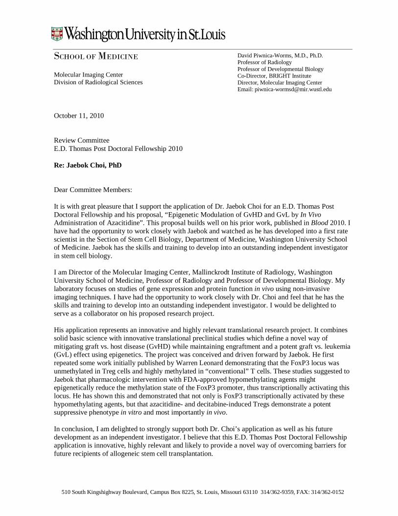
510 South Kingshighway Boulevard, Campus Box 8225, St. Louis, Missouri 63110 314/362-9359, FAX: 314/362-0152
SCHOOL OF MEDICINE Molecular Imaging Center Division of Radiological Sciences October 11, 2010 Review Committee E.D. Thomas Post Doctoral Fellowship 2010 Re: Jaebok Choi, PhD Dear Committee Members: It is with great pleasure that I support the application of Dr. Jaebok Choi for an E.D. Thomas Post Doctoral Fellowship and his proposal, “Epigenetic Modulation of GvHD and GvL by In Vivo Administration of Azacitidine”. This proposal builds well on his prior work, published in Blood 2010. I have had the opportunity to work closely with Jaebok and watched as he has developed into a first rate scientist in the Section of Stem Cell Biology, Department of Medicine, Washington University School of Medicine. Jaebok has the skills and training to develop into an outstanding independent investigator in stem cell biology. I am Director of the Molecular Imaging Center, Mallinckrodt Institute of Radiology, Washington University School of Medicine, Professor of Radiology and Professor of Developmental Biology. My laboratory focuses on studies of gene expression and protein function in vivo using non-invasive imaging techniques. I have had the opportunity to work closely with Dr. Choi and feel that he has the skills and training to develop into an outstanding independent investigator. I would be delighted to serve as a collaborator on his proposed research project. His application represents an innovative and highly relevant translational research project. It combines solid basic science with innovative translational preclinical studies which define a novel way of mitigating graft vs. host disease (GvHD) while maintaining engraftment and a potent graft vs. leukemia (GvL) effect using epigenetics. The project was conceived and driven forward by Jaebok. He first repeated some work initially published by Warren Leonard demonstrating that the FoxP3 locus was unmethylated in Treg cells and highly methylated in “conventional” T cells. These studies suggested to Jaebok that pharmacologic intervention with FDA-approved hypomethylating agents might epigenetically reduce the methylation state of the FoxP3 promoter, thus transcriptionally activating this locus. He has shown this and demonstrated that not only is FoxP3 transcriptionally activated by these hypomethylating agents, but that azacitidine- and decitabine-induced Tregs demonstrate a potent suppressive phenotype in vitro and most importantly in vivo. In conclusion, I am delighted to strongly support both Dr. Choi’s application as well as his future development as an independent investigator. I believe that this E.D. Thomas Post Doctoral Fellowship application is innovative, highly relevant and likely to provide a novel way of overcoming barriers for future recipients of allogeneic stem cell transplantation.
David Piwnica-Worms, M.D., Ph.D. Professor of Radiology Professor of Developmental Biology Co-Director, BRIGHT Institute Director, Molecular Imaging Center Email: [email protected]

2
510 South Kingshighway Boulevard, Campus Box 8225, St. Louis, Missouri 63110 314/362-9359, FAX: 314/362-0152
Sincerely yours,
David Piwnica-Worms Professor

October 13, 2010 Jaebok Choi, PhD Research Instructor Internal Medicine/Division of Oncology Washington University School of Medicine 660 South Euclid Avenue, Campus Box 8007 St. Louis, MO 63110 Dear Jaebok: I am excited about participating on your “Epigenetic Modulation of GvHD and GvL by In Vivo Administration of Azacitidine” proposal and am happy to offer my services and support in any way needed. As Director of the Department of Medicine's Biostatistics Consulting Center, I have extensive experience in experimental design and data analysis of biomedical research in many different clinical and research areas. I am honored to be involved with this important project, and am happy to commit the resources and staff of the consulting core to help you with this project. I cannot state strongly enough my enthusiasm for this work. I wish you success in this endeavor. Sincerely,
William D. Shannon, Ph.D. Associate Professor of Biostatistics in Medicine Director of the Dept. of Medicine's Biostatistics Consulting Center