circulationcircep.ahajournals.org/content/circae/early/2008/01/01/circep.108...circulation journa l...
TRANSCRIPT

Circulation
Journal of the American Heart Association
Arrhythmia and Electrophysiology
Alpha-1-Syntrophin Mutation and the Long QT Syndrome: a disease of sodium channel
disruption
Geru Wu, MD, PhD 1, Tomohiko Ai, MD, PhD 1, Jeffrey J. Kim, MD2, Bhagyalaxmi
Mohapatra, PhD2, Yutao Xi, PhD1, Zhaohui Li, MD, PhD 2, Shahrzad Abbasi, MS1,
Enkhsaikhan Purevjav, MD, PhD2, Kaveh Samani, MD1, Michael J Ackerman, MD, PhD3,
Ming Qi, PhD4, Arthur J. Moss, MD4, Wataru Shimizu, MD5, Jeffrey A. Towbin, MD2, Jie
Cheng, MD, PhD1, Matteo Vatta, PhD2
1Electrophysiology Research Laboratory, Texas Heart Institute/St. Luke’s Episcopal Hospital,
Houston, TX; 2Pediatric Cardiology, Texas Children’s Hospital/Baylor College of Medicine,
Houston, TX; 3Departments of Medicine, Pediatrics, and Molecular Pharmacology &
Experimental Therapeutics/Divisions of Cardiovascular Diseases and Pediatric Cardiology,
Mayo Clinic, Rochester, MN; 4Heart Research Follow-up Program, University of Rochester
Medical Center, Rochester, NY 14642-8653, USA; 5Division of Cardiology, National
Cardiovascular Center, Suita, Japan.
Correspondence to:
Matteo Vatta, PhD BCM/TCH 1102 Bates St., F.C. 430.04, Houston, Texas 77030 Phone: 832-824-4153, Fax: 832-825-4153, E-mail: [email protected] or Tomohiko Ai, MD, PhD THI/SLEH 6770 Bertner Ave. MC 2-255, Houston, TX 77030 Phone: (832) 355-8864, Fax: (832) 355-8880, E-mail: [email protected]
1
by guest on May 28, 2018
http://circep.ahajournals.org/D
ownloaded from
Circulation
Journal of the American Heart Association
Arrhythmia and Electrophysiology
Short title: Mutant Alpha-1-Syntrophin alters hNav1.5 in Long QT Syndrome
Funding Sources: This work was supported by the National Heart, Lung, and Blood Institute
(HL078807; M.V.), and the National Institute of Child and Health Development HD42569
(MJA). TA and JC were recipients of the Roderick D. MacDonald General Research Fund
Awards. JC was in part supported by a Cardiovascular Initiative Grant from the St. Luke’s
Episcopal Hospital/Texas Heart Institute. JAT was supported by the Texas Children’s
Foundation Chair in Pediatric Cardiac Research. This work was presented in part at the 2007
American Heart Association (AHA) Annual Scientific Sessions, Orlando, FL.
Conflict of Interest Disclosures: None
Acknowledgement
The authors are grateful to the patients and their family members who participated in
this study. In addition, we are grateful to Corina Rosales for her editorial assistance and to
Jennifer L. Robinson, LQTS Study Coordinator for registry information assistance.
Key Words
SNTA, alpha-1 syntrophin, SCN5A, hNav1.5, Long QT Syndrome, Arrhythmias; seizure
2
by guest on May 28, 2018
http://circep.ahajournals.org/D
ownloaded from

Circulation
Journal of the American Heart Association
Arrhythmia and Electrophysiology
Abstract
Background: Long QT syndrome (LQTS) is an inherited disorder associated with sudden
cardiac death. The cytoskeletal protein alpha-1-syntrophin (SNTA1) is known to interact with
the cardiac sodium channel (hNav1.5) and we hypothesized that SNTA1 mutations might
cause phenotypical LQTS in patients with genotypically normal hNav1.5 by secondarily
disturbing sodium channel function.
Methods and Results: Mutational analysis of SNTA1 was performed on 39 LQTS patients
(QTc ≥ 480ms) with previously negative genetic-screening for the known LQTS-causing
genes. We identified a novel A257G-SNTA1 missense mutation, which affects a highly
conserved residue, in 3 unrelated LQTS probands but not in 400 ethnic-matched control
alleles. Only one of these probands had a preexisting family history of LQTS and sudden
death with an additional intronic variant in KCNQ1. Electrophysiological analysis was
performed using HEK-293 cells stably expressing hNav1.5 and transiently transfected with
either wild type (WT) or mutant SNTA1, and in neonatal rat cardiomyocytes transiently
expressing with either WT or mutant SNTA1. In both HEK293 cells and neonatal rat
cardiomyocytes increased peak sodium currents were noted along with a 10 mV negative shift
of the onset and peak of currents of the I-V relationships. In addition, A257G-SNTA1 shifted
the steady-state activation (Vh) leftward by 9.4 mV, while the voltage-dependent inactivation
kinetics and the late sodium currents were similar to WT-SNTA1.
Conclusion: SNTA1 is a new susceptibility gene for LQTS. A257G-SNTA1 can cause gain-of-
function of Nav1.5 similar to the LQT3.
(Word = 238; max 250)
4
by guest on May 28, 2018
http://circep.ahajournals.org/D
ownloaded from
Circulation
Journal of the American Heart Association
Arrhythmia and Electrophysiology
Introduction
Long QT syndrome (LQTS) is an inherited disorder that can cause sudden cardiac
death (SCD). To date, hundreds of genetic mutations and single nucleotide polymorphisms in
11 distinct LQTS-susceptibility genes have been reported. Electrophysiological studies using
in vitro cell expression systems and genetically-engineered animal models have suggested that
“gain of function” or “loss of function” in the ion channels that are essential to generate action
potentials (APs) may account for the LQTS phenotypes.1 The functional modifications of the
ion channels are primarily caused by defects in the genes encoding the pore-forming subunits
(KCNQ1, KCNH2, SCN5A, KCNJ2, and CACNA1C) or the beta subunits (KCNE1, KCNE2,
and SCN4B) of the ion channels except for the gene responsible for LQT4. 2 Unlike the other
genes, LQT4 is caused by the malfunction of ankyrin B that is involved in the cellular
organization of the sodium/calcium exchanger and inositol-1,4,5-triphosphate receptors. 3
However, in 20-30% of cases, genetic analysis fails to identify the responsible gene
for the LQTS phenotypes in affected patients. 4 Recently, Vatta et al. and Cronk et al. reported
that mutations in caveolin-3 identified in the patients with LQTS or the infants succumbing to
sudden infant death syndrome (SIDS) can affect human cardiac sodium channel (hNav1.5)
gating kinetics and generate sustained currents, probably by direct protein-protein
interactions.5,6 In addition, mutations in the A-kinase anchoring protein 9 gene (AKAP9) have
been identified in LQTS. 9 AKAP9 determines the subcellular localization of protein kinase A
(PKA) and the phosphorylation of the IKs potassium channel α subunit (KCNQ1) to which it
assembles. 7 These studies proposed a novel concept that the defects of non-ion channel
proteins or channel interacting proteins (ChIPs) can affect ion channel gating kinetics, thereby
causing secondary channel dysfunction leading to LQTS. Hence, this concept uncovers a
5
by guest on May 28, 2018
http://circep.ahajournals.org/D
ownloaded from
Circulation
Journal of the American Heart Association
Arrhythmia and Electrophysiology
cascade or domino effect that disturbs the “final common pathway” 8 that causes arrhythmias,
ion channels, and focuses attention on a novel class of proteins and candidate genes to explain
the residual 25% of LQTS that remain genotype negative.
Syntrophins are cytoplasmic submembraneous proteins that are components of the
dystrophin-associated protein complex. 9 Several syntrophin isoforms (α1, β1, β2, γ1 and γ2)
have been identified in the heart, skeletal muscle and neurons. 10-14 The PDZ domain of
syntrophin-α1 (SNTA1), the most abundant isoform in the heart, has been reported to bind to
the C-terminal domain of murine cardiac voltage-gated sodium channels (SkM2). 15 Ou et al.
demonstrated that syntrophin-γ2 (SNTG2) affects hNav1.5 gating kinetics by protein-protein
interaction via PDZ domain in the distal C-terminus of the SCN5A-encoded sodium channel
pore-forming subunit. 16 Notably, SNTG2 shares structural similarity with SNTA1. These
observations compelled us to hypothesize that SNTA1 might be a new candidate gene
responsible for the LQTS in patients whose genetic screenings were negative for the already
known subtypes operating by secondary disruption of sodium channel function.
Methods
Patients Demographics and Genetic Screening
We previously enrolled 364 unrelated probands clinically diagnosed with LQTS. 5 Among
them, 39 genotype-negative LQTS patients (26 females; 66.7%) presenting a resting QTc ≥
480 ms and a LQTS diagnostic score ≥ 3 were selected for further investigation. 17 All
patients underwent physical examination, family history, and ECG analysis. The average age
at diagnosis was 23.6 ± 6.3 years (range: 1–84 years) and the average QTc = 537 ± 19 ms
(range: 480–670 ms).
6
by guest on May 28, 2018
http://circep.ahajournals.org/D
ownloaded from

Circulation
Journal of the American Heart Association
Arrhythmia and Electrophysiology
Blood was obtained after the written informed consent was obtained from all subjects.
Genomic DNA was extracted from peripheral blood lymphocytes as previously described. 5
Using polymerase chain reaction (PCR), and direct DNA sequencing, open reading
frame/splice site mutational analysis was performed on the SNTA1 gene (chromosome
20q11.2; 8 exons). PCR amplification was performed using primers designed to flank the
regions of this gene. All patients were, also, screened for the known LQTS-susceptibility
genes (LQT1-9). All SNTA1 genetic variants regarded as putative LQTS-associated mutations
were required to change a conserved residue or splice site, altering the primary amino acid
structure of the encoded protein. In addition, these genetic variants were required to be absent
in at least 400 ethnic-matched reference alleles in order to be considered as mutations.
Synonymous single nucleotide polymorphisms (SNPs) were excluded from consideration.
SNTA1 gene synthesis and mutagenesis
Wild-type (WT) human SNTA1 was synthesized based upon the previously deposited
sequence (Gene bank Accession #NM_003098; GenScript Corporation, Piscataway, NJ) and
subcloned into the pcDNA3.1/CT-GFP-TOPO vector (Invitrogen, Carlsbad, CA). Site-
directed mutagenesis was performed with the QuikChange Site-Directed Mutagenesis Kit
(Stratagene, La Jolla, CA) using the vector containing the WT-SNTA1 as a template. The
primers used for the mutagenesis are available upon request. PCR and bacteria
transformations were performed according to the manufacturer’s instructions. The mutated
A257G-SNTA1 clones were sequenced to ensure the presence of the mutation and the absence
of other substitutions introduced by DNA polymerase.
7
by guest on May 28, 2018
http://circep.ahajournals.org/D
ownloaded from

Circulation
Journal of the American Heart Association
Arrhythmia and Electrophysiology
HEK-293 cell preparation and transient expression of WT and mutant SNTA1
Stable HEK-293 cell lines expressing consistent sodium currents were established from one
single cell transfected with the vector containing Flag-tagged hNav1.5 (clone cells). The clone
cells were transiently transfected with the vector containing WT- or A257G-SNTA1 using the
Lipofectamine 2000 transfection reagent (Qiagen, Valencia, CA). To express β1-subunit of
human cardiac sodium channel (hβ1), the clone cells were transiently transfected with the
pIRES vector carrying hβ1 and CD8 (kindly provided by Dr. Naomasa Makita, Hokkaido
University, Sapporo, Japan) in conjunction with SNTA1. The cells were incubated at 37°C for
2~3 days before use.
Neonatal rat cardiomyocyte isolation and transient expression of WT and mutant SNTA1
All procedures were approved by the Institutional Animal Care and Use Committee at the
Texas Heart Institute. Neonatal rat cardiomyocytes were isolated according to the procedures
described elsewhere (see supplemental methods).
Patch-clamp experiments
Patch-clamp experiments were performed as previously described. 18 Macroscopic sodium
currents were recorded at ambient temperature (22-24°C). Step-pulse voltages were generated
with Axopatch 200B amplifier using pClamp9.0 software (Axon Instruments, Sunnyvale,
CA). Currents were filtered at 10 kHz with a built-in four-pole Bessel filter and fed to a
computer at a sampling frequency of 20 kHz. The details are described in the supplemental
methods.
8
by guest on May 28, 2018
http://circep.ahajournals.org/D
ownloaded from

Circulation
Journal of the American Heart Association
Arrhythmia and Electrophysiology
Co-Immunoprecipitation
Co-Immunoprecipitation was performed in HEK293 cells stably expressing Flag-tagged
hNav1.5 and transfected with either GFP-tagged WT-SNTA1 or A257G-SNTA1 as previously
reported (see supplemental methods). 5
Immunohistochemistry
Immunohistochemical staining was performed by standard techniques described elsewhere.
Briefly, the HEK-293 cells stably expressing hNav1.5 were transfected with GFP-tagged WT-
SNTA1. The cells were fixed with 4% paraformaldehyde and incubated with 0.5% Triton X-
100, then sequentially stained with anti-SCN5A sodium channel antibody (1:100 dilution,
Santa Cruz sc-23174) for 1 hour at room temperature, biotinylated anti-goat IgG (1:100
dilution, VECTOR BA-2000 ) for 30 minutes, and anti-streptavidin conjugated with Texas
Red (1:100 dilution, ZYMED 43-4317) for 30 minutes. The results were examined with
TCS-SP2 confocal laser-scanning microscope (Leica Microsystems).
Data Analysis
The sodium channel gating kinetics were analyzed using Clampfit (Axon Instruments,
Sunnyvale, CA) and Igor software (Wavemetrics, Lake Oswego, OR). Data were presented as
mean ± SE (median) and comparisons were made using nonparametric test (Mann-Whitney
test) with p<0.05 considered significant.
The authors had full access to and take full responsibility for the integrity of the data. All
authors have read and agree to the manuscript as written.
9
by guest on May 28, 2018
http://circep.ahajournals.org/D
ownloaded from
Circulation
Journal of the American Heart Association
Arrhythmia and Electrophysiology
Results
Clinical Evaluation and Mutation Analysis
Mutational analysis of SNTA1 in 39 unrelated patients with genotype negative for the
previously known LQTS subtypes was performed and one novel missense mutation (A257G)
was identified in 3 unrelated patients (2 females). This mutation involves a highly conserved
residue across several species (Figure 1A-B).
The proband is a 17-year-old Caucasian male presenting with congenital LQTS. His
ECG obtained at 6 hours after birth demonstrated marked QT-prolongation with late onset T-
wave pattern (QTc = 550 ms, Figure 1C, Table 1, case LQT-249 III: 2). The proband had a
syncopal attack at 3 years of age without previous symptoms of prodromal illness, vomiting,
diarrhea, fever, or upper respiratory infection. He collapsed while playing in a baby pool after
excessive running in a very hot day and remained unconscious for few minutes. The patient
underwent Holter monitoring, which showed the average heart rate of 128 bpm (99-167) and
prolonged QT-intervals (260-280 ms). No arrhythmias were documented during the
recording. The patient was treated with propranolol. In addition to the SNTA1 mutation, a
second variant was identified in KCNQ1 (IVS7+5G>A), which is present in all affected
members along with the SNTA1 mutation. Although this variant is of unknown significance,
computer predictions analyzed in our laboratory, using the
http://www.fruitfly.org/seq_tools/splice.html software 20 suggests the possible creation of a
cryptic splicing site leading to an in-frame insertion of 25 amino acids in one of the KCNQ1
alleles. This suggests that the KCNQ1 variant may add to the clinical phenotype caused by
SNTA1.
10
by guest on May 28, 2018
http://circep.ahajournals.org/D
ownloaded from

Circulation
Journal of the American Heart Association
Arrhythmia and Electrophysiology
A257G mutation was, also, identified in the proband’s sister (subject III:1) and mother
(subject II:2). Both of them have been apparently healthy. The sister’s ECG obtained at birth
showed QT-prolongation (QTc: 500 ms) with normal T-wave pattern in V1-3 and inverted T-
waves in V4-6. Her Holter ECG demonstrated normal sinus rhythm, no arrhythmias and
prolonged QT intervals. The mother’s ECG obtained at 27 years of age showed mild QT-
prolongation with asymmetrical peaked T-wave pattern (QTc = 460 ms; Figure 1C). The
proband’s maternal uncle died suddenly at age 25 during physical exertion. He had prior
history of syncope and prolonged QT interval (QTc = 470 ms). No autopsy sample was
available to us. The proband’s father was negative for ECG and genetic screening (Figure
1C; Table 1).
The A257G was, also, identified in a 37-year-old and a 27-year-old unrelated female with
unremarkable family history of either LQTS or SCD. Both probands showed QT-prolongation
at baseline ECG (QTc = 480 ms). Their parents were negative for both ECG and genetic
screening analysis consistent with de novo genetic change.
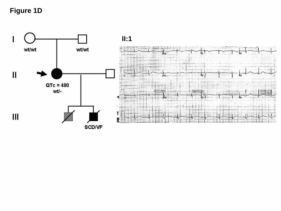
Subject LQT-682 was identified subsequently to screening after her 11-year-old son died
suddenly due to ventricular fibrillation registered at the time of the code at the hospital where
he was visiting the brother admitted for a car accident. The negative autopsy findings of
structural heart diseases led to the diagnosis of suspected LQTS, but no sample was available
to us. LQT-682 proband had a longstanding history of seizure disorder with normal seizure
work-up and positive EEG. The proband was treated with phenytoin for her seizure since she
was 12-years-old.
11
by guest on May 28, 2018
http://circep.ahajournals.org/D
ownloaded from

Circulation
Journal of the American Heart Association
Arrhythmia and Electrophysiology
The ECG (Figure 1D) and Holter ECG were obtained in the absence of beta blockers
but in the presence of phenytoin, and they demonstrated intermittent QT prolongation.
Echocardiography assessment was normal.
Analysis of 400 ethnic-matched control alleles did not identify the A257G mutation.
Immunostaining
HEK-293 cells stably expressing Flag-tagged hNav1.5 and GFP-tagged WT-SNTA1 were
fixed and stained with anti-Flag antibody. Figure 2A demonstrates that the green fluorescence
(SNTA1, middle panel) and red fluorescence (hNav1.5, right panel) when merged give the
orange-yellow areas suggesting colocalizaton of hNav1.5 and SNTA1, consistent with
possible association in a protein complex.
Co-Immunoprecipitation assay of hNav1.5 and SNTA1
Previous studies showed that the PDZ domain of SNTG2, highly homologous to SNTA1
binds the C-terminal domain of Nav1.5 and affect its gating kinetics. 16 Therefore, we
performed co-immunoprecipitation assay using HEK-293 cells stably expressing Flag-tagged
hNav1.5 and transiently transfected with either GFP-tagged WT- or A257G-SNTA. The anti-
GFP antibody detected SNTA1 (87kDa) in the co-immunoprecipitants with the anti-Flag
antibody (Figure 2B, lane 4 and 5), while the anti-Flag antibody detected hNav1.5 (260kDa
in the co-immunoprecipitants with the anti-GFP (Figure 2B, lane 1 and 2) for both WT-
SNTA1 and A257G-SNTA1, suggesting that human SNTA1 can physically interact with
hNav1.5, and that perturbations in SNTA1 might alter hNav1.5 function.
12
by guest on May 28, 2018
http://circep.ahajournals.org/D
ownloaded from
Circulation
Journal of the American Heart Association
Arrhythmia and Electrophysiology
Electrophysiological experiments
Figure 3A shows the superimposed whole-cell current traces obtained from HEK-293 cells
stably expressing consistent hNav1.5 currents and transiently transfected with WT- or A257G-
SNTA1. Figure 3B shows the current-voltage (I-V) relationships and demonstrates peak
current densities nearly twice as large in the cells expressing A257G-SNAT1 compared to
WT. In addition, A257G-SNAT1 shifted the onset and peak of the currents toward more
negative potentials by 10mV compared to the WT. Figure 3C shows the effects of A257G-
SNAT1 on the voltage-dependent kinetics of steady-state activation and inactivation. The half
maximal voltage of the steady-state activation (Vh) was shifted leftward by 8.3 mV in the cells
expressing A257G-SNAT1. The slope factor (k) was not affected by A257G-SNAT1. Steady-
state inactivation was examined by a pre-pulse protocol. The normalized currents were plotted
as a function of the membrane potentials and fitted by the Boltzman equation. Voltage-
dependent inactivation kinetics were not significantly affected by A257G-SNAT1. The
leftward shift of steady-state activation without shift of the inactivation kinetics increased the
window currents (Figure 3C inset). These biophysical parameters were, also, studied in the
presence of β1-subunit of the human cardiac sodium channel (hβ1); hβ1 did not significantly
modulate the effects of A257G-SNTA1 on the hNav1.5 gating kinetics (Table II).
The recovery time-course from fast inactivation was, studied using a two-pulse
protocol. Figure 3D shows the fraction of channels that recovered from the fast inactivation.
The data were fitted by a double-exponential equation; the fast components of the time
constant (τf) were significantly slower in the cells expressing A257G-SNTA1 than WT. The
slow components of the time constant (τs) and the fraction of fast and slow components were
comparable between the WT-SNTA1 and the A257G-SNTA1.
13
by guest on May 28, 2018
http://circep.ahajournals.org/D
ownloaded from
Circulation
Journal of the American Heart Association
Arrhythmia and Electrophysiology
Delay of sodium current decay and/or generation of sustained sodium currents have
been proposed as the pathophysiological mechanisms responsible for the LQT3 phenotype.
We estimated the time course of whole-cell current decay and the late sodium currents. Since
the I-V curve was shifted, the comparison of the current decay at a same membrane potential
might not simply express the time-dependent kinetics. The decay of the peak sodium currents
was analyzed with a double exponential fit. Figure 3E shows that A257G-SNTA1 slightly
delayed the macroscopic current decay compared to the WT-SNTA1. While the slow
component of time constant was comparable between WT-SNTA1 and A257G-SNTA1,
A257G-SNTA1 significantly slowed the fast component of time constant (Table II). The late
sodium currents were evaluated by a long depolarization pulse (2000 ms at -20 mV from a
holding potential of -140 mV) using 30 μM tetrodotoxin (TTX). No TTX-sensitive sustained
currents were detected in the cells expressing WT-SNTA1 and A257G-SNTA1 (data not
shown). Table II summarizes the effects of WT-SNTA1 and A257G-SNTA1 on the Nav1.5
gating kinetics.
Since the cellular environment of HEK-293 cells might be far different from
myocardial cells, we studied the effects of WT-SNTA1 and A257G-SNTA1 on hNav1.5 in
neonatal rat cardiomyocytes. The cardiomyocytes were isolated from neonatal rats (3-5 days
old) and transfected with GFP-tagged WT-SNTA1 and the A257G-SNTA1 using the
nucleofector. The cardiomyocytes showing green fluorescence were used for the patch-clamp
experiments. In native cardiomyocytes, A257G-SNTA1 increased the peak sodium currents
and altered the kinetics consistently with that observed in HEK-293 cells. Figure 4A shows
the representative whole-cell current traces obtained from the cardiomyocytes transiently
transfected with either WT-SNTA1 or A257G-SNTA1. Figure 4B shows the current-voltage
14
by guest on May 28, 2018
http://circep.ahajournals.org/D
ownloaded from

Circulation
Journal of the American Heart Association
Arrhythmia and Electrophysiology
(I-V) relationships. The peak current densities were significantly larger (1.6 fold) in cells
expressing A257G-SNTA1 compared to the WT. The onset and peak of the currents of the I-
V relationships were shifted toward negative by approximately 10 mV in cells expressing the
A257G-SNTA1 compared to WT. Figure 4C shows the effects of A257G-SNTA1 on the
voltage-dependency of the steady-state activation and the inactivation. The Vh of activation
curve was shifted leftward by 9.4 mV in the cells transfected with A257G-SNTA1; on the
contrary, voltage-dependent inactivation kinetics were not significantly affected by A257G-
SNTA1. The persistent sodium currents were examined using a long depolarization pulse
protocol (Figure 4D). The 30 μM TTX-sensitive persistent sodium currents were comparable
between the WT and the A257G mutant (% over the peak currents: WT, 0.25 ± 0.09%, n = 4;
A257G, 0.22 ± 0.04%, n = 4, N.S.). Figure 4E demonstrates that A257G-SNTA1 delayed the
macroscopic current decay (peak currents) compared to the WT-SNTA1. While the slow
component of time constant was comparable between WT and mutant SNTA1, A257G-
SNTA1 significantly increased the fast component of the time constant (Table II).
Discussion
Until recently the pathophysiological mechanisms of inherited arrhythmias were believed to
stem merely from primary alterations in ion channels leading to the term “ion
chanelopathies”. However, this paradigm has been challenged by the identification of altered
channel interacting proteins (ChIPs), which can directly modify ion channel functions. In
particular, our recent findings demonstrating that caveolin-3, which co-fractionates with
dystrophin and the dystrophin glycoprotein complex (DGC), binds Nav1.5 in human heart and
if mutated leads to LQTS, 5 has focused our attention on the other DGC proteins such as
15
by guest on May 28, 2018
http://circep.ahajournals.org/D
ownloaded from
Circulation
Journal of the American Heart Association
Arrhythmia and Electrophysiology
SNTA. SNTA1 is an integral part of the DGC, and regulates the Nav1.5 by forming a
multiprotein complex through its direct binding to dystrophin. 21 Our study, for the first time,
demonstrates that alteration of SNTA1 can affect the Nav1.5 gating kinetics, leading to the
LQTS phenotype.
How does A257G-SNTA1 affect the gating kinetics of Nav1.5?
A257G-SNTA1 caused gain-of-function of the Nav1.5 through the leftward shift of the
activation curve by 8-9 mV without shift of the inactivation curve, which might increase the
channel availability. The extent of this shift is equal to or larger than previously reported
values for SCN5A mutations (N1325S, L619F; Figure 3C and 4C). 22,23 In addition, the subtle
but significant delay of the fast component of whole-cell current decay (Figure 3E and 4E)
and the significantly larger peak current density for A257G-SNTA1 might contribute to an
increase of total inward currents.
Ou et al. reported that SNTG2, which is highly homologous to SNTA1, can attenuate
sodium current densities and shift the voltage-dependent activation kinetics compared to the
control.16 Therefore, it is possible that WT-SNTA1 also attenuates sodium currents, while
A257G-SNTA1 abolished the ability of WT-SNTA1 to decrease the currents. In order to
prevent the endogenous gene expression level affecting the comparison of the current
densities in a transiently transfection system, we established stable HEK-293 cell lines that
generate consistent sodium current densities from one single clone, and we transfected them
with WT-SNTA1 and A257G-SNTA1 by consistent protocols. Therefore, it is reasonable to
speculate that A257G-SNTA1 might increase the open-probability of the channel compared to
the WT. Although single channel recordings need to be studied to elucidate the detail
16
by guest on May 28, 2018
http://circep.ahajournals.org/D
ownloaded from
Circulation
Journal of the American Heart Association
Arrhythmia and Electrophysiology
underlying mechanisms, our data indicate that these biophysical modifications by the A257G-
SNTA1 can cause gain of function of the cardiac sodium channels in comparison to the WT-
SNTA1.
How does SNTA1 interact with Nav1.5?
Previous studies demonstrated that the PDZ domain of syntrophins (human SNTG2
and rodent SNTA1) can bind to the C-terminal domain of cardiac sodium channels (hNav1.5
and rodent Skm2). 15,16 Our study demonstrated that human SNTA1 can directly interact with
hNav1.5 (Figure 2B), consistently with these reports and suggesting direct protein-protein
interaction as the mechanism by which A257G-SNTA1 modifies the hNav1.5 function.
The previous study using the dystrophin-deficient mdx mice suggested that the
syntrophin-dystrophin complex plays critical roles in the regulation of the sodium channel
gating kinetics, as well as the ECG phenotype (conduction disturbances and QT-
prolongation). 24 Therefore, even secondary alterations of the DGC may result in primary
hNav1.5 defects via SNTA1 in cardiomyocytes. In addition, the effect of A257G-SNTA1 on
hNav1.5 in HEK-293 cells demonstrates that SNTA1 plays a key role in hNav1.5 regulation.
Further studies using genetically-engineered animal models and addressing the detailed
mechanism of Nav1.5 function regulation by SNTA1 and/or the involvement of other
syntrophin associated proteins, such as caveolin-3, dystrobrevin and nitric oxide synthase 1, 24
are warranted.
Study limitations. There are several significant limitations in this study: (1) the study
population is small and linkage analysis could not be performed; (2) we could not perform
17
by guest on May 28, 2018
http://circep.ahajournals.org/D
ownloaded from

Circulation
Journal of the American Heart Association
Arrhythmia and Electrophysiology
functional studies on the KCNQ1 intronic variant and therefore we cannot exclude that this
intronic variant or other unknown genes can be accountable for the patients’ phenotypes
although we examined and excluded all known LQTS genes; and (3) the data was obtained by
in vitro experiments using cell lines expressing only the α- and β1-subunit of the cardiac
sodium channels or neonatal rat cardiomyocytes over-expressing human SNTA1, which is far
different from the physiological conditions in the actual human heart. Although further studies
are warranted to explore the links between our data and the phenotypes of the affected
patients, our study does indicate that SNTA1 now joins ANKB, CAV3, and AKAP9 (yotiao) as
“non-primary channelopathic” mechanisms for LQTS. 2,3,5
Conclusions:
We identified a novel mutation of SNTA1, A257G, in three unrelated LQTS patients whose
genetic screenings were negative for LQT1-9. Electrophysiologic study suggests that
biophysical modifications of the Nav1.5 by A257G-SNTA1 can lead to “gain-of-function,”
through three mechanisms: (1) increase of channel avilability by leftward shift of activation
kinetics; (2) delay of current decay and (3) increase of current density. The molecular and
electrophysiological evidences implicate A257G-SNTA1 in the pathogenesis of LQTS as a
novel, albeit rare, LQTS-susceptibility gene. Although further studies are warranted to
understand the detail mechanisms leading to LQTS phenotypes in patients with the A257G-
SNTA1, our study demonstrated that SNTA1, as integral part of the DGC, plays an important
role in regulating hNav1.5 function. Mutation in SNTA1 may not only account for the
development of cardiac arrhythmias in affected patients but also potentially serve as novel
target(s) of therapeutic interventions.
18
by guest on May 28, 2018
http://circep.ahajournals.org/D
ownloaded from
Circulation
Journal of the American Heart Association
Arrhythmia and Electrophysiology
References:
1. Shah M, Akar FG, Tomaselli GF. Molecular basis of arrhythmias. Circulation.
2005;112:2517-2529.
2. Saenen JB, Vrints CJ. Molecular aspects of the congenital and acquired Long QT
Syndrome: Clinical implications. J Mol Cell Cardiol. 2008 Feb 9 [Epub ahead of
print]
3. Mohler PJ, Schott JJ, Gramolini AO, Dilly KW, Guatimosim S, duBell WH, Song LS,
Haurogne K, Kyndt F, Ali ME, Rogers TB, Lederer WJ, Escande D, Le Marec H,
Bennett V. Ankyrin-B mutation causes type 4 long-QT cardiac arrhythmia and sudden
cardiac death. Nature. 2003;421:634-639.
4. Schwartz PJ. Management of long QT syndrome. Nat Clin Pract Cardiovasc Med.
2005;2:346-351.
5. Vatta M, Ackerman MJ, Ye B, Makielski JC, Ughanze EE, Taylor EW, Tester DJ,
Balijepalli RC, Foell JD, Li Z, Kamp TJ, Towbin JA. Mutant caveolin-3 induces
persistent late sodium current and is associated with long-QT syndrome. Circulation.
2006;114:2104-2112.
6. Cronk LB, Ye B, Kaku T, Tester DJ, Vatta M, Makielski JC, Ackerman MJ. Novel
mechanism for sudden infant death syndrome: persistent late sodium current
secondary to mutations in caveolin-3. Heart Rhythm. 2007;4:161-166.
19
by guest on May 28, 2018
http://circep.ahajournals.org/D
ownloaded from
Circulation
Journal of the American Heart Association
Arrhythmia and Electrophysiology
7. Chen L, Marquardt ML, Tester DJ, Sampson KJ, Ackerman MJ, Kass RS. Mutation of
an A-kinase-anchoring protein causes long-QT syndrome. Proc Natl Acad Sci U S A.
2007 Dec 26;104(52):20990-57.
8. J.A. Towbin, Cardiac arrhythmias: the genetic connection, J Cardiac Electrophysiol
11 (2000), pp. 601–602.
9. Guglieri M, Magri F, Comi GP. Molecular etiopathogenesis of limb girdle muscular
and congenital muscular dystrophies: boundaries and contiguities. Clin Chim Acta.
2005;361:54-79.
10. Adams ME, Butler MH, Dwyer TM, Peters MF, Murnane AA, Froehner SC. Two
forms of mouse syntrophin, a 58 kd dystrophin-associated protein, differ in primary
structure and tissue distribution. Neuron. 1993;11:531-540.
11. Adams ME, Dwyer TM, Dowler LL, White RA, Froehner SC. Mouse alpha 1- and
beta 2-syntrophin gene structure, chromosome localization, and homology with a discs
large domain. J Biol Chem. 1995;270:25859-25865.
12. Ahn AH, Yoshida M, Anderson MS, Feener CA, Selig S, Hagiwara Y, Ozawa E,
Kunkel LM. Cloning of human basic A1, a distinct 59-kDa dystrophin-associated
protein encoded on chromosome 8q23-24. Proc Natl Acad Sci U S A. 1994;91:4446-
4450.
13. Yang B, Ibraghimov-Beskrovnaya O, Moomaw CR, Slaughter CA, Campbell KP.
Heterogeneity of the 59-kDa dystrophin-associated protein revealed by cDNA cloning
and expression. J Biol Chem. 1994;269:6040-6044.
14. Piluso G, Mirabella M, Ricci E, Belsito A, Abbondanza C, Servidei S, Puca AA,
Tonali P, Puca GA, Nigro V. Gamma1- and gamma2-syntrophins, two novel
20
by guest on May 28, 2018
http://circep.ahajournals.org/D
ownloaded from
Circulation
Journal of the American Heart Association
Arrhythmia and Electrophysiology
dystrophin-binding proteins localized in neuronal cells. J Biol Chem. 2000;275:15851-
15860.
15. Gee SH, Madhavan R, Levinson SR, Caldwell JH, Sealock R, Froehner SC.
Interaction of muscle and brain sodium channels with multiple members of the
syntrophin family of dystrophin-associated proteins. J Neurosci. 1998;18:128-137.
16. Ou Y, Strege P, Miller SM, Makielski J, Ackerman M, Gibbons SJ, Farrugia G.
Syntrophin gamma 2 regulates SCN5A gating by a PDZ domain-mediated interaction.
J Biol Chem. 2003;278:1915-1923.
17. Schwartz PJ, Moss AJ, Vincent GM, Crampton RS. Diagnostic criteria for the long
QT syndrome. An update. Circulation. 1993;88:782-784.
18. Shuraih M, Ai T, Vatta M, Sohma Y, Merkle EM, Taylor EW, Li Z, Razavi M,
Towbin JA, Cheng J. A common SCN5A variant alters its pharmacological
responsiveness to class I antiarrhythmic agents. Journal of Cardiovascular
Electrophysiology 2007 Apr;18(4):434-40
19. Moss AJ, Zareba W, Benhorin J, Locati EH, Hall WJ, Robinson JL, Schwartz PJ,
Towbin JA, Vincent GM, Lehmann MH. ECG T-wave patterns in genetically distinct
forms of the hereditary long QT syndrome. Circulation. 1995 Nov 15;92(10):2929-34
20. Reese MG, Eeckman, FH, Kulp, D, Haussler, D, 1997. ``Improved Splice Site
Detection in Genie''. J Comp Biol 4(3), 311-23.
21. Gavillet B, Rougier JS, Domenighetti AA, Behar R, Boixel C, Ruchat P, Lehr HA,
Pedrazzini T, Abriel H. Cardiac sodium channel Nav1.5 is regulated by a multiprotein
complex composed of syntrophins and dystrophin. Circ Res. 2006;99:407-414.
21
by guest on May 28, 2018
http://circep.ahajournals.org/D
ownloaded from
Circulation
Journal of the American Heart Association
Arrhythmia and Electrophysiology
22. Wang DW, Yazawa K, George AL, Jr., Bennett PB. Characterization of human
cardiac Na+ channel mutations in the congenital long QT syndrome. Proc Natl Acad
Sci U S A. 1996;93:13200-13205.
23. Wehrens XH, Rossenbacker T, Jongbloed RJ, Gewillig M, Heidbuchel H, Doevendans
PA, Vos MA, Wellens HJ, Kass RS. A novel mutation L619F in the cardiac Na+
channel SCN5A associated with long-QT syndrome (LQT3): a role for the I-II linker
in inactivation gating. Hum Mutat. 2003;21:552.
24. Aarnoudse AJ, Newton-Cheh C, de Bakker PI, Straus SM, Kors JA, Hofman A,
Uitterlinden AG, Witteman JC, Stricker BH. Common NOS1AP variants are
associated with a prolonged QTc interval in the Rotterdam Study. Circulation.
2007;116:10-16.
22
by guest on May 28, 2018
http://circep.ahajournals.org/D
ownloaded from

Circulation
Journal of the American Heart Association
Arrhythmia and Electrophysiology
Figure Legend
Figure 1: SNTA1 Mutational Analysis in LQTS.
Sequencing analysis demonstrates (A) a novel nucleotide variant in SNTA1 leading to a non-
synonymous change in three patients and (B) amino acid conservation analysis of the A257G-
SNTA1 variant, which modifies a highly conserved amino acid in SNTA1. (C) LQT-249
family pedigree and ECG recording showing a pattern with features similar to LQT3 and a
prolonged QT interval in proband III: 2, his sister III:1 and his mother II:2 bearing the
A257G-SNTA1 mutation. The ECG of the proband demonstrates a prolonged QT interval and
late onset T wave.19 (D) LQT-682 family pedigree and ECG recording showing a pattern with
features similar to LQT3 and a prolonged QT interval in proband II: 1 bearing the A257G-
SNTA1 mutation.
Figure 2: hNav1.5 and SNTA1 may form a protein complex in HEK-293 cells.
(A) Immunohistochemical staining: The left panel shows the merged images of green fluorescence
(mid) and red fluorescence (right) images. The green fluorescence depicts the GFP-tagged SNTA1
and the red fluorescence depicts the Flag-tagged hNav1.5. (B) Co-immunoprecipitation assays. IP
depicts immunoprecipitants; (+), transfected with the genes indicated in the left column; (-) non-
transfected with the genes.
Figure 3: The effects of WT-SNTA1 and A257G-SNTA1 on hNav1.5 in HEK-293 cells.
(A) Superimposed whole-cell current traces induced by a step-pulse protocol from a holding
potential of –140 mV. (B) I-V relationships. (C) Voltage-dependence of peak conductance and
steady-state fast inactivation. Conductance G(V) was calculated by the equation: G(V) = I / (Vm -
23
by guest on May 28, 2018
http://circep.ahajournals.org/D
ownloaded from

Circulation
Journal of the American Heart Association
Arrhythmia and Electrophysiology
Erev), where I is the peak currents, Erev is the measured reversal potential, Vm is the membrane
potential. The normalized peak conductance was plotted as a function of membrane potentials.
Steady-state inactivation was estimated by pre-pulse protocols (500 ms) from a holding potential of
–140 mV. The normalized peak currents were plotted as a function of membrane potentials. Steady-
state activation and inactivation were fitted with the Boltzmann equation: y = [1 + exp ((Vh -
Vm)/k)]-1, where y represents variables; Vh, midpoint; k, slope factor; Vm, membrane potential. The
inset indicates the magnified illustration of the window currents. The solid lines represent WT-
SNTA1 and the dotted lines represent A257G-SNTA1. (D) Recovery from the fast-inactivation
estimated by a double pulse protocol. Cells were depolarized at 0 mV for 500 ms from a holding
potential of –140 mV, then stepped to –140 mV for various duration before the 2nd pulse (20 ms at
–20 mV). The fractional recovery was calculated as the ratio of peak currents at the second pulse.
The recovery time course was fitted with a double exponential function: I(t)/Imax= C - Af x exp (-
t/τf) - As x exp (-t/τs), where t is the recovery time, Af and As are the fraction of fast and slow
components, τf and τs are the time constants of fast and slow components of recovery. (E)
Macroscopic current decay was fit with a double exponential function: I(t)/Imax = C - Af exp(-t/τf) -
Asexp(-t/τs), where Imax is the peak current, t is the time, Af and As are the fraction of fast and slow
components, τf and τs are the time constants of fast and slow components, respectively. The data
were represented as mean ± S.E.
Figure 4: The effects of WT-SNTA1 and A257G-SNTA1 on Nav1.5 in neonatal rat
cardiomyocytes. (A) Superimposed whole-cell current traces induced by a step-pulse protocol
from a holding potential of –140 mV. (B) I-V relationships. (C) Voltage-dependence of peak
conductance and steady-state fast inactivation. (D) Magnified current traces of the TTX-
24
by guest on May 28, 2018
http://circep.ahajournals.org/D
ownloaded from

Circulation
Journal of the American Heart Association
Arrhythmia and Electrophysiology
sensitive late sodium currents induced by a long depolarization-pulse protocol (2000 ms at –
20 mV from a holding potential of –140 mV). The dotted lines indicate zero current levels.
(E) Macroscopic current decay was fit with a double exponential function. The data were
represented as mean ± S.E.
25
by guest on May 28, 2018
http://circep.ahajournals.org/D
ownloaded from

Circulation
Journal of the American Heart Association
Arrhythmia and Electrophysiology
26
Table 1. Demographics of the LQTS patients
Case# Gene
Analysis Age/Gender Race Symptom Onset ECG
age QTc (ms) T-wave morphology
LQT-249-II:1 n/a 25§/M W syncope/SCD uk 470 n/a LQT-249-II:2 A257G 45/F W none 460 asymmetric peaked LQT-249-III:1 A257G 13/F W none 500 inverted
LQT-249-III:2* A257G 17/F W syncope with
pulmonary arrest 3 550 late onset LQT-682* A257G 37/F W recurrent seizures 480 n/a LQT-755* A257G 27/F W none 480 n/a
*proband; §age at SCD; W, white; SCD, sudden cardiac death; uk, unknown
by guest on May 28, 2018
http://circep.ahajournals.org/D
ownloaded from

Circulation
Journal of the American Heart Association
Arrhythmia and Electrophysiology
Table II. Effects of WT and A257G-SNTA1 on the Nav1.5 gating kinetics
HEK without β1 subunit HEK with β1 subunit Neonatal cardiomyocyte
WT A257G p WT A257G p WT A257G p
Current density(pA/pF) -97.8 ± 9.1 (-95.4) -179.6 ± 18.4 (-182.0)** 0.000 -151.0 ± 25.4 (-140.7) -336.9 ± 70.0 (-356.7)* 0.018 -316.3 ± 51.1 (-305.7) -517.5 ± 70.7 (-567.5)* 0.041 n 15 13 12 9 6 6
Activation
Vh (mV) -43.5 ± 1.5 (-44.0) -51.7 ± 1.6 (-51.5)** 0.002 -43.1 ± 1.3 (-42.7) -50.8 ± 1.5 (-48.9)** 0.001 -47.9 ± 1.7 (-48.4) -57.3 ± 3.1 (-55.0)* 0.015
k 7.87 ± 0.31 (7.47) 7.91 ± 0.62 (7.49) 0.892 8.80 ± 0.47 (8.98) 8.22 ± 0.24 (8.10) 0.464 7.56 ± 0.37 (7.54) 7.06 ± 0.14 (7.09) 0.24
n 15 13 12 9 6 6
Fast-inactivation
Vh (mV) -97.4 ± 1.8 (-96.7) -101.3 ± 2.0 (-102.1) 0.190 -104.6± 7.7 (-102.8) -105.9 ± 5.8 (-106.0) 0.430 -91.9 ± 2.3 (-91.2) -91.4 ± 3.1 (-92.1) 0.943
k 5.18 ± 0.19 (5.39) 5.44 ± 0.19 (5.49) 0.740 5.47 ± 0.17 (5.59) 5.59 ± 0.23 (5.76) 0.650 6.43 ± 0.26 (6.71) 7.10 ± 0.13 (6.92) 0.065
n 12 11 13 14 8 5
Recovery from fast- inactivation
τf (ms) 2.92 ± 0.30 (2.59) 4.33 ± 0.56 (3.63)* 0.033
τs (ms) 33.43 ± 4.57 (29.86) 41.34 ± 2.14 (40.93) 0.094
Af (%) 93.4 ±3.6 (91.6) 87.2 ±2.8 (87.46) 0.169
As (%) 20.4 ±2.7 (19.8) 23.4 ±1.8 (22.09) 0.169
n 13 14
Current decay
τf (ms) 0.92 ± 0.04 (0.94) 1.06 ± 0.04 (1.01)* 0.033 0.80 ± 0.07 (0.74) 1.12 ± 0.08 (1.12)* 0.026
τs (ms) 8.57 ± 0.99 (7.76) 8.97 ±1.00 (9.99) 0.586 5.90 ± 1.78 (3.53) 4.23 ± 0.43 (3.96) 0.589
n 15 13 6 6
Vh, midpoint voltage of maximal activation/inactivation; k, slope factor; τf and τs time constants of fast and slow components of recovery; Af and As, fraction of fast and slow components; n, number of patches. Numbers in parentheses represent the median of data. *p<0.05; **p<0.01 vs. WT.
27
by guest on May 28, 2018 http://circep.ahajournals.org/ Downloaded from

Circulation
Journal of the American Heart Association
Arrhythmia and Electrophysiology
SNTA1 reverse sequence
HUM: ADGQDTLFLRAKDEAS A RSWATAIQAQVNTLRAT: ADGQDTLFLRAKDEAS A RSWAGAIQAQISTFMOU: ADGQDAVFLRAKDEAS A RSWAGAIQAQIGTF DOG: ADGQDTLFLRAKDEAS A KSWAAAIQAQVNALCOW: ADGRDTLFLRAKDEAS A KSWAAAIQAQVNTLRAB: ADGQDTIFLRAKDEAS A RSWAGAIQAQINALAPE: AEGQDTLFLRAKDEAS A RSWGSAIQAQVNAL
AAAAAAA
Figure 1
A
B
by guest on May 28, 2018 http://circep.ahajournals.org/ Downloaded from

Circulation
Journal of the American Heart Association
Arrhythmia and Electrophysiology
Figure 1C
III:1 III:2
II:2
by guest on May 28, 2018 http://circep.ahajournals.org/ Downloaded from
Circulation
Journal of the American Heart Association
Arrhythmia and Electrophysiology
Figure 1D
II:1
by guest on May 28, 2018 http://circep.ahajournals.org/ Downloaded from

Circulation
Journal of the American Heart Association
Arrhythmia and Electrophysiology
100 μm
Merged SNTA1 SCN5A
IP GFP IP FLAG
Anti-GFP
Anti-FLAG
+ + ++ - -- + -
+ + ++ - -- + -
hNav1.5-FLAGWT-SNTA1-GFPA257G-SNTA1-GFP
1 2 3 4 5 6
Figure 2A
B
by guest on May 28, 2018 http://circep.ahajournals.org/ Downloaded from

Circulation
Journal of the American Heart Association
Arrhythmia and Electrophysiology (p
A/p
F)
100
(mV)
-100
○ WT● A257G
0
-200
○ WT● A257G
-140 mV
0 mV500 ms 20 ms
Δt
Nor
mal
ized
cur
rent
s
1 10 100 1000
0
1.0
(ms)
○ WT● A257G
Nor
mal
ized
cur
rent
s
-140 mV
500 ms
-140 mV
20 ms
-120 -80 -40(mV)
0
1.0
0
5 ms
0.5 nA
5 ms
0.5 nA
WT
A257G
5 ms
Nor
mal
ized
cur
rent
s
A257G
WT
0
-1
Figure 3A
D
B
E
C
by guest on May 28, 2018 http://circep.ahajournals.org/ Downloaded from

Circulation
Journal of the American Heart Association
Arrhythmia and Electrophysiology
-600
200
(pA
/pF)
-100
○ WT● A257G
100(mV)
Nor
mal
ized
cur
rent
s
-140 mV
500 ms
-140 mV
20 ms
-120 -80 -40 0(mV)
0
1.0
○ WT● A257G
5 ms
1 nA
WT
5 ms1 nA
A257G
40 pA
50 ms
50 ms
40 pA
WT
A257G5 ms
0
-1
Nor
mal
ized
cur
rent
s
A257G
WT
A
D
B
E
CFigure 4
by guest on May 28, 2018 http://circep.ahajournals.org/ Downloaded from
Wataru Shimizu, Jeffrey A. Towbin, Jie Cheng and Matteo VattaAbbasi, Enkhsaikhan Purevjav, Kaveh Samani, Michael J. Ackerman, Ming Qi, Arthur J. Moss,
Geru Wu, Tomohiko Ai, Jeffrey J. Kim, Bhagyalaxmi Mohapatra, Yutao Xi, Zhaohui Li, Shahrzaddisruption
Alpha-1-Syntrophin Mutation and the Long QT Syndrome: a disease of sodium channel
Print ISSN: 1941-3149. Online ISSN: 1941-3084 Copyright © 2008 American Heart Association, Inc. All rights reserved.
Dallas, TX 75231is published by the American Heart Association, 7272 Greenville Avenue,Circulation: Arrhythmia and Electrophysiology
published online January 1, 2008;Circ Arrhythm Electrophysiol.
http://circep.ahajournals.org/content/early/2008/01/01/CIRCEP.108.769224World Wide Web at:
The online version of this article, along with updated information and services, is located on the
http://circep.ahajournals.org//subscriptions/
is online at: Circulation: Arrhythmia and Electrophysiology Information about subscribing to Subscriptions:
http://www.lww.com/reprints Information about reprints can be found online at: Reprints:
document. Permissions and Rights Question and Answerinformation about this process is available in the
requested is located, click Request Permissions in the middle column of the Web page under Services. FurtherCenter, not the Editorial Office. Once the online version of the published article for which permission is being
can be obtained via RightsLink, a service of the Copyright ClearanceCirculation: Arrhythmia and Electrophysiology Requests for permissions to reproduce figures, tables, or portions of articles originally published inPermissions:
by guest on May 28, 2018
http://circep.ahajournals.org/D
ownloaded from