journal of american science, 2011;7(9) ... · effect of methyl tetrahydrofolate reductase (mthfr)...
TRANSCRIPT

Journal of American Science, 2011;7(9) http://www.americanscience.org
929
Effect of Methyl Tetrahydrofolate Reductase (MTHFR) C766T Polymorphism on Promoter Methylation and Protein Expression of P16 Gene in Primary Ovarian Carcinoma
Naglaa R. AbdRaboh *1; Fatehia A. Bayoumi 2 and Ahmed H. El Barkouky3
1 Biochemistry Department, Faculty of Medicine Ain Shams University, Cairo, Egypt, 2Pathology Department, Faculty of Medicine, Zagazig University, Dubai, UAE 3Pathology Department in Baraha hospital, Dubai, UAE
*[email protected] Abstract: Several malignant tumors are associated with decreased expression of tumor suppressor gene P16 and one of the common causes of this suppression is epigenetic modification as methylation of promoter region; promoter methylation is triggered by enzymatic activity related to methylation reaction as MTHFR. This study was aimed to elucidate the relationship between P16 protein expression and promoter methylation in primary ovarian tumors and the liability for gene methylation in patients with mutated MTHFR. This study was conducted on 32 samples of ovarian cancer tissues and 18 samples of normal ovarian tissues were used as control group. Genomic DNA extracted from tissues was subjected to amplification with polymerase chain reaction using specific primers followed by restriction fragment polymorphism for detection of MTHFR C766T Polymorphism, after treatment with sodium bisulfite P16 promoter methylation was analyzed using methylation specific PCR (MSP). Immunohistochemical analysis was performed to examine the association of P16 methylation with protein expression. Obtained results revealed that promoter methylation of p16 was positive in 43.8% of malignant samples in contrast to 16.7 % in normal ovarian samples (p<0.05). There was significant association between promoter methylation and lack of p16 protein expression (p=0.03). Regarding MTHFR C766T Polymorphism T containing genotypes (CT+TT) constitutes 87.5% of malignant samples in comparison to 66.7% in normal samples., but there was a reverse correlation between T allele, T containing genotypes (CT+TT) was frequency and p16 promoter methylation as 27.5% of (CT+TT) genotypes was methylated in comparison to 60% of CC genotype, but no association was detected between MTHFR C766T Polymorphism and P16 protein expression or clinicopathological criteria of the tumor. Our result reflects a probable effect of MTHFR C766T Polymorphism on level of P16 promoter methylation but not on protein expression, and possibly MTHFR C766T Polymorphism and P16 promoter methylation have separate pathogenic role in ovarian cancer. [Naglaa R. AbdRaboh; Fatehia A. Bayoumi and Ahmed H. El Barkouky Effect of Methyl Tetrahydrofolate Reductase (MTHFR) C766T Polymorphism on Promoter Methylation and Protein Expression of P16 Gene in Primary Ovarian Carcinoma. Journal of American Science 2011;7(9):929-940]. (ISSN: 1545-1003). http://www.americanscience.org. Keywords: MTHFR C677T polymorphism, Promoter Methylation, P16 Expression, Ovarian Cancer.
1. Introduction
Tumor suppressor gene p16 (INK4A, MTS-1, or CDKN2A) located at 9p21, it is composed of 3 exons which encode 156 amino acids protein that is involved in cell cycle control (Kamb et al., 1994). P16 protein interacts with cyclin-dependent kinase CDK4 and CDK6 which bind to cylcin D regulating progression of cell from G1 to S phases. Binding of p16 to CDKs inhibits the formation of CDK/cyclin D complex, resulting in cell cycle arrest at G1 phase (Liggett and Sidransky 1998). Aberrant DNA methylation is recognized as one of the most common molecular abnormalities in human cancer (Herman and Baylin 2000). This epigenetic modification occurs at the cytosines of CpG dinucleotides, which often exist in clusters called CpG islands. When methylation of these sites occurs in the promoter region of a gene, it can result in chromatin condensation and gene silencing (Issa 2003). In cancer cells, aberrant methylation
frequently has been reported in tumor suppressor genes, DNA repair genes, and genes related to cancer metastasis and invasion (Esteller et al., 2001).
Among the common targets for aberrant DNA methylation is the P16 gene (Ahuja et al., 1997). The silencing of these functionally important genes leads to shift of cells from a normal cellular cycle to a state of high proliferation that favors tumor development and progression (Esteller 2005). Promoter methylation, in addition to gene deletion and point mutation of p16 locus, has been found to be one of the main mechanisms of p16 transcriptional inactivation (Huang et al., 2011). Hypermethylation of the 5'CpG islands of p16 gene promoter region has been reported to be linked to loss of protein expression in several human carcinomas including ovarian cancer (Hu et al., 2010; Malhotra et al., 2010; Shima et al., 2011).
Promoter methylation is important feature in carcinogenesis (Agrawal et al., 2007) however

Journal of American Science, 2011;7(9) http://www.americanscience.org
930
the precipitating factor of aberrant DNA methylation is not completely understood. Methylenetetrahydrofolate reductase (MTHFR; EC 1.5.1.20) is a key enzyme in availability of methyl group that is needed for DNA methylation (Choi and Mason; 2002). It is responsible for conversion of methylene-THF to methyl-THF which is used in remethylation of sulfur-containing amino acid homocysteine to methionine. Endogenous methionine is then catabolized to produce the universal methyl donor S-adenosylmethionine (SAM) (Van der Put et al., 1998). The MTHFR gene is highly polymorphic in the general population, with the most common functional variant of 677 C to T. This polymorphism results in an alanine to valine substitution, leading to a reduction in enzyme activity [Langevin et al., 2009].
The reduction in enzyme activity impairs the folate metabolism, leading to genomic DNA hypomethylation (Ulrich et al., 2005) coexistent with gene-specific promoter hypermethylation (Oyama et al., 2004), as is frequently observed in cancer. Results on the effect of this polymorphism on cancer susceptibility are not consistent. Different types of associations have been reported in solid and hematological malignancies. The modulator role the MTHFR-677T allele has been suggested in the risk of developing human tumors as colorectal, gastric, and endometrial neoplasm and leukemia (Wiemels et al., 2001). It might be speculated therefore that MTHFR polymorphisms, and the consequent decrease in enzyme function, either protect against the development of cancer by providing more folate for DNA synthesis and repair, or increase the risk by reducing the availability of methyl groups (Friso and Choi; 2005).
So far the association of MTHFR C677T polymorphism, folate level and P16 methylation along with protein expression is hardly studied. Kamiya, et al., (1998) have advocated that folate metabolism can affect carcinogenesis through the increasing the expression of P16. Conversely, Wettergren et al., (2010) found no link between any of the MTHFR C677T polymorphic variants and P16 methylation status in colorectal cancer
Ovarian cancer accounts for approximately 3 percent of all cancers in women and is the fifth leading cause of cancer-related death among women in the United States. It has the highest mortality of all cancers of the female reproductive system. This reflects, in part, a lack of early symptoms and proven ovarian cancer screening tests. Thus, ovarian cancer is often diagnosed at an advanced stage, after the cancer has spread beyond the ovary (Ries et al.; 2005). This indicates the need of more studies on the genetic bases and pathogenesis of ovarian cancer.
The present study was conducted to investigate the association of MTHFR C677T polymorphism with p16 promoter methylation and expression to underline the probable involvement in pathogenesis of ovarian carcinomas. 2. Materials and subjects: Tissue samples from 50 females age range from 35 years to 70 years at diagnosis, 32 were diagnosed as primary ovarian carcinoma and 18 with normal ovary excised in patient with other gynecological lesions as multiple fibromatosis and endometrial carcinoma from pathology department in Al Baraha hospital; Dubai between 2007 and 2009 using appropriate informed consent from each patient obtained after a formal approval from institute ethics committee. Information on tumor size, histological grade and stage was abstracted from medical records. Histological types included serous cystadenocarcinoma, mucinous, endometroid, and undifferentiated carcinomas. DNA extraction: Tissue samples were subjected to DNA extraction by digestion with proteinase K and RNase followed by phenol/choloroform extraction and ethanol precipitation (Sambrook and Russell 2001).The methylation status of 5’ CPG islands of P16 gene was assessed by bisulfate modification of DNA and methylation specific PCR (MSP) according to the method of Herman et al. (1996). The treatment by bisulfite converts unmethylated cytosine to thymine while keeping methylated cytosine unchanged. Briefly, approximately 2 μg genomic DNA in a volume of 50 ul was denatured by 2 M NaOH at a 10:1 ratio and the sample was incubated at 37°C for 10 min. Then 30 ul of 0.1 M hydroquinone and 520 ul of 5 M Na bisulfite solution, pH 5.0, both freshly prepared were added to each sample and incubated at 50°C for 16 h. After incubation, the DNA samples were purified from the bisulfite solution using column desulphonation and elution buffer using EZ DNA Methylation™ Kit, Zymo Research. The DNA purity and concentration were determined by spectrophotometer measurement of absorbance at 260 and 280 nm. PCR Amplification and Primers for p16 methylation: Methylation-specific PCR (MSP) was performed on the sodium bisulfite-treated DNA samples to amplify the promoter region of the p16 gene. Two pairs of PCR primers were used in the amplification, one for methylated sequences and one for unmethylated sequences. The forward and reverse primers for the p16 unmethylated product (124 bp) were sense:

Journal of American Science, 2011;7(9) http://www.americanscience.org
931
5’GGTAGTTAGGAAGGTTGTATTGT3’ antisense: 5’TCCCTACTCCCAACCACA3’, for the methylated product (126 bp), the primers were p16 methylated sense 5’TTGGTAGTTAGGAAGGTTGTATCGC3’, p16 methylated antisense: 5’ TCCCTACTCCCAACCGCG3’ (antisense) (Herman et al., 1996). Amplification of the promoter region of the p16 gene was carried out using a 96-well Gene Amp PCR System 9700 thermocycler (Applied Biosystems). 50 µl PCR reaction mixture containing 2 µl of bisulfite-treated genomic DNA, dinucleotide triphospates (dNTPs) (each at 200 µM), primers (50 pmol each per reaction), 2.5 mM MgCl2 and 1.25 U Taq in 1X PCR buffer (All reagents were supplied with the Promega). The PCR conditions were as follows: initial denaturation and hot start at 95°C for 5 min, then 40 cycles consisting of 30 s at 95°C, 30 s at 60°C (unmethylated reactions) or 65°C (methylated reactions) and 1 min at 72°C followed by a final 5-min extension at 72°C. The PCR products were examined on 2% agarose gel stained with ethidium bromide and visualized under UV illumination. PCR amplification of methylated and unmethylated products was carried out separately in two tubes with the same PCR conditions and reagents (except the primers). MTHFR C677T Polymorphism Detection: For genotype analysis, the MTHFR gene was amplified in DNA extracted from all tissue samples by polymerase chain reaction followed by restriction enzyme digestion Hinf I (Promega, UK). Primer sequences, PCR conditions and restriction enzyme digestion will be as follows; (oligonucleotides were synthesized by Promega, UK).
The forward primer is 5TGAAGGAGAAGGTGTCTGCGGGA-3' and reverse primer 'AGGACGGTGCGGTGAGAGTG-3'. PCR was carried out in a 25-μL reaction volume containing 100 ng of genomic DNA, 0.4 mmol/L of each primer, 0.2 mmol/L dNTPs, 2 mmol/L MgCl2 in 10% PCR buffer and 1U of DNA polymerase (Promega, UK). PCR involved an initial 2 min denaturation at 93°C, 35 cycles of denaturing at 93°C for 1 min, annealing at 58°C for 1 min and extension at 72°C for 1 min, with a final extension at 72°C for 4 min. Aliquots of 12.5 μL of the PCR products were digested with 5U Hinf1 (Promega, UK) for overnight at 37° C. RFLP products will be analyzed on 2.5% agarose gel and stained with ethidium bromide (Josef et al., 2002). Immunohistochemical analysis: Freshly removed tissue samples were immediately fixed in 10% buffered formalin for 24 hours, embedded in paraffin, cut into sections 5-μm in thickness and mounted on slides coated with poly-L-lysine. The sections were deparaffinized by
heating at 60℃ for 30 min, treated with xylene, and dehydrated in alcohol. Endogenous peroxidase was blocked with 0.03% H2O2 in methanol for 20 min. The antigenic sites were unmasked by means of pressure cooker treatment for 15 min in 10 mmol/L Citrate buffer (pH 6.0). The sections were then incubated with p16 primary antibody (p16 lNK4a
specific monoclonal antibodies) were added for 45 min at room temperature. For detection, biotinylated horse anti-mouse or antirabbit sera (Vectastain) were applied as secondary antibodies for 30 min, followed by incubation with the avidin-biotin complex (Vectastain) for 30 min. The reaction was developed using the chromogen 3-3’ diaminodenzidine mixed with hydrogen peroxide in acetate buffer, and the sections were counterstained with hematoxylin (Malhotra et al., 2010). Ki67 immnunostaining was performed to assess the proliferative activity of the tumor cells. Statistical analysis: The frequency comparisons of methylation, expression, genotype and alleles were analyzed by using Pearson chi-square or Fisher’s exact test, statistical values of P ≤0.05 were considered as significant. Odd ratios (ORs) and 95% confidence interval (CIs) were computed using the relevant 2 x 2 contingency tables for comparing p16 methylation and expression in malignant and control groups taking homozygous wild type CC genotype a reference. All statistical tests were performed using SPSS version 17 software. 3. Results: Promoter methylation and protein expression was evaluated in 50 ovarian tissue specimens, 32 tissue samples from female patients diagnosed as ovarian cancer with mean age (53.8 + 9.4 years), and 18 samples from females with normal ovarian tissues as control group with mean age (53.7 + 10.9 years). Methylation analysis of p16 promoter: As shown in figure (1) Methylation status was evaluated in ovarian cancer and normal ovarian tissues using MSP. A band of methylated DNA was found in 43.8 % (14/32) of malignant tissues compared to 16.7% (3/18) in normal ovarian tissues, a statistical significance was found (P=0.049), patient age at diagnosis was not significantly different (P>0.05) between patients with positive methylation (mean 53.5 years) and patients without methyaltion (mean age 54.4 years). Expression of p16 protein: (Figure 4-6).
As shown in figures (3–6) the p16 immunohistochemical staining results were

Journal of American Science, 2011;7(9) http://www.americanscience.org
932
interpreted as follows: positive (+), if positive immunohistochemical staining in both nuclei and cytoplasm is present in more than 50% tumor cells; and negative (-), if positive p16 immunohistochemical staining is present in less than 10% tumor cells.
Immunostaining of the formalin-fixed, paraffin-embedded sections was reviewed by two independent observers, and strong nuclear as well as cytoplasmic staining was considered a positive reaction. Immunohistochemical analysis was performed to evaluate difference in protein expression of p16 in tumor tissues and control group there was a significant (p value <0.05) loss of p16 protein expression in 87.5% of malignant subjects (28/32) as compared to 55.6% (10/18) in control group. As shown in table (1): the frequency of p16 methylation, protein expression and odd ratio for ovarian cancer samples in comparison to normal control group. Effect of p16 promoter methylation on protein expression: P16 promoter methylation was matched up to protein expression and results were summarized in table (2). Our data showed that 34% (17/50) of the total analyzed samples were positive for methylation, while the lack of expression was detected in 76% (38/50) of total samples, when investigating the relation of methylation and loss of expression, we found 94.1% of methylated samples lacking protein expression and lost only in 66.7% of unmethylated samples and p value was 0.03 this correlation become non significant when we compared control and malignant groups separately as smaller number decreased statistical power. Genotyping of MTHFR C677T polymorphism in malignant and control groups:
As shown in figure (2) the CC genotype gave one band of 198 bp, CT gave three bands (198bp, 175bp and 23bp), and CC gave two bands (175bp and 23bp). As shown in table (3) no significant difference was found in between frequency of MTHFR C677T genotypes in cases with ovarian carcinoma and control group. Although not statistically significant when we pooled T allele containing genotypes (CT, and TT) in comparison to homozygous CC allele we found marked increase risk of malignancy in T containing genotypes which constitutes 87.5% (28/32) of malignant samples in comparison to 66.7% (12/18) in normal samples with odd ratio 3.5 (95%CI=0 .8-14.7). Genotyping of MTHFR C677T polymorphism versus p16 methylation, protein expression: As shown in table (4) a reverse association between T allele and T containing genotype with p16 methylation, as T containing genotypes was found in 27.5% (11/40) of methylated versus 72.5% (25/40) in unmethylated samples with OR= 0.25 (CI95%=0.06-1). Similar figure was recorded when polymorphism is correlated to methylation in malignant group only as T containing genotypes were 35.7% (10/28) methylated compared to 64.3% (18/28) unmethylated OR 0.18 (5% CI=0.02-2). Mild increase of p16 protein expression was noticed in samples with T containing genotype but with no statistical significance or risk association. Relationship of p16 methylation, protein expression, and MTHFR C677T polymorphism with clinicopathological parameters: As shown in table (5) there was no significant correlation between p16 promoter methylation, p16 protein expression, MTHFR C677T polymorphism and any of clinicopathological criteria of malignant samples. These parameters included patient’s age, tumor size, histological grade, and TNM staging. Except for mild increase risk for advanced tumor stage (3, 4) 35.7% in methylated versus 27.8% in unmethylated samples.
Table 1: The frequency of p16 promoter methylation , and protein expression in malignant and normal ovarian tissues
Group Unmethylated
p16 n (%)
Methylated p16
n (%)
OR
(95%CI)
P value
Negative expression
n (%)
Positive expression
n (%)
OR
(95%CI)
P value
Control 15/18 (83.3) 3/18 (16.7) 4.4
(1.2-15.1) 0.049 *
10/18 (55.6)
8/18 (44.4) 3.6
(1.2-10.1) 0.017*
Malignant 18/32 (56.2) 14/32 (43.8) 28/32 (87.5)
4/32 (12.5)
*Significant values. P < 0.05, % = percentage values, n= number, OR= odd ratio, 95% CI= 95% confidence interval.

Journal of American Science, 2011;7(9) http://www.americanscience.org
933
Table 2: The analysis of association of p16 expression and gene methylation in ovarian tissues
Expression (n) Unmethylated p16
n (%) Methylated p16
n (%) P value
All samples (50) Negative Positive Total
22 (66.7) 11(33.3) 33(100)
16 (94.1)
1(5.9) 17(100)
0.03*
Malignant group (32) Negative Positive
Total
14(77.8) 4(22.2) 18(100)
14(100)
0(0) 14(100)
0.08
Control group (18) Negative Positive Total
8(53.3) 7(46.7) 15(100)
2(66.7) 1(33.3) 3(100)
0.6
*Significant values. P < 0.05, percentage values are shown in parentheses, n= number. Table (3): Frequency of MTHFR C677T polymorphism in malignant and control groups
Normal control
n (%) Malignant cases
n (%) OR 95% CI P
CC genotype CT genotype TT genotype
6/18(33.3) 11/18 (61.1)
1/18(5.6)
4/32 (12.5) 25/32 (78.1)
3 (9.4) 0.2
CC Genotype
n (%) 6/18(33.3) 4/32 (12.5)
3.5 0.83-14.7 0.14 CT+TT Genotypes
n (%) 12/18 (66.7) 28/32 (87.5)
C Allele 23/36 (64) 33/64 (51) 1.6
0.7-3.8 0.3
T Allele 13/36 (36) 31/64 (48)
OR= odd ratio, 95% CI= 95% confidence interval, *Significant values. P < 0.05, percentage values are shown in parentheses, n= number. Table (4): The MTHFR C677T polymorphism and promoter methylation and protein expression of p16 gene:
CC Genotype
n (%)
CT+TT Genotypes
n (%)
OR 95% CI
P C Allele n (%)
T Allele n (%)
OR 95% CI
P
All samples
Unmethylated p16
4/10 (40) 25/40 (62.5) 0.25
0.06-1.07 0.06
34/56 (61)
32/44 (73)
0.6
0.25-1.4 0.28
Methylated p16
6/10 (60) 11/40 (27.5)
22/56 (39)
12/44 (27)
Malignant cases
Unmethylated p16
0/4(0) 18/28 (64.3) 0.18
0.02-2 0.17
18/33 (55)
20/31 (65)
0.7
0.24-1.8 0.45
Methylated p16
4/4(100) 10/28(35.7) 15/33 (45)
11/31 (35)
All samples
Negative expression
8/10 (80) 30/40 (75) 1.3
0.24-7.3 0.55
44/56 (79)
32/44 (73) 1.37
0.55-3.45 0.6
Positive expression
2/10 (20) 10/40 (25) 12/56 (21)
12/44 (27)
Malignant cases
Negative expression
4/4 (100) 24/28(85.7) 0.9
0.7-1 0.56
30/33 (90)
26/31 (84) 1.9
0.4-8 0.46
Positive expression
0/4 (0) 4/28(14.3) 3/33 (10)
5/31 (16)
OR= odd ratio, 95% CI= 95% confidence interval, p <0.05 = statistical significance.

Journal of American Science, 2011;7(9) http://www.americanscience.org
934
Table (5): Clinicopathological criteria of ovarian cancer patients in relation to promoter methylation and protein expression of p16 gene and MTHFR C677T polymorphism:
Parameter (n) Unmethylated
p16 n (%)
Methylated p16
n (%)
OR (95% CI)
P value
Negative expression
n (%)
Positive expression
n (%)
OR (95% CI)
P value
CC n
(%)
CT+TT n (%)
OR (95% CI)
P value
Grade: Low(18) High(14)
10 (71.4) 4 (28.6)
8 (44.4) 10 (55.6)
0.32 (0.07-1.4)
0.12
2 (50) 2 (50)
16 (57.1) 12 (42.9) 1.3
(0.17-10.8) 0.6
2(50) 2 (50)
16 (57.1) 12 (42.9)
0.8 (0.09-6.11)
0.6
Stage Early(22) late(10)
9 (64.3) 5 (35.7)
13 (72.2) 5 (27.8)
0.7 (0.15-3.11)
0.7
4 (100) 0 ( 0)
18 (64.3) 10 (53.7)
0.64
(0.5-0.9)
0.2
2(50) 2 (50)
20 (71.4) 8 (28.6)
0.4 (0.04-3.35)
0.37
Tumor Small(20) Large(12)
10 (71.4) 4 (28.6)
10 (55.6) 8 (44.4)
0.5 (0.11-2.2)
0.29
2 ( 50) 2 (50)
18 (64.3) 10 (35.7)
0.6
(0.07-4.5)
0.5
3 (75) 1 (25)
16 (57.1) 12 (42.9)
0.8 (0.2-24)
0.6
Age ˂ 45(7) ˃ 45(25) Mean + SD
3 (21.4) 11 (78.6) 53.5 + 9.3
4 (22.2) 14 (78.8)
54.4 + 11.3
1.04 (0.19-5.69)
0.65
3(25) 11(75)
6 (21.4) 22 (78.6)
0.8
(0.07-9.4)
0.6
1
(14.3) 3 (12)
6 (85.7) 22 (88)
0.8
(0.07-9.3)
0.6
Histological types
Serous (14) 10 (71.4) 4 (28.6) 12 (85.7) 2 (14.3) 2 (50) 18
(64.3)
Mucinous (6) 4 (66.7) 2 (33.3) 0.43 6 (100) 0 (0) 0.5 0 (0) 4 (14.3) 0.09 Endometroid (4)
2 (50) 2 (50) 4 (100) 0 (0) 0 (0) 4 (14.3)
Undifferentiated (8)
4 (50) 4 (50) 6 (75) 2 (25) 2 (50) 2 (7.1)
OR= odd ratio, 95% CI= 95% confidence interval, p <0.05 = statistical significance. Figure 1: methylation specific polymerase chain reaction of p16 gene in ovarian cancer and normal tissues:
M: polymerase chain reaction PCR with primers for methylated p16 gene, showing positive 126 bp bands in lanes 2,3,6,8,10. U: polymerase chain reaction PCR repeated for the same samples with primers for unmethylated p16 gene, showing positive 124 bp bands in lanes 4,5,7,9,11, lane I in both gels is 50 bp DNA marker. Figure (2): Agarose Gel electrophoresis for MTHFR C677T polymorphism: Lane 1 (DNA marker), lane 2,4,5 (TT ) genotype. Lane 3 and 6 (CC) genotype. Lane 7 and 8 (CT) genotype.
Journal of American Science, 2011;7(9) http://www.americanscience.org
935
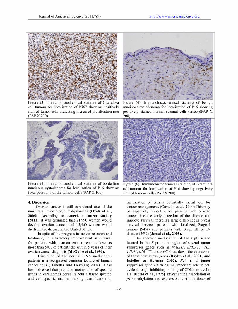
Figure (3): Immunohistochemical staining of Granulosa cell tumour for localization of Ki67 showing positively stained tumor cells indicating increased proliferation rate (PAP X 200)
Figure (4): Immunohistochemical staining of benign mucinous cystadenoma for localization of P16 showing positively stained normal stromal cells (arrow)(PAP X 200).
Figure (5): Immunohistochemical staining of borderline mucinous cystadenoma for localization of P16 showing focal positivity of the tumour cells (PAP X 100)
Figure (6): Immunohistochemical staining of Granulosa cell tumour for localization of P16 showing negatively stained tumour cells (PAP X 200)
4. Discussion: Ovarian cancer is still considered one of the most fatal gynecologic malignancies (Ozols et al., 2005). According to American cancer society (2011), it was estimated that 21,990 women would develop ovarian cancer, and 15,460 women would die from the disease in the United States. In spite of the progress in cancer research and treatment, no satisfactory improvement in survival for patients with ovarian cancer remains low; as more than 50% of patients die within 5 years of their ovarian cancer diagnosis (McGuire et al., 1996). Disruption of the normal DNA methylation patterns is a recognized common feature of human cancer cells ( Esteller and Herman; 2002). It has been observed that promoter methylation of specific genes in carcinomas occur in both a tissue specific and cell specific manner making identification of
methylation patterns a potentially useful tool for cancer management. (Costello et al., 2000) This may be especially important for patients with ovarian cancer, because early detection of the disease can improve survival; there is a large difference in 5-year survival between patients with localized, Stage I tumors (94%) and patients with Stage III or IV disease (29%) (Jemal et al., 2005). The aberrant methylation of the CpG island located in the 5′-promoter region of several tumor suppressor genes such as hMLH1, BRCA1, VHL, CDH1, p16INK4a, and APC shuts down the expression of these contiguous genes (Baylin et al., 2001; and Esteller & Herman 2002). P16 is a tumor suppressor gene which has an important role in cell cycle through inhibiting binding of CDK4 to cyclin D1 (Merlo et al., 1995). Investigating association of p16 methylation and expression is still in focus of

Journal of American Science, 2011;7(9) http://www.americanscience.org
936
interest in carcinogenesis and prognosis of human malignancy. In the current study using MSP there was significant increase in frequency of promoter methylation of p16 in malignant ovarian tissue (43.8 %) as compared to normal control group (16.7%). Similar results were also reported by other investigators as Andrew et al., 2006 who recorded positive methylation in 41.6% of malignancy and 21.1% in borderline ovarian tumor. Another study on 249 patients with ovarian cancer was performed by Katsaros et al., (2004) who found 40% positive in malignant tissues. But previous results using PCR- RFLP which is less sensitive than MSP showed less frequent as McCluskey et al., (1999) who found 5% of methylation, and 33% methylation was reported by Li et al., (2006) in ovarian cancer tissues. Increased p16 promoter methylation has been identified to be a ubiquitous mechanism of gene silencing that play a significant role in tumorigenesis of several human cancers not only ovarian including oral squamous cell carcinoma (Kaur et al., 2010), hepatocellular carcinoma (Fukai et al., 2005), colon (Malhotra et al. 2010), breast (Nielsen et al., 2001), gastric (Hu et al., 2010), and cervical carcinomas (Huang et al., 2011). The mechanism of p16 methylation linked carcinogenesis is mostly transcriptional silencing of gene expression (Kriegl et al., 2011). Clinical studies indicated that p16 expression is undetectable in about a third of ovarian cancer cases (Sui et al., 2000; and Havrilesky et al., 2001), and that patients with low p16 expression have poor response to chemotherapy and unfavorable survival outcome (Kudoh et al., 2002) Cell culture experiments demonstrate that reintroducing functional p16 into p16-null ovarian cancer cells results in inhibition of cell growth and increases in apoptosis, suggesting that p16 plays a role in ovarian cancer progression (Wolf et al., 1999). Li et al., (2006) found significant reduction of expression in ovarian cancer than normal tissues and lack of expression was mainly in methylated samples. In the present study we used immunohistochemistry to evaluate p16 protein expression, our results detected loss of expression in 87.5% (28/32) of malignant samples , while in normal samples it was 55.6% (10/18)., and there was significant correlation between promoter methylation and loss of protein expression as 94% (16/17) of total methylated samples (normal and malignant) showed loss of p16 protein expression, while it was found in 66% (22/33) of unmethylated samples, in malignant samples loss of expression was found in 100% (14/14) of methylated and 77.8% (14/18) of unmethylated samples, this indicates that
methylation increase p16 inactivation but it is not the only reason, it may be a result of other mechanism exhibited by previous works such as transcriptional errors (Chen et al., 1997) or posttranslational mechanisms as gene deletion or mutation (Fujita et al., 1997). Numerous previous studies investigated the relationship of inactivation or epigenetic changes of p16 gene with prognosis of ovarian tumors and other human tumors. Kudoh et al., (2002) found that ovarian cancer with low expression didn’t respond well to chemotherapy, and were associated with poor prognosis, similar findings were reported by Andrew et al., (2006); but they found significant increase risk of disease progression and overall survival but this was not available in our study. Moreover the prognostic value of p16 methylation was proved in many malignant tumors to be an indicator of poor prognosis and/or biomarker for response to chemotherapy (Goto et al., 2010; Wettergren et al., 2008; Csepregi et al., 2010; and Endo et al., 2011). Kaur et al., (2010) found significant association of p16 methylation and nodal involvement in oral squamous cell carcinoma. Decrease expression of p16 protein was significantly correlated with large tumor size in leiomyosarcoma (Kawaguchi et al., 2003). In our study no correlation was found between p16 methyaltion, expression, and various clinicopathological factors including age, tumor stage, and grade. This disagreement may be caused by the heterogenous nature of ovarian tumors and small size of investigated samples. Limited number of studies investigated the association of MTHFR C677T and ovarian cancer with controversial results (Terry et al., 2010; and Magnowski et al., 2010). In the current study we recorded more frequency of T allele and T containing genotypes (CT+TT) in ovarian cancer tissues (48%, 87.5%) than in normal ovarian tissues (36%, 66.7%) respectively with OR 1.6 (0.7-3.8) for T alleles and OR 3.5 (0.8-14.7)for (CT+TT) genotype showing the potential conflict of MTHFR C677T mutation on ovarian carcinogenesis. This may be explained by the fact that among 677TT (Val/Val) individuals, the MTHFR enzyme is less efficient in converting 5, 10-methylenetetrahydrofolate to 5-methyltetrahydrofolate, thus potentially preventing depletion of 5, l0-methylenetetrahydrofolate, a cofactor for de novo DNA synthesis, especially dTMP. As a result, cells may be less prone to "dNTP stress" which has been shown to promote cancer-associated genetic alterations due to alterations in the pool of nucleotide precursors available for DNA synthesis. Alteration of these precursor pools induced by methyl (folate) deficiency significantly increases the uracil content

Journal of American Science, 2011;7(9) http://www.americanscience.org
937
and the frequency of chromosome breakages in human leukocyte DNA (Deloughery et al., 1996). MTHFR C677T genotype has been analyzed in relation to promoter methylation of p16 in tumors from different sites with controversial results, Tao et al., (2009) found no relation to p16 promoter methylation in breast cancer; Curtin et al., (2007) found an increased likelihood of highly CpG-methylated phenotype in colon tumors for those with one or two variant MTHFR 1298C alleles, and the association was modified by high-risk dietary profiles (low folate and methionine intake and high alcohol use). Similarly, MTHFR C677T, A1298C, or MTR C2756G genotypes were not associated with E-cadherin and p16 promoter methylation in esophageal (Wang et al., 2008) and cervical cancers (Kang et al., 2005). A pooled analysis including 725 cases esophageal carcinoma and 1531 controls showed a significant association between the MTHFR 677 TT genotype and susceptibility to esophageal cancer [Langevin et al., 2009]. Lu et al., (2011) found that diet folate intake had different effects on the prognosis of esophageal carcinoma by different genotypes of MTHFR C677T. The preventive effect of folate intake was more evident in patients carrying MTHFR 677CC genotype. In our research we analyzed MTHFR C677T polymorphism and compared its frequency to p16 methylation and expression in ovarian cancer. we demonstrated marked decrease of p16 promoter methylation in CT+ TT genotypes than CC genotype in all studied individuals, and in separate malignant and control groups with OR 0.25 (0.06-1.07), and 0.18 (0.02-2) respectively with no significant difference between malignant and control group regarding decline of p16 methylation (data not shown). This coincide with finding of Chiusolo et al., (2006) in multiple myeloma who demonstrated that MTHFR 677CC is associated with a higher prevalence of p16 hypermethylation. In our study no clear association was found between polymorphism and p16 protein expression or any of clinicopatholgical criteria of ovarian carcinomas. We conclude that p16 methylation is an important carcinogenic factor on ovarian tissue through inhibition of gene expression, and there is potential effect of T allele and T containing genotype of MTHFR C677T polymorphism on degree of methylation of p16 gene, as MTHFR C677T polymorphism was associated with reduction in P16 promoter methylation and increased expression of p16 gene, meanwhile it is linked to increased risk of ovarian cancer so mostly carcinogenic effect is not through modifying the methylation of p16 gene. However a remarkable risk of ovarian carcinogenesis was noticed in separate analysis of the effect of
MTHFR mutation and p16 methylation on ovarian tissues. This may reflect the possible interaction of other genetic, environmental and dietary factors that can affect DNA expression and epigenetic changes that needs to be further declared. Acknowledgments:
The authors like to express sincere appreciation to Dubai medical College for the financial support given by medical research fund committee and allowance of using laboratories of biochemistry and molecular biology. Corresponding author Naglaa R. AbdRaboh 1Biochemistry department, Faculty of Medicine Ain Shams University, Cairo, Egypt [email protected] References: Agrawal A.; Murphy R.F.; and Agrawal D.K.
(2007). DNA methylation in breast and colorectal cancers. Mod Pathol; 20:711–21.
Ahuja N.; Mohan A.L.; Li Q.; Stolker J.M.; Herman J.G.; Hamilton S.R.; Baylin S.B.; and Issa J.P. (1997). Association between CpG island methylation and microsatellite instability in colorectal cancer. Cancer Res; 57: 3370-3374.
American cancer society (2011). Genetics of Breast and Ovarian Cancer. http://www.cancer.gov/cancertopics/types/ovarian US national cancer institute.
Andrew W.; Dionyssios K.; Haigang C.; Irene A. R.; Alicia B.; Manuela P.; Rakesh S.;Yan Z.; Agyenim A.; Daniel Z.; and Herbert Y. (2006). Aberrant promoter methylation of multiple genes in malignant ovarian tumors and in ovarian tumors with low malignant potential.Cancer.177:299-308.
Baylin S. B.; Esteller M.; Rountree M. R.; Bachman K. E.; Schuebel K.; and Herman J. G. (2001). Aberrant patterns of DNA methylation, chromatin formation and gene expression in cancer. Hum. Mol. Genet., 10: 687-692.
Chen Y.J.; Chang J.G.; Shin L.S.; Chen P.H.; Endo M.; Whang-Peng J.; and Chen Y.M. (1997). Frequent detection of aberrant RNA transcripts of CDKN2 gene in human gastric adenocarcinoma. Int.J. Cancer. 71:350-354.
Chiusolo P.; Farina G.; Putzulu R.; Reddiconto G.; Fiorini A.; De Stefano V.; Rossi E.; Palladino M.; Leone G.; and Sica S.(2006). Analysis of MTHFR polymorphisms and P16 methylation and their correlation with clinical-biological features of multiple myeloma. Ann Hematol. 85(7):474-7.

Journal of American Science, 2011;7(9) http://www.americanscience.org
938
Choi S.W.; and Mason J.B.; (2002). Folate status: effects on pathways of colorectal carcinogenesis. J Nutr; 132:2413–8S.
Costello J.F.; Fruhwald M.C.; Smiraglia D.J.; Costello J.F.; Frühwald M.C.; Smiraglia D.J.; Rush L.J.; Robertson G.P.; Gao X; and Wright F.A.(2000). Aberrant CpG-island methylation has non-random and tumour-type-specific patterns. Nat Genet. 24: 132-138.
Csepregi A.; Ebert M.P.; Rocken C.; Schneider-Stock R.; Hoffmann J.; Schulz H.U.; Roessner A.; and Malfertheiner P.(2010). Promoter methylation of CDKN2A and lack of p16 expression characterize patients with hepatocellular carcinoma. BMC Cancer. 22; 10:317.
Curtin K, Slattery ML, Ulrich CM, Wolff R.K.; Herrick J. S.; Caan B. J.; and Slatteryand M.L. (2007). Genetic polymorphisms in one-carbon metabolism: associations with CpG island methylator phenotype (CIMP) in colon cancer and the modifying effects of diet. Carcinogenesis 28:1672–9.
Deloughery T.G.; Evans A.; Sadeghi A.; McWilliams J.; Henner W.D.; and Taylor L.M. (1996). Common mutation in methylenetetrahydrofolate reductase. Correlation with homocysteine metabolism and late-onset vascular disease. Circulation 94:3074–78.
Endo M.; Kobayashi C.; Setsu N.; Takahashi Y.; Kohashi K.; Yamamoto H.; Tamiya S.; Matsuda S.; Iwamoto Y.; Tsuneyoshi M.; and Oda Y. (2011). Prognostic significance of p14ARF, p15INK4b and p16INK4a inactivation in malignant peripheral nerve sheath tumors. Clin Cancer Res. 17(11):3771-82.
Esteller M. (2005). Aberrant DNA methylation as a cancer-inducing mechanism. Annu Rev Pharmacol.; 45: 629-656.
Esteller M.; and Herman J. G. (2002). Cancer as an epigenetic disease: DNA methylation and chromatin alterations in human tumors. J. Pathol., 196: 1-7.
Esteller M.; Corn P.G.; Baylin S.B.; and Herman J.G. (2001). A gene hypermethylation profile of human cancer. Cancer Res.; 61: 3225-3229.
Friso S.; and Choi S.W. (2005). Gene-nutrient interactions in one-carbon metabolism. Curr Drug Metab 6:37–46.
Fujita M.; Enomoto T.; Haba T.; Nakashima R.; Sasaki M.; Yoshino K.; Wada H.; Buzard G.S.; Matsuzaki N.; Wakasa K.; and Murata Y. (1997). Alteration of p16 and p15 genes in common epithelial ovarian tumors. Int J Cancer; 74:148-155.
Fukai K.; Yokosuka O.; Imazeki F.; Tada M.; Mikata R.; Miyazaki M.; Ochiai T.; and Saisho H. (2005). Methylation status of p14ARF,
p15INK4b, and p16INK4a genes in human hepatocellular carcinoma. Liver Int. 25(6):1209-16.
Goto T.; Mizukami H.; Shirahata A.; Yokomizo K.; Kitamura Y.H.; Sakuraba K.; Saito M.; Ishibashi K.; Kigawa G.; Nemoto H.; Sanada Y.; and Hibi K. (2010). Methylation of the p16 gene is frequently detected in lymphatic-invasive gastric cancer. Anticancer Res. 30(7):2701-3.
Havrilesky L.J.; Alvarez A.A.; Whitaker R.S.; Marks J.R.; and Berchuck A. (2001). Loss of expression of the p16 tumor suppressor gene is more frequent in advanced ovarian cancers lacking p53 mutations. Gynecol. Oncol. 83 (3): 491–500.
Herman J.G.; and Baylin S.B. (2000). Promoter-region hypermethylation and gene silencing in human cancer. In: Vogt PAJaPK (ed). DNA Methylation and Cancer, 1st edn. Springer-Verlag: New York, Berlin, Heidelberg, pp. 35–50.
Herman J.G.; Graff J.R.; Myöhänen S.; Nelkin B.D.; and Baylin S.B. (1996). Methylation-specific PCR: a novel PCR assay for methylation status of CpG islands. Proc Natl Acad Sci USA.; 93(18):9821-6.
Hu S.L.; Kong X.Y.; Cheng Z.D.; Sun Y.B.; Shen G.; Xu W.P.; Wu L.; Xu X.C.; Jiang X.D.; and Huang D.B. (2010). Promoter methylation of p16, Runx3, DAPK and CHFR genes is frequent in gastric carcinoma. Tumori. 96(5):726-33.
Huang L.W.; Pan H.S.; Lin Y.H.; Seow K.M.; Chen H.J.; and Hwang J.L. (2011). P16 methylation is an early event in cervical carcinogenesis. Int J Gynecol Cancer. 21(3):452-6.
Issa J-P.J. (2003). Methylation and prognosis: of molecular clocks and hypermethylator phenotypes. Clin Cancer Res.; 9: 2879-2881.
Jemal A.; Murray T.; Ward E.; Samuels A.; Tiwari R.C.; Ghafoor A.; Feuer E.J.; and Thun M.J. (2005). Cancer statistics 2005. CA Cancer J Clin. 55: 10-30.
Josef S.; David W.R.; Nina I.; and Kaaren A.J. (2002). MOLECULAR CLONING: Rapid Isolation of Mammalian DNA. Cold Spring Harbour Laboratory Press. New York 2002; 628-630.
Kamb A.; Gruis N.A, Weaver-Feldhaus J.; Liu Q.; Harshman K.; Tavtigian S.V.; Stockert E.; Day R.S 3rd.; Johnson B.E.; and Skolnick M.H. (1994). A cell cycle regulator potentially involved in genesis of many tumor types. Science; 264: 436-440.
Kamiya H.; Kawakami K.; Miyanaga T.; Omura K.; Oda M.; Murakami S.; and Watanabe Y. (1998). A methylenetetrahydrofolate reductase polymorphism is associated with expression of p16 in human lung cancer. Oncol. Rep. 5 (4):911–4.

Journal of American Science, 2011;7(9) http://www.americanscience.org
939
Kang S.; Kim J.W.; Kang G.H.; Park N.H.; Song Y.S.; Kang S.B.; and Lee H.P. (2005). Polymorphism in folate- and methionine-metabolizing enzyme and aberrant CpG island hypermethylation in uterine cervical cancer. Gynecol Oncol; 96:173–80.
Katsarosa D.; Chob W.; Singalc R.; Fracchiolia S.; Rigault de la Longraisa I.A.; Arisioa R.; Massobrioa M.; Smithc M.; Zhengb W.; Glassc J.; and Yu H. (2004). Methylation of tumor suppressor gene p16 and prognosis of epithelial ovarian cancer. Gynecologic Oncology 94(3): 685-692.
Kaur J.; Demokan S.; Trpathi S.C.; Macha M.A.; Begum S.; Begum S.; Califano J.; and Ralhan R. (2010). Promoter hypermethylation in Indian primary oral squamous cell carcinoma. Int J Cancer, 15; 127(10): 2367-2373.
Kawaguchi K.; Oda Y.; Saito T.; Yamamoto H.; Tamiya S.; Takahira T.; Miyajima K.; Iwamoto Y.; and Tsuneyoshi M. (2003). Mechanisms of inactivation of the p16INK4a gene in leiomyosarcoma of soft tissue: decreased p16 expression correlates with promoter methylation and poor prognosis. J Pathol. 201(3):487-95.
Kriegl L.; Neumann J.; Vieth M.; Greten F.R.; Reu S.; Jung A.; and Kirchner T. (2011). Up and down regulation of p16 (Ink4a) expression in BRAF-mutated polyps/adenomas indicates a senescence barrier in the serrated route to colon cancer. Mod Pathol. 24(7):1015-22.
Kudoh K.; Ichikawa Y.; Yoshida S.; Hirai M.; Kikuchi Y.; Nagata I.; Miwa M.; and Uchida K. (2002). Inactivation of p16/CDKN2 and p15/MTS2 is associated with prognosis and response to chemotherapy in ovarian cancer. Int J Cancer. 99(4):579-82.
Langevin S.M.; Lin D.; Matsuo K.; Gao C.M.; Takezaki T.; Stolzenberg-Solomon R.Z.; Vasavi M.; Hasan Q.; and Taioli E. (2009). Review and pooled analysis of studies on MTHFR C677T polymorphism and esophageal cancer. Toxicol Lett.; 184:73–80.
Li M.; Huang Z.J.; Dong W.H.; Li X.Y.; Wang X.Y.; He X.H.; Wang H.; and Wang Z.H. (2006). Zhonghua Fu Chan Ke Za Zhi. 41(6):408-12.
Liggett W.H.; and Sidransky D. (1998). Role of the p16 tumor suppressor gene in cancer. J. Clin. Oncol; 16:1197–1206.
Lu C.; Xie H.; Wang F.; Shen H.; and Wang J. (2011). Diet folate, DNA methylation and genetic polymorphisms of MTHFR C677T in association with the prognosis of esophageal squamous cell carcinoma. BMC Cancer. 11: 91.
Magnowski P.; Seremak-Mrozikiewicz A.; Nowak-Markwitz E.; Kurzawiska G.; Drews K.;
and Spaczyski M. (2010). No association between MTHFR 677C>T polymorphism and ovarian cancer risk in BRCA1 mutation carriers in Wielkopolska region. Ginekol Pol. 81(7):506-10.
Malhotra P.; Kochhar R.; Vaiphei K.; Wig J.D.; and Mahmood S. (2010). Aberrant promoter methylation of p16 in colorectal adenocarcinoma in North Indian patients. World J Gastrointest Oncol. 2(7): 295-303.
McCluskey L.L.; Chen C.; Delgadillo E.; Felix J.C.; Muderspach L.I.; and Dubeau L. (1999). Differences in p16 gene methylation and expression in benign and malignant ovarian tumors. Gynecol Oncol .72: 87-92.
McGuire W.P.; Hoskins W.J.; Brady M.F.; Kucera P.R.; Partridge E.E.; Look K.Y.; Clarke-Pearson D.L.; and Davidson M. (1996). Cyclophosphamide and cisplatin compared with paclitaxel and cisplatin in patients with Stage III and Stage IV ovarian cancer. N Engl J Med. 334: 1-6.
Merlo A.; Herman J.G.; Mao L.; Lee D.J.; Gabrielson E.; Burger P.C.; Baylin S.B.; and Sidransky D. (1995). 5' CpG island methylation is associated with transcriptional silencing of the tumor suppressor p16/ CDKN2/MTS1 in human cancers. Nat Med; 1: 686-692.
Nielsen N.H.; Roos G.; Emdin S.O.; and Landberg G. (2001). Methylation of the p16 (Ink4a) tumor suppressor gene 5'-CpG island in breast cancer. Cancer Lett; 163: 59-69.
Oyama K.; Kawakami K.; Maeda K.; Ishiguro K.; and Watanabe G. (2004). The association between methylenetetrahydrofolate reductase polymorphism and promoter methylation in proximal colon cancer. Anticancer Res. 24:649–54.
Ozols R.F.; Schwartz P.E.; and Eifel P.J. (2005). ovarian cancer, peritoneal carcinoma, and fallopian tube carcinoma. In: DeVita VT Jr., Hellman S, Rosenberg SA, editors. Cancer: Principles and Practice of Oncology, 7th ed. Philadelphia: Lippincott, Williams and Wilkins; Section 32.4: 1364-1398.
Ries L.; Melbert D.; Krapcho M.; Stinchcomb D., Howlader N.; Horner M.J.;, Mariotto A.; Miller B.A.; Feuer E.J.; Altekruse S.F.; Lewis D.R.; Clegg L.; Eisner M.P.; Reichman M.; and Edwards B.K. (2008). SEER Cancer Statistics Review, 1975-2005, National Cancer Institute. Bethesda, MD http://seer.cancer.gov/csr/1975_2005/, based on November 2007 SEER data submission, posted to the SEER web site.
Sambrook J.; and Russell D.W. (2001). Preparation and analysis of eukaryotic genomic DNA. In: Sambrook J, Russell DW, editors. Molecular

Journal of American Science, 2011;7(9) http://www.americanscience.org
940
cloning: a laboratory manual. 3rd ed. New york: cold spring harbor laboratory press, pp 6.4-6.12.
Shima K.; Nosho K.; Baba Y.; Cantor M.; Meyerhardt J.A.; Giovannucci E.L.; Fuchs C.S.; and Ogino S. (2011). Prognostic significance of CDKN2A (p16) promoter methylation and loss of expression in 902 colorectal cancers: Cohort study and literature review. Int J Cancer.128(5):1080-94.
Sui L.; Dong Y.; Ohno M.; Goto M.; Inohara T.; Sugimoto K.; Tai Y.; Hando T.; and Tokuda M. (2000). Inverse expression of Cdk4 and p16 in epithelial ovarian tumors. Gynecol. Oncol. 79 (2): 230–237.
Tao M.; Shields P.G.; Nie J.; Marian C.; Ambrosone C.B.; McCann S.E.; Mary P. M.; Krishnan S.S.; Xie B.; Edge S.B.; Winston J.; Vito D.; Trevisan M.; and Freudenheim J.L. (2009). DNA Promoter Methylation in Breast Tumors: No Association with Genetic Polymorphisms in MTHFR and MTR. Cancer epidemiol Biomarkers Prev; 18 (3).
Terry K.L.; Tworoger S.S.; Goode E.L.; Gates M.A.; Titus-Ernstoff L.; Kelemen L.E.; Sellers T.A.; Hankinson S.E.; and Cramer D.W. (2010). MTHFR polymorphisms in relation to ovarian cancer risk. Gynecol Oncol. 119(2):319-24.
Ulrich C.M.; Curtin K.; Samowitz W.; Bigler j.; Potter J.D.; Caan B.; and Slattery M.L. (2005). MTHFR variants reduce the risk of G: C->A: T transition mutations within the p53 tumor suppressor gene in colon tumors. J. Nutr. 135:2462–7.
Van der Put N. M.; Gabreels F.; Stevens E. M.; Smeitink J. A.; Trijbels F. J.; Eskes T. K.; van den Heuvel L. P.; and Blom H. J. ( 1998). A second common mutation in the methylenetetrahydrofolate reductase gene: an additional risk factor for neural-tube defects. Am. J. Hum. Genet. 62: 1044-1051.
Wang J.M.; Sasco A.; Fu C.W.; Xue H.; Guo G.; Hua Z.; Zhou Q.; Jiang Q.; and Xu B.(2008). Aberrant DNA methylation of P16, MGMT, and hMLH1 genes in combination with MTHFR C677T genetic polymorphism in esophageal squamous cell carcinoma. Cancer Epidemiol Biomarkers Prev. 17:118–25.
Wettergren Y.; Odin E.; Carlsson G.; and Gustavsson B. (2010). MTHFR, MTR, and MTRR Polymorphisms in Relation to p16INK4A Hypermethylation in Mucosa of Patients with Colorectal Cancer. Mol Med. 16(9-10): 425–432.
Wiemels J. L.; Smith R. N.; Taylor G. M.; Eden O. B.; Alexander F. E.; and Greaves M. F.(2001). United Kingdom Childhood Cancer Study investigators. Methylenetetrahydrofolate reductase (MTHFR) polymorphisms and risk of molecularly defined subtypes of childhood acute leukemia. Proc. Natl. Acad. Sci. USA, 98: 4004-4009.
Wolf J.K.; Mills G.B.; Bazzet L.; Bast R.C.; Roth J.A.; and Gershenson D.M. (1999). Adenovirus-mediated p53 growth inhibition of ovarian cancer cells is independent of endogenous p53 status. Gynecol. Oncol. 75 (2): 261–266.
8/22/2011