journal of biochemistry and cell biology · effect of tartrazine on thyroid gland of male rat and...
TRANSCRIPT

Effect of Tartrazine on Thyroid Gland of Male Rat and Ameliorating Roleof Curcumin (Histological and Immunohistochemical Study)Heba M Abdel-Aziz1*, Zeinab M Alazouny1, Karima F Abdelfadeel1 and Aisha A Abohashem2
1Department of Histology and Cell Biology, Faculty of Medicine, Zagazig University, Zagazig, Egypt2Department of Forensic Medicine and Clinical Toxicology, Faculty of Medicine, Zagazig University, Zagazig, Egypt*Corresponding author: Heba M Abdel-Aziz, Lecturer in Department of Histology and Cell Biology, Faculty of Medicine, Zagazig University, Zagazig, Egypt, Tel:00201281547665, E-mail: [email protected] date: October 15, 2018; Accepted date: October 25, 2018; Published date: November 1, 2018
Copyright: © 2019 Abdel-Aziz HM, et al. This is an open-access article distributed under the terms of the Creative Commons Attribution License, which permitsunrestricted use, distribution, and reproduction in any medium, provided the original author and source are credited.
Abstract
Tartrazine and curcumin are commonly used as food additives and also, in drugs and cosmetics. The presentstudy aimed to evaluate the effect of tartrazine on the structure and function of the thyroid glands of adult malealbino rats and the possible protective role of curcumin intake. Forty five adult male albino rats were classified intofour groups: Control, treated that received tartrazine, protective received curcumin with tartrazine and recovery. Atthe end of the experiment, blood samples were subjected to a hormonal assay of total T3, T4 and thyrotropin (TSH).The tissue malondialdehyde (MDA), superoxide dismutase (SOD) catalase (CAT) and glutathione (GSH) wereestimated. Additionally, the thyroid gland tissue samples were processed for histological and immunohistochemicalstudy. The results obtained were analyzed morphometrically and statistically. The treated group showeddeterioration of thyroid functions with various degrees of thyroid structural alterations as vacuolated cytoplasm, darknuclei and vacuolated colloid, epithelial stratification and increase in the immune expression of caspase 3 andnuclear factor kappa B (NF-kB p65). Administration of curcumin showed improvement in the biochemical parametersand thyroid structure than in recovery group. Curcumin have a relevant protective role against tartrazine-inducedthyroid gland damage.
Keywords: Tartrazine; Curcumin; NF-kB p65; Thyroid gland
IntroductionFood additives play a vital role in increasing the taste, tint, quality
and price of foods and classified into 6 main groups: Preservatives,nutritive, flavouring, colouring, texturizing and miscellaneouscompounds [1]. Colour additives are defined as any dyes or pigmentthat can impart color to food, drugs or cosmetics to enhance the visualappeal [2]. Some of these substances are derived from the naturalcolors such as carotene and chlorophyll and others are synthetic suchas indigotin, allura red and tartrazine [3].
Synthetic food additive tartrazine is a bright yellow azo dye that ismore stable and a cheaper alternative to natural food dyes. It is knownby other names such as FD and C Yellow No. 5 and E 102 Europe [4]. Itis commonly seen in medication, cosmetics and food products such ascandy, soft drinks (Mountain Dew), energy drinks, flavored corn chips,cereals (corn flakes), cake mixes, ice cream, ice pops, chewing gum,marzipan, jam, jelly, yogurt noodles, potato chips, biscuits and in somehoney products [5]. It has been implicated to be responsible for allergicreactions [6] and has harmful effects in learning and memoryfunctions in animals [2]. In toxicological studies on rats, someresearchers revealed that it induces several adverse effects on kidney,liver and blood cells [1]. Some behavioral changes among children thatintake coloured foods contained tartrazine such as irritability,restlessness and sleep disturbance [5].
The use of natural colors with or without synthetic colors as foodcoloring agents is considered essential for promoting health status andminimizing risks of adverse effects in consumers. Widespread usage ofnatural colors in the food industry occurred due to mounting demands
by consumers for safer foods with natural additives. Currently, 13natural food colors are approved for coloring foods one of which iscurcumin [7]. Curcumin (curry powder) is the active ingredient of thedietary spice turmeric and is extracted from the rhizomes of Curcumalonga which belongs to ginger family. It has several pharmacologicaland biological properties such as being anti-inflammatory, antiviral,antimicrobial and antifungal agent [8]. In recent years, numerousstudies have shown that oral administration of curcumin significantlyameliorated collagen-induced arthritis [9]. Moreover, curcumin showschemopreventive actions [10] and were used in many cancerstreatment including colorectal cancer, breast cancer, skin cancer, andoral cancer [11]. Curcumin was reported to have a protective effectagainst cyclosporine-induced nephrotoxicity in rats [12]. It is also usedin Asian and African traditional medicine to treat several mild ormoderate health problems such as aches; wounds; sprains; and liverdisorders [13]. Thyroid gland is one of the most important endocrineorgans and almost all cells of the body are target sites for its hormones(T3,T4) and considered as body’s primary regulator of metabolism[14]. Nuclear Factor Kappa B (NF-Kb) is a heterodimer proteincomplex, and it usually consists of two proteins, a p65 subunit and ap50 subunit and other subunits [15]. Caspase 3 is a cytosolic proteinfound in the cells as an inactive 32 kDa proenzyme. It is activated byproteolytic cleavage into two active subunits only when cells undergoapoptosis [16]. The present study was directed to evaluate the effect oftartrazine on thyroid gland of adult male albino rats from histological,immunohistochemical and biochemical point of view. Additionally, thepossible protective effect of curcumin intake was also investigated.
Jour
nal o
f Bioc
hemistry and Cell Biology
Journal of Biochemistry and CellBiology Abdel-Aziz et al., J Biochem Cell Biol 2019, 2:1
Research Article Open Access
J Biochem Cell Biol, an open access journal Volume 2 • Issue 1 • 1000111

Materials and Methods
ChemicalsTartrazine: (CAS 1934-21-0, Purity 86.7%) was purchased in the
form of powder from Sigma Aldrich (Germany). Its chemical formula:Trisodium salt: Trisodium 5-hydroxy-1-(4-sulfonatophenyl)-4-(E)-(4sulfonatophenyl) diazenyl-1H-pyrazole-3-carboxylate,
Curcumin: (CAS 458-37-7, Purity: ≥ 95%) was purchased in theform of powder from El-goumhoria Co. for trading chemicals,medicines and medical appliances, Egypt. Its chemical formula: 1,7-bis(4-hydroxy-3methoxyphenyl)-1E,6E-heptadiene-3,5-dione. MolecularFormula: C21H20O6. Formula Weight: 368.4.
AnimalsForty five healthy adult male albino rats with average body weight
(200-250 g) were used and housed in stainless steel cages at AnimalHouse, Faculty of Medicine, Zagazig University. They were kept atroom temperature, fed standard balanced diet and allowed water ad-libitum. The experiment was performed in accordance with the (Guidefor the Care and Use of Laboratory Animals). The experimentalprotocol was approved by the Ethical Committee of Zagazig University.
Experimental designAfter an acclimatization period of 1 week, rats were randomly
divided into four equal groups; I, II, III and IV.
Group I (Control group): Included 15 rats, further subdivided intothree equal subgroups ((five rats each):
Subgroup Ia (Negative control group): Were left withoutintervention to measure the basic parameters.
Subgroup Ib (Positive control group; distilled water): Received 1 mldistilled water once daily by orogastric tube (the solvent of bothtartrazine and curcumin) for thirty days.
Subgroup Ic: (Positive control group; curcumin group): Receivedcurcumin dissolved into 1 ml distilled water at a dose of (100 mg/ kgonce daily) by orogastric tube for thirty days.
Group II (treated group): Included 10 rats that were receivedtartrazine dissolved in 1 ml distilled water at concentration of 300mg/kg given once daily by orogastric tube for thirty days [4].
Group III (protective group): Included 10 rats that were receivedtartrazine solution at the same dose as group II in concomitant withcurcumin dissolved in 1ml distilled in a dose of (100 mg/kg water oncedaily) by orogastric tube [8] for thirty days [17].
Group IV (recovery group): Included 10 rats that were receivedtartrazine solution at the same dose as group II once daily for thirtydays. At a day 31 of treatment, the rats were left without any treatmentfor further thirty days as a recovery period [18].
General observations of animals: During the experimental period,clinical signs and general appearances, which included the amount offood and water consumed, were checked daily. Mortalities of the ratswere recorded as it occurred.
The body weight of each animal was assessed before and at the endof experiment. The percentage of body weight gain was calculated asfollows according to previous reports [19].
Mean final body weight - Mean initial body weight × 100
Mean initial body weightAt the time of sacrifice, rats were anesthetized with intraperitoneal
injection of sodium pentobarbital (Nembutal, 30 mg/kg body weight).Venous blood samples (2 ml each) were withdrawn from the retro-orbital sinus for hormonal assay.
The thyroid glands were dissected out in two stages to avoid tissuedamage. First, the neck was opened by a longitudinal incision, thefascia was removed, and the trachea was cut by a horizontal planesuperior and inferior to the thyroid. It appeared as two small reddishoval masses on each side of the trachea [20,21]. Thyroid glands fromeach rat were quickly excised into specimens.
Histological Study1 cm3 specimens were processed with paraffin technique [22] to
investigate the histological and immunohistochemical results throughthe light microscopic examination.
The specimens were fixed in 10% neutral-buffered formalin,dehydrated, embedded in paraffin and then the sections were cut at 4-5µm by a microtome then stained with Haematoxylin and eosin (H andE). Also, the Periodic Acid and Schiff (PAS) staining procedure wereused according to [22] to differentially stain the colloid inside thethyroid follicles [23].
1 mm specimens; were immediately fixed in 3% glutaraldehydebuffered with 0.1 M phosphate at 4°C (pH 7.4) then post fixed in 1%osmium tetroxide for 2 hrs at 4°C. The specimens were thendehydrated and embedded in epoxy resin. Semi-thin sections (1 µmthick) were stained with toluidine blue [24].
Immunnohistochemical studyImmunohistochemical detection of caspase-3 and NF-kB p65 was
carried out using streptavidin-biotin complex immunoperoxidasesystem. Serial sections of paraffin-embedded specimens weredeparaffinized on charged slides. The sections were incubated in 0.1%hydrogen peroxide for 30 min to block the endogenous peroxidase.The sections were then incubated with 1.5% non-immunized goatserum for 30 min at room temperature, and then incubated withdiluted primary antibody (1:500 dilutions) [25] .
Primary antibody for Caspase 3: A rabbit polyclonal antibody of IgGtype was carried out for localization of Caspase 3 (GTX 110543) [26]or NF-kB p65 (GTX102090) for 30 min at room temperature [27].
The sections were then washed 3-times with (PBS, pH 7.4) for 30min and then incubated with biotinylated goat anti-mouseimmunoglobulin serum for 60 min. After being gently washed withPBS, the sections were incubated with avidin-biotin peroxidasecomplex. Ultimately, sites for peroxidase binding were detected usingDAB (3,30-diaminobenzidine) substrate) [28]. Tissue sections werethen counterstained with hematoxylin and subjected to lightmicroscopy analyses and morphometric measures. The kits were werepurchased from Sigma-Aldrich, St. Louis.
Biochemical analysis of oxidant and antioxidant markers in thyroidtissue: Thyroid gland specimens were processed for determination theoxidative markers stress in tissues (Oxidant markers: Malondialdehyde(MDA) and Superoxide Dismutase (SOD); Antioxidant markers:Catalase (CAT) and reduced glutathione (GSH)). Those thyroid tissues
Citation: Abdel-Aziz HM, Alazouny ZM, Abdelfadeel KF, Abohashem AA (2019) Effect of Tartrazine on Thyroid Gland of Male Rat andAmeliorating Role of Curcumin (Histological and Immunohistochemical Study). J Biochem Cell Biol 2: 111.
Page 2 of 9
J Biochem Cell Biol, an open access journal Volume 2 • Issue 1 • 1000111

were washed with ice-cold saline, blot-dried, suspended in phosphatebuffer (pH 6) at 5-times then processed in a Potter-Elvehjemhomogenizer. The raw homogenate was then frozen at 85°C until usedin the various assays [29,30]. Measuring the levels of MDA was basedon the method of thiobarbituric acid (TBA) [31]. The superoxidedismutase (SOD) activity (in µ/g tissue) was determined according to[32]. Catalase (CAT) activity (in µ/g tissue) was assessed by means ofthe method of Aebi [33]. Reduced glutathione (GSH) was determinedaccording to the method of Tipple and Rogers [34]. All the above-mentioned kits were purchased from Biodiagnostic and conducted atBiochemistry Department, Faculty of Medicine- Zagazig University.
Biochemical Hormonal assay study: The blood samples wereobtained through heparinized capillary tubes and allowed to clot atroom temperature in a water bath for 15 min. The supernatant serumwas collected in a dry tube. The sera were quickly removed and kept at-20°C until the assay. Serum total T3 (triiodothyronine), T4(thyroxine) and thyroid stimulating hormone (TSH) levels weremeasured using RIA Kit (Diagnostic Products Corporation, LA, USA).
Morphometric StudyThe image analyzer computer system Leica Qwin 500 (Cambridge,
UK, Leica Microsystems Imaging Solutions, Ltd) in the imageanalyzing unit of the Pathology Department, Faculty of Dentist, CairoUniversity, Egypt.
• Haematoxylin and eosin stained sections were used for themeasurement of follicular epithelial height. This was performed in5 non overlapping fields from 5 different sections of 5 different ratsin each group at × 400.
• Periodic acid Schiff (PAS) reaction for the demonstration of theoptical density of colloid of thyroid follicle.
• Immune stained sections were used for the measurement of opticaldensity of immunoexpression in caspase-3 and NF-kB p65. Thiswas performed in 5 non overlapping fields from 5 differentsections of 5 different rats in each group at × 400.
Statistical analysisStatistical analysis was performed using statistical Package for the
Social Sciences (SPSS) version 20 for Windows software system (SPSSInc, Chicago, IL, USA). Results were articulated as mean ± standarderror (SE) and all statistical comparisons were made by means of aone-way ANOVA test. A P value <0.05 was considered significant.When the difference between groups was significant, post hoc analysiswas carried out by applying the LSD.
Results
General observation, food intake and body weightTreatment with tartrazine did not affect mortality and food intake
when compared to the control group. As a function of growth, bodyweight of experimental animals was monitored in comparison with thecontrol group. Our data showed that the body weight (%) of the rats ofcontrol group and curcumin protective group was non-significantlyincreased at the end of the experiment. However, there was asignificant retardation (p<0.05) in it in rats of tartrazine treated andrecovery groups. Results are presented in (Table 1).
Group (Ia) (Ib) (Ic) (II) III IVF P. value
Parameters Mean ± SD Mean ± SD Mean ± SD Mean ± SD Mean ± SD Mean ± SD
Body weight (%) 25.16 ± 0.46 24.99 ± 0.49 25.1 ± 0.47 10.33 ± 3.07a 24.9 ± 16b 23.87 ± 0.88a,c 674.04 <0.001
Results: Values are expressed as mean ± standard deviation (SD) of n=10 rats/group, aSignificant as compared with the Ia,Ib and Ic groups, P<0.05. bSignificant ascompared with II group, P<0.05. cSignificant as compared with III group, P<0.05.
Table 1: Statistical results of the body weight percent in the studied groups.
Histological ResultsBoth subgroups (a,b and c) of the control group revealed the same
histological and immunohistochemical features. H and E stainedsections of thyroid glands from the control group showed that thyroidparenchyma was composed of multiple and relatively rounded follicles.Mostly they are of moderate size and lined by single layer of cubicalepithelium with rounded nuclei. The follicular lumen was filled withhomogenous acidophilic colloid. Interfollicular cells and blood vesselsare seen in connective tissue between the follicles (Figure 1a).Tartarzine-treated group showed that majority of the follicles werelined by tall columnar cells with rod shaped nuclei. Some follicles werelined by more than one layer of follicular cells. Multiple pyknoticnuclei were seen in follicular lining epithelium. Many follicular cellsappeared with vacuolated cytoplasm. Reduction in amount of colloidwith increased marginal vacuolation were also observed in somefollicles. Other follicles appeared empty with no colloid in theirlumina. Disorganized and damaged follicles were observed. Congestedblood vessels in between the follicles were also noticed (Figure 1b). Incurcumine-protective group sections Administration of tartarzine
concomitantly with curcumin ameliorated thyroid follicles. Most ofthyroid follicles were similar to that of the control group. They werelined by single layer of low cubical cells with rounded nuclei and theirluminae were filled with acidophilic colloid. However, some vacuoleswere seen within the colloid in some follicles. Few follicular cellsappeared with vacuolated cytoplasm and some (Figure 1c). Therecovery group showed that many thyroid follicles were lined by tallcolumnar follicular cells. However, some follicles were lined by lowcuboidal epithelium. Some follicles were lined by more than one layerof follicular cells. Reduced amount of colloid with increased marginalvacuolation were also seen in some follicles. Some empty follicles andcongested blood vessels were noticed (Figure 1d).
Toluidine blue staining of control group showed that thyroidfollicles were lined by cuboidal cells with rounded vesicular nuclei.These follicles were filled with homogenous colloid. Parafollicular Ccells were resting on the basement membrane and exhibited large palenuclei. Vascular connective tissue septa were noticed between thefollicles (Figure 2a). Tartarzine-treated group showed variable shapedthyroid follicles, some of them were involuted. Wide interfollicular
Citation: Abdel-Aziz HM, Alazouny ZM, Abdelfadeel KF, Abohashem AA (2019) Effect of Tartrazine on Thyroid Gland of Male Rat andAmeliorating Role of Curcumin (Histological and Immunohistochemical Study). J Biochem Cell Biol 2: 111.
Page 3 of 9
J Biochem Cell Biol, an open access journal Volume 2 • Issue 1 • 1000111

spaces with interfollicular cellular infiltration and congested bloodvessel were detected. Hyperplasia of epithelial lining of some folliclesand darkly stained nuclei of follicular epithelium were also noticed(Figures 2b and 2c). Curcumine-protective group showed nearlynormal thyroid follicles. They were filled with homogenous colloid andlined by cuboidal cells with vesicular rounded nuclei. However, somefollicles revealed hyperplasia of their epithelial lining (Figure 2d). Therecovery group revealed variable shaped thyroid follicles, some of themwere involuted. Some follicles had darkly stained nuclei, others hadnormal cuboidal cells with vesicular nuclei. Intefollicular spacescontaining many blood vessels and cellular infiltration were seen(Figure 2e).
Periodic acid Schiff (PAS) reaction of control group showed thethyroid glands from control group exhibited the magenta coloredcolloid in the follicular lumina (Figure 3a). Tartarzine-treated grouprevealed marked decrease in the amount of PAS positive colloid inmany follicles, many follicles were completely empty (Figure 3b).Curcumine-protective group showed PAS positive colloid in manyfollicles with few marginal vacuolation (v), few follicles werecompletely empty (Figure 3c). Recovery group revealed PAS positivevacuolated colloid in few follicles, many follicles were completelyempty (Figure 3d).
Immunohistochemical stainingCaspase-3 protein immunoreactions showed in control group
sections a weak expression in the cytoplasm of few follicular cells(Figure 4a). Tartarzine-treated group revealed a strong expression ofcaspase-3 protein in the cytoplasm of most follicular cells (Figure 4b).Curcumine-protective group showed mild increase in expression ofcaspase-3 protein in the cytoplasm of follicular cells (Figure 4c).Recovery group revealed moderate increase in expression of caspase-3protein in the cytoplasm of follicular cells (Figure 4d).
Nuclear factor kappa-b (NF-kB) showed control group a weakimmunoreaction of NF-kB protein in the nuclei and cytoplasm of fewfollicular cells (Figure 5a). Tartarzine-treated group revealed a strongimmunoreaction of NF-kB protein mostly in the nuclei of mostfollicular cells. Few cells showed positive cytoplasmic expression ofNF-KB protein (Figure 5b). Curcumine-protective group showed mildincreased immunoreaction of NF-kB protein in the nuclei andcytoplasm of follicular cells (Figure 5c). Recovery group exhibitedmoderate increased immunoreaction of NF-kB protein in the nucleiand cytoplasm of follicular cells (Figure 5d).
Biochemical ResultsStatistical analysis of the mean of hormonal assay in comparison
with the control group showed a significant increase in total T3 and T4while a significant decrease in the TSH hormones in both tartarzinetreated and recovery group(p<0.05). However, there was a non-significant difference between control and curcumin protective group(p>0.05) (Table 2).
Figure 1: Photomicrographs of H and E stained sections of thyroidof: Control group (1a): showing that most thyroid follicles (F) are ofmoderate size and lined by single layer of cubical epithelium withrounded nuclei (arrow). The follicular lumen is filled withhomogenous acidophilic colloid (co). Interfollicular cells (arrowhead) and blood capillaries bc) are seen in connective tissuebetween the follicles. Tartarzine-treated group (1b): Showing thatthe majority of the follicles are lined by tall columnar cells with rodshaped nuclei (arrow). Some follicles are lined by more than onelayer of follicular cells (double arrows). Multiple pyknotic nuclei areseen (arrow head). Many follicular cells appear with vacuolatedcytoplasm (zigzag arrow). Reduction in amount of colloid withincreased marginal vacuolation (v) is also observed in somefollicles. Other follicles appear empty with no colloid in theirluminae (E). Disorganized and damaged follicles are seen (asterix).Congested blood vessels (bv) in between the follicles are alsonoticed. Curcumine-protective Group (1c): Showing that most ofthyroid follicles are lined by single layer of cubical cells withrounded nuclei (arrow) and their luminae are filled with acidophiliccolloid (co). However, few follicular cells appear with vacuolatedcytoplasm (zigzag arrow). Some vacuoles can be seen within thecolloid in some follicles (v). Recovery group (1d): Showing thatmany thyroid follicles are lined by tall columnar follicular cells(arrow). Some follicles are lined by more than one layer of follicularcells (double arrows). Some pyknotic nuclei (arrow head) with inthe epithelial lining of some follicles are seen. Many follicular cellsappear with vacuolated cytoplasm (zigzag arrow). Increasedmarginal vacuolation (v) within the colloid and some congestedblood vessels (bv) are noticed. (H and E 40X, Scale bar 300 μm).
Statistical analysis of the mean of oxidative stress markers in thyroidtissues in comparison with the control group showed a highlysignificant increase in both MDA and SOD while a highly significantdecrease in CAT and GSH in both tartarzine treated and recoverygroup (p< 0.001). However, there was a non-significant difference in allparameters between control and curcumin protective group (p>0.05)(Tables 3 and 4).
Citation: Abdel-Aziz HM, Alazouny ZM, Abdelfadeel KF, Abohashem AA (2019) Effect of Tartrazine on Thyroid Gland of Male Rat andAmeliorating Role of Curcumin (Histological and Immunohistochemical Study). J Biochem Cell Biol 2: 111.
Page 4 of 9
J Biochem Cell Biol, an open access journal Volume 2 • Issue 1 • 1000111

Figure 2: Photomicrographs of toluidine blue stained sections of:Control group (5a): Showing thyroid follicles filled withhomogenous colloid (C) and lined by cuboidal cells with roundedvesicular nuclei (f). Parafollicular C cell (pa) resting on thebasement membrane have large pale nuclei (n). Vascular connectivetissue septa (vs) are noticed between the follicles. Tartarzine-Treated group (5b and 5c): Showing variable shaped thyroid follicles(F), some of them are involuted (arrow). Note, wide interfollicularspaces (S) with interfollicular cellular infilteration (if) andcongested blood vessel (bv). Hyperplasia of epithelial lining of somefollicles (double arrow) and darkly stained nuclei of follicularepithelium (n) were noticed. Curcumine-Protective group (5d):Showing nearly normal thyroid follicles. (F). They are filled withhomogenous colloid (C) and lined by cuboidal cells with vesicularrounded nuclei. (f). However, some follicles show hyperplasia oftheir epithelial lining (double arrow). Recovery group (5d):Showing: variable shaped thyroid follicles (F), some of them areinvoluted (arrow). Somefollicles have darkly stained nuclei (n),others have normal cuboidal cells with vesicular nuclei (f). Notice,intefollicular spaces (S) containing many blood vessels (bv) andcellular infiltration (if). (Toludine blue 40X, Scale bar 100 μm).
Figure 3: Photomicrographs of PAS stained sections of thyroid of:Control group (2a) showing the magenta coloured colloid in thefollicular lumina (arrow). Tartarzine treated group (2b): Showingmarked decrease in the amount of PAS positive colloid in manyfollicles, some follicles are completely empty (asterisks).Curcumine-protective Group (2c): showingPAS positivecolloid inmany follicles (arrow) with few marginal vacuolation (v). Fewfollicles are completely empty (asterisks). Recovery group (2d):Showing PAS positive vacuolated colloid (v) in few follicles (arrow).Many follicles are completely empty (asterisks). (PAS 40X, Scale bar300 μm).
Morphometrical resultsStatistical analysis of the mean of follicular epithelial height in
comparison with the control group showed a significant increase inboth tartarzine treated and recovery group (p<0.05). However, therewas a non-significant increase in curcumin protective group (p>0.05).
Group
Parameters
(Ia) (Ib) (Ic) II III IVF P. value
Mean ± SD Mean ± SD Mean ± SD Mean ± SD Mean ± SD Mean ± SD
T3 0.88 ± 0.32 0.90 ± 0.3 0.97 ± 0.35 1.63 ± 0.17a 0.95 ± 0.28b 1.56 ± 0.11a,c 81.628 <0.001
T4 2.7 ± 0.43 2.65 ± 0.52 2.66 ± 0.44 4.85 ± 1.044a 2.78 ± 0.27b 5.14 ± 0.7a,c 143 <0.001
TSH 0.185 ± 0.075 0.189 ± 0.057 0.196 ± 0.064 0.069 ± 0.026a 0.174 ± 0.074b 0.11 ± 0.03a,c 36.4 <0.001
Results: Values are expressed as mean ± standard deviation (SD) of n=10 rats/group. T3: Triiodothyronine-T4: Thyroxine-TSH: Thyroid Stimulating Hormone.aSignificant as compared with the Ia,Ib and Ic groups, P<0.05. bSignificant as compared with II group, P<0.05. cSignificant as compared with III group, P<0.05.
Table 2: Statistical results of the hormonal assay (T3,T4 and TSH) in the studied groups.
Group
parameters
(Ia) (Ib) (Ic) II III IVF P. value
Mean ± SD Mean ± SD Mean ± SD Mean ± SD Mean ± SD Mean ± SD
Citation: Abdel-Aziz HM, Alazouny ZM, Abdelfadeel KF, Abohashem AA (2019) Effect of Tartrazine on Thyroid Gland of Male Rat andAmeliorating Role of Curcumin (Histological and Immunohistochemical Study). J Biochem Cell Biol 2: 111.
Page 5 of 9
J Biochem Cell Biol, an open access journal Volume 2 • Issue 1 • 1000111

MDA (nmol/g tissue) 1.8 ± 0.18 1.8 ± 0.19 1.8 ± 0.15 2.8 ± 0.105a 1.8 ± 1.7b 2.5 ± 0.26a,c 94.196 <0.001
SOD (µ/g tissue) 76.5 ± 1.29 76.5 ± 1.30 76.5 ± 1.34 110.75 ± 0.96a 77.25 ± 1.7b 106 ± 2.1a,c 517.4 <0.001
CAT (µ/g tissue) 128 ± 2.16 128 ± 2.20 128 ± 2.17 84 ± 1.7a 127 ± 2.5b 97 ± 1. 29a,c 495.011 <0.001
GSH (nmol/ tissue) 57 ± 2.18 57 ± 2.19 57 ± 2.20 128 ± 2.16a 56.5 ± 1.29b 47 ± 10.99a,c 42.784 <0.001
Results: Values are expressed as mean ± standard deviation (SD) of n=10 rats/group. MDA: Malondialdehyde; SOD: Superoxide Dismutase; CAT: Catalase; GSH:Growth Stimulating Hormone. aSignificant as compared with the Ia,Ib and Ic groups, P<0.05. bSignificant as compared with II group, P<0.05. cSignificant as comparedwith III group, P<0.05.
Table 3: Statistical results of oxidative and antioxidant biomarkers levels in thyroid gland tissue in the studied groups.
Figure 4: Photomicrographs of immunohistochemical stainedsections for caspase-3 protein in thyroid gland of: Control group(2a): Showing a weak expression of caspase-3 protein in thecytoplasm of few follicular cells (arrow). Treated group (2b):Showing strong expression of caspase-3 protein in the cytoplasm ofmost follicular cells (arrow). Protective group (2c): Showing mildincreased expression of caspase-3 protein in the cytoplasm offollicular cells (arrow). Recovery group (2d): Showing moderateincreased expression of caspase-3 protein in the cytoplasm offollicular cells (arrow). (Immunoperoxidase technique forcaspase-3, scale bar 300 μm).
Statistical analysis of the mean of optical density of follicular colloidin comparison with the control group showed a significant decrease inboth tartarzine treated and recovery group (p<0.05). However, therewas a non-significant decrease in curcumin protective group (p>0.05).
Figure 5: Photomicrographs of immunohistochemical stainedsections for NF-kB protein in thyroid gland of: Control group. (3a):Showing a weak immunoreaction in the nuclei (arrow) andcytoplasm (curved arrow) of few follicular cells. Treated group (3b):Showing strong expression of NF-kB protein mostly in the nuclei ofmost follicular cells (arrow). Few cells show positive cytoplasmicexpression of NF-KB protein (curved arrow). Protective group (3c):Showing mild increased expression of NF-kB protein in the nuclei(arrow) and cytoplasm (curved arrow) of follicular cells. Recoverygroup (3d): Showing moderate increased expression of NF-kBprotein in the nuclei (arrow) and cytoplasm (curved arrow) offollicular cells. (Immunoperoxidase technique for NF-kB, scale bar300 μm).
Statistical analysis of the mean of area % of the immune reaction ofcaspase-3 and NF-kB in comparison with the control group showed asignificant increase in both tartarzine treated and recovery group(p<0.05). However, there was a non-significant increase in curcuminprotective group (p>0.05).
Group
parameters
Ia Ib Ic II III IVF P. value
Mean ± SD Mean ± SD Mean ± SD Mean ± SD Mean ± SD Mean ± SD
Epithelial height (Mm) 10.28 ± 0.7 10.7 ± 0.75 10.37 ± 0.56 14.27 ± 0.75a 10.8 ± 1.41b 12.27 ± 0.74a,c 427.5 <0.001
Citation: Abdel-Aziz HM, Alazouny ZM, Abdelfadeel KF, Abohashem AA (2019) Effect of Tartrazine on Thyroid Gland of Male Rat andAmeliorating Role of Curcumin (Histological and Immunohistochemical Study). J Biochem Cell Biol 2: 111.
Page 6 of 9
J Biochem Cell Biol, an open access journal Volume 2 • Issue 1 • 1000111
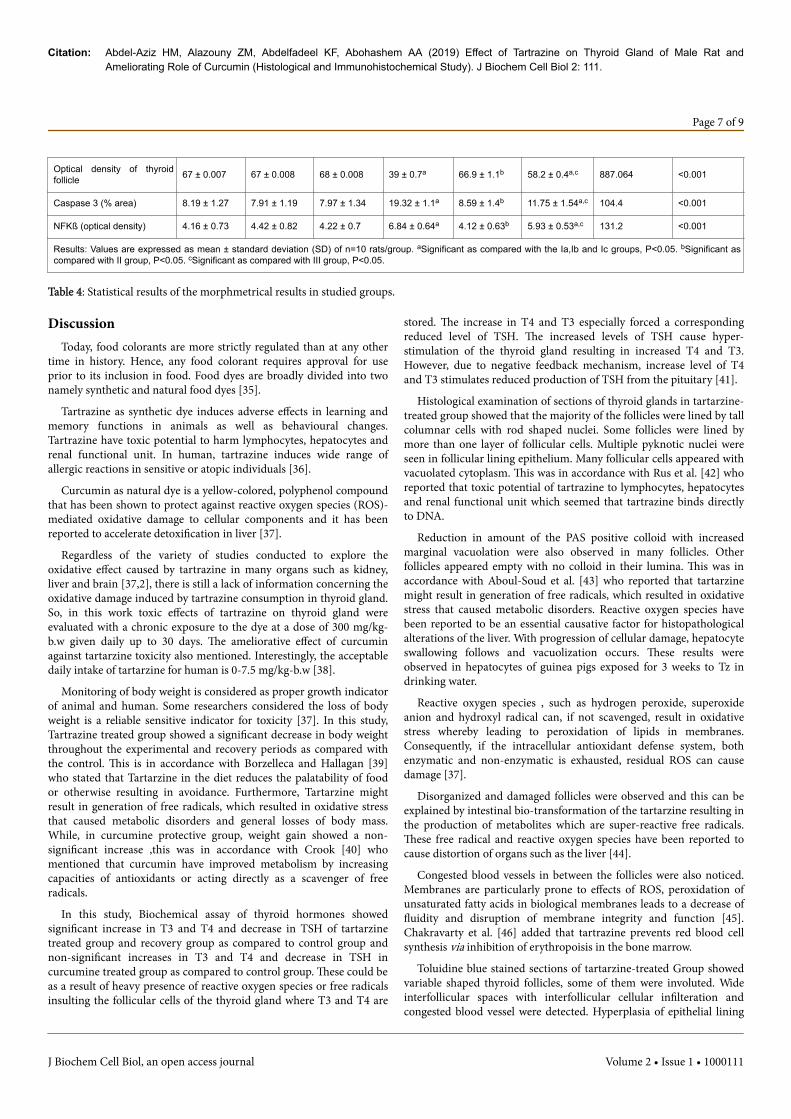
Optical density of thyroidfollicle 67 ± 0.007 67 ± 0.008 68 ± 0.008 39 ± 0.7a 66.9 ± 1.1b 58.2 ± 0.4a,c 887.064 <0.001
Caspase 3 (% area) 8.19 ± 1.27 7.91 ± 1.19 7.97 ± 1.34 19.32 ± 1.1a 8.59 ± 1.4b 11.75 ± 1.54a,c 104.4 <0.001
NFKß (optical density) 4.16 ± 0.73 4.42 ± 0.82 4.22 ± 0.7 6.84 ± 0.64a 4.12 ± 0.63b 5.93 ± 0.53a,c 131.2 <0.001
Results: Values are expressed as mean ± standard deviation (SD) of n=10 rats/group. aSignificant as compared with the Ia,Ib and Ic groups, P<0.05. bSignificant ascompared with II group, P<0.05. cSignificant as compared with III group, P<0.05.
Table 4: Statistical results of the morphmetrical results in studied groups.
DiscussionToday, food colorants are more strictly regulated than at any other
time in history. Hence, any food colorant requires approval for useprior to its inclusion in food. Food dyes are broadly divided into twonamely synthetic and natural food dyes [35].
Tartrazine as synthetic dye induces adverse effects in learning andmemory functions in animals as well as behavioural changes.Tartrazine have toxic potential to harm lymphocytes, hepatocytes andrenal functional unit. In human, tartrazine induces wide range ofallergic reactions in sensitive or atopic individuals [36].
Curcumin as natural dye is a yellow-colored, polyphenol compoundthat has been shown to protect against reactive oxygen species (ROS)-mediated oxidative damage to cellular components and it has beenreported to accelerate detoxification in liver [37].
Regardless of the variety of studies conducted to explore theoxidative effect caused by tartrazine in many organs such as kidney,liver and brain [37,2], there is still a lack of information concerning theoxidative damage induced by tartrazine consumption in thyroid gland.So, in this work toxic effects of tartrazine on thyroid gland wereevaluated with a chronic exposure to the dye at a dose of 300 mg/kg-b.w given daily up to 30 days. The ameliorative effect of curcuminagainst tartarzine toxicity also mentioned. Interestingly, the acceptabledaily intake of tartarzine for human is 0-7.5 mg/kg-b.w [38].
Monitoring of body weight is considered as proper growth indicatorof animal and human. Some researchers considered the loss of bodyweight is a reliable sensitive indicator for toxicity [37]. In this study,Tartrazine treated group showed a significant decrease in body weightthroughout the experimental and recovery periods as compared withthe control. This is in accordance with Borzelleca and Hallagan [39]who stated that Tartarzine in the diet reduces the palatability of foodor otherwise resulting in avoidance. Furthermore, Tartarzine mightresult in generation of free radicals, which resulted in oxidative stressthat caused metabolic disorders and general losses of body mass.While, in curcumine protective group, weight gain showed a non-significant increase ,this was in accordance with Crook [40] whomentioned that curcumin have improved metabolism by increasingcapacities of antioxidants or acting directly as a scavenger of freeradicals.
In this study, Biochemical assay of thyroid hormones showedsignificant increase in T3 and T4 and decrease in TSH of tartarzinetreated group and recovery group as compared to control group andnon-significant increases in T3 and T4 and decrease in TSH incurcumine treated group as compared to control group. These could beas a result of heavy presence of reactive oxygen species or free radicalsinsulting the follicular cells of the thyroid gland where T3 and T4 are
stored. The increase in T4 and T3 especially forced a correspondingreduced level of TSH. The increased levels of TSH cause hyper-stimulation of the thyroid gland resulting in increased T4 and T3.However, due to negative feedback mechanism, increase level of T4and T3 stimulates reduced production of TSH from the pituitary [41].
Histological examination of sections of thyroid glands in tartarzine-treated group showed that the majority of the follicles were lined by tallcolumnar cells with rod shaped nuclei. Some follicles were lined bymore than one layer of follicular cells. Multiple pyknotic nuclei wereseen in follicular lining epithelium. Many follicular cells appeared withvacuolated cytoplasm. This was in accordance with Rus et al. [42] whoreported that toxic potential of tartrazine to lymphocytes, hepatocytesand renal functional unit which seemed that tartrazine binds directlyto DNA.
Reduction in amount of the PAS positive colloid with increasedmarginal vacuolation were also observed in many follicles. Otherfollicles appeared empty with no colloid in their lumina. This was inaccordance with Aboul-Soud et al. [43] who reported that tartarzinemight result in generation of free radicals, which resulted in oxidativestress that caused metabolic disorders. Reactive oxygen species havebeen reported to be an essential causative factor for histopathologicalalterations of the liver. With progression of cellular damage, hepatocyteswallowing follows and vacuolization occurs. These results wereobserved in hepatocytes of guinea pigs exposed for 3 weeks to Tz indrinking water.
Reactive oxygen species , such as hydrogen peroxide, superoxideanion and hydroxyl radical can, if not scavenged, result in oxidativestress whereby leading to peroxidation of lipids in membranes.Consequently, if the intracellular antioxidant defense system, bothenzymatic and non-enzymatic is exhausted, residual ROS can causedamage [37].
Disorganized and damaged follicles were observed and this can beexplained by intestinal bio-transformation of the tartarzine resulting inthe production of metabolites which are super-reactive free radicals.These free radical and reactive oxygen species have been reported tocause distortion of organs such as the liver [44].
Congested blood vessels in between the follicles were also noticed.Membranes are particularly prone to effects of ROS, peroxidation ofunsaturated fatty acids in biological membranes leads to a decrease offluidity and disruption of membrane integrity and function [45].Chakravarty et al. [46] added that tartrazine prevents red blood cellsynthesis via inhibition of erythropoisis in the bone marrow.
Toluidine blue stained sections of tartarzine-treated Group showedvariable shaped thyroid follicles, some of them were involuted. Wideinterfollicular spaces with interfollicular cellular infilteration andcongested blood vessel were detected. Hyperplasia of epithelial lining
Citation: Abdel-Aziz HM, Alazouny ZM, Abdelfadeel KF, Abohashem AA (2019) Effect of Tartrazine on Thyroid Gland of Male Rat andAmeliorating Role of Curcumin (Histological and Immunohistochemical Study). J Biochem Cell Biol 2: 111.
Page 7 of 9
J Biochem Cell Biol, an open access journal Volume 2 • Issue 1 • 1000111

of some follicles and darkly stained nuclei of follicular epithelium werealso noticed. Identical histopathological alterations have been observedin hepatocytes of guinea pigs exposed for 3 weeks to tartarzine indrinking water, infiltration of Kupffer cells, congested blood vesselswith various areas of hemorrhage in liver exposed to tartarzine [47].
H and E, PAS and toluidine stained sections of thyroid glands ofCurcumine-protective Group showed that most of thyroid follicleswere similar to that of the control group. They were lined by singlelayer of low cubical cells with rounded nuclei and their luminae werefilled with acidophilic colloid. Curcumin exerts its protective effectsagainst severe oxidative damage via: (1) its powerful antioxidantproperty, whereby scavenges oxygen free radicals, and (2) its ability toincrease intracellular GSH levels, which consequently lead to theefficient control of levels of lipid peroxidation [48].
Immunohistochemical Caspase-3 stained sections of thyroid glandof control group showed a weak expression of caspase-3 protein in thecytoplasm of few follicular cells. In Tartarzine-treated Group revealed astrong expression of caspase-3 protein in the cytoplasm of mostfollicular cells. In Curcumine-protective Group showed mild increasein expression of caspase-3 protein in the cytoplasm of follicular cells.In Recovery Group, revealed moderate increase in expression ofcaspase-3 protein in the cytoplasm of follicular cells. Curcuminprotection against tartrazine-induced oxidative injuries and lipidperoxidation occurs via enhancement of the antioxidant defensesystem. The distinctive protective effect of curcumin is thought to bevia expression of a gene subset since it has been shown to regulate geneexpression of insulin-like growth factor, B-cell CLL/lymphoma [46].
In tartarzine-treated group there was a strong immunoreaction ofNF-kB protein mostly in the nuclei of most follicular cells. Few cellsshowed positive cytoplasmic expression for it. Barnes and Karin [15]concluded that in unstimulated cells, NF-kB is found in cytoplasm andis bound to IkBa and IkBb, which prevent it from entering the nuclei.When these cells are stimulated, specific kinases phosphorylate IkB,causing its rapid degradation by proteasomes. The release of NF-kBfrom IkB results in the passage of NF-kB into the nucleus, where itbinds to specific sequences in the promoter regions of inflammatorygenes. In contrary to curcumin-protective group that showed mildincreased immunoreaction of NF-kB protein in the nuclei andcytoplasm of follicular cells.
Concerning the recovery group, our study revealed that there wasincomplete recovery from tartarzine effects. This result can beexplained by binding of cellular copper and iron to the artificial foodcolorants, resulting in its tissues accumulation [48]. Soares et al. [49]also explained the incomplete recovery by the significant genotoxiceffect of tartarzine that might generate carcinogenesis with itsprolonged consumption.
ConclusionWe provided evidence for the potential of CUR, as a natural food
coloring agent, to minimize or prevent oxidative stress, commonlytaking place due to consumption of various potentially hazardoustartarzine coloring foods. CUR can thus delay the onset of formationof potentially damaging ROS, eventually leading to a better capacity inmaintaining nutritional food quality and more efficient cellular redoxstate homeostasis.
Conflict of InterestThere is no potential conflict of interest among the authors.
References1. Amin KA, El-Shehri FS (2018) Toxicological and safety assessment of
tartrazine as a synthetic food additive on health biomarkers: A review. AfrJ Biotechol 17: 139-149.
2. Gao Y, Li C, Shen J, Yin H, An X, et al. (2011) Effect of food azo dyetartrazine on learning and memory functions in mice and rats, and thepossible mechanisms involved. J Food Sci 76: T125-T129.
3. Ali FA, Abdelgayed AS, EL-Tawil SO, BaNeer MA (2016) Toxicologicaland histopathological studies on the effect of tartrazine in male albinorats. Int J Bio Biomol Agri Biotech Eng 10: 473-478.
4. Boussada M, Lamine JA, Bini ID, Abidi N, Lasram M, et al. (2017)Assessment of a sub-chronic consumption of tartrazine (E102) on spermand oxidative stress features in Wistar rat. Int Food Res 24: 1473-1481.
5. Visweswaran B, Krishnamoorthy G (2012) Oxidative stress by tartrazinein the testis of wistar rats. Int J Pharm Biol Sci 2: 44-49.
6. Moutinho IL, Bertges LC, Assis RV (2007) Prolonged use of the food dyetartrazine (fd&c yellow no 5) and its effects on the gastric mucosa ofwistar rats. Braz J Biol 67: 141-145.
7. Carocho M, Barreiro MF, Morales P, Ferreira ICFR (2014) Addingmolecules to food, pros and cons: A review on synthetic and natural foodadditives. Comp Rev Food Sci Food Safety 13: 377-399.
8. Benzer F, Kandemir FM, Ozkaraca M, Kucukler, S, Caglayan C (2018)Curcumin ameliorates doxorubicin‐induced cardiotoxicity by abrogationof inflammation, apoptosis oxidative DNA damage, and protein oxidationin rats. J Biochem Mol Toxicol 32: e22030.
9. Dou Y, Luo J, Wu X, Wei Z, Tong B, et al. (2018) Curcumin attenuatescollagen-induced inflammatory response through the “gut-brain axis”. JNeuroinflammation 15: 1-6.
10. Sharma RA, Gescher AJ, Steward WP (2005) Curcumin: The story so far.Eur J Cancer 41: 1955-1968.
11. Sa G, Das T (2008) Anti-cancer effects of curcumin: Cycle of life anddeath. Cell Div 3: 1-14.
12. Huang J, Yao X, Weng G, Qi H, Ye X (2018) Protective effect of curcuminagainst cyclosporine a induced rat nephrotoxicity. Mol Med Rep 17:6038-6044.
13. El-Twab SMA, Abdul-Hamid M (2016) Curcumin mitigates lithium-induced thyroid dysfunction by modulating antioxidant status, apoptosisand inflammatory cytokines. J Basic Appl Zool 76: 7-19.
14. Mohamed DA, Elnegris HM (2015) Histological study of thyroid glandafter experimental exposure to low frequency electromagnetic fields inadult male albino rat and possible protective role of vitamin E. J CytolHistol 6: 374.
15. Barnes PJ, Karin M (1997) Nuclear factor-κB-a pivotal transcriptionfactor in chronic inflammatory diseases. N Engl J Med 336: 1066-1071.
16. Fan TJ, Han LH, Cong RS, Liang J (2005) Caspase family proteases andapoptosis. Acta Biochim Biophys Sin (Shanghai) 37: 719-727.
17. Hashish EA, Elgaml SA (2016) Hepatoprotective and nephroprotectiveeffect of curcumin against copper toxicity in rats. Indian J Clin Biochem31: 270-277.
18. Al-Shinnawy MS, Elkattan NA (2013) Assessment of the changes in somediagnostic parameters in male albino rats fed on an azo dye. Int J EnvironSci Eng 4: 85-90.
19. Le Grange D, Doyle PM, Swanson SA, Ludwig K, Glunz C, et al. (2012)Calculation of expected body weight in adolescents with eating disorders.Pediatrics 129: e438-e446.
20. El-Bakry RH, Tawfik SM (2014) Histological study of the effect ofpotassium dichromate on the thyroid follicular cells of adult male albinorat and the possible protective role of ascorbic acid (vitamin C). JMicroscopy Ultrastruct 2:137-150.
Citation: Abdel-Aziz HM, Alazouny ZM, Abdelfadeel KF, Abohashem AA (2019) Effect of Tartrazine on Thyroid Gland of Male Rat andAmeliorating Role of Curcumin (Histological and Immunohistochemical Study). J Biochem Cell Biol 2: 111.
Page 8 of 9
J Biochem Cell Biol, an open access journal Volume 2 • Issue 1 • 1000111

21. Selim AO, El-Haleem MRA, Ibrahim IH (2012) Effect of sodium fluorideon the thyroid gland of growing male albino rats: Histological andbiochemical study. Egypt J Histol 35: 470-482.
22. Bancroft JD, Gamble A (2018) Theory and practice of histologicaltechniques. (8th edtn). Churchil, Livingstone, New York, London.
23. Mahmood T, Qureshi IZ, Iqbal MJ (2010) Histopathological andbiochemical changes in rat thyroid following acute exposure tohexavalent chromium. Histol Histopathol 25: 1355-1370.
24. Glauert AM, Lewis PR (2014) Biological specimen preparation fortransmission electron microscopy. Princeton University Press, USA.
25. Javois LC (1999) Immunocytochemical methods and protocols. Totowa:Humana Press, USA.
26. Mancini M, Nicholson DW, Roy S, Thornberry NA, Peterson EP, et al.(1998) The caspase-3 precursor has a cytosolic and mitochondrialdistribution: implications for apoptotic signaling. J Cell Biol 140:1485-1495.
27. Maguire O, O'Loughlin K, Minderman H (2015) Simultaneousassessment of NF-κB/p65 phosphorylation and nuclear localization usingimaging flow cytometry. J Immunol Methods 423: 3-11.
28. Sui X, Sui Y, Wang Y (2018) LARP7 in papillary thyroid carcinomainduces NIS expression through suppression of the SHH signalingpathway. Mol Med Rep 17: 7521-7528.
29. Helewski KJ, Kowalczyk-Ziomek GI, Czecior E, Swietochowska E,Wielkoszynski T, et al. (2010) Administration of low doses of TNFaprotects rat liver from ischemic damage and re-perfusion injury. J PhysiolPharmacol 61: 273-278.
30. Negahdary M, Bezhgi M, Ajdary M (2015) Effects of silymarin onoxidative stress markers in rats treated with magnesium oxidenanoparticles. Annu Res Rev Biol 5: 254.
31. Olszewska-Słonina DM, Mątewski D, Czajkowski R, Olszewski KJ, WoŸniak A, et al. (2011) The concentration of thiobarbituric acid reactivesubstances (TBARS) and paraoxonase activity in blood of patients withosteoarthrosis after endoprosthesis implantation. Med Sci Monit 17:CR498-CR504.
32. Nishikimi M (1975) Oxidation of ascorbic acid with superoxide aniongenerated by the xanthine-xanthine oxidase system. Biochem Biophys ResCommun 63: 463-468.
33. Aebi H (1984) Catalase in vitro. Methods Enzymol 105: 121-126.34. Tipple TE, Rogers LK (2012) Methods for the determination of plasma or
tissue glutathione levels. Methods Mol Biol 889: 315-324.35. Sharma A, Goyal RP, Chakravarty G, Sharma S (2015) Effects of chocolate
brown. Science Academic 57: 183-198.36. Sasaki YF, Kawaguchi S, Kamaya A, Ohshita M, Kabasawa K (2002) The
comet assay with 8 mouse organs: Results with 39 currently used foodadditives. Mutat Res 519: 103-119.
37. Amin H, Abdel-Hameid H, Abd-Elsttar AH (2010) Effect of food azodyes tartrazine and carmoisine on biochemical parameters related torenal, hepatic function and oxidative stress biomarkers in young malerats. Food Chemi Toxicol 48: 2994-2999.
38. Alison D, Collins P (2001) Colouring our foods in the last and nextmillennium. Int. J Food Sci Technol 35: 5-22.
39. Borzelleca JF, Hallagan JB (1988) A chronic toxicity/carcinogenicity studyof FD & C Yellow No. 5 (tartrazine) in mice. Food Chem Toxicol 26:189-194.
40. Crook AM (2006) Thyroid function. In clinical chemistry and metabolicmedicine. (7th edtn), Hodder Arnold publishers, London.
41. Sayari S, Molaei Z, Torabi Z (2018) The relationship between subclinicalhypothyroidism and serum levels of uric acid and creatinine in childrenaged 2-14 years. Ann Pediatr Endocrinol Metab 23: 38-42.
42. Rus V, Gherman C, Miclăuş V, Mihalca A, Nadăş GC et al. (2010)Comparative toxicity of food dyes on liver and kidney in guinea pigs: Ahistopathological study. Ann RSCB 15: 161-165.
43. Aboul-Soud MA, Al-Othman, AM, El-Desoky GE, Al-Othman ZA, YusufK, et al. (2011) Hepatoprotective effects of vitamin E/selenium againstmalathion-induced injuries on the antioxidant status and apoptosis-related gene expression in rats. J Toxicol Sci 36: 285-296.
44. Suzuki Y, Ishihara M, Segami T, Ito M (1998) Antiulcer effects ofantioxidants, quercetin, alpha tocopherol, nifedipine and tetracycline inrats. Jpn J Pharmacol 78: 435-441.
45. Chakravarty G, Goyal RP, Sharma S, Sharma A (2005) Haematologicalchanges induced by a common non-permitted food colour malachitegreen (MG) in swiss albino mice. Indian J Environ Sci 9:113-117.
46. El-Desoky GE, Abdel-Ghaffar A, Al-Othman ZA, Habila MA, Al-SheikhYA, et al. (2017) Curcumin protects against tartrazine-mediated oxidativestress and hepatotoxicity in male rats. Eur Rev Med Pharmacol Sci 21:635-645.
47. Al-Rubaei ZM, Mohammad TU, Ali LK (2014) Effects of local curcuminon oxidative stress and total antioxidant capacity in vivo study. Pak J BiolSci 17: 1237-1241.
48. Stevens LJ, Kuczek T, Burgess JR, Stochelski MA, Arnold LE, et al. (2013)Mechanisms of behavioral, atopic, and other reactions to artificial foodcolors in children. Nutr Rev 71: 268-281.
49. Soares BM, Araujo TM, Ramos JA, Pinto LC, Khayat BM, et al. (2015)Effects on DNA repair in human lymphocytes exposed to the food dyetartrazine yellow. Anticancer Res 35: 1465-1474.
Citation: Abdel-Aziz HM, Alazouny ZM, Abdelfadeel KF, Abohashem AA (2019) Effect of Tartrazine on Thyroid Gland of Male Rat andAmeliorating Role of Curcumin (Histological and Immunohistochemical Study). J Biochem Cell Biol 2: 111.
Page 9 of 9
J Biochem Cell Biol, an open access journal Volume 2 • Issue 1 • 1000111