journal of controlled release - forside - helse bergen · a national centre for ultrasound in...
TRANSCRIPT

Journal of Controlled Release 243 (2016) 172–181
Contents lists available at ScienceDirect
Journal of Controlled Release
j ourna l homepage: www.e lsev ie r .com/ locate / jconre l
A human clinical trial using ultrasound and microbubbles to enhancegemcitabine treatment of inoperable pancreatic cancer☆
Georg Dimcevski a,b,⁎, Spiros Kotopoulis a,b, Tormod Bjånes c, Dag Hoem d, Jan Schjøt c,e, Bjørn Tore Gjertsen f,g,Martin Biermann h,b, Anders Molven i,j, Halfdan Sorbye k,e, Emmet McCormack e,g,Michiel Postema l,m, Odd Helge Gilja a,b
a National Centre for Ultrasound in Gastroenterology, Haukeland University Hospital, Bergen, Norwayb Department of Clinical Medicine, University of Bergen, Bergen, Norwayc Laboratory of Clinical Biochemistry, Section of Clinical Pharmacology, Haukeland University Hospital, Bergen, Norwayd Department of Surgical Sciences, Haukeland University Hospital, Norwaye Department of Clinical Science, University of Bergen, Bergen, Norwayf Centre for Cancer Biomarkers, CCBIO, Department of Clinical Science, University of Bergen, Bergen, Norwayg Department of Internal Medicine, Hematology Section, Haukeland University Hospital, Bergen, Norwayh Department of Radiology, Haukeland University Hospital, Bergen, Norwayi Department of Pathology, Haukeland University Hospital, Bergen, Norwayj Gade Laboratory for Pathology, Department of Clinical Medicine, University of Bergen, Bergen, Norwayk Department of Oncology, Haukeland University Hospital, Bergen, Norwayl Institute of Fundamental Technological Research, Polish Academy of Sciences, Warszawa, Polandm School of Electrical and Information Engineering, Chamber of Mines Building, University of the Witwatersrand, Johannesburg, South Africa
☆ ClinicalTrials.gov number: NCT01674556⁎ Corresponding author at: Department of Medicine, H
5021 Bergen, Norway.E-mail address: [email protected] (G
http://dx.doi.org/10.1016/j.jconrel.2016.10.0070168-3659/© 2016 The Authors. Published by Elsevier B.V
a b s t r a c t
a r t i c l e i n f oArticle history:Received 2 July 2016Received in revised form 7 October 2016Accepted 10 October 2016Available online 12 October 2016
Background: The primary aim of our study was to evaluate the safety and potential toxicity of gemcitabine com-bined with microbubbles under sonication in inoperable pancreatic cancer patients. The secondary aim was toevaluate a novel image-guided microbubble-based therapy, based on commercially available technology, to-wards improving chemotherapeutic efficacy, preserving patient performance status, and prolonging survival.Methods: Ten patients were enrolled and treated in this Phase I clinical trial. Gemcitabine was infused intrave-nously over 30 min. Subsequently, patients were treated using a commercial clinical ultrasound scanner for31.5 min. SonoVue®was injected intravenously (0.5 ml followed by 5 ml saline every 3.5 min) during the ultra-sound treatment with the aim of inducing sonoporation, thus enhancing therapeutic efficacy.Results: The combined therapeutic regimen did not induce any additional toxicity or increased frequency of sideeffects when compared to gemcitabine chemotherapy alone (historical controls). Combination treated patients(n = 10) tolerated an increased number of gemcitabine cycles compared with historical controls (n = 63 pa-tients; average of 8.3 ± 6.0 cycles, versus 13.8 ± 5.6 cycles, p = 0.008, unpaired t-test). In five patients, themaximum tumour diameter was decreased from the first to last treatment. The median survival in our patients(n = 10) was also increased from 8.9 months to 17.6 months (p = 0.011).Conclusions: It is possible to combine ultrasound, microbubbles, and chemotherapy in a clinical setting usingcommercially available equipment with no additional toxicities. This combined treatmentmay improve the clin-ical efficacy of gemcitabine, prolong the quality of life, and extend survival in patients with pancreatic ductaladenocarcinoma.
© 2016 The Authors. Published by Elsevier B.V. This is an open access article under the CC BY-NC-ND license(http://creativecommons.org/licenses/by-nc-nd/4.0/).
Keywords:UltrasoundMicrobubblesSonoporationPancreatic cancerImage-guided therapyClinical trial
1. Introduction
A diagnosis of pancreatic ductal adenocarcinoma (PDAC) carries oneof the most dismal prognoses in all of medicine. Currently the 4th most
aukeland University Hospital,
. Dimcevski).
. This is an open access article under
lethal cancer in the western world, it has an average 5-year survival ofapproximately 5% and is predictedwithin the decade to become the sec-ond greatest cause of cancer death [1]. Surgery provides the only possi-bility for cure, however N85% of newly diagnosed pancreatic tumoursare considered unresectable due to locally advanced disease with en-casement of large blood vessels or metastasis. Furthermore, the preva-lence of extreme desmoplasia generally renders the disease resistantto chemo-radiative approaches [2]. Untreated, locally advanced PDACpatients have a median survival of 6–10 months and 3–5 months for
the CC BY-NC-ND license (http://creativecommons.org/licenses/by-nc-nd/4.0/).

Table 1Clinico-pathological characteristics of all pancreatic cancer patients. There was no statisti-cally significant difference between the sonoporation treated cohort and historical controlgroup in age, body mass index and blood chemistry. CA19-9 was not recorded in the his-torical control cohort.
Variables (unit)
Sonoporation(n = 10)
Control(n = 63)
Start of treatment End of treatment Start of treatment
Age (years) 58.8 (±9.8) 59.5 (±10) 64.8 (±14.0)Gender (%)(male/female)
30/70 54/46
Body Mass Index(kg/m2)
23.7 (±4.3) 23.9 (±5.1) 22.9 (±3.05)
ECOG performancestatus (%)0 50 10 711 50 80 292 0 10
Histological type AdenocarcinomaStage
Locally advanced 70 NA 55Metastatic 30 45
Blood chemistryB-hemoglobin (g/dL) 13.4 (±1.5) 11.9 (±0.9) 12.6 (±1.5)ALAT (U/L) 45.2 (±21.8) 59.7 (±42.9) 71.2 (±59.6)LD (mg/dL) 151.4 (±27.6) 209.6 (±46.0) 177.7 (±49.4)Bilirubin (μmol/L) 14.5 (±8.46) 7.3 (±4.0) 37.3 (±66.0)CA 125 (U/mL) 54.1 (±39.6) 62 (±60.1) 90.0 (±100.5)CA19-9 (U/mL)a 248.5 (±380.8) 117.1 (±202.9) NA
Comments:Obligatory lab values for chemotherapy inclusion: B-Hemoglobin N10, Neutrophils(polymorphoneuclear leukocytes) N3.5, Platelets N150, Bilirubin N75.
a One sonoporation treatedpatient exhibited abnormally highCA19-9 values at 4608U/mL hence not included in average CA19-9 values.
173G. Dimcevski et al. / Journal of Controlled Release 243 (2016) 172–181
patients with metastatic disease [3–5] highlighting the immediate anddire need for novel therapeutic interventions.
Gemcitabine has been the standard chemotherapeutic used inrecent years and the most effective single agent. Compared to 5-fluorouracil, gemcitabine extends the survival by approximately onemonth whilst also improving clinical symptoms [6]. Recently,FOLFIRINOX (bolus and infusion of 5-fluorouracil, leucovorin,irinotecan, and oxaliplatin) emerged as a new chemotherapeutic optionfor patients with metastatic pancreatic cancer and an Eastern Coopera-tive Oncology Group (ECOG) performance status of 0–1. For this cohortof patients FOLFIRINOX is now the reference treatment. However,owing to the demonstrable toxicities and side effects of this therapy,gemcitabine is still the standard of care in patients with poor perfor-mance status or contraindication to FOLFIRINOX [7]. Furthermore, thecombination of nanoparticle albumin-bound paclitaxel (nab-paclitaxel)and gemcitabine provides another new therapeutic option resultingwith improved median survival of 1.8 months, compared togemcitabine alone [8]. Despite these novel interventions, the reportedincreases in survival areminimal andwe continue ourwait for a therapythat will impact survival, provide a bridge to reductive surgery and ulti-mately cure PDAC.
Diagnostic ultrasound (US) imaging has been used in the clinic for N50 years [9,10], with detection of pancreatic lesions dating back to thelate 1960s [11]. Over the past 30 years, the use of ultrasound to detectPDAC has significantly increased [11–13]. Contrast-enhanced ultra-sound uses stabilised gas microbubbles (MBs) to enhance the signal-to-noise ratio of the vasculature and allows clinicians to better visualisetissue perfusion. Twenty years ago, researchers discover that upon ap-plication of ultrasound these microbubbles volumetrically oscillate. Ifthese oscillating microbubbles were in the vicinity of cells, small porescould be formed increasing the uptake of macromolecules significantly[14–16]. Henceforth, the use of ultrasound and microbubbles to invokebiomechanical effects that increase the permeability of the vascular bar-rier and/or the extravasation of drug in a specific location is now com-monly known as “sonoporation”.
Numerous researchers have shown in vitro and in vivo thatsonoporation is a viable technique to improve drug delivery and im-prove therapeutic efficacy in various cell lines derived from pharyngeal[17], glioma [18], prostate [19,20], melanoma [21], and pancreatic can-cer [22]. Sonoporation has also been used to open the blood brain barri-er [23,24]. In general, sonoporation research is split into two camps: A)high-intensity, i.e., using inertial cavitation [9,25–27] and/or taking ad-vantage of the thermal effects [28,29], and B) low-intensity, i.e., usingstable cavitation [30,31] and non-thermal effects [32–34].
The use of high-intensity ultrasound without MB has previouslybeen evaluated clinically and shown considerable success for pain ther-apy [35,36], ablation of breast fibroadenomas [37], opening the blood-brain barrier [38] and treatment of pancreatic adenocarcinoma [39].Nevertheless, to our knowledge, there has been no clinical trial evaluat-ing the efficacy of low-intensity ultrasound in combination withmicrobubbles to improve the chemotherapeutic efficacy in patientswith PDAC.
We have previously demonstrated in vitro and preclinically in anorthotopic model of PDAC, enhanced treatment effects of gemcitabinewith concurrent exposure to SonoVue® MB and US at low acoustic in-tensities [40]. Based on these preclinical results we initiated an openlabel phase I, single centre, safety evaluation study in PDAC patientsby combining an ultrasound contrast agent and gemcitabine under son-ication at clinical diagnostic conditions.
The primary objective of this study was to evaluate the safety andpotential toxicity of gemcitabine combined with ultrasound contrastagent under ultrasound treatment in inoperable pancreatic cancer pa-tients. The secondary objective was to evaluate a novel image-guidedmicrobubble-based therapy, based on commercially available technolo-gy, towards improving chemotherapeutic efficacy, preserving patientperformance status and prolonging survival.
2. Material and methods
2.1. Subjects
Over a 23-month period (January 2012–November 2013), we re-cruited ten consecutive voluntary patients with inoperable pancreaticcancer (ICD-10 C25.0–3) at Haukeland University Hospital. All had his-tologically verified, locally advanced (non-resectable Stage III) or meta-static (Stage IV) pancreatic adenocarcinoma. Needle biopsies wereobtained either from the primary tumour or from a metastatic lesion.The tissuewas processed in the diagnostic pathology laboratory accord-ing to standard routines (formalin-fixation, paraffin-embedment, stain-ing with hematoxylin and eosin). The histology was evaluated by asenior pathologist with special competence in gastrointestinal patholo-gy. Patients were ambulatory with an Eastern Cooperative OncologyGroup (ECOG) performance status 0–1 (Table 1). Patients had to meetthe standard criteria at our hospital for treatment with gemcitabineand no known intolerance to gemcitabine or SonoVue® (Bracco Imag-ing Scandinavia AB, Oslo, Norway) ultrasound contrast agent [45].
Historical data from PDAC patients undergoing equal gemcitabinetreatment following the same inclusion and exclusion criteria, between2009 and 2011 atHaukelandUniversityHospital, were used for compar-ison of treatment tolerance, safety, and overall survival. The only differ-ence in treatment between the historical control group and our treatedgroup was the addition of ultrasound and microbubbles following che-motherapeutic infusion. Gemcitabine was considered the standard ofcare for the treatment time period of the control patients and through-out this clinical study.
2.2. Chemotherapeutic and microbubble dosage
Two experienced oncologists, not participating in the study,were re-sponsible for the chemotherapeutic treatment. The only divergencefrom normal administration practice was relocation to the researchunit. We used the standard recommended treatment protocol of
174 G. Dimcevski et al. / Journal of Controlled Release 243 (2016) 172–181
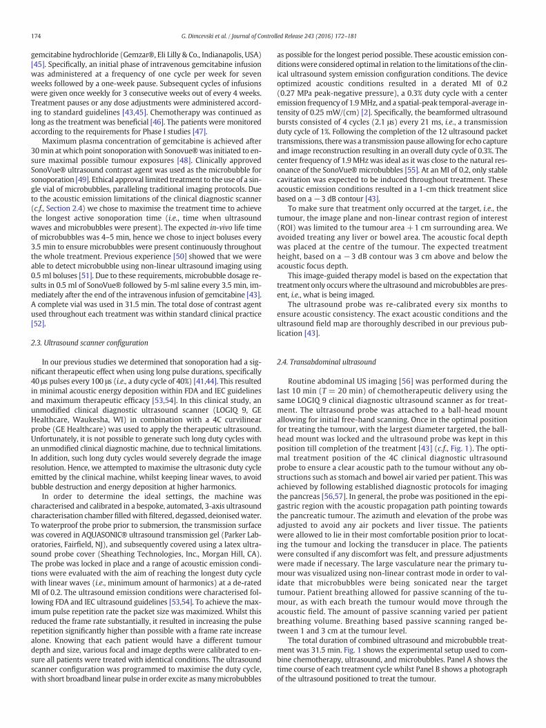
gemcitabine hydrochloride (Gemzar®, Eli Lilly & Co., Indianapolis, USA)[45]. Specifically, an initial phase of intravenous gemcitabine infusionwas administered at a frequency of one cycle per week for sevenweeks followed by a one-week pause. Subsequent cycles of infusionswere given once weekly for 3 consecutive weeks out of every 4 weeks.Treatment pauses or any dose adjustments were administered accord-ing to standard guidelines [43,45]. Chemotherapy was continued aslong as the treatment was beneficial [46]. The patients were monitoredaccording to the requirements for Phase I studies [47].
Maximum plasma concentration of gemcitabine is achieved after30min atwhich point sonoporationwith Sonovue®was initiated to en-sure maximal possible tumour exposures [48]. Clinically approvedSonoVue® ultrasound contrast agent was used as the microbubble forsonoporation [49]. Ethical approval limited treatment to the use of a sin-gle vial of microbubbles, paralleling traditional imaging protocols. Dueto the acoustic emission limitations of the clinical diagnostic scanner(c.f., Section 2.4) we chose to maximise the treatment time to achievethe longest active sonoporation time (i.e., time when ultrasoundwaves and microbubbles were present). The expected in-vivo life timeof microbubbles was 4–5 min, hence we chose to inject boluses every3.5 min to ensure microbubbles were present continuously throughoutthe whole treatment. Previous experience [50] showed that we wereable to detect microbubble using non-linear ultrasound imaging using0.5 ml boluses [51]. Due to these requirements, microbubble dosage re-sults in 0.5 ml of SonoVue® followed by 5-ml saline every 3.5 min, im-mediately after the end of the intravenous infusion of gemcitabine [43].A complete vial was used in 31.5 min. The total dose of contrast agentused throughout each treatment was within standard clinical practice[52].
2.3. Ultrasound scanner configuration
In our previous studies we determined that sonoporation had a sig-nificant therapeutic effect when using long pulse durations, specifically40 μs pulses every 100 μs (i.e., a duty cycle of 40%) [41,44]. This resultedin minimal acoustic energy deposition within FDA and IEC guidelinesand maximum therapeutic efficacy [53,54]. In this clinical study, anunmodified clinical diagnostic ultrasound scanner (LOGIQ 9, GEHealthcare, Waukesha, WI) in combination with a 4C curvilinearprobe (GE Healthcare) was used to apply the therapeutic ultrasound.Unfortunately, it is not possible to generate such long duty cycles withan unmodified clinical diagnostic machine, due to technical limitations.In addition, such long duty cycles would severely degrade the imageresolution. Hence, we attempted to maximise the ultrasonic duty cycleemitted by the clinical machine, whilst keeping linear waves, to avoidbubble destruction and energy deposition at higher harmonics.
In order to determine the ideal settings, the machine wascharacterised and calibrated in a bespoke, automated, 3-axis ultrasoundcharacterisation chamberfilledwithfiltered, degassed, deionisedwater.To waterproof the probe prior to submersion, the transmission surfacewas covered in AQUASONIC® ultrasound transmission gel (Parker Lab-oratories, Fairfield, NJ), and subsequently covered using a latex ultra-sound probe cover (Sheathing Technologies, Inc., Morgan Hill, CA).The probe was locked in place and a range of acoustic emission condi-tions were evaluated with the aim of reaching the longest duty cyclewith linear waves (i.e., minimum amount of harmonics) at a de-ratedMI of 0.2. The ultrasound emission conditions were characterised fol-lowing FDA and IEC ultrasound guidelines [53,54]. To achieve the max-imum pulse repetition rate the packet size was maximized. Whilst thisreduced the frame rate substantially, it resulted in increasing the pulserepetition significantly higher than possible with a frame rate increasealone. Knowing that each patient would have a different tumourdepth and size, various focal and image depths were calibrated to en-sure all patients were treated with identical conditions. The ultrasoundscanner configuration was programmed to maximise the duty cycle,with short broadband linear pulse in order excite asmanymicrobubbles
as possible for the longest period possible. These acoustic emission con-ditionswere considered optimal in relation to the limitations of the clin-ical ultrasound system emission configuration conditions. The deviceoptimized acoustic conditions resulted in a derated MI of 0.2(0.27 MPa peak-negative pressure), a 0.3% duty cycle with a centeremission frequency of 1.9MHz, and a spatial-peak temporal-average in-tensity of 0.25 mW/(cm) [2]. Specifically, the beamformed ultrasoundbursts consisted of 4 cycles (2.1 μs) every 21 ms, i.e., a transmissionduty cycle of 1%. Following the completion of the 12 ultrasound packettransmissions, therewas a transmission pause allowing for echo captureand image reconstruction resulting in an overall duty cycle of 0.3%. Thecenter frequency of 1.9MHzwas ideal as it was close to the natural res-onance of the SonoVue®microbubbles [55]. At an MI of 0.2, only stablecavitation was expected to be induced throughout treatment. Theseacoustic emission conditions resulted in a 1-cm thick treatment slicebased on a −3 dB contour [43].
To make sure that treatment only occurred at the target, i.e., thetumour, the image plane and non-linear contrast region of interest(ROI) was limited to the tumour area +1 cm surrounding area. Weavoided treating any liver or bowel area. The acoustic focal depthwas placed at the centre of the tumour. The expected treatmentheight, based on a −3 dB contour was 3 cm above and below theacoustic focus depth.
This image-guided therapy model is based on the expectation thattreatment only occurswhere the ultrasound andmicrobubbles are pres-ent, i.e., what is being imaged.
The ultrasound probe was re-calibrated every six months toensure acoustic consistency. The exact acoustic conditions and theultrasound field map are thoroughly described in our previous pub-lication [43].
2.4. Transabdominal ultrasound
Routine abdominal US imaging [56] was performed during thelast 10 min (T = 20 min) of chemotherapeutic delivery using thesame LOGIQ 9 clinical diagnostic ultrasound scanner as for treat-ment. The ultrasound probe was attached to a ball-head mountallowing for initial free-hand scanning. Once in the optimal positionfor treating the tumour, with the largest diameter targeted, the ball-head mount was locked and the ultrasound probe was kept in thisposition till completion of the treatment [43] (c.f., Fig. 1). The opti-mal treatment position of the 4C clinical diagnostic ultrasoundprobe to ensure a clear acoustic path to the tumour without any ob-structions such as stomach and bowel air varied per patient. This wasachieved by following established diagnostic protocols for imagingthe pancreas [56,57]. In general, the probe was positioned in the epi-gastric region with the acoustic propagation path pointing towardsthe pancreatic tumour. The azimuth and elevation of the probe wasadjusted to avoid any air pockets and liver tissue. The patientswere allowed to lie in their most comfortable position prior to locat-ing the tumour and locking the transducer in place. The patientswere consulted if any discomfort was felt, and pressure adjustmentswere made if necessary. The large vasculature near the primary tu-mour was visualized using non-linear contrast mode in order to val-idate that microbubbles were being sonicated near the targettumour. Patient breathing allowed for passive scanning of the tu-mour, as with each breath the tumour would move through theacoustic field. The amount of passive scanning varied per patientbreathing volume. Breathing based passive scanning ranged be-tween 1 and 3 cm at the tumour level.
The total duration of combined ultrasound and microbubble treat-ment was 31.5 min. Fig. 1 shows the experimental setup used to com-bine chemotherapy, ultrasound, and microbubbles. Panel A shows thetime course of each treatment cycle whilst Panel B shows a photographof the ultrasound positioned to treat the tumour.

Fig. 1. (A) Treatment procedure flow chart with timings of chemotherapeutics, ultrasound exposure, and microbubble infusion. Using the current protocol, the treatment duration was61.5 min. The first 30 min were reserved for chemotherapeutic infusion and the last 31.5 min were reserved for ultrasound and microbubble treatment. Abdominal imaging wasperformed for the last 10 min of infusion. Every 3.5 min, 0.5 ml of SonoVue® microbubbles were injected. (B) Photograph of patient with PDAC undergoing treatment using a clinicallyavailable diagnostic scanner. The ultrasound probe was locked in position using a mechanical arm targeted at the primary tumour for the full 31.5 min of ultrasound and microbubbletreatment.
175G. Dimcevski et al. / Journal of Controlled Release 243 (2016) 172–181
2.5. Pharmacokinetic evaluations
Analytical methods for pharmacokinetic (PK) evaluations were de-veloped in parallel to the clinical study [58]. Whole blood sampleswere collected sequentially into prechilled heparinized tubes at the fol-lowing time-points: T = 0, 30, 60, 120, 180 and 240 min. Tubes werespiked with the cytidine deaminase inhibitor tetrahydrouridine to pre-vent deamination of gemcitabine to dFdU [58]. Plasma andmononucle-ar cells were separated from whole blood as described previously.Concentrations of gemcitabine and dFdU were measured in plasmausing in-house LC-MS/MS methods [58].
2.6. Monitoring
All patients underwent dual-phase computed tomography (CT) im-aging ≤3 weeks before study inclusion. Routine abdominal CT was per-formed every 8th week where maximum tumour diameter wasquantified by independent radiologists. Tumour size and developmentwas characterised according to the Response Evaluation Criteria inSolid Tumours (RECIST). Positron emission tomography (PET) imagingwith F-18-fluoro-deoxyglucose (FDG)was performed prior to the treat-ment to determine if metastases were present.
Assessment of clinical state during the treatment also included anevaluation of the clinical benefit response and if surgical resectioncould be performed [46,59]. ECOG performance status was used as aproxy to monitor the effectiveness of the combined treatment. TheECOG scale describes patients' level of functioning in terms of their
ability to care for themselves, daily activity, and physical ability [46].An ECOG grade of 0 indicates a patient who is fully active and able tocarry on all pre-disease performance without restriction. An ECOGgrade of 1 indicates that a patient is restricted in physical strenuous ac-tivity but ambulatory and able to carry out work of a light or sedentarynature, e.g., light housework, officework. An ECOGgrade of 2 indicates apatient is ambulatory and capable of all self-care but unable to carry outany work activities. The patient is up and about N50% of waking hours.An ECOG grade of 3 indicates a patient capable of limited self-care andconfided to bed or chair N50% of waking hours. Hence, the longer a pa-tient stayed below an ECOG grade of 3, themore effective the treatmentwas considered indicating an extended period of well-being. When apatient reaches an ECOG grade of 3, they are no-longer able to undergogemcitabine chemotherapy.
Select patients also underwent diagnostic contrast-enhanced ultra-sound following established clinical procedures [60]. Blood analysiswas performed to evaluate if there was any acute toxicity.
2.7. Statistical analysis
The results are expressed asmean values± SD, unless otherwise in-dicated. Continuous data was analysed using t-tests, or Mann-Whitneytests if data were not normally distributed. Gehan-Breslow-Wilcoxontest and Log-rank (Mantel-Cox) test were used to compare survival.Variance is expressed through 95% confidence intervals. p b 0.05 wasconsidered statistical significant. Patients removed from the study dueto improvement were considered as intention to treat in the survivalstatistical analysis.

176 G. Dimcevski et al. / Journal of Controlled Release 243 (2016) 172–181
3. Results
3.1. Tumour targeting
The established guidelines for imaging the pancreas [56,57] allowedus to target the primary PDAC tumour, independent of tumour depthand size. Fig. 2 shows four representative ultrasound images of thePDAC tumours from our treated patient cohort captured prior toswitching the diagnostic ultrasound scanner settings to “treatmentmode”. In these images tumour depths ranges from 3.1 cm to 8.9 cm in-dicating that shallow or deep tumour did not inhibit tumour visualisa-tion or targeting.
3.2. Toxicity evaluation
The direct parameters used to evaluate the toxicity of our treatmentwere clinical parameters including vital signs, ECG and blood chemistry.Overall, all data indicated that gemcitabine in combination with US didnot induce any unexpected deviation or additional toxicities than che-motherapy alone.
One patientwas hospitalised for a serious adverse event (SAE) unre-lated to protocol therapy. Four SAEs occurred during protocol therapy.Two patients had symptoms indicating biliary obstruction and necessi-tated hospitalisation and rescheduling of the treatment. One was treat-ed for pneumonia and one had fever due to cholangitis. The mostfrequent possibly treatment-related toxicities i.e., adverse events (AE)were abdominal pain (n=9), nausea (n=7), fever (n=6), neutrope-nia (n=6), and fatigue (n=6) as described in Fig. 3. These eventswereregistered as possibly related to protocol therapy. Since all the reported
Fig. 2. Representative ultrasound images showing the PDAC tumour in four of the ultrasound angreen dotted lines. The ultrasound transducer was positioned to ensure no obstructions of the apatient and treatment. Distance 1 and 2 indicate the tumourwidth and height respectively. Valuthis figure legend, the reader is referred to the web version of this article.)
toxicities are expected side effects of gemcitabine, they were evaluatedas gemcitabine related. All other AE were probably related to progres-sion of underlying disease. There were no treatment-related deaths.
3.3. Blood biochemistry
No additional toxicity was observed. Blood values changed as ex-pected. CA 19-9 and CA 125 levels decreased in 5 out of 8 patients mea-sured, and 7 of the 10 patients, respectively.
When evaluating the levels of cancer marker CA 125 we observed adecline following combined treatment. A total of four out of ten patientswent from elevated to normal counts and only a single patient wentfrom normal to elevated counts. Whilst fewer measurements weremade in the CA 19-9 counts a similar trend was also observed wherethree patientswent fromelevated counts to normal counts,five patientsshowed a decrease, two patients showed an increase, and only a singlepatient went from normal to elevated counts. No correlation betweentumour size change and cancer marker count was observed (Supple-mental Fig. 1).
Bilirubin, LD, ALAT and other liver parameters were in line with theexpected variation under gemcitabine treatment. Thesewere all consid-ered to be normal blood biochemistry changes as expected from chemo-therapy and disease course.
3.4. Clinical benefit and response assessment
The followingmethodswere applied to evaluate the responses in theten patients: RECIST, tumour size, ECOG grade and treatment cycles[59,61].
dmicrobubble treated patients. Tumour height andwidth are indicated by the yellow andcoustical beam path to the tumour. This resulted in a unique ultrasound probe position pere D indicated the tumour centroid depth. (For interpretation of the references to colour in

Fig. 3. Percentage of patients with PDAC treated with sonoporation that experienced a given adverse event. This graph shows all the adverse events experienced by all patients regardlessof severity grade, or direct correlation to the treatment. All adverse events were already associated with gemcitabine treatment alone, indicating that addition of ultrasound andmicrobubbles did not induce or increase the frequency of new adverse events.
177G. Dimcevski et al. / Journal of Controlled Release 243 (2016) 172–181
The patients considered to be positive clinical responders were reg-ularly evaluated by the Dept. of Oncology for FOLFIRINOX treatment orconsolidative radiation therapy and surgery. After 12 treatment cycles,one patient was down-staged from 8.6 cm to 4.2 cm in tumour sizeand thereby became available for potentially curative therapy. Shewas removed from the clinical trial and underwent radiation therapyand subsequent pancreatectomy. Five patients exhibited partial re-sponses as evidenced by reduction in tumour diameter. As a result,they were offered either consolidative radiation therapy or FOLFIRINOXtreatment.
Fig. 4 shows the effect of our combined treatment on the tumoursize. The green lines indicate the patient tumour size recession or stabi-lization from the start to the end of the treatment, whereas the red linesindicate tumour size increase. When the line ends, this indicates thatthe patient was removed from the clinical trial.
An average of 13.8 ± 5.6 and median 12.5 (range 5–26) treatmentcycles of protocol therapy were delivered per patient. In comparison,our historical control group treated with the same chemotherapeuticprotocol of gemcitabine alone received an average of 8.3 ± 6.0 andme-dian 7 (range 1–28) treatment cycles (p=0.008). Fig. 5A shows awhis-ker plot depicting the number and range of treatment cycles.
Fig. 5B shows the survival curve of the combined treatment groupcompared to the historical control group. The number of treatment cy-cles and days of survival in our patient group are summarised in
Fig. 4. Maximum tumour size as function of time for all ten patients with inoperablepancreatic adenocarcinoma. Green lines indicate tumour size recession or stabilization.Red/orange or grey lines indicate tumour size increase. Colour gradient indicates linearregression fit of tumour growth gradient (lighter = shallower). Five out of ten patients(50%) showed tumour size reduction during treatment. A reduction in tumour size mayallow for surgical resection; the only current curative option. The star (*) indicateswhich patients showed tumour size reduction and were evaluated for consolidativeradiation therapy or FOLFIRINOX treatment. (For interpretation of the references tocolour in this figure legend, the reader is referred to the web version of this article.)
Table 2. Both Gehan-Breslow-Wilcoxon test and Log-rank (Mantel-Cox) test showed that the survival was significantly different withp = 0.0043 and p = 0.011, respectively.
3.5. Gemcitabine pharmacokinetics
Concentration profiles of gemcitabine and dFdU in plasma sampleswere in accordance with previous studies of gemcitabine-infusions of800–1000 mg/m2 administered to breast, lung, pancreatic and patientswith various other solid tumours [48,62]. This demonstrates that thecombination regimen did not seem to alter the systemic pharmacoki-netics of gemcitabine. A representative concentration profile from oneof the patients is shown in Supplemental Fig. 2.
4. Discussion
To our knowledge, this is the first human trial evaluating the use oflow intensity ultrasound and microbubbles to treat cancer. All previousstudies have only been performed in vitro or pre-clinically. Clinical stud-ies using ultrasound for therapy have been focused on high-intensity ul-trasound without microbubbles, or for pain treatment. Hence the effectof low intensity sonoporation therapy for PDAC in humans is unknown[44,63–65].
In our previous study [43], we presented the experimental protocolfocusing on the technical aspects of implementing low-intensitysonoporation using a clinical diagnostic ultrasound scanner. We alsopresented pilot results of five patients briefly discussing the number ofcycles and tumour sizes. In the currentworkwe present the final resultsand clinical data of all 10 patients, including a comparison of overall sur-vival. In addition,we provide a toxicity report regarding the safety of thestudy following 138 treatment cycles.
The primary aim of this Phase I study was to evaluate the safety andpotential toxicity, when combing microbubbles, ultrasound, and a che-motherapeutic agent in patients with PDAC. Hence, in this clinical trialwe only evaluated a total of ten patients, as required by the NMA. Over-all, all data clearly indicated that this combination did not induce anyadditional toxicities.
4.1. Cancer markers
These results indicate that chemotherapy in combination withmicrobubbles andultrasoundmayhave a positive impact on tumour de-velopment. It iswell known that there are correlations between CA 19-9decline and both overall survival and time to treatment failure in pa-tients treated with gemcitabine alone [66]. The limited number of pa-tients in our Phase-I-trial does not allow us to make any furtherconclusions.

Fig. 5. (A) Whisker plot comparing the number of treatment cycles undergone in patients with pancreatic adenocarcinoma. Patients treated with sonoporation showed a statisticallysignificant increase in number of treatment cycles (p = 0.008, unpaired t-test) indicating inhibited tumour progression and extended period of well-being (B) Survival plot comparingpatients treated with ultrasound, microbubbles, and gemcitabine vs gemcitabine alone. The survival curve indicated that the combined treatment group had near twice as high mediansurvival compared to treatment with gemcitabine alone; from a median of 8.9 months to 17.6 months (p = 0.011, Log Rank test).
178 G. Dimcevski et al. / Journal of Controlled Release 243 (2016) 172–181
4.2. Adverse events
In our present work, we present all adverse events experiencedby the patients independent of grade and severity (Fig. 3). This alsoincludes adverse events due to the actual malignancy, or personalexperiences. Other clinical studies typically only register adverseevents that can be directly correlated to the treatment itself, with oc-currences above 10% and grades ≥3 [45]. As adverse events are rarelyregistered clinically, we were unable to directly compare with ourhistorical group. To aid comparison we have compared to valuesavailable in literature (c.f., Supplemental Fig. 3). In this Figure, weobserve a 40% difference of abdominal pain. The primary symptomsof pancreatic cancer are abdominal pain and weight loss [67], as a re-sult this symptom is rarely recorded. Nine out of our ten patients ex-hibited abdominal pain prior to treatment, hence we do not attributethis adverse event as treatment related. In contrast, in studies whereweight loss was recorded, it was observed in nearly all patients. Inour treated patient cohort, only 20% (2 patients) exhibited weightloss. Throughout this study all AE had already been previously asso-ciated with gemcitabine chemotherapy alone. This strongly suggeststhat there is no additional toxicity when combining ultrasound andmicrobubbles with gemcitabine chemotherapy.
4.3. Overall survival and well being
When the patients' health deteriorates, and their ECOG status risesabove 2, they are no longer able to undergo therapy. Hence, numberof treatment cycles indirectly represents the physical well-being of thepatients. Our clinical trial group was able to undergo 66% more cyclesthan the historical control group. It is important to note that the analysisof treatment cycles is biased against the sonoporation group as four out
Table 2Number of cycles and days survival as of diagnosis for patients with pancreatic cancertreated with ultrasound, microbubbles, and gemcitabine. The number of treatment cyclesranged from 5 to 26 cycles whereas survival ranged from 207 to over 1333 days.
Patient Number of treatment cycles Days survival
P1 26 443P2a 11 207P3 10 774P4 16 513P5 16 859P6a 11 412P7a 12 1333P8a 18 543P9 5 464P10a 13 865Average 13.8 641Median 12.5 528SD 5.7 322
a Patients removed from the study due to improvement.
of ten patients were removed from the study due to reduction of the tu-mour size. If these patients had continued treatment, the number oftreatment cycles would be higher. This suggests that chemotherapy incombination with ultrasound and microbubbles may prolong the phys-ical health and ambulatory status of patients with pancreatic cancer.Due to the study design, our data may not be directly comparable tothe historical control cohort; hence these results should be interpretedwith caution.
When evaluating survival, our results showed a mean survival of21.4 months andmedian survival of 17.6 months. This was significantlylonger than our historical control group (8.9 months) and literaturevalues (6.7months) [5].Whilst these results should be interpreted care-fully, we argue that chemotherapy in combination with ultrasound andmicrobubbles probably increases survival in patients with pancreaticcancer.
4.4. Other chemotherapeutic options
Whilst gemcitabine is no longer considered at the forefront of che-motherapeutic treatment for PDAC, it was the first choice treatmentwhen this clinical trial was initiated [68]. Other drugs and drug-combi-nations such as FOLFIRINOX and Gemcitabine+ nab-Paclitaxel are nowconsidered state-of-the-art [7,8]. As this trial was initiated usingGemcitabine we could not modify the protocol when other drugs anddrug combinations reached the forefront of PDAC chemotherapeutictreatment. Gemcitabine is still commonly usedworldwide for the treat-ment of PDAC, hence this protocolmay allow for easier implementation.
When we compare median survivals of these patient groups fromliterature we see that FOLFIRINOX results in median survival of 11.7months while gemcitabine + nab-Paclitaxel give a median survival of12.2 months [69]. The observed median survival in our study farsurpassed both these values using a less effective drug (Graphical Ab-stract). As sonoporation is not limited to any specific drug, inducingsonoporation with a more effective chemotherapeutic may further im-prove the therapeutic efficacy. In the case of combined chemotherapeu-tics, sonoporation could either be induced during or after infusion of alldrugs, or at a time point where all chemotherapeutics are in thebloodstream.
4.5. Tumour perfusion
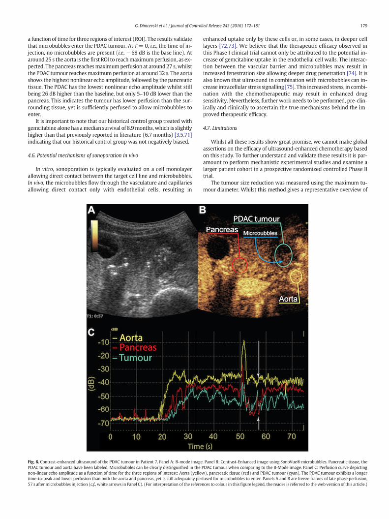
PDAC is well known to be a hypovascular tumour [70], meaning ithas less perfusion than the tissue surrounding it. This is falsely correlat-ed to no perfusion.Nevertheless, in the clinicalfield it iswell known thatPDAC still exhibits perfusion. An example of such hypovascular perfu-sion can be seen the Supplemental video 1 and Fig. 6. Fig. 6 shows a B-Mode image, contrast-enhanced image, and a perfusion curve of theaorta, healthy pancreatic tissue and the primary PDAC tumour.Microbubbles can be clearly distinguished in the primary PDAC tumourwhen comparing the primary PDAC tumour area in Fig. 6 A vs. B. Theperfusion curve Fig. 6C, depicts non-linear contrast echo amplitude as
179G. Dimcevski et al. / Journal of Controlled Release 243 (2016) 172–181
a function of time for three regions of interest (ROI). The results validatethat microbubbles enter the PDAC tumour. At T= 0, i.e., the time of in-jection, no microbubbles are present (i.e, −68 dB is the base line). Ataround 25 s the aorta is the first ROI to reachmaximumperfusion, as ex-pected. The pancreas reachesmaximumperfusion at around27 s,whilstthe PDAC tumour reachesmaximumperfusion at around 32 s. The aortashows the highest nonlinear echo amplitude, followed by the pancreatictissue. The PDAC has the lowest nonlinear echo amplitude whilst stillbeing 26 dB higher than the baseline, but only 5–10 dB lower than thepancreas. This indicates the tumour has lower perfusion than the sur-rounding tissue, yet is sufficiently perfused to allow microbubbles toenter.
It is important to note that our historical control group treated withgemcitabine alone has amedian survival of 8.9months, which is slightlyhigher than that previously reported in literature (6.7 months) [3,5,71]indicating that our historical control group was not negatively biased.
4.6. Potential mechanisms of sonoporation in vivo
In vitro, sonoporation is typically evaluated on a cell monolayerallowing direct contact between the target cell line and microbubbles.In vivo, the microbubbles flow through the vasculature and capillariesallowing direct contact only with endothelial cells, resulting in
Fig. 6. Contrast-enhanced ultrasound of the PDAC tumour in Patient 7. Panel A: B-mode imagePDAC tumour and aorta have been labeled. Microbubbles can be clearly distinguished in thenon-linear echo amplitude as a function of time for the three regions of interest: Aorta (yellowtime-to-peak and lower perfusion than both the aorta and pancreas, yet is still adequately per57 s after microbubbles injection (c.f.,white arrows in Panel C). (For interpretation of the refere
enhanced uptake only by these cells or, in some cases, in deeper celllayers [72,73]. We believe that the therapeutic efficacy observed inthis Phase I clinical trial cannot only be attributed to the potential in-crease of gemcitabine uptake in the endothelial cell walls. The interac-tion between the vascular barrier and microbubbles may result inincreased fenestration size allowing deeper drug penetration [74]. It isalso known that ultrasound in combination with microbubbles can in-crease intracellular stress signalling [75]. This increased stress, in combi-nation with the chemotherapeutic may result in enhanced drugsensitivity. Nevertheless, further work needs to be performed, pre-clin-ically and clinically to ascertain the true mechanisms behind the im-proved therapeutic efficacy.
4.7. Limitations
Whilst all these results show great promise, we cannot make globalassertions on the efficacy of ultrasound-enhanced chemotherapy basedon this study. To further understand and validate these results it is par-amount to perform mechanistic experimental studies and examine alarger patient cohort in a prospective randomized controlled Phase IItrial.
The tumour size reduction was measured using the maximum tu-mour diameter. Whilst this method gives a representative overview of
. Panel B: Contrast-Enhanced image using SonoVue®microbubbles. Pancreatic tissue, thePDAC tumour when comparing to the B-Mode image. Panel C: Perfusion curve depicting), pancreatic tissue (red) and PDAC tumour (cyan). The PDAC tumour exhibits a longer
fused for microbubbles to enter. Panels A and B are freeze frames of late phase perfusion,nces to colour in this figure legend, the reader is referred to the web version of this article.)

180 G. Dimcevski et al. / Journal of Controlled Release 243 (2016) 172–181
tumour progression it does not take into account the 3D structuralchange of the tumour. In our opinion, future work should address thetreatment effect on the tumour volume and not only the maximaldiameter.
The primary limitations of this study are that only a single 2D slice ofthe tumour was treated. Using a 3D ultrasound probewith further opti-mized acoustic conditions and modifying the microbubble type andconcentration may improve the therapeutic efficacy [44].
The ultrasound emission conditions used here were severely limitedby the clinical diagnostic scanner. In previous studies, longer duty cycleshave shown to have a better therapeutic effect than short duty cycles[76]. Future work should aim to determine the ultrasound conditionsthat induce the highest therapeutic effect and to allow implementationof such conditions in the clinic.
There is currently no consensus on what is considered an idealmicrobubble dose. At high dosages, the microbubbles may interactmore with each other than the cells due to secondary Bjerknes forces[42], whereas at low concentrations, there may not be enoughmicrobubbles to interact with the cells. Future work should evaluateand optimise the microbubble type and dosage.
In thefield of sonoporation, it is typically assumed that the enhancedeffect is due to the increase in local drug concentrations. In ourwork,wedid not evaluate if the local drug concentrationwas increased and if thiscould be the reason for the enhanced effect. Future work should evalu-ate if there is an increase in local drug concentration, or if the improvedtherapeutic efficacy is due to increase or decrease in perfusion, or otherintracellular responses.
5. Conclusion
In conclusion, our study indicated that chemotherapy in combina-tion with ultrasound and microbubbles seems to be safe. No additionaltoxicity was observed when compared to chemotherapy alone. In ourpatient cohort, sonoporation has the additional benefit of improvingthe number of treatment cycles the patients were able to undergo andcorrespondingly extending the period of well-being. Significantly in-creased survival was also observed compared to a historical cohort ofpatients. Acknowledging the small treatment group with sub-optimaltreatment conditions in this study, a larger study with improved acous-tic conditions and microbubble delivery is essential to improve our un-derstanding and validating our results. Nevertheless, in our opinionthese novel results show great promise for ultrasound andmicrobubbleenhanced therapy.
Supplementary data to this article can be found online at http://dx.doi.org/10.1016/j.jconrel.2016.10.007.
Ethical considerations
The protocolwas approved by the Regional Ethics Committee (2011/1601/REK vest) and the Norwegian Medicines Agency (NMA). Thestudy was performed in accordance with the Helsinki Declaration. Allsubjects signed an informed consent.
Conflict of interest disclosure statement
I declare no conflict of interest Georg Dimcevski, Date: 06/10/2016,Bergen Norway.
Acknowledgements
This study has received initial financial support from the NorwegianCancer Society (11007001), Helse Vest (911779), and MedViz (03-2014) (http://medviz.uib.no/), a research consortium from HaukelandUniversity Hospital, University of Bergen and Christian Michelsen Re-search AS. We give our special acknowledgement to our chemotherapyadministrating oncologists Nils Glenjen and Hämmerling Katrin. And
special thanks to our nurses (Hilde Sælensminde, Torill Våge, MarianneLehmann, Mari Holsen, Elisabeth Bjerkan) at the Clinical Trial Unit atHaukeland University Hospital for taking care of our patients.
References
[1] World Cancer Report. 2014.[2] A. Neesse, P. Michl, K.K. Frese, C. Feig, N. Cook, M.A. Jacobetz, M.P. Lolkema, M.
Buchholz, K.P. Olive, T.M. Gress, D.A. Tuveson, Stromal biology and therapy in pan-creatic cancer, Gut 60 (2011) 861–868.
[3] Kreftregisteret, Cancer in Norway, 2010.[4] N. Alexakis, C. Halloran, M. Raraty, P. Ghaneh, R. Sutton, J.P. Neoptolemos, Current
standards of surgery for pancreatic cancer, Br J Surg 91 (2004) 1410–1427.[5] M. Malvezzi, P. Bertuccio, F. Levi, V.C. La, E. Negri, European cancer mortality predic-
tions for the year 2013, Ann. Oncol. 24 (2013) 792–800.[6] H.A. Burris III, M.J. Moore, J. Andersen, M.R. Green, M.L. Rothenberg, M.R. Modiano,
M.C. Cripps, R.K. Portenoy, A.M. Storniolo, P. Tarassoff, R. Nelson, F.A. Dorr, C.D.Stephens, D.D. Von Hoff, Improvements in survival and clinical benefit withgemcitabine as first-line therapy for patients with advanced pancreas cancer: a ran-domized trial, J. Clin. Oncol. 15 (1997) 2403–2413.
[7] T. Conroy, C. Gavoille, E. Samalin, M. Ychou, M. Ducreux, The role of the FOLFIRINOXregimen for advanced pancreatic cancer, Curr. Oncol. Rep. 15 (2013) 182–189.
[8] S.M. Hoy, Albumin-bound paclitaxel: a review of its use for the first-line combina-tion treatment of metastatic pancreatic cancer, Drugs 74 (2014) 1757–1768.
[9] B.B. Goldberg, R. Gramiak, A.K. Freimanis, Early history of diagnostic ultrasound: therole of American radiologists, AJR Am. J. Roentgenol. 160 (1993) 189–194.
[10] S. Ødegaard, Gilja OH, Gregersen H, Basic and New Aspects of Gastrointestinal Ultra-sonography, 2005.
[11] B.J. Ostrum, B.B. Goldberg, H.J. Isard, A-mode ultrasound differentiation of soft-tissuemasses, Radiology 88 (1967) 745–749.
[12] G.A. Zamboni, M.C. Ambrosetti, M. D'Onofrio, M.R. Pozzi, Ultrasonography of thepancreas, Radiol. Clin. N. Am. 50 (2012) 395–406.
[13] Y. Wei, X.L. Yu, P. Liang, Z.G. Cheng, Z.Y. Han, F.Y. Liu, J. Yu, Guiding and controllingpercutaneous pancreas biopsies with contrast-enhanced ultrasound: target lesionsare not localized on B-mode ultrasound, Ultrasound Med. Biol. 41 (2015)1561–1569.
[14] D.L. Miller, J. Quddus, Sonoporation of monolayer cells by diagnostic ultrasound ac-tivation of contrast-agent gas bodies, Ultrasound Med. Biol. 26 (2000) 661–667.
[15] J. Wu, J.P. Ross, J.F. Chiu, Reparable sonoporation generated by microstreaming, J.Acoust. Soc. Am. 111 (2002) 1460–1464.
[16] S. Bao, B.D. Thrall, D.L. Miller, Transfection of a reporter plasmid into cultured cellsby sonoporation in vitro, Ultrasound Med. Biol. 23 (1997) 953–959.
[17] B.H. Lammertink, C. Bos, K.M. van der Wurff-Jacobs, G. Storm, C.T. Moonen, R.Deckers, Increase of intracellular cisplatin levels and radiosensitization by ultra-sound in combination with microbubbles, J. Control. Release 238 (2016) 157–165.
[18] M. Derieppe, K. Rojek, J.M. Escoffre, B.D. de Senneville, C. Moonen, C. Bos, Recruit-ment of endocytosis in sonopermeabilization-mediated drug delivery: a real-timestudy, Phys. Biol. 12 (2015) 046010.
[19] v. WA, S. PC, A. Healey, S. Kvale, N. Bush, J. Bamber, d.L. DC, Acoustic cluster therapy(ACT) enhances the therapeutic efficacy of paclitaxel and Abraxane(R) for treatmentof human prostate adenocarcinoma in mice, J. Control. Release 236 (2016) 15–21.
[20] S. Eggen, S.M. Fagerland, Y. Morch, R. Hansen, K. Sovik, S. Berg, H. Furu, A.D. Bohn,M.B. Lilledahl, A. Angelsen, B. Angelsen, D.C. de Lange, Ultrasound-enhanced drugdelivery in prostate cancer xenografts by nanoparticles stabilizing microbubbles, J.Control. Release 187 (2014) 39–49.
[21] I. Lentacker, B. Geers, J. Demeester, S.C. De Smedt, N.N. Sanders, Design and evalua-tion of doxorubicin-containing microbubbles for ultrasound-triggered doxorubicindelivery: cytotoxicity and mechanisms involved, Mol. Ther. 18 (2010) 101–108.
[22] B. Theek, M. Baues, T. Ojha, D. Mockel, S.K. Veettil, J. Steitz, v. BL, G. Storm, F.Kiessling, T. Lammers, Sonoporation enhances liposome accumulation and penetra-tion in tumors with low EPR, J. Control. Release 231 (2016) 77–85.
[23] M. Bazan-Peregrino, C.D. Arvanitis, B. Rifai, L.W. Seymour, C.C. Coussios, Ultrasound-induced cavitation enhances the delivery and therapeutic efficacy of an oncolyticvirus in an in vitro model, J. Control. Release 157 (2012) 235–242.
[24] N. McDannold, C.D. Arvanitis, N. Vykhodtseva, M.S. Livingstone, Temporary disrup-tion of the blood-brain barrier by use of ultrasound andmicrobubbles: safety and ef-ficacy evaluation in rhesus macaques, Cancer Res. 72 (2012) 3652–3663.
[25] S.M. Fix, M.A. Borden, P.A. Dayton, Therapeutic gas delivery via microbubbles and li-posomes, J. Control. Release 209 (2015) 139–149.
[26] F. Prieur, A. Pillon, J.L. Mestas, V. Cartron, P. Cebe, N. Chansard, M. Lafond, C. Lafon,Enhancement of fluorescent probe penetration into tumors in vivo using unseededinertial cavitation, Ultrasound Med. Biol. 42 (2016) 1706–1713.
[27] I. Lentacker, B. Geers, J. Demeester, S.C. De Smedt, N.N. Sanders, Design and evalua-tion of doxorubicin-containing microbubbles for ultrasound-triggered doxorubicindelivery: cytotoxicity and mechanisms involved, Mol. Ther. 18 (2010) 101–108.
[28] B.D. de Senneville, C. Moonen, M. Ries, MRI-Guided HIFUmethods for the ablation ofliver and renal cancers, Adv. Exp. Med. Biol. 880 (2016) 43–63.
[29] Ebbini ES, ter HG. Ultrasound-guided therapeutic focused ultrasound: current statusand future directions. Int. J. Hyperth. 2015;31:77–89.
[30] G. Shapiro, A.W. Wong, M. Bez, F. Yang, S. Tam, L. Even, D. Sheyn, S. Ben-David, W.Tawackoli, G. Pelled, K.W. Ferrara, D. Gazit, Multiparameter evaluation of in vivogene delivery using ultrasound-guided, microbubble-enhanced sonoporation, J.Control. Release 223 (2016) 157–164.
181G. Dimcevski et al. / Journal of Controlled Release 243 (2016) 172–181
[31] I. De C, G. Lajoinie, M. Versluis, D.S. SC, I. Lentacker, Sonoprinting and the importanceof microbubble loading for the ultrasound mediated cellular delivery of nanoparti-cles, Biomaterials 83 (2016) 294–307.
[32] M. Postema, S. Kotopoulis, A. Delalande, Odd Helge Gilja. Sonoporation: whymicrobubbles create pores, Ultraschall in Med 2012 33 (1) (2016) 97–98.
[33] v. RT, I. Skachkov, I. Beekers, L. KR, V. JD, K. TJ, D. Bera, Y. Luan, v. der Steen AF, d. JN,K. Kooiman, Viability of endothelial cells after ultrasound-mediated sonoporation:influence of targeting, oscillation, and displacement of microbubbles, J. Control. Re-lease 238 (2016) 197–211.
[34] A. Delalande, C. Leduc, P. Midoux, M. Postema, C. Pichon, Efficient gene delivery bysonoporation is associated with microbubble entry into cells and the clathrin-de-pendent endocytosis pathway, Ultrasound Med. Biol. 41 (2015) 1913–1926.
[35] M. Chan, K. Dennis, Y. Huang, C. Mougenot, E. Chow, C. DeAngelis, J. Coccagna, A.Sahgal, K. Hynynen, G. Czarnota, W. Chu, Magnetic resonance-guided high-intensi-ty-focused ultrasound for palliation of painful skeletal metastases: a pilot study,Technol Cancer Res Treat (2016).
[36] A. Waspe, Y. Huang, R. Endre, J. Amaral, J. de Ruiter, F. Campbell, C. Mougenot, K.Hynynen, G. Czarnota, J. Drake, M. Temple, Magnetic resonance guided focused ul-trasound for noninvasive pain therapy of osteoid osteoma in children, Journal ofTherapeutic Ultrasound 3 (Suppl 1) (2015) O48 2015.
[37] K. Hynynen, O. Pomeroy, D.N. Smith, P.E. Huber, N.J. McDannold, J. Kettenbach, J.Baum, S. Singer, F.A. Jolesz, MR imaging-guided focused ultrasound surgery offibroadenomas in the breast: a feasibility study, Radiology 219 (2001) 176–185.
[38] A. Carpentier, M. Canney, A. Vignot, V. Reina, K. Beccaria, C. Horodyckid, C. Karachi,D. Leclercq, C. Lafon, J.Y. Chapelon, L. Capelle, P. Cornu, M. Sanson, K. Hoang-Xuan,J.Y. Delattre, A. Idbaih, Clinical trial of blood-brain barrier disruption by pulsed ultra-sound, Sci. Transl. Med. 8 (2016) 343re2.
[39] M. Anzidei, B.C. Marincola, M. Bezzi, G. Brachetti, F. Nudo, E. Cortesi, P. Berloco, C.Catalano, A. Napoli, Magnetic resonance-guided high-intensity focused ultrasoundtreatment of locally advanced pancreatic adenocarcinoma: preliminary experiencefor pain palliation and local tumor control, Investig. Radiol. 49 (2014) 759–765.
[40] A. Delalande, S. Kotopoulis, T. Rovers, C. Pichon, M. Postema, Sonoporation at a lowmechanical index, Bubble Science, Engineering & Technology 3 (2011) 3–12.
[41] A. Delalande, S. Kotopoulis, M. Postema, P. Midoux, C. Pichon, Sonoporation: mech-anistic insights and ongoing challenges for gene transfer, Gene 525 (2013) 191–199.
[42] S. Kotopoulis, M. Postema,Microfoam formation in a capillary, Ultrasonics 50 (2010)260–268.
[43] S. Kotopoulis, G. Dimcevski, O.H. Gilja, D. Hoem, M. Postema, Treatment of humanpancreatic cancer using combined ultrasound, microbubbles, and gemcitabine: aclinical case study, Med. Phys. 40 (2013) 072902.
[44] S. Kotopoulis, A. Delalande, M. Popa, V. Mamaeva, G. Dimcevski, O.H. Gilja, M.Postema, B.T. Gjertsen, E. McCormack, Sonoporation-enhanced chemotherapy sig-nificantly reduces primary tumour burden in an orthotopic pancreatic cancer xeno-graft, Mol. Imaging Biol. 16 (2014) 53–62.
[45] Highlights of Prescribing Information: Gemzar, Eli Lilly and Company, Indianapolis,Indiana, 2010.
[46] M.M. Oken, R.H. Creech, D.C. Tormey, J. Horton, T.E. Davis, E.T. McFadden, P.P.Carbone, Toxicity and response criteria of the eastern cooperative oncology group,Am. J. Clin. Oncol. 5 (1982) 649–655.
[47] European Medicines Agency, Guideline for good clinical practice, ICH Topic E 6 (R1)(2002).
[48] L.S. Tham, L.Z. Wang, R.A. Soo, H.S. Lee, S.C. Lee, B.C. Goh, N.H. Holford, Does satura-ble formation of gemcitabine triphosphate occur in patients? Cancer Chemother.Pharmacol. 63 (2008) 55–64.
[49] M. Schneider, Characteristics of SonoVue®, Echocardiography 16 (1999) 743–746.[50] S. Schafer, K. Nylund, F. Saevik, T. Engjom, M. Mezl, R. Jirik, G. Dimcevski, O.H. Gilja,
K. Tonnies, Semi-automatic motion compensation of contrast-enhanced ultrasoundimages from abdominal organs for perfusion analysis, Comput. Biol. Med. 63 (2015)229–237.
[51] F. Piscaglia, C. Nolsoe, D. CF, C. DO, G. OH, B. NM, T. Albrecht, L. Barozzi, M. Bertolotto,O. Catalano, M. Claudon, C. DA, C. JM, M. D'Onofrio, D. FM, J. Eyding, M. Giovannini,M. Hocke, A. Ignee, J. EM, K. AS, N. Lassau, E. Leen, G. Mathis, A. Saftoiu, G. Seidel, S.PS, H. GT, D. Timmerman,W. HP, The EFSUMB Guidelines and Recommendations onthe Clinical Practice of Contrast Enhanced Ultrasound (CEUS): update 2011 on non-hepatic applications, Ultraschall Med. (2011).
[52] M. Claudon, D. Cosgrove, T. Albrecht, L. Bolondi, M. Bosio, F. Calliada, J.M. Correas, K.Darge, C. Dietrich, M. D'Onofrio, D.H. Evans, C. Filice, L. Greiner, K. Jager, N. Jong, E.Leen, R. Lencioni, D. Lindsell, A. Martegani, S. Meairs, C. Nolsoe, F. Piscaglia, P.Ricci, G. Seidel, B. Skjoldbye, L. Solbiati, L. Thorelius, F. Tranquart, H.P. Weskott, T.Whittingham, Guidelines and good clinical practice recommendations for contrastenhanced ultrasound (CEUS) - update 2008, Ultraschall Med. 29 (2008) 28–44.
[53] U.S. Department of Health and Human Services, Information for ManufacturersSeeking Marketing Clearance of Diagnostic Ultrasound Systems and Transducers(Food and Drug Administration), 2008.
[54] International Electrotechnical Commission, Ultrasonics - Hydrophones Part 2: Cali-bration for Ultrasonic Fields up to 40 MHz, Report No. 62127-2 ed1.0, IEC, Geneva,Switzerland, 2013.
[55] d. JN, M. Emmer, v. WA, M. Versluis, Ultrasonic characterization of ultrasound con-trast agents, Med. Biol. Eng. Comput. 47 (2009) 861–873.
[56] G. Dimcevski, F.G. Erchinger, R. Havre, O.H. Gilja, Ultrasonography in diagnosingchronic pancreatitis: new aspects, World J. Gastroenterol. 19 (2013) 7247–7257.
[57] F. Erchinger, G. Dimcevski, T. Engjom, O.H. Gilja, Transabdominal ultrasound of thepancreas: basic and new aspects, Imaging in Medicine 3 (2011) 411–422.
[58] T. Bjanes, T. Kamceva, T. Eide, B. Riedel, J. Schjott, A. Svardal, Preanalytical stability ofgemcitabine and its metabolite 2′, 2′-difluoro-2′-deoxyuridine in whole blood-assessed by liquid chromatography tandem mass spectrometry, J. Pharm. Sci.(2015).
[59] S.A. Sohaib, B. Turner, J.A. Hanson, M. Farquharson, R.T. Oliver, R.H. Reznek, CT as-sessment of tumour response to treatment: comparison of linear, cross-sectionaland volumetric measures of tumour size, Br. J. Radiol. 73 (2000) 1178–1184.
[60] C.P. Nolsoe, T. Lorentzen, International guidelines for contrast-enhanced ultrasonog-raphy: ultrasound imaging in the new millennium, Ultrasonography 35 (2016)89–103.
[61] R.L. Wahl, H. Jacene, Y. Kasamon, M.A. Lodge, From RECIST to PERCIST: evolving con-siderations for PET response criteria in solid tumors, J. Nucl. Med. 50 (Suppl 1)(2009) 122S–150S.
[62] J.L. Abbruzzese, R. Grunewald, E.A. Weeks, D. Gravel, T. Adams, B. Nowak, S.Mineishi, P. Tarassoff, W. Satterlee, M.N. Raber, A phase I clinical, plasma, and cellu-lar pharmacology study of gemcitabine, J. Clin. Oncol. 9 (1991) 491–498.
[63] H. Inoue, Y. Arai, T. Kishida, M. Shin-Ya, R. Terauchi, S. Nakagawa, M. Saito, S.Tsuchida, A. Inoue, T. Shirai, H. Fujiwara, O. Mazda, T. Kubo, Sonoporation-mediatedtransduction of siRNA ameliorated experimental arthritis using 3 MHz pulsed ultra-sound, Ultrasonics 54 (2014) 874–881.
[64] H. Dewitte, L.S. Van, C. Heirman, K. Thielemans, S.C. De Smedt, K. Breckpot, I.Lentacker, The potential of antigen and TriMix sonoporation using mRNA-loadedmicrobubbles for ultrasound-triggered cancer immunotherapy, J. Control. Release194 (2014) 28–36.
[65] I. Skachkov, Y. Luan, A.F. van der Steen, d. JN, K. Kooiman, Targetedmicrobubbleme-diated sonoporation of endothelial cells in vivo, IEEE Trans. Ultrason. Ferroelectr.Freq. Control 61 (2014) 1661–1667.
[66] A.H. Ko, J. Hwang, A.P. Venook, J.L. Abbruzzese, E.K. Bergsland, M.A. Tempero, SerumCA19-9 response as a surrogate for clinical outcome in patients receiving fixed-doserate gemcitabine for advanced pancreatic cancer, Br. J. Cancer 93 (2005) 195–199.
[67] A. Vincent, J. Herman, R. Schulick, R.H. Hruban, M. Goggins, Pancreatic cancer, Lancet378 (2011) 607–620.
[68] N. Makrilia, K.N. Syrigos, M.W. Saif, Updates on treatment of gemcitabine-refractorypancreatic adenocarcinoma. Highlights from the “2011 ASCO annual meeting”. Chi-cago, IL, USA; June 3-7, 2011, JOP 12 (2011) 351–354.
[69] P.F. Peddi, S. Lubner, R. McWilliams, B.R. Tan, J. Picus, S.M. Sorscher, R. Suresh, A.C.Lockhart, J. Wang, C. Menias, F. Gao, D. Linehan, A. Wang-Gillam, Multi-institutionalexperience with FOLFIRINOX in pancreatic adenocarcinoma, JOP 13 (2012)497–501.
[70] A. Neesse, S. Krug, T.M. Gress, D.A. Tuveson, P. Michl, Emerging concepts in pancre-atic cancer medicine: targeting the tumor stroma, Onco Targets Ther 7 (2013)33–43.
[71] C. Bosetti, P. Bertuccio, M. Malvezzi, F. Levi, L. Chatenoud, E. Negri, V.C. La, Cancermortality in Europe, 2005-2009, and an overview of trends since 1980, Ann.Oncol. 24 (2013) 2657–2671.
[72] A. Bouakaz, A. Zeghimi, A.A. Doinikov, Sonoporation: concept and mechanisms, Adv.Exp. Med. Biol. 880 (2016) 175–189.
[73] B. Helfield, X. Chen, S.C. Watkins, F.S. Villanueva, Biophysical insight into mecha-nisms of sonoporation, Proc. Natl. Acad. Sci. U. S. A. 113 (2016) 9983–9988.
[74] F. Yuan, M. Dellian, D. Fukumura, M. Leunig, D.A. Berk, V.P. Torchilin, R.K. Jain, Vas-cular permeability in a human tumor xenograft: molecular size dependence andcutoff size, Cancer Res. 55 (1995) 3752–3756.
[75] S. Kotopoulis, R. Haugsez, M. Mujic, A. Sulen, S.E. Gullaksen, E. McCormack, O.H.Gilja, M. Postema, B.T. Gjertsen, Evaluation of the effects of clinical diagnostic ultra-sound in combination with ultrasound contrast agents on cell stress: Single cellanalysis of intracellular phospho-signaling pathways in blood cancer cells and nor-mal blood leukocytes. Ultrasonics Symposium (IUS), IEEE International (3–6 Sept.2014).
[76] P. Reuter, J. Masomi, H. Kuntze, I. Fischer, K. Helling, C. Sommer, B. Alessandri, A.Heimann, T. Gerriets, J. Marx, O. Kempski, M. Nedelmann, Low-frequency therapeu-tic ultrasound with varied duty cycle: effects on the ischemic brain and the innerear, Ultrasound Med. Biol. 36 (2010) 1188–1195.