journal of controlled release - laysan biolaysanbio.com/clientuploads/journal of controlled release...
TRANSCRIPT
Journal of Controlled Release 160 (2012) 264–273
Contents lists available at SciVerse ScienceDirect
Journal of Controlled Release
j ourna l homepage: www.e lsev ie r .com/ locate / jconre l
NANOMEDICIN
E
Multifunctional PEGylated 2C5-immunoliposomes containing pH-sensitive bondsand TAT peptide for enhanced tumor cell internalization and cytotoxicity
Erez Koren 1, Anjali Apte 1, Ankur Jani 1, Vladimir P. Torchilin ⁎Center for Pharmaceutical Biotechnology and Nanomedicine, Northeastern University, 360 Huntington Avenue, Boston, MA 02115, USA
⁎ Corresponding author at: Department of Pharmaceumaceutical Biotechnology and Nanomedicine, NortheastSciences Building, 360 Huntington Avenue, Boston, MA3206; fax: +1 617 373 7509, +1 617 373 8886.
E-mail address: [email protected] (V.P. Torchilin)1 Tel.: +1 617 373 3206; fax: +1 617 373 8886.
0168-3659/$ – see front matter © 2011 Elsevier B.V. Alldoi:10.1016/j.jconrel.2011.12.002
a b s t r a c t
a r t i c l e i n f oArticle history:Received 2 September 2011Accepted 5 December 2011Available online 13 December 2011
Keywords:Long-circulating liposomesTAT peptidemAb 2C5pH-sensitive PEG-PE conjugateDoxil®Multifunctional carriers
pH-sensitive PEGylated (with PEG-PE) long-circulating liposomes (HSPC:cholesterol and Doxil®), modifiedwith cell-penetrating TAT peptide (TATp) moieties and cancer-specific mAb 2C5 were prepared. A degradablepH-sensitive hydrazone bond between a long shielding PEG chains and PE (PEG2k-Hz-PE) was introduced.TATp was conjugated with a short PEG1k-PE spacer and mAb 2C5 was attached to a long PEG chain (2C5-PEG3.4k-PE). The “shielding” effect of TATp by long PEG chains was investigated using three liposomal models.At normal pH, surface TATp moieties are “hidden” by the long PEG chains. Upon the exposure to lowered pH,this multifunctional carrier exposes TATp moieties after the degradation of the hydrazone bond and removalof the long PEG chains. Enhanced cellular uptake of the TATp-containing immunoliposomes was observed invitro after pre-treatment at lowered pH (using flow cytometry and fluorescence microscopy techniques). Thepresence of mAb 2C5 on the liposome surface further enhanced the interaction between the carrier andtumor cells but not normal cells. Furthermore, multifunctional immuno-Doxil® preparation showed in-creased cellular cytotoxicity of B16-F10, HeLa and MCF-7 cells when pre-incubated at lower pH, indicatingTATp exposure and activity. In conclusion, a multifunctional immunoliposomal nanocarrier containing apH-sensitive PEG-PE component, TATp, and the cancer cell-specific mAb 2C5 promotes enhanced cytotoxicityand carrier internalization by cancer cells and demonstrates the potential for intracellular drug delivery afterexposure to lowered pH environment, typical of solid tumors.
© 2011 Elsevier B.V. All rights reserved.
1. Introduction
The engineering of a multifunctional pharmaceutical nanocarrieris based on a number of properties that can act either simultaneouslyor sequentially to significantly enhance the efficacy of a variety oftherapeutic and diagnostic protocols [1]. Among the drug deliverysystems (DDS) developed, liposomes have demonstrated substantialpromise as carriers for the delivery of soluble/non-soluble drugs andother therapeutic/diagnostic agents [2]. It is known that PEGylated li-posomes are not readily taken up by the macrophages of the reticulo-endothelial system (RES) and hence stay in the circulation for arelatively long period of time [3,4]. This long circulating effect enablesthese small-sized carriers to “passively” accumulate in tumor tissue,due to the enhanced permeation and retention (EPR) effect [5]. Thiseffect is based on the spontaneous penetration of circulating macro-molecules, particulate drug carriers, and molecular aggregates intothe interstitium through the leaky vasculature at certain pathological
tical Sciences, Center for Phar-ern University, 312 Mugar Life02115, USA. Tel.: +1 617 373
.
rights reserved.
sites. This effect is typical of solid tumors, infarcts and inflammationzones [5–7].
A designed multifunctional carrier with an “active” targeted drugdelivery approach, based on one or more functional groups on thecarrier's surface, can further enhance its efficient accumulation inthe target site and also enable delivery of the therapeutic/diagnosticagent to a specific cell organelle. Since many anticancer drugs, DNAand other therapeutic agents have their effects only in a specific cel-lular organelle (e.g. pro-apoptotic drugs in the mitochondrial mem-brane, gene therapy in the nuclear or mitochondrial genomes),enhancement of intracellular delivery of drug carriers can sharply in-crease the efficiency of a variety of treatment protocols. However, thereceptor-mediated endocytosis of drugs, drug carriers, and DNA leadsto their lysosomal delivery and subsequent considerably high degra-dation. These approaches for direct intra-cytoplasmic delivery thatcircumvent the endocytic pathway should be helpful.
Cell penetrating peptides (CPPs) (e.g. TAT-peptide, penetratin,poly-arginine, Antp and VP22) have demonstrated a capability for de-livery of large variety of biologically active cargoes such as proteins,DNA, antibodies, contrast (imaging) agents, toxins and nanoparticu-lar drug carriers, including liposomes, to the cell interior by traversingthe cell's plasma membrane independent of a membrane receptor [8].These peptides show no cell-type specificity and basically rely on thepositively charged sequences of amino acids (mostly arginine and
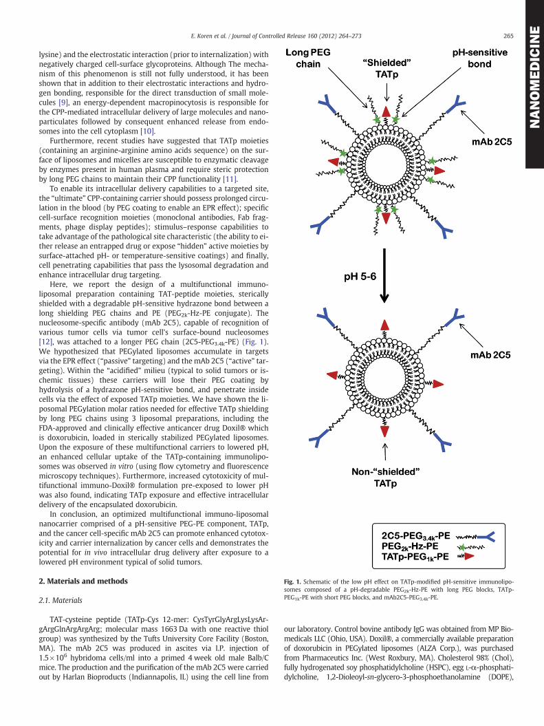
Fig. 1. Schematic of the low pH effect on TATp-modified pH-sensitive immunolipo-somes composed of a pH-degradable PEG2k-Hz-PE with long PEG blocks, TATp-PEG1k-PE with short PEG blocks, and mAb2C5-PEG3.4k-PE.
265E. Koren et al. / Journal of Controlled Release 160 (2012) 264–273
NANOMEDICIN
E
lysine) and the electrostatic interaction (prior to internalization) withnegatively charged cell-surface glycoproteins. Although The mecha-nism of this phenomenon is still not fully understood, it has beenshown that in addition to their electrostatic interactions and hydro-gen bonding, responsible for the direct transduction of small mole-cules [9], an energy-dependent macropinocytosis is responsible forthe CPP-mediated intracellular delivery of large molecules and nano-particulates followed by consequent enhanced release from endo-somes into the cell cytoplasm [10].
Furthermore, recent studies have suggested that TATp moieties(containing an arginine-arginine amino acids sequence) on the sur-face of liposomes and micelles are susceptible to enzymatic cleavageby enzymes present in human plasma and require steric protectionby long PEG chains to maintain their CPP functionality [11].
To enable its intracellular delivery capabilities to a targeted site,the “ultimate” CPP-containing carrier should possess prolonged circu-lation in the blood (by PEG coating to enable an EPR effect); specificcell-surface recognition moieties (monoclonal antibodies, Fab frag-ments, phage display peptides); stimulus–response capabilities totake advantage of the pathological site characteristic (the ability to ei-ther release an entrapped drug or expose “hidden” active moieties bysurface-attached pH- or temperature-sensitive coatings) and finally,cell penetrating capabilities that pass the lysosomal degradation andenhance intracellular drug targeting.
Here, we report the design of a multifunctional immuno-liposomal preparation containing TAT-peptide moieties, stericallyshielded with a degradable pH-sensitive hydrazone bond between along shielding PEG chains and PE (PEG2k-Hz-PE conjugate). Thenucleosome-specific antibody (mAb 2C5), capable of recognition ofvarious tumor cells via tumor cell's surface-bound nucleosomes[12], was attached to a longer PEG chain (2C5-PEG3.4k-PE) (Fig. 1).We hypothesized that PEGylated liposomes accumulate in targetsvia the EPR effect (“passive” targeting) and the mAb 2C5 (“active” tar-geting). Within the “acidified” milieu (typical to solid tumors or is-chemic tissues) these carriers will lose their PEG coating byhydrolysis of a hydrazone pH-sensitive bond, and penetrate insidecells via the effect of exposed TATp moieties. We have shown the li-posomal PEGylation molar ratios needed for effective TATp shieldingby long PEG chains using 3 liposomal preparations, including theFDA-approved and clinically effective anticancer drug Doxil® whichis doxorubicin, loaded in sterically stabilized PEGylated liposomes.Upon the exposure of these multifunctional carriers to lowered pH,an enhanced cellular uptake of the TATp-containing immunolipo-somes was observed in vitro (using flow cytometry and fluorescencemicroscopy techniques). Furthermore, increased cytotoxicity of mul-tifunctional immuno-Doxil® formulation pre-exposed to lower pHwas also found, indicating TATp exposure and effective intracellulardelivery of the encapsulated doxorubicin.
In conclusion, an optimized multifunctional immuno-liposomalnanocarrier comprised of a pH-sensitive PEG-PE component, TATp,and the cancer cell-specific mAb 2C5 can promote enhanced cytotox-icity and carrier internalization by cancer cells and demonstrates thepotential for in vivo intracellular drug delivery after exposure to alowered pH environment typical of solid tumors.
2. Materials and methods
2.1. Materials
TAT-cysteine peptide (TATp-Cys 12-mer: CysTyrGlyArgLysLysAr-gArgGlnArgArgArg; molecular mass 1663 Da with one reactive thiolgroup) was synthesized by the Tufts University Core Facility (Boston,MA). The mAb 2C5 was produced in ascites via I.P. injection of1.5×106 hybridoma cells/ml into a primed 4 week old male Balb/Cmice. The production and the purification of the mAb 2C5 were carriedout by Harlan Bioproducts (Indiannapolis, IL) using the cell line from
our laboratory. Control bovine antibody IgG was obtained from MP Bio-medicals LLC (Ohio, USA). Doxil®, a commercially available preparationof doxorubicin in PEGylated liposomes (ALZA Corp.), was purchasedfrom Pharmaceutics Inc. (West Roxbury, MA). Cholesterol 98% (Chol),fully hydrogenated soy phosphatidylcholine (HSPC), egg L-α-phosphati-dylcholine, 1,2-Dioleoyl-sn-glycero-3-phosphoethanolamine (DOPE),

266 E. Koren et al. / Journal of Controlled Release 160 (2012) 264–273
NANOMEDICIN
E
1,2-dipalmitoyl-sn-glycero-3-phosphothioethanolamine (Sodium Salt)(DPPE-SH), NHS-PEG1000-maleimide, diacyllipid polyethylene glycols(PEG1000-PE, PEG2000-PE), rhodamine-phosphatidylethanolamine (Rh-PE) andphosphatidylthioethanol (DPPE-SH)were purchased fromAvantiPolar Lipids (Alabaster, AL, USA). Poly-oxyethylene 3400-bis(p-nitrophe-nyl carbonate) [PEG(pNP)2] was purchased from Laysan Bio. Inc. (Arab,Alabama). Triethylamine (TEA) andfluorenylmethyloxycarbonyl chloride(FMOC-Cl) were purchased from Fluka (AG, Switzerland). N-(4-Acetyl-phenylmaleimidewas fromAcros Organics (Fairlawn, NJ, USA), methoxypoly(ethylene) glycol butyraldehyde (MW 2000), mPEG-SH – from Lay-san Bio, Inc. (Huntsville, AL, USA), and 4-(4-N-maleimidophenyl) butyricacid hydrazide hydrochloride (MPBH) from Pierce Biotechnology, Inc.(Rockford, IL, USA). Fluoromount-Gwas fromSouthernBiotechnologyAs-sociates Inc. (Birmingham, AL), and CellTiter Blue cell viability assay wasfrom Promega, Madison, WI. TATp-PEG1k-PE (TATp-conjugate), PEG2k-Hz-PE and pNP-PEG3.4k-PE were synthesized in-house (see below).
Cell lines (Human fibroblasts, 4T1, MCF-7, B16-F10, and HeLa)were purchased from the American Type Culture Collection (Manas-sas, VA). All cell culture media, DMEM, heat-inactivated fetal bovineserum (FBS), and concentrated solutions of penicillin/streptomycinstock solutions were from Cellgro® (Herndon, VA). All other chemi-cals and solvents were of analytical grade, purchased from ThermoFisher Scientific and used without further purification.
2.2. Methods
2.2.1. Synthesis of TATp-PEG1k-PE (TATp conjugate)The TATp-PEG1000-PE conjugate was synthesized as described pre-
viously [13] with some modifications. Briefly, an approximately 1.5-fold molar excess of NHS-PEG1k-maleimide was reacted with DOPEby stirring for 2 h in chloroform at room temperature with a 3-foldmolar excess of triethylamine. A 2-fold molar excess of TATp-Cyswas then added, and the reaction was continued with stirring over-night. The solvent was evaporated, and the product was freeze-dried overnight. The excess of TATp-Cys was separated from theproduct by gel filtration chromatography. Fractions were collectedand monitored by TLC using silica plates (mobile phase of chloro-form/methanol 80:20% v/v), and TATp-PEG-PE was visualized withphosphomolybdic acid and Dragendorff spray reagents. In order toconfirm TATp conjugation and presence, we also analyzed for TATpconjugation using HPLC, as previously described [14].
2.2.2. Synthesis of PEG2k-hydrazone-PEAn aldehyde-derived hydrazone-based PEG2k-Hz-PE conjugate (pH-
sensitive conjugate) was synthesized by a two-steps method as previ-ously described [15] with modifications. For step I (synthesis of acylhydrazide-PEG derivative) 40 μmol of mPEG-SH in chloroform wasmixed with a two molar excess of the acyl hydrazide cross-linkerMPBH in presence of 5 molar excess of triethylamine over lipid. Follow-ing 2 h of stirring at room temperature, product was dialyzed (Spectra/Por 6 dialysis membrane, MWCO 1 K, Spectrum Laboratories, RanchoDominguez, CA) against deionized water, analyzed by TLC, freeze-driedand stored as a chloroform solution at−80 °C. For step II of the synthesis(activation of phospholipidwith 4-acetyl phenylmaleimide) 40 mmol of4-acetyl phenyl maleimide were reacted with 27 mmol of 1,2-dipalmi-toyl-sn-glycero-3-phosphothioethanolamine (DPPE-SH) in the presenceof triethylamine overnight with continuous stirring. The activated phos-pholipid was separated on a silica gel column using chloroform:metha-nol mobile phase (9:1 v/v). The fractions containing product wereidentified by TLC analysis, pooled, concentrated, freeze-dried and storedas a chloroform solution at −80 °C.
For the synthesis of the PEG-HZ-PE conjugate, a hydrazide activat-ed PEG derivative was reacted over-night with a 1.5 M excess of theactivated phospholipid with constant stirring at room temperature.The conjugate was separated and purified by size-exclusion gel chro-matography using Sepharose-CL4B media.
2.2.3. Synthesis of pNP-PEG3.4k-PE and mAb 2C5 modificationIn order to attach mAb 2C5 to the liposomal preparations, we first
conjugated the mAb to the distal ends of PEG blocks via p-nitrophenylcarbonyl (pNP) groups (using a pNP-PEG3.4k-PE conju-gate) to form immunomicelles. Using the post-insertion methodwith micelles [16,17] we formed ligand-coupled liposomes (dis-cussed below). First, we synthesized and purified pNP-PEG3.4k-PEaccording to an established method as previously described [18].Briefly, the synthesis includes the interaction of PE with a 10-foldmolar excess of PEG-(pNP)2 in chloroform in the presence of triethy-lamine. Organic solvents were removed, pNP-PEG3.4k-PE micelleswere formed and separated from free PEG and pNP on a CL-4B col-umn. The pNP-PEG3.4k-PE product was freeze-dried, extracted withchloroform and stored at −80 °C.
For antibody conjugates with PEG3.4k-PE (mAb 2C5 or non-specificIgG), a 40 molar excess of pNP-PEG3.4k-PE dispersed in a 10 mg/mLmicellar solution in 5 mM Na-citrate, 150 mM NaCl, pH 5.0, wasadded to an equal volume of a 1 mg/mL solution of protein in100 mM Tris-buffered saline (TBS), pH 8.5. The mixtures were incu-bated at pH 8.5 for 24 h at 4 °C.
2.2.4. Preparation of liposomesTwo PEGylated liposomal formulations were prepared. For all, we
first formed the liposomes and then attached a variety of surface-decorating polymers in different molar ratios to their surfaces(using post-insertion technique) (see below).
For the first liposomal preparation, a lipid film was obtained froma mixture of egg-phosphatidylcholine, cholesterol (Egg-PC:cholester-ol, 7:3 molar ratio) in chloroform. Chloroform was removed by venti-lation using N2 gas followed by freeze-drying. The film was hydratedwith an appropriate buffer and vortexed at room temperature for5 min. The second liposomal formulation was of Doxil®-mimickingcomposition but contained no doxorubicin. For this preparation, weused the same lipid components and the same concentrationsas found in Doxil®. A lipid film was obtained from N-(carbonyl-meth-oxypolyethylene glycol 2000)-1,2-distearoyl-sn-glycero-3-phos-phoethanolamine sodium salt (mPEG-DSPE, 3.19 mg/mL), fullyhydrogenated soy phosphatidylcholine (HSPC, 9.58 mg/mL), and cho-lesterol (3.19 mg/mL). Following chloroform removal with N2 gas andfreeze drying, the lipid film was hydrated in HEPES-buffered saline(HBS), pH 7.4. Both liposomal formulations were sonicated with aprobe-type sonicator at 11 W power for 30 min until approximately100 nm liposomes with narrow size distribution formed (see below).
2.2.5. Modification of liposomesWith the post-insertion method [16,17] we efficiently decorated
liposomal surfaces with a variety of polymeric conjugates. In orderto “add” a function to the three pre-formed liposomal preparations(Egg-PC:cholesterol, HSPC:cholesterol and Doxil®), liposomes wereincubated overnight with different micellar combinations from thefollowing: TATp-PEG1k-PE (2.5 mol%), PEG2k-PE (2.5–15 mol%),PEG5k-PE (2.5–15 mol%), PEG2k-Hz-PE (15 mol%), 2C5-PEG3.4k-PE(2.5 mol%), IgG-PEG3.4k-PE (2.5 mol%) and rhodamine-PE (1 mol%).Subsequently, preparations were dialyzed against water for 24 h.The optimal multifunctional liposomal formulation used for charac-terization and/or cell culture experiments included TATp-PEG1k-PE(2.5 mol%), 2C5-PEG3.4k-PE (2.5 mol%) and PEG2k-Hz-PE (15 mol%).Multifunctional immunoliposomes were pre-incubated for 30 min atpH 5.0 and 7.4 prior to analyses and in vitro studies.
2.2.6. Characterization of liposomes
2.2.6.1. Size and zeta-potential measurements. Liposome size measure-ments and size distribution analysis were performed by dynamic lightscattering (DLS) using a Coulter® N4-Plus Submicron Particle Sizer(Coulter Corporation, Miami, FL). In all cases, size distribution was
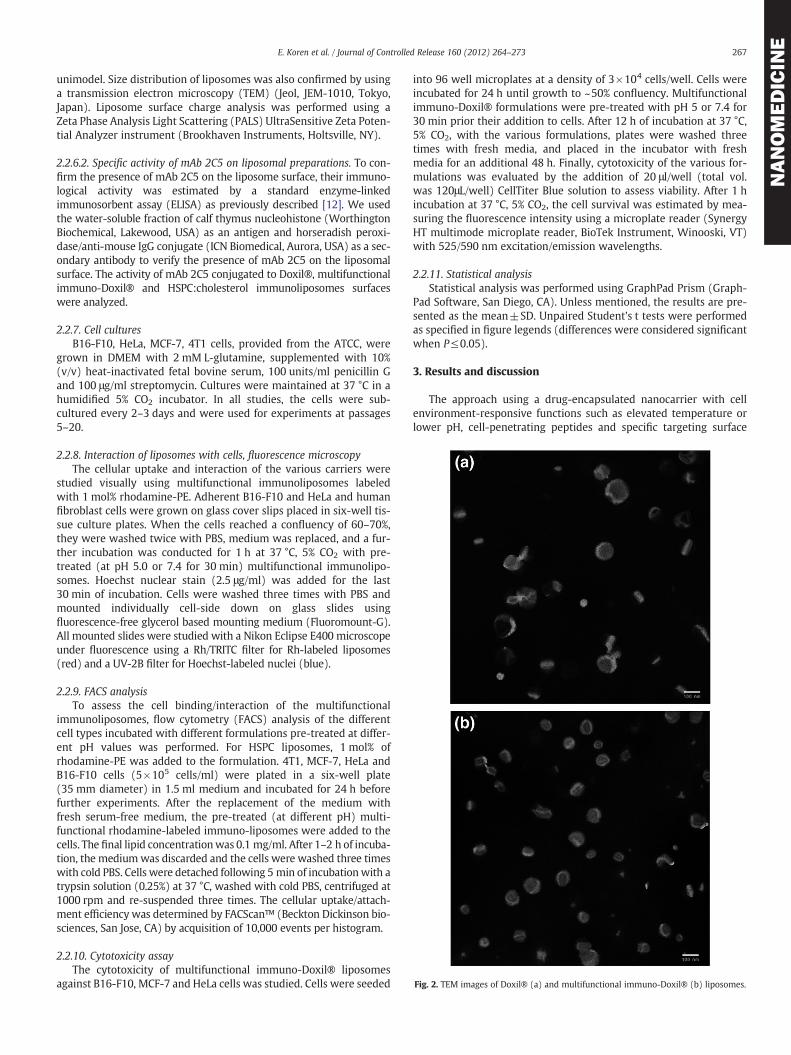
Fig. 2. TEM images of Doxil® (a) and multifunctional immuno-Doxil® (b) liposomes.
267E. Koren et al. / Journal of Controlled Release 160 (2012) 264–273
NANOMEDICIN
E
unimodel. Size distribution of liposomes was also confirmed by usinga transmission electron microscopy (TEM) (Jeol, JEM-1010, Tokyo,Japan). Liposome surface charge analysis was performed using aZeta Phase Analysis Light Scattering (PALS) UltraSensitive Zeta Poten-tial Analyzer instrument (Brookhaven Instruments, Holtsville, NY).
2.2.6.2. Specific activity of mAb 2C5 on liposomal preparations. To con-firm the presence of mAb 2C5 on the liposome surface, their immuno-logical activity was estimated by a standard enzyme-linkedimmunosorbent assay (ELISA) as previously described [12]. We usedthe water-soluble fraction of calf thymus nucleohistone (WorthingtonBiochemical, Lakewood, USA) as an antigen and horseradish peroxi-dase/anti-mouse IgG conjugate (ICN Biomedical, Aurora, USA) as a sec-ondary antibody to verify the presence of mAb 2C5 on the liposomalsurface. The activity of mAb 2C5 conjugated to Doxil®, multifunctionalimmuno-Doxil® and HSPC:cholesterol immunoliposomes surfaceswere analyzed.
2.2.7. Cell culturesB16-F10, HeLa, MCF-7, 4T1 cells, provided from the ATCC, were
grown in DMEM with 2 mM L-glutamine, supplemented with 10%(v/v) heat-inactivated fetal bovine serum, 100 units/ml penicillin Gand 100 μg/ml streptomycin. Cultures were maintained at 37 °C in ahumidified 5% CO2 incubator. In all studies, the cells were sub-cultured every 2–3 days and were used for experiments at passages5–20.
2.2.8. Interaction of liposomes with cells, fluorescence microscopyThe cellular uptake and interaction of the various carriers were
studied visually using multifunctional immunoliposomes labeledwith 1 mol% rhodamine-PE. Adherent B16-F10 and HeLa and humanfibroblast cells were grown on glass cover slips placed in six-well tis-sue culture plates. When the cells reached a confluency of 60–70%,they were washed twice with PBS, medium was replaced, and a fur-ther incubation was conducted for 1 h at 37 °C, 5% CO2 with pre-treated (at pH 5.0 or 7.4 for 30 min) multifunctional immunolipo-somes. Hoechst nuclear stain (2.5 μg/ml) was added for the last30 min of incubation. Cells were washed three times with PBS andmounted individually cell-side down on glass slides usingfluorescence-free glycerol based mounting medium (Fluoromount-G).All mounted slides were studied with a Nikon Eclipse E400 microscopeunder fluorescence using a Rh/TRITC filter for Rh-labeled liposomes(red) and a UV-2B filter for Hoechst-labeled nuclei (blue).
2.2.9. FACS analysisTo assess the cell binding/interaction of the multifunctional
immunoliposomes, flow cytometry (FACS) analysis of the differentcell types incubated with different formulations pre-treated at differ-ent pH values was performed. For HSPC liposomes, 1 mol% ofrhodamine-PE was added to the formulation. 4T1, MCF-7, HeLa andB16-F10 cells (5×10⁵ cells/ml) were plated in a six-well plate(35 mm diameter) in 1.5 ml medium and incubated for 24 h beforefurther experiments. After the replacement of the medium withfresh serum-free medium, the pre-treated (at different pH) multi-functional rhodamine-labeled immuno-liposomes were added to thecells. The final lipid concentrationwas 0.1 mg/ml. After 1–2 h of incuba-tion, the mediumwas discarded and the cells were washed three timeswith cold PBS. Cells were detached following 5 min of incubationwith atrypsin solution (0.25%) at 37 °C, washed with cold PBS, centrifuged at1000 rpm and re-suspended three times. The cellular uptake/attach-ment efficiency was determined by FACScan™ (Beckton Dickinson bio-sciences, San Jose, CA) by acquisition of 10,000 events per histogram.
2.2.10. Cytotoxicity assayThe cytotoxicity of multifunctional immuno-Doxil® liposomes
against B16-F10, MCF-7 and HeLa cells was studied. Cells were seeded
into 96 well microplates at a density of 3×10⁴ cells/well. Cells wereincubated for 24 h until growth to ~50% confluency. Multifunctionalimmuno-Doxil® formulations were pre-treated with pH 5 or 7.4 for30 min prior their addition to cells. After 12 h of incubation at 37 °C,5% CO2, with the various formulations, plates were washed threetimes with fresh media, and placed in the incubator with freshmedia for an additional 48 h. Finally, cytotoxicity of the various for-mulations was evaluated by the addition of 20 μl/well (total vol.was 120μL/well) CellTiter Blue solution to assess viability. After 1 hincubation at 37 °C, 5% CO2, the cell survival was estimated by mea-suring the fluorescence intensity using a microplate reader (SynergyHT multimode microplate reader, BioTek Instrument, Winooski, VT)with 525/590 nm excitation/emission wavelengths.
2.2.11. Statistical analysisStatistical analysis was performed using GraphPad Prism (Graph-
Pad Software, San Diego, CA). Unless mentioned, the results are pre-sented as the mean±SD. Unpaired Student's t tests were performedas specified in figure legends (differences were considered significantwhen P≤0.05).
3. Results and discussion
The approach using a drug-encapsulated nanocarrier with cellenvironment-responsive functions such as elevated temperature orlower pH, cell-penetrating peptides and specific targeting surface
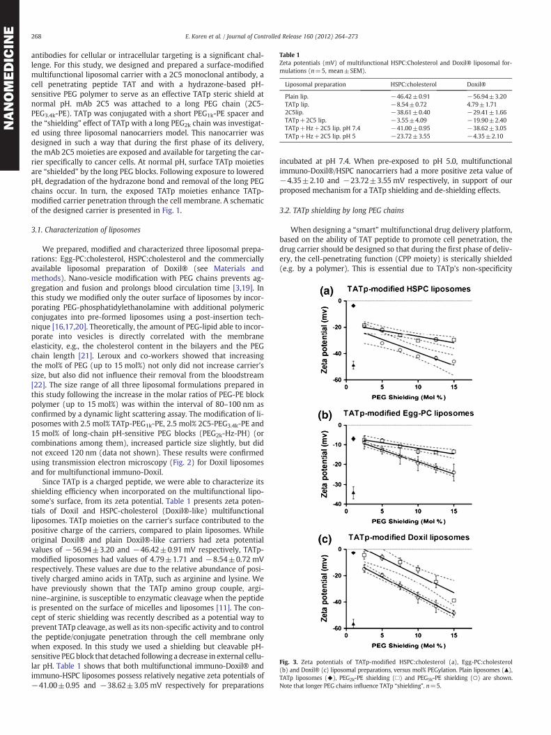
Fig. 3. Zeta potentials of TATp-modified HSPC:cholesterol (a), Egg-PC:cholesterol(b) and Doxil® (c) liposomal preparations, versus mol% PEGylation. Plain liposomes (▲),TATp liposomes (◆), PEG2k-PE shielding (□) and PEG5k-PE shielding (○) are shown.Note that longer PEG chains influence TATp “shielding”. n=5.
Table 1Zeta potentials (mV) of multifunctional HSPC:Cholesterol and Doxil® liposomal for-mulations (n=5, mean±SEM).
Liposomal preparation HSPC:cholesterol Doxil®
Plain lip. −46.42±0.91 −56.94±3.20TATp lip. −8.54±0.72 4.79±1.712C5lip. −38.61±0.40 −29.41±1.66TATp+2C5 lip. −3.55±4.09 −19.90±2.40TATp+Hz+2C5 lip. pH 7.4 −41.00±0.95 −38.62±3.05TATp+Hz+2C5 lip. pH 5 −23.72±3.55 −4.35±2.10
268 E. Koren et al. / Journal of Controlled Release 160 (2012) 264–273
NANOMEDICIN
E
antibodies for cellular or intracellular targeting is a significant chal-lenge. For this study, we designed and prepared a surface-modifiedmultifunctional liposomal carrier with a 2C5 monoclonal antibody, acell penetrating peptide TAT and with a hydrazone-based pH-sensitive PEG polymer to serve as an effective TATp steric shield atnormal pH. mAb 2C5 was attached to a long PEG chain (2C5-PEG3.4k-PE). TATp was conjugated with a short PEG1k-PE spacer andthe “shielding” effect of TATp with a long PEG2k chain was investigat-ed using three liposomal nanocarriers model. This nanocarrier wasdesigned in such a way that during the first phase of its delivery,the mAb 2C5 moieties are exposed and available for targeting the car-rier specifically to cancer cells. At normal pH, surface TATp moietiesare “shielded” by the long PEG blocks. Following exposure to loweredpH, degradation of the hydrazone bond and removal of the long PEGchains occur. In turn, the exposed TATp moieties enhance TATp-modified carrier penetration through the cell membrane. A schematicof the designed carrier is presented in Fig. 1.
3.1. Characterization of liposomes
We prepared, modified and characterized three liposomal prepa-rations: Egg-PC:cholesterol, HSPC:cholesterol and the commerciallyavailable liposomal preparation of Doxil® (see Materials andmethods). Nano-vesicle modification with PEG chains prevents ag-gregation and fusion and prolongs blood circulation time [3,19]. Inthis study we modified only the outer surface of liposomes by incor-porating PEG-phosphatidylethanolamine with additional polymericconjugates into pre-formed liposomes using a post-insertion tech-nique [16,17,20]. Theoretically, the amount of PEG-lipid able to incor-porate into vesicles is directly correlated with the membraneelasticity, e.g., the cholesterol content in the bilayers and the PEGchain length [21]. Leroux and co-workers showed that increasingthe mol% of PEG (up to 15 mol%) not only did not increase carrier'ssize, but also did not influence their removal from the bloodstream[22]. The size range of all three liposomal formulations prepared inthis study following the increase in the molar ratios of PEG-PE blockpolymer (up to 15 mol%) was within the interval of 80–100 nm asconfirmed by a dynamic light scattering assay. The modification of li-posomes with 2.5 mol% TATp-PEG1k-PE, 2.5 mol% 2C5-PEG3.4k-PE and15 mol% of long-chain pH-sensitive PEG blocks (PEG2k-Hz-PH) (orcombinations among them), increased particle size slightly, but didnot exceed 120 nm (data not shown). These results were confirmedusing transmission electron microscopy (Fig. 2) for Doxil liposomesand for multifunctional immuno-Doxil.
Since TATp is a charged peptide, we were able to characterize itsshielding efficiency when incorporated on the multifunctional lipo-some's surface, from its zeta potential. Table 1 presents zeta poten-tials of Doxil and HSPC-cholesterol (Doxil®-like) multifunctionalliposomes. TATp moieties on the carrier's surface contributed to thepositive charge of the carriers, compared to plain liposomes. Whileoriginal Doxil® and plain Doxil®-like carriers had zeta potentialvalues of −56.94±3.20 and −46.42±0.91 mV respectively, TATp-modified liposomes had values of 4.79±1.71 and −8.54±0.72 mVrespectively. These values are due to the relative abundance of posi-tively charged amino acids in TATp, such as arginine and lysine. Wehave previously shown that the TATp amino group couple, argi-nine–arginine, is susceptible to enzymatic cleavage when the peptideis presented on the surface of micelles and liposomes [11]. The con-cept of steric shielding was recently described as a potential way toprevent TATp cleavage, as well as its non-specific activity and to controlthe peptide/conjugate penetration through the cell membrane onlywhen exposed. In this study we used a shielding but cleavable pH-sensitive PEGblock that detached following a decrease in external cellu-lar pH. Table 1 shows that both multifunctional immuno-Doxil® andimmuno-HSPC liposomes possess relatively negative zeta potentials of−41.00±0.95 and −38.62±3.05 mV respectively for preparations
incubated at pH 7.4. When pre-exposed to pH 5.0, multifunctionalimmuno-Doxil®/HSPC nanocarriers had a more positive zeta value of−4.35±2.10 and −23.72±3.55 mV respectively, in support of ourproposed mechanism for a TATp shielding and de-shielding effects.
3.2. TATp shielding by long PEG chains
When designing a “smart”multifunctional drug delivery platform,based on the ability of TAT peptide to promote cell penetration, thedrug carrier should be designed so that during the first phase of deliv-ery, the cell-penetrating function (CPP moiety) is sterically shielded(e.g. by a polymer). This is essential due to TATp's non-specificity
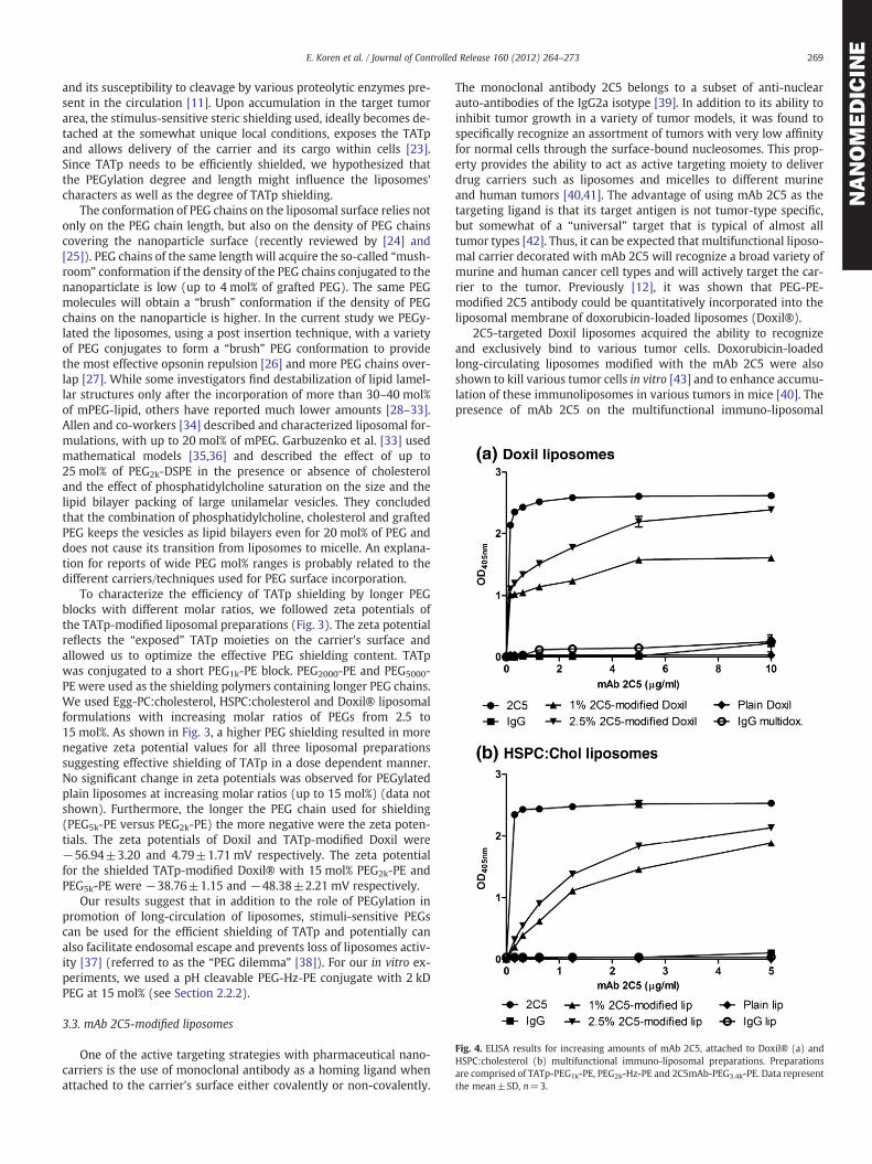
Fig. 4. ELISA results for increasing amounts of mAb 2C5, attached to Doxil® (a) andHSPC:cholesterol (b) multifunctional immuno-liposomal preparations. Preparationsare comprised of TATp-PEG1k-PE, PEG2k-Hz-PE and 2C5mAb-PEG3.4k-PE. Data representthe mean±SD, n=3.
269E. Koren et al. / Journal of Controlled Release 160 (2012) 264–273
NANOMEDICIN
E
and its susceptibility to cleavage by various proteolytic enzymes pre-sent in the circulation [11]. Upon accumulation in the target tumorarea, the stimulus-sensitive steric shielding used, ideally becomes de-tached at the somewhat unique local conditions, exposes the TATpand allows delivery of the carrier and its cargo within cells [23].Since TATp needs to be efficiently shielded, we hypothesized thatthe PEGylation degree and length might influence the liposomes'characters as well as the degree of TATp shielding.
The conformation of PEG chains on the liposomal surface relies notonly on the PEG chain length, but also on the density of PEG chainscovering the nanoparticle surface (recently reviewed by [24] and[25]). PEG chains of the same length will acquire the so-called “mush-room” conformation if the density of the PEG chains conjugated to thenanoparticlate is low (up to 4 mol% of grafted PEG). The same PEGmolecules will obtain a “brush” conformation if the density of PEGchains on the nanoparticle is higher. In the current study we PEGy-lated the liposomes, using a post insertion technique, with a varietyof PEG conjugates to form a “brush” PEG conformation to providethe most effective opsonin repulsion [26] and more PEG chains over-lap [27]. While some investigators find destabilization of lipid lamel-lar structures only after the incorporation of more than 30–40 mol%of mPEG-lipid, others have reported much lower amounts [28–33].Allen and co-workers [34] described and characterized liposomal for-mulations, with up to 20 mol% of mPEG. Garbuzenko et al. [33] usedmathematical models [35,36] and described the effect of up to25 mol% of PEG2k-DSPE in the presence or absence of cholesteroland the effect of phosphatidylcholine saturation on the size and thelipid bilayer packing of large unilamelar vesicles. They concludedthat the combination of phosphatidylcholine, cholesterol and graftedPEG keeps the vesicles as lipid bilayers even for 20 mol% of PEG anddoes not cause its transition from liposomes to micelle. An explana-tion for reports of wide PEG mol% ranges is probably related to thedifferent carriers/techniques used for PEG surface incorporation.
To characterize the efficiency of TATp shielding by longer PEGblocks with different molar ratios, we followed zeta potentials ofthe TATp-modified liposomal preparations (Fig. 3). The zeta potentialreflects the “exposed” TATp moieties on the carrier's surface andallowed us to optimize the effective PEG shielding content. TATpwas conjugated to a short PEG1k-PE block. PEG2000-PE and PEG5000-PE were used as the shielding polymers containing longer PEG chains.We used Egg-PC:cholesterol, HSPC:cholesterol and Doxil® liposomalformulations with increasing molar ratios of PEGs from 2.5 to15 mol%. As shown in Fig. 3, a higher PEG shielding resulted in morenegative zeta potential values for all three liposomal preparationssuggesting effective shielding of TATp in a dose dependent manner.No significant change in zeta potentials was observed for PEGylatedplain liposomes at increasing molar ratios (up to 15 mol%) (data notshown). Furthermore, the longer the PEG chain used for shielding(PEG5k-PE versus PEG2k-PE) the more negative were the zeta poten-tials. The zeta potentials of Doxil and TATp-modified Doxil were−56.94±3.20 and 4.79±1.71 mV respectively. The zeta potentialfor the shielded TATp-modified Doxil® with 15 mol% PEG2k-PE andPEG5k-PE were −38.76±1.15 and −48.38±2.21 mV respectively.
Our results suggest that in addition to the role of PEGylation inpromotion of long-circulation of liposomes, stimuli-sensitive PEGscan be used for the efficient shielding of TATp and potentially canalso facilitate endosomal escape and prevents loss of liposomes activ-ity [37] (referred to as the “PEG dilemma” [38]). For our in vitro ex-periments, we used a pH cleavable PEG-Hz-PE conjugate with 2 kDPEG at 15 mol% (see Section 2.2.2).
3.3. mAb 2C5-modified liposomes
One of the active targeting strategies with pharmaceutical nano-carriers is the use of monoclonal antibody as a homing ligand whenattached to the carrier's surface either covalently or non-covalently.
The monoclonal antibody 2C5 belongs to a subset of anti-nuclearauto-antibodies of the IgG2a isotype [39]. In addition to its ability toinhibit tumor growth in a variety of tumor models, it was found tospecifically recognize an assortment of tumors with very low affinityfor normal cells through the surface-bound nucleosomes. This prop-erty provides the ability to act as active targeting moiety to deliverdrug carriers such as liposomes and micelles to different murineand human tumors [40,41]. The advantage of using mAb 2C5 as thetargeting ligand is that its target antigen is not tumor-type specific,but somewhat of a “universal” target that is typical of almost alltumor types [42]. Thus, it can be expected that multifunctional liposo-mal carrier decorated with mAb 2C5 will recognize a broad variety ofmurine and human cancer cell types and will actively target the car-rier to the tumor. Previously [12], it was shown that PEG-PE-modified 2C5 antibody could be quantitatively incorporated into theliposomal membrane of doxorubicin-loaded liposomes (Doxil®).
2C5-targeted Doxil liposomes acquired the ability to recognizeand exclusively bind to various tumor cells. Doxorubicin-loadedlong-circulating liposomes modified with the mAb 2C5 were alsoshown to kill various tumor cells in vitro [43] and to enhance accumu-lation of these immunoliposomes in various tumors in mice [40]. Thepresence of mAb 2C5 on the multifunctional immuno-liposomal

270 E. Koren et al. / Journal of Controlled Release 160 (2012) 264–273
NANOMEDICIN
E
surfaces was confirmed by ELISA using nucleosomes as binding sub-strate [12]. To prevent the mAb from being hidden in the PEG corona,a long PEG spacer (3.4 kDa) was conjugated to the antibody. BothDoxil®- and HSPC:cholesterol-2C5 modified multifunctional lipo-somes demonstrated immunoreactivity toward the antigen, com-pared to the IgG-modified carriers (Fig. 4). The ELISA experimentsclearly demonstrated that mAb 2C5 was attached to the carrier's sur-face to a major extent and retained the specific activity required forthe successful targeting of liposomes to cancer cells (see below).The production of multifunctional immuno-liposomes did not notice-able change the liposome size but showed slight increase in netcharge (see Table 1).
3.4. Specific binding and uptake of multifunctional immunoliposomes bycancer cells in vitro
The combination of three targeting components on a single carriershould be considered a challenge. TAT peptide on the surface of lipo-somes was shown to enhance their efficient intracellular delivery[13]. Furthermore, the use of a combination of TATp and a PEG“shielding” by pH-sensitive PEG blocks demonstrated the ability tocontrol TATp exposure and prevented its enzymatic degradationwhen attached to liposomes or micelles [11,15]. The addition ofmAb 2C5, conjugated to a long PEG block, on the surface of such adesigned carrier should enhance liposome targeting and interactionwith cancer cells [42]. In order to mimic the acidic environment
Fig. 5. Fluorescence microscopy showing the internalization of 1 mol% rhodamine-PE-labeled Doxil®-like liposomes by fibroblasts, MCF-7 and by B16-F10 cells. Plain li-posomes (a), TATp-modified liposomes (b), 2C5-modified liposomes (c), TATp-2C5-Hydrazone-modified liposomes pre-incubated at pH 7.4 (d) and TATp-2C5-Hydrazone-modified liposomes pre-incubated at pH 5 (e) are shown. (red-rhodamine; blue-Hoechstnuclei staining).
Fig. 6. Flow cytometry. Representative histogram plots of MCF-7 cells (a) incubated for1 h with rhodamine-labeled Doxil®-like immunoliposomes (HSPC:cholesterol) andB16-F10, Hela and 4T1 cells (b–d respectively), incubated for 2 h with multifunctionalimmuno-Doxil®. Multifunctional carriers were incubated with cells following theirpre-exposure to normal or acidic conditions for 30 min.

271E. Koren et al. / Journal of Controlled Release 160 (2012) 264–273
NANOMEDICIN
E
typical in the solid tumor milieu [44,45], multifunctional immunoli-posomes were pre-incubated at pH 5.0 or 7.4 before incubationwith cells. Liposomal interaction with cells was measured using fluo-rescence microscopy and flow cytometry.
Fluorescence microscopy showed that while TATp-modified lipo-somes were significantly internalized by normal fibroblasts and byMCF-7 and B16-F10 cells, 2C5-modified liposomes were internalizedonly by the cancer cells. Fig. 5 clearly shows that pre-exposed multi-functional immunoliposomes to acidic conditions for 30 min en-hanced internalization of the liposomal formulations to the cells,compared with liposomes incubated at pH 7.4. These results suggestthat although TATp cell penetrating properties were shielded at pH7.4, short exposure to lower pH cleaved the hydrazone pH-sensitivebond, exposed TATp moieties and promoted their activity.
Our in vitro results suggest that PEG-shielding of TATp moieties onthe surface of liposomes will prevent or reduce the carrier's non-specific interaction with cells and allow targeting to cancer cells bythe mAb 2C5.
To further investigate multifunctional immuno-liposome internal-ization, we performed a series of experiments using flow cytometry(Fig. 6). As expected, the analysis of the geometric mean fluorescencerevealed a 2-fold stronger binding of 2C5-modified immunolipo-somes and 6-fold stronger interaction of TATp-modified liposomeswith both B16-F10 and MCF-7 cells, when compared with plain lipo-somes. Binding experiments with multifunctional immunoliposomespre-treated at pH 5.0 or 7.4 for 30 min showed a significant increasein fluorescence for the pH 5.0 pre-incubated group (6.68 and 7.44-fold stronger binding for B16-F10 and MCF-7 respectively) when
Fig. 7. In vitro cytotoxicity of various multifunctional immuno-Doxil® preparations. CommerPEG2k-Hz-PE (15 mol%) conjugates solely or in combination. The cytotoxicity of each prepashow the cytotoxicity of the different preparations over a range of concentrations of doxorub
compared to pH 7.4 conditions (2.39 and 2.21-fold stronger bindingfor B16-F10 and MCF-7 respectively versus plain liposomes) suggest-ing that the TATp was exposed and allowed liposomes to interactmore efficiently with these cell lines. The increase in the multifunc-tional immuno-liposomes binding, incubated at pH 7.4, canbe explained as the mAb 2C5 contribution to the nanocarrier-cellinteraction.
3.5. In vitro cytotoxicity of multifunctional immuno-Doxil® liposomes
The liposomal formulation of doxorubicin (Doxil®) is nano-sized,with long-circulating properties, resulting from PEGylation. Its target-ing dependsmostly on the EPR effect [5,46] to passively deliver doxoru-bicin into tumor site. Due to the variety of side-effects of doxorubicin,many attempts have been made to decrease the dose administratedwhilemaintaining its therapeutic effect. Active targeting of this efficientdrug carrier is expected to improve its safety parameters and its clinicalparameters. It has been shown that 2C5 mAb-modified Doxil® in-creased the cytotoxicity drug effect with a reduction in the LC50 [43].Furthermore, the attachment of the cell penetrating peptide, TATp to aDoxil preparation enhanced the cytotoxicity profile in vitro [11]. ThepH-sensitive TAT-modified PEGylated liposomes also enhanced trans-fection of tumor cells in vivo [47]. It was also observed that TATp activitywas diminished due to proteolytic cleavage. The authors concluded thatTATp should be sterically shielded and described the shielding of thispeptide by long PEG blocks [14].
With this in mind, we tested the cytotoxicity of our pH-sensitivemultifunctional immuno-Doxil® liposomal preparation using an
cial Doxil® was grafted with TATp-PEG1k-PE (2.5 mol%), 2C5-PEG3.4k-PE (2.5 mol%) andration was expressed as % survival with untreated cells considered 100%. Upper panelsicin. Lower panels compare survival of cells at 25 μg/ml of doxorubicin. n=5, *pb0.05.
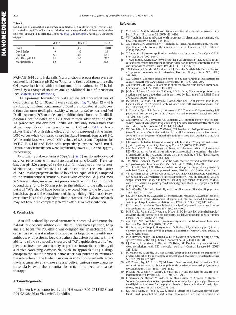
Table 2LC50 values of unmodified and surface modified Doxil® multifunctional immunolipo-somes following 12 h. of incubation. Medium was changed and additional 48 h incuba-tion was followed in normal media (see Materials and methods). Results are presentedas μg/ml.
MCF-7 B16-F10 HeLa
Doxil 38.0 3.5 100.0Doxil-TATp 1.0 1.0 1.8Doxil-2C5 27.0 3.0 65.0MultiDox pH 7.4 8.8 3.0 70.0MultiDox pH 5 2.0 1.2 9.0
272 E. Koren et al. / Journal of Controlled Release 160 (2012) 264–273
NANOMEDICIN
E
MCF-7, B16-F10 and HeLa cells. Multifunctional preparations were in-cubated for 30 min at pH 5.0 or 7.4 prior to their addition to the cells.Cells were incubated with the liposomal formulations for 12 h, fol-lowed by a change of medium and an additional 48 h of incubation(see Materials and methods).
The liposomal formulations with equivalent concentrations ofdoxorubicin at 1.5 to 100 μg/ml were evaluated (Fig. 7). After 12+48 hincubation, multifunctional immuno-Doxil pre-incubated at acidic con-ditions demonstrated higher toxicity when comprised to non-modifiedDoxil liposomes, 2C5-modified and multifunctional immuno-Doxil® li-posomes, pre-incubated at pH 7.4 prior to their addition to the cells.TATp-modified non-shielded Doxil® was the only formulation thatshowed superior cytotoxicity and a lower LC50 value. Table 2 clearlyshows that a TATp shielding effect at pH 7.4 is expressed at the higherLC50 values when compared to pre-incubated formulations at pH 5.0.While multi-Doxil® showed LC50 values of 8.8, 3 and 70 μM/ml forMCF-7, B16-F10 and HeLa cells respectively, pre-incubated multi-Doxil® at acidic incubation were significantly lower (2, 1.2 and 9 μg/ml,respectively).
Cytotoxicity of doxorubicin at 25 μg/ml (Fig. 7) significantly loweredsurvival percentage with multifunctional immuno-Doxil® (Pre-incu-bated at pH 5.0) compared to the other formulations but not to non-shielded TATp-Doxil formulation for all three cell lines. The high toxicityof TATp-Doxil® preparation should have been equal or less, comparedto the multifunctional immuno-Doxil® with exposed TATp and mAb2C5. Nevertheless, since we have pre-exposed the formulations to acid-ic conditions for only 30 min prior to the addition to the cells, at thispoint all TATp should have been fully exposed (due to the hydrazonebond cleavage and the detachment of the “shielding” PEG block). How-ever, since it is a time-dependent kinetic reaction, the hydrazone bondsmay not have been completely cleaved after 30 min of incubation.
4. Conclusion
Amultifunctional liposomal nanocarrier, decorated with monoclo-nal anti-nucleosome antibody 2C5, the cell-penetrating peptide, TATpand a pH-sensitive PEG-shield was designed and characterized. Thiscarrier can act as a stimulus-sensitive carrier targeted with antitumorantibody, with systemic long circulation characteristics and with theability to show site-specific exposure of TAT peptide after a brief ex-posure to lower pH, and thereby to promote intracellular delivery ofa carrier containing doxorubicin. Such an approach using a drug-encapsulated multifunctional nanocarrier can potentially minimizethe interaction of the loaded nanocarrier with non-target cells, effec-tively accumulate at a tumor site and deliver various cargo drugs in-tracellularly with the potential for much improved anti-cancertherapy.
Acknowledgments
This work was supported by the NIH grants RO1 CA121838 andRO1 CA128486 to Vladimir P. Torchilin.
References
[1] V. Torchilin, Multifunctional and stimuli-sensitive pharmaceutical nanocarriers,Eur. J. Pharm. Biopharm. 71 (2009) 431–444.
[2] V.P. Torchilin, Recent advances with liposomes as pharmaceutical carriers, Nat.Rev. Drug Discov. 4 (2005) 145–160.
[3] A.L. Klibanov, K. Maruyama, V.P. Torchilin, L. Huang, Amphipathic polyethylene-glycols effectively prolong the circulation time of liposomes, FEBS Lett. 268(1990) 235–237.
[4] Y. Barenholz, Liposome application: problems and prospects, Curr. Opin. ColloidInterface Sci. 6 (2001) 66–77.
[5] Y. Matsumura, H. Maeda, A new concept for macromolecular therapeutics in can-cer chemotherapy: mechanism of tumoritropic accumulation of proteins and theantitumor agent smancs, Cancer Res. 46 (1986) 6387–6392.
[6] T.N. Palmer, V.J. Caride, M.A. Caldecourt, J. Twickler, V. Abdullah, The mechanismof liposome accumulation in infarction, Biochim. Biophys. Acta 797 (1984)363–368.
[7] A.A. Gabizon, Liposome circulation time and tumor targeting: implications forcancer chemotherapy, Adv. Drug Delivery Rev. 16 (1995) 285–294.
[8] A.D. Frankel, C.O. Pabo, Cellular uptake of the tat protein from human immunode-ficiency virus, Cell 55 (1988) 1189–1193.
[9] J.C. Mai, H. Shen, S.C. Watkins, T. Cheng, P.D. Robbins, Efficiency of protein trans-duction is cell type-dependent and is enhanced by dextran sulfate, J. Biol. Chem.277 (2002) 30208–30218.
[10] J.S. Wadia, R.V. Stan, S.F. Dowdy, Transducible TAT-HA fusogenic peptide en-hances escape of TAT-fusion proteins after lipid raft macropinocytosis, Nat.Med. 10 (2004) 310–315.
[11] E. Koren, A. Apte, R.R. Sawant, J. Grunwald, V.P. Torchilin, Cell-penetrating TATpeptide in drug delivery systems: proteolytic stability requirements, Drug Deliv.18 (2011) 377–384.
[12] A.N. Lukyanov, T.A. Elbayoumi, A.R. Chakilam, V.P. Torchilin, Tumor-targeted lipo-somes: doxorubicin-loaded long-circulating liposomes modified with anti-cancerantibody, J. Control. Release 100 (2004) 135–144.
[13] V.P. Torchilin, R. Rammohan, V. Weissig, T.S. Levchenko, TAT peptide on the sur-face of liposomes affords their efficient intracellular delivery even at low temper-ature and in the presence of metabolic inhibitors, Proc. Natl. Acad. Sci. U. S. A. 98(2001) 8786–8791.
[14] J. Grunwald, T. Rejtar, R. Sawant, Z. Wang, V.P. Torchilin, TAT peptide and its con-jugates: proteolytic stability, Bioconjug Chem. 20 (2009) 1531–1537.
[15] A.A. Kale, V.P. Torchilin, Design, synthesis, and characterization of pH-sensitivePEG-PE conjugates for stimuli-sensitive pharmaceutical nanocarriers: the effectof substitutes at the hydrazone linkage on the ph stability of PEG-PE conjugates,Bioconjug Chem. 18 (2007) 363–370.
[16] T.M. Allen, P. Sapra, E. Moase, Use of the post-insertion method for the formationof ligand-coupled liposomes, Cell. Mol. Biol. Lett. 7 (2002) 889–894.
[17] T. Ishida, D.L. Iden, T.M. Allen, A combinatorial approach to producing stericallystabilized (Stealth) immunoliposomal drugs, FEBS Lett. 460 (1999) 129–133.
[18] V.P. Torchilin, T.S. Levchenko, A.N. Lukyanov, B.A. Khaw, A.L. Klibanov, R. Rammohan,G.P. Samokhin, K.R. Whiteman, p-Nitrophenylcarbonyl-PEG-PE-liposomes: fast andsimple attachment of specific ligands, including monoclonal antibodies, to distalends of PEG chains via p-nitrophenylcarbonyl groups, Biochim. Biophys. Acta 1511(2001) 397–411.
[19] M.C. Woodle, D.D. Lasic, Sterically stabilized liposomes, Biochim. Biophys. Acta1113 (1992) 171–199.
[20] P.S. Uster, T.M. Allen, B.E. Daniel, C.J. Mendez, M.S. Newman, G.Z. Zhu, Insertion ofpoly(ethylene glycol) derivatized phospholipid into pre-formed liposomes re-sults in prolonged in vivo circulation time, FEBS Lett. 386 (1996) 243–246.
[21] K. Hristova, D. Needham, Phase behavior of a lipid/polymer-lipid mixture in aque-ous medium, Macromolecules 28 (1995) 991–1002.
[22] M.N. Khalid, P. Simard, D. Hoarau, A. Dragomir, J.C. Leroux, Long circulating poly(-ethylene glycol)-decorated lipid nanocapsules deliver docetaxel to solid tumors,Pharm. Res. 23 (2006) 752–758.
[23] A.A. Kale, V.P. Torchilin, Environment-responsive multifunctional liposomes,Methods Mol. Biol. 605 (2010) 213–242.
[24] U.S. Schubert, K. Knop, R. Hoogenboom, D. Fischer, Poly(ethylene glycol) in drugdelivery: pros and cons as well as potential alternatives, Angew. Chem. Int. Ed. 49(2010) 6288–6308.
[25] M.D. Howard, M. Jay, T.D. Dziubla, X. Lu, PEGylation of nanocarrier drug deliverysystems: state of the art, J. Biomed. Nanotechnol. 4 (2008) 133–148.
[26] P.J. Photos, L. Bacakova, B. Discher, F.S. Bates, D.E. Discher, Polymer vesicles invivo: correlations with PEG molecular weight, J. Control. Release 90 (2003)323–334.
[27] M. Malmsten, K. Emoto, J.M. Van Alstine, Effect of chain density on inhibition ofprotein adsorption by poly (ethylene glycol) based coatings* 1, J. Colloid InterfaceSci. 202 (1998) 507–517.
[28] A.K. Kenworthy, S.A. Simon, T.J. McIntosh, Structure and phase behavior of lipidsuspensions containing phospholipids with covalently attached poly(ethyleneglycol), Biophys. J. 68 (1995) 1903–1920.
[29] D. Lasic, M. Woodle, F. Martin, T. Valentincic, Phase behavior of stealth-lipid–lecithin mixtures, Period. Biol. 93 (1991) 287–290.
[30] K. Shimada, S. Matsuo, Y. Sadzuka, A. Miyagishima, Y. Nozawa, S. Hirota, T.Sonobe, Determination of incorporated amounts of poly(ethylene glycol)-deriva-tized lipids in liposomes for the physicochemical characterization of stealth lipo-somes, Int. J. Pharm. 203 (2000) 255–263.
[31] F.K. Bedu-Addo, P. Tang, Y. Xu, L. Huang, Effects of polyethyleneglycol chainlength and phospholipid acyl chain composition on the interaction of

273E. Koren et al. / Journal of Controlled Release 160 (2012) 264–273
NANOMEDICIN
E
polyethyleneglycol-phospholipid conjugates with phospholipid: implications inliposomal drug delivery, Pharm. Res. 13 (1996) 710–717.
[32] R.M. Sawant, J.P. Hurley, S. Salmaso, A. Kale, E. Tolcheva, T.S. Levchenko, V.P.Torchilin, “SMART” drug delivery systems: double-targeted pH-responsive phar-maceutical nanocarriers, Bioconjug Chem. 17 (2006) 943–949.
[33] O. Garbuzenko, Y. Barenholz, A. Priev, Effect of grafted PEG on liposome size andon compressibility and packing of lipid bilayer, Chem. Phys. Lipids 135 (2005)117–129.
[34] J.X. Zhang, S. Zalipsky, N. Mullah, M. Pechar, T.M. Allen, Pharmaco attributes ofdioleoylphosphatidylethanolamine/cholesterylhemisuccinate liposomes contain-ing different types of cleavable lipopolymers, Pharmacol. Res. 49 (2004) 185–198.
[35] K. Hristova, D. Needham, The influence of polymer-grafted lipids on the physical-properties of lipid bilayers – a theoretical-study, J. Colloid Interface Sci. 168(1994) 302–314.
[36] P.G. Degennes, Polymers at an interface – a simplified view, Adv. Colloid Interface27 (1987) 189–209.
[37] K. Remaut, B. Lucas, K. Braeckmans, J. Demeester, S.C. De Smedt, Pegylation of li-posomes favours the endosomal degradation of the delivered phosphodiester ol-igonucleotides, J. Control. Release 117 (2007) 256–266.
[38] H. Hatakeyama, H. Akita, H. Harashima, A multifunctional envelope type nano de-vice (MEND) for gene delivery to tumours based on the EPR effect: a strategy forovercoming the PEG dilemma, Adv. Drug Deliv. Rev. 63 (2011) 152–160.
[39] L.Z. Iakoubov, V.P. Torchilin, A novel class of antitumor antibodies: nucleosome-restricted antinuclear autoantibodies (ANA) from healthy aged nonautoimmunemice, Oncol. Res. 9 (1997) 439–446.
[40] T.A. Elbayoumi, V.P. Torchilin, Enhanced accumulation of long-circulating lipo-somes modified with the nucleosome-specific monoclonal antibody 2C5 in vari-ous tumours in mice: gamma-imaging studies, Eur. J. Nucl. Med. Mol. Imaging33 (2006) 1196–1205.
[41] V.P. Torchilin, A.N. Lukyanov, Z. Gao, B. Papahadjopoulos-Sternberg, Immunomi-celles: targeted pharmaceutical carriers for poorly soluble drugs, Proc. Natl.Acad. Sci. U. S. A. 100 (2003) 6039–6044.
[42] L.Z. Iakoubov, V.P. Torchilin, Nucleosome-releasing treatment makes survivingtumor cells better targets for nucleosome-specific anticancer antibodies, CancerDetect Prev. 22 (1998) 470–475.
[43] T.A. Elbayoumi, V.P. Torchilin, Enhanced cytotoxicity of monoclonal anticancerantibody 2C5-modified doxorubicin-loaded PEGylated liposomes against varioustumor cell lines, Eur. J. Pharm. Sci. 32 (2007) 159–168.
[44] J.L. Wike-Hooley, J. Haveman, H.S. Reinhold, The relevance of tumour pH to thetreatment of malignant disease, Radiother. Oncol. 2 (1984) 343–366.
[45] L.E. Gerweck, K. Seetharaman, Cellular pH gradient in tumor versus normal tis-sue: potential exploitation for the treatment of cancer, Cancer Res. 56 (1996)1194–1198.
[46] A. Gabizon, R. Catane, B. Uziely, B. Kaufman, T. Safra, R. Cohen, F. Martin, A. Huang,Y. Barenholz, Prolonged circulation time and enhanced accumulation in malig-nant exudates of doxorubicin encapsulated in polyethylene-glycol coated lipo-somes, Cancer Res. 54 (1994) 987–992.
[47] A.A. Kale, V.P. Torchilin, Enhanced transfection of tumor cells in vivo using“Smart” pH-sensitive TAT-modified pegylated liposomes, J. Drug Target. 15(2007) 538–545.