journal of immunological methods - core.ac.uk · journal of immunological methods 430 (2016)...
TRANSCRIPT

Journal of Immunological Methods 430 (2016) 43–50
Contents lists available at ScienceDirect
Journal of Immunological Methods
j ourna l homepage: www.e lsev ie r .com/ locate / j im
Research paper
T-cell libraries allow simple parallel generation of multiplepeptide-specific human T-cell clones
Sarah M. Theaker a, Cristina Rius a, Alexander Greenshields-Watson a, Angharad Lloyd a, Andrew Trimby a,Anna Fuller a, John J. Miles a,b, David K. Cole a, Mark Peakman c, Andrew K. Sewell a,⁎,1, Garry Dolton a,1
a Division of Infection and Immunity, Cardiff University School of Medicine, Cardiff, UKb QIMR Berghofer Medical Research Institute, Brisbane, Queensland 4029, Australiac Peter Gorer Department of Immunobiology, King's College London Faculty of Life Sciences & Medicine, Guy's Hospital, London, UK
Abbreviations: APC, Antigen presenting cells; CDH3, cDC, dendritic cell; EBOV-Z, Zaire Ebola virus; EBV, Epstelinked immunosorbent assay; ELISpot, enzyme-linkEngrailed-2; flu, influenza A; FBS, foetal bovine serum; GAase; gp, glycoprotein; HA, haemagglutinin; HLA, humanspecific glucose-6-phosphatase catalytic subunit-relategrowth factor 2 mRNA binding protein 3; InsB, insuliassociated antigen; MHC, major histocompatibility complcleoprotein; PAP-3, prostatic acid phosphatase-3; PBMC,cells; PHA, phytohemagglutinin; pMHC, peptide–MHCforming cells; TCR, T-cell receptor; T1D, Type 1 diabetes.⁎ Corresponding author at: Division of Infection and
School of Medicine, Henry Wellcome Building, UniversityCardiff CF14 4XN, UK.
E-mail address: [email protected] (A.K. Sewell).1 These authors contributed equally.
http://dx.doi.org/10.1016/j.jim.2016.01.0140022-1759/© 2016 The Authors. Published by Elsevier B.V
a b s t r a c t
a r t i c l e i n f oArticle history:Received 23 December 2015Received in revised form 26 January 2016Accepted 26 January 2016Available online 28 January 2016
Isolation of peptide-specific T-cell clones is highly desirable for determining the role of T-cells in human disease,as well as for the development of therapies and diagnostics. However, generation of monoclonal T-cells with therequired specificity is challenging and time-consuming. Here we describe a library-based strategy for the simpleparallel detection and isolation of multiple peptide-specific human T-cell clones from CD8+ or CD4+ polyclonalT-cell populations. T-cells were first amplified by CD3/CD28 microbeads in a 96U-well library format, priorto screening for desired peptide recognition. T-cells from peptide-reactive wells were then subjected tocytokine-mediated enrichment followed by single-cell cloning, with the entire process from sample to validatedclone taking as little as 6 weeks. Overall, T-cell libraries represent an efficient and relatively rapid tool for thegeneration of peptide-specific T-cell clones, with applications shown here in infectious disease (Epstein–Barrvirus, influenza A, and Ebola virus), autoimmunity (type 1 diabetes) and cancer.
© 2016 The Authors. Published by Elsevier B.V. This is an open access article under the CC BY license(http://creativecommons.org/licenses/by/4.0/).
Keywords:EbolaLibraryPeptide-specificT-cell cloneTumourType 1 diabetes
1. Introduction
Classical CD8+ (cytotoxic) and CD4+ (helper) T-cell subsets scan foranomalies in the proteome by recognising peptides presented by majorhistocompatibility complex class I (MHC-I) and class II (MHC-II)proteins, respectively, through their heterodimeric αβ T-cell receptors(TCRs) (Attaf et al., 2015b). The TCR repertoire of a person is clono-typically diverse (Attaf et al., 2015a), with individual clonotypesexhibiting peptide cross-reactivity (Wooldridge et al., 2012; Sewell,2012), thereby enabling the host to combat invading pathogens and
adherin-3; 51Cr, chromium-51;in–Barr virus; ELISA, enzyme-ed immunospot assay; EN2,D65, glutamic acid decarboxyl-leukocyte antigen; IGRP, islet-d protein; IMP-3, Insulin-liken β chain; MAGE, melanoma-ex; MP, matrix protein; NP, nu-peripheral blood mononuclear; PPI, preproinsulin; SFC, spot
Immunity, Cardiff UniversityHospital of Wales, Heath Park,
. This is an open access article under
occasionally neoplasms. T-cells can also play a detrimental role in hosthealth, as is seen during autoimmune disease (Gomez-Tourino et al.,2015; Salou et al., 2015) and organ transplant rejection (Lin et al., 2015).
T-cell clones provide a good experimental system to address re-search hypotheses without the ambiguities associated with polyclonalpopulations of T-cells, and also enable production of monoclonal T-cellreceptors (TCRs) for immunotherapy approaches. However, generationof T-cell clones can be challenging,with factors such as sample availabil-ity, clonotype frequency, and access to suitable detection systems all im-pinging on the isolation of desired peptide-specific T-cell populations.
Here we describe a library-based strategy for the relatively rapidgeneration of peptide-specific human T-cell clones from polyclonalpopulations of CD8+ or CD4+ T-cells. Firstly, CD3/CD28 microbeadswere used to amplify the T-cells (Trickett and Kwan, 2003) in a 96U-well library format, fromwhich theywere subsequently screened for re-activity against desired peptides via enzyme-linked immunospot assay(ELISpot). T-cells from peptide-reactive wells were then enrichedusing cytokine-mediated strategies, subjected to single-cell cloning,and grown to sufficient numbers for functional validation, with the en-tire process from blood to validated clone taking as little as 6weeks.Wehave now used this T-cell library approach to generate many hundredsof different T-cell clones, without the need for access to peptide-MHC(pMHC)multimers or autologous dendritic cells (DCs). Ourmethodolo-gy is not only compatible with small sample sizes (e.g. 1ml of blood or asmall biopsy), but also permits the isolation of potentially rare T-cell
the CC BY license (http://creativecommons.org/licenses/by/4.0/).
44 S.M. Theaker et al. / Journal of Immunological Methods 430 (2016) 43–50
clonotypes from diverse polyclonal T-cell populations. Overall, T-celllibraries represent a simple and efficient tool for the simultaneous de-tection and isolation of multiple peptide-specific T-cell clones, with ex-amples shown here for infectious disease (Epstein–Barr virus, influenzaA, and Ebola virus), autoimmunity (type 1 diabetes) and cancer.
2. Materials and methods
2.1. T-cell subset isolation
Buffy coats from healthy human leukocyte antigen (HLA)-A*0201+
(HLA-A2+) donors were obtained from the Welsh Blood Service.Peripheral blood was obtained from a healthy HLA-DRB*0101+ (HLA-DR1+) donor, an HLA-A2+ donor with type 1 diabetes (T1D), and ahealthy HLA-A2+ donor who had previously participated in a clinicaltrial for an Ebola virus (EBOV) DNA vaccine (EBODNA012-00-VP)(Martin et al., 2006). Informed consent was obtained from all donors,and blood was collected according to institutional guidelines. Isolationof peripheral blood mononuclear cells (PBMC) was carried out by den-sity gradient centrifugation. On day 1 of the described method, T-cellswere enriched from fresh or frozen PBMC by positive selection withanti-CD8+ or -CD4+ microbeads, according to the manufacturer's in-structions (Miltenyi Biotec, Bergisch Gladbach, Germany).
2.2. Tumour lines and immortalised cell lines
All HLA-A2+ tumour lines (Mel 624, Mel 526, MM909.24 andMCF-7)were maintained in culture at 37 °C as adherent monolayers with R10media (RPMI 1640 medium supplemented with 10% foetal bovineserum (FBS), 100 U/ml penicillin, 100 μg/ml streptomycin, and 2 mM L-Glutamine (Life Technologies, Paisley, UK)). T2 cells expressing eitherHLA-A2 (T2) or T2 cells transduced with HLA-DR1 (T2-DR1s) were cul-tured at 37 °C as suspension cells in R10.
2.3. Production of HLA-DR1 expressing T2 cells
HLA-DR1was cloned into the pRRLSIN.cPPT.PGK-GFP.WPRE transfervector (Addgene #12252). Integrase proficient lentivirus stocks wereproduced by co-transfecting 293T/17 cells via calcium phosphate pre-cipitation with the transfer vector and packaging plasmids: pCMV-dR8.74 (Addgene #22036) and pMD2.G (Addgene #12259). Lentiviruscontaining supernatant was collected after 24 h and 48 h incubations.The lentivirus stocks were concentrated by ultracentrifugation beforebeing used to transduce T2 (174 × CEM) cells. The surface expressionof DR1 was assessed using a mouse anti-human unconjugated HLA-DRantibody (clone L243, 0.5 mg/ml; Biolegend®, London, UK) and a goatanti-mouse polyclonal conjugated antibody (BD Biosciences, Oxford,UK). Populations were then enriched using the above antibodies andanti-fluorochrome microbeads (Miltenyi Biotec). Monoclonal popula-tions were generated by single-cell cloning of the HLA-DR1 enrichedpopulation.
2.4. Establishing CD8+ and CD4+ T-cell libraries
An overview of our T-cell library methodology is illustrated in Fig. 1.Following enrichment from PBMC on day 1, T-cells were immediatelyseeded (range of densities tested from300 to 1500 cells perwell) acrossmultiple (typically 1 to 6) 96U-well plates with Human T-ActivatorCD3/CD28 Dynabeads® (Life Technologies) (Trickett and Kwan, 2003)at a 1:2 cell:bead ratio, in 20 IU IL-2 T-cell media (R10 media supple-mented with 1X MEM non-essential amino acids, 1 mM sodium pyru-vate, 10 mM HEPES buffer (Life Technologies), and 20 IU/ml IL-2(aldesleukin, brand name Proleukin®; Prometheus, San Diego, CA). Li-brary plates were spun before culture at 300 G for 5 min, and thenmaintained (at 37 °C) by feeding on days 3 and 6 with 20 IU and200 IU IL-2 T-cell media, respectively. From day 9 onwards, libraries
were maintained by feeding every 3 days with 200 IU IL-2 T-cellmedia (+25ng/ml IL-15 (PeproTech, RockyHill, NJ) for CD8+ libraries).Between days 14 and 17 of culture, 3 random wells from each libraryplate were counted to establish an average T-cell number per well.Using this representative count, enough cells were removed from eachlibrary well to provide approximately 2.5 × 10⁴ cells per well for anELISpot screen. These cells were rested in 96U well plates by washingin R0 (recipe as for R10 but with no serum), and then culturing for24 h in R5 (recipe as for R10 but with 5% FBS). Rested library cellswere then screened ± peptide(s) (10−5 to 10−6 M) via ELISpot, using5 × 104 antigen presenting cells (APC) (T2 or T2-DR1s) per well. Thisrelatively high level of peptide was used in order to ensure capture ofall responses, although the use of 10−7 and 10−8Mpeptide alsoworkedwell (data not shown).Wewere concerned that the use of high concen-trations of peptide for screening might result in the generation of T-cellclones that were only capable of recognising targets displaying highdensities of cognate peptide. These worries were unfounded as theclones generated by this method were often capable of recognisinglower levels of peptide, as demonstrated by the peptide titration datain the relevant figures. ELISpot screens were carried out according tothe manufacturer's instructions (Mabtech, Nacka, Sweden), and anAID ELISpot reader (AID, Strassberg, Germany) was used to read thenumber of spot forming cells (SFC) present in each well. If the limit ofdetection was exceeded, and individual spots could not be accuratelydiscerned by the reader, peptide-reactive wells were enumerated byeye. Cells from peptide-reactive wells of the screen, with a SFC increaseof ≥20 from the corresponding “no peptide”well, were either pooled orkept as individual wells.
2.5. Isolating T-cell clones
Peptide-specific (IFNγ or IFNγ/TNFα secreting) T-cells were isolatedfrom the positive library well(s) by resting the cells in R5 media (asabove), stimulating the rested cells with 10−5 M peptide for 4 h, andthen using an IFNγ or dual IFNγ/TNFα capturemethod to isolate the ac-tivated T-cells, according to the manufacturer's instructions (MiltenyiBiotec). The cells were then cloned to the single-cell level by dilution,or expanded as anenriched line. T-cell clones and linesweremaintainedat 37 °C in either 20 IU or 200 IU IL-2 T-cell media (+IL-15 for CD8+
cells). T-cells were stimulated fortnightlywith 1 μg/ml phytohemagglu-tinin (PHA) (Alere, Cheshire, UK), in the presence of irradiated(3100 Gy) allogeneic feeder cells (PBMC) from three healthy donors(5 × 10⁴ per well).
2.6. Peptides
The peptides and their knownHLA restriction are listed in Table 1. Inaddition to generating T-cell clones specific for established T-cellepitopes, we were also interested in using the technique to verify newepitopes. In this respect, we used the T-cell library strategy to testnew peptide epitopes from Engrailed-2 (EN2) (Morgan et al., 2011),influenza A (flu) haemagglutinin (HA) (Babon et al., 2012), and the5T4 oncofetal protein (Starzynska et al., 1994).
2.7. Clone validation
Peptide-specificity of the T-cell clones was determined by quantify-ing either MIP-1β or IFNγ release from peptide-stimulated T-cells in anenzyme-linked immunosorbent assay (ELISA), according to themanufacturer's instructions (R&D Systems,Minneapolis,MN). Typically,6 × 104 APC perwell, 3 × 10⁴ rested T-cells perwell, and 10−5M of pep-tidewas used for each ELISA. Clone sensitivity (dose–response) was de-termined by titrating the peptide (ranging from 10−5 M to 10−10 M) ina MIP-1β ELISA. MIP-1β and IFNγ concentrations were calculated bysubtracting the appropriate “no peptide” control wells. Where possible,staining with pMHCmultimer (Dolton et al., 2015; Tungatt et al., 2015)

Fig. 1. Overview of T-cell library methodology. (A–C) T-cells were enriched from fresh or frozen peripheral blood mononuclear cells (PBMC) via magnetic separation using anti-CD8+ or-CD4+ microbeads, prior to seeding into multiple 96U-well plates (range tested from 300–1500 cells per well) with CD3/CD28 beads at a 1:2 cell:bead ratio. (D) Approximately 2 weeksafter initial T-cell activation with the beads, libraries were screened ± peptide(s) by IFNγ enzyme-linked immunospot assay (ELISpot). (E & F) Peptide-reactive wells identified from thescreenwere then enriched for peptide-specific T-cells using either an IFNγ or dual IFNγ/TNFα capturemethod. (G) T-cells were then cloned to the single-cell level * or expanded as a line.(H) Clone validation was performed by peptide titration (dose–response), phenotyping, pMHC multimer staining, clonotyping and cytotoxicity assays.
45S.M. Theaker et al. / Journal of Immunological Methods 430 (2016) 43–50
was used to confirm TCR binding to peptide via HLA-A2 presentation. Inthe case of tumour-specific clones, a chromium (51Cr)-release cytotox-icity assay (Tungatt et al., 2015) (PerkinElmer, Waltham, MA) was car-ried out to determine if the T-cell clones were capable of tumour cellkilling. Percentage (%) specific lysis was calculated using the followingequation: (experimental release− spontaneous release) / (maximal re-lease − spontaneous release) × 100.
3. Results
3.1. Generation of a T1D-relevant CD8+ T-cell clone from limitedstarting material
One of the main challenges in identifying and isolating peptide-specific T-cells frompatients is that there is often limited cell availabilitydue to sample sharing between researchers, ethical limitations on thesize/volume of the sample that can be taken, and also the nature ofthe tissue source, such as with biopsies. To demonstrate that T-cell li-braries can be used to overcome this limitation, 1 ml of blood from anHLA-A2+ donor with T1D was used to make a library consisting of 96wells with 1000 CD8+ T-cells per well. After 14 days, and an approxi-mate 300- to 400-fold expansion of T-cells, the library was screened
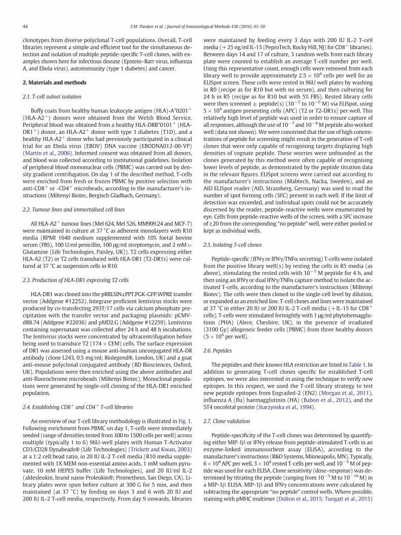
against two pools of HLA-A2-restricted peptides using IFNγ ELISpot.Peptides from Epstein–Barr virus (EBV) (BMFL1280–288 (Steven et al.,1997)) and flu (matrix protein (MP)58–66 (Bednarek et al., 1991))were used to test the feasibility of screening in this manner, as robustT-cell responses are elicited in HLA-A2+ people exposed to these virus-es. Background release of IFNγ without added peptide was observed insome wells (Fig. 2A, top), however this did not preclude the identifica-tion of peptide-reactive wells: 30wells of the 48 screened (17/48) werepositive for the EBV peptide, and 2/48 for the flu peptide (Fig. 2A, mid-dle). More interestingly, 1/96 of the wells screened with a pool of fourwell-characterised T1D-relevant peptides (preproinsulin (PPI)15–24(Skowera et al., 2008), insulin β chain (InsB)10–18 (Pinkse et al., 2005),glutamic acid decarboxylase (GAD65)114–123 (Panina-Bordignon et al.,1995), and islet-specific glucose-6-phosphatase catalytic subunit-related protein (IGRP)265–273 (Jarchum et al., 2008)) was peptide-reactive (Fig. 2A, bottom). One peptide-reactive well from each of thethree screens was enriched based on IFNγ production in response tostimulation with relevant peptide(s), prior to T-cell cloning. pMHCtetramer staining was used to confirm the specificities of the EBV andflu clones (Fig. 2B). We established that the T1D peptide-reactiveclone (GD.InsB.4)was specific for the InsB peptide, as shown via peptidedose–response (MIP-1β ELISA) and pMHC dextramer staining (Fig. 2C).
Table 1List of peptides used in this study. HLA restriction and full peptide sequence are listed in all but 3 cases. Sequences of the newHLA-A*0201-restricted Engrailed-2-derived epitopes, and thenew HLA-DRB*0101-restricted epitopes from influenza haemagglutinin and 5T4 oncofetal protein will be published in other studies we are currently preparing.
Origin ProteinAmino acidresidues
Peptidesequence
HLArestriction
Reference
Epstein–Barr virus BMLF1 lytic protein 280–288 GLCTLVAML A*0201 Steven et al. (1997)Influenza A Matrix protein (MP) 58–66 GILGFVFTL A*0201 Bednarek et al. (1991)Influenza A Haemagglutinin (HA) Putative DRB*0101 Babon et al. (2012)Zaire Ebola virus Nucleoprotein 150–158 FLSFASLFL A*0201 Sundar et al. (2007)Zaire Ebola virus Nucleoprotein (NP) 202–210 RLMRTNFLI A*0201 Sundar et al. (2007)Zaire Ebola virus Nucleoprotein 404–412 KLTEAITAA A*0201 Sundar et al. (2007)Type 1 diabetes Glutamic acid decarboxylase (GAD65) 114–123 VMNILLQYVV A*0201 Panina-Bordignon et al. (1995)Type 1 diabetes Insulin β chain (InsB) 10–18 HLVEALYLV A*0201 Pinkse et al. (2005)Type 1 diabetes Islet-specific glucose-6-phosphatase catalytic subunit-related protein (IGRP) 265–273 VLFGLGFAI A*0201 Jarchum et al. (2008)Type 1 diabetes Preproinsulin (PPI) 15–24 ALWGPDPAAA A*0201 Skowera et al. (2008)Tumour Cadherin-3/P-Cadherin (CDH3) 655–663 FILPVLGAV A*0201 Imai et al. (2008)Tumour Engrailed-2 (EN2) Putative A*0201 Morgan et al. (2011)Tumour Glycoprotein 100 (gp100) 280–288 YLEPGPVTA A*0201 Kawakami et al. (1995)Tumour Insulin-like growth factor 2 mRNA binding protein 3 (IMP-3) 199–207 RLLVPTQFV A*0201 Tomita et al. (2011)Tumour Melanoma-associated antigen-1 (MAGE-A1) 278–286 KVLEYVIKV A*0201 Pascolo et al. (2001)Tumour Melanoma-associated antigen-3 (MAGE-A3) 112–120 KVAELVHFL A*0201 Chinnasamy et al. (2011)
Melanoma-associated antigen 3 (MAGE-A3) 240–248 YLEYRQVPG A*0201 Graff-Dubois et al. (2002)Tumour NY-BR-1 904–912 SLSKILDTV A*0201 Wang et al. (2006)Tumour Oncofetal protein, 5CT4 Putative DRB*0101 Starzynska et al. (1994)Tumour Prostatic acid phosphatase-3 (PAP-3) 299–307 ALDVYNGLL A*0201 Harada et al. (2003)Tumour Prostein 31–39 CLAAGITYV A*0201 Kiessling et al. (2004)
46 S.M. Theaker et al. / Journal of Immunological Methods 430 (2016) 43–50
All the InsB clones that were grown stained with the same TCR variableβ chain antibody (data not shown), suggesting they were likely to bederived from the same precursor. Together, these data verify thatpeptide-specific T-cells of interest can be successfully isolated frompatient samples even when cells are in short supply.
3.2. Generation of tumour-specific T-cell clones from potentially rarepopulations using CD8+ T-cell libraries
In addition to the limitations associated with sample size, the pro-duction of T-cell clones is often made more difficult when the peptide-specific T-cells of interest occur at naturally low frequencies. This isoften the case with tumour-reactive T-cells (Sharpe and Mount, 2015)(recognising tumour-associated antigens) in PBMC, as a result of thymicselection reducing the presence of “self” reactive T-cells in the pe-riphery (Klein et al., 2014). Thus, with the aim of isolating raretumour-specific T-cells, a CD8+ T cell library (576 wells at 1000cells per well) was generated from the PBMC of a healthy HLA-A2+
donor, and screened via IFNγ ELISpot against a pool of five HLA-A2-restricted tumour peptides (melanoma-associated antigen-3(MAGE-A3)112–120 (Chinnasamy et al., 2011), MAGE-A3240–248
(Graff-Dubois et al., 2002), cadherin-3/P-cadherin (CDH3)655–663(Imai et al., 2008), NY-BR-1904–912 (Wang et al., 2006) and glycopro-tein 100 (gp100)280–288 (Kawakami et al., 1995)). Fig. 3A shows thepositive library wells (10/576) from this library screen, which weresubsequently pooled, and specificity for the gp100 peptide deter-mined (by IFNγ ELISpot) prior to enrichment and cloning (Fig. 3B).From this library, a gp100-specific clone (THEAK.gp100) was pro-duced, and its reactivity confirmed via a peptide dose–response ex-periment using MIP-1β ELISA (Fig. 3C). THEAK.gp100 was able tokill multiple HLA-A2+ melanoma cell lines (Mel 624, Mel 526 andMM909.24) in a 51Cr-release assay after 18 h, at a T-cell:tumourcell ratio of 10:1 (Fig. 3D).
Next, a second CD8+ T-cell library (288 wells at 500 cells per well)was produced from a different healthy HLA-A2+ donor, but this timescreened against two separate pools of HLA-A2-restricted tumour pep-tides, (Pool 1: prostatic acid phosphatase-3 (PAP-3)299–307 (Haradaet al., 2003), melanoma-associated antigen-1 (MAGE-A1)278–286(Pascolo et al., 2001), MAGE-A3112–120, prostein31–39 (Kiessling et al.,2004), insulin-like growth factor 2 mRNA binding protein 3 (IMP-
3199–207) (Tomita et al., 2011), and CDH3655–663; Pool 2: six putativepeptides from EN2 (EN2-1, -2, -3, -4, -5, and -6)). ELISpot data for thepeptide-reactive wells (1/288 for pool 1, and 13/288 for pool 2) isshown in Fig. 3E. T-cells were then cloned from the positive wells, andscreened against individual peptides by IFNγ ELISA (Fig. 3F). Two cloneswere produced; one recognising the CDH3 peptide (GD.FIL.6/30), andthe other recognising a putative EN2-3 peptide (GD.RPA.2/30), as con-firmed by a peptide dose–response (MIP-1β ELISA) (Fig. 3G). TheCDH3-specific clone was shown to specifically kill an HLA-A2+ breastcancer cell line (MCF-7), and not an HLA-A2+ metastatic melanomacell line (MM909.24; obtained from the Center for Cancer ImmuneTherapy, Herlev Hospital, Copenhagen, Denmark) in a 51Cr-releaseassay after 4 h (Fig. 3H). Collectively, these data show that obtainingtumour-reactive T-cell clones using this method is not hindered by pre-dicted low clonotype frequencies.
3.3. Generation of T-cell clones from CD4+ T-cell libraries
To further illustrate the versatility of the T-cell library method, aCD4+ T-cell library (192 wells at 1000 cells per well), was generatedfrom a healthy HLA-DR1+ donor, and simultaneously screened viaIFNγ ELISpot for reactivity against two HLA-DR1-restricted peptidepools. The first peptide pool contained three putative peptides fromHA of flu (Flu-1, -2, and -3), and the second peptide pool containedfive putative peptides from 5T4 oncofetal protein (5T4-2, -12, -20, -38,and -PMS). Positive wells from the screen (3/48 for the flu pool, and9/144 for the 5T4 pool: shown in Fig. 4A) were enriched based onIFNγ production in response to peptide(s), and then expanded oncewith PHA and irradiated allogeneic feeder cells to produce lines. Thelines were subsequently screened against individual peptides in anIFNγ ELISpot (Fig. 4B), and then cloned to the single-cell level. Fromthis, three 5T4-clones (GD.C112.DC, GD.D821.DC and GD.D104.DC)were generated and tested against decreasing doses of peptide viaMIP-1β ELISA, in order to establish their sensitivity to the correspondingepitope (Fig. 4C). Thus, these data indicate that our T-cell library strate-gy can also successfully produce CD4+ T-cells with desired specificities.It is noteworthy that autologous EBV immortalised B-cell lines were ini-tially used for the screening of CD4+ libraries, but these induced highnumbers of positive wells (data not shown), presumably because of T-cells with reactivity against EBV.
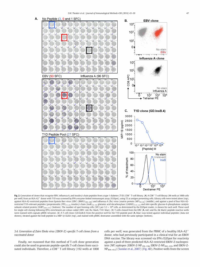
Fig. 2.Generation of clones that recognise EBV, influenza A, and insulin β chain peptides from a type 1 diabetes (T1D) CD8+ T-cell library. (A) A CD8+ T-cell library (96 wells at 1000 cellsper well) from an HLA-A2+ donor with T1Dwas screened by IFNγ enzyme-linked immunospot assay (ELISpot), using T2 as antigen presenting cells. Library cells were tested alone (top),against HLA-A2-restricted peptides from Epstein Barr virus (EBV) (BMFL1280–288) and influenza A (flu) virus (matrix protein (MP)58–66) (middle), and against a pool of four HLA-A2-restricted T1D-relevant peptides (preproinsulin (PPI)15–24, insulin β chain (InsB)10–18, glutamic acid decarboxylase (GAD65)114–123 and islet-specific glucose-6-phosphatase catalyticsubunit-related protein (IGRP)265–273) (bottom). The number of spot forming cells (SFC) per 3.3 × 10⁴ cells, as determined by the ELISpot reader, is shown for each well. Those usedfor single-cell cloning following IFNγ enrichment are colour coded (EBV: red, flu: black, T1D: blue). (B) T-cells cloned from the EBV (A, red) and flu (A, black) peptide-reactive wellswere stained with cognate pMHC tetramer. (C) A T-cell clone (GD.InsB.4) from the positive well for the T1D peptide pool (A, blue) was tested against individual peptides (data notshown), titrated against the InsB peptide in a MIP-1β ELISA (top), and stained with pMHC dextramer assembled with the same epitope (bottom).
47S.M. Theaker et al. / Journal of Immunological Methods 430 (2016) 43–50
3.4. Generation of Zaire Ebola virus (EBOV-Z)-specific T-cell clones from avaccinated donor
Finally, we reasoned that this method of T-cell clone generationcould also be used to generate peptide-specific T-cell clones from vacci-nated individuals. Therefore, a CD8+ T cell library (192 wells at 1000
cells per well) was generated from the PBMC of a healthy HLA-A2+
donor, who had previously participated in a clinical trial for an EBOVDNA vaccine. The library was screened via IFNγ ELISpot for reactivityagainst a pool of three predicted HLA-A2-restricted EBOV-Z nucleopro-tein (NP) epitopes (EBOV-Z-NP150–158, EBOV-Z-NP202–210, and EBOV-Z-NP404–412) (Sundar et al., 2007) (Fig. 4D). Positive wells from the screen
Fig. 3. CD8+ T-cell clone generation from T-cell libraries screened with tumour peptides. A CD8+ T-cell library (576wells at 1000 cells per well) from an HLA-A2+ donor was screened±an HLA-A2-restricted tumour peptide pool by IFNγ enzyme-linked immunospot assay (ELISpot), using T2 as antigen presenting cells. (A) Spot forming cells (SFC) per 2.5 × 10⁴ cells isshown for the 10 peptide-reactive wells. (B) Positive wells were then pooled, and tested for individual peptide-specificity by IFNγ ELISpot. SFC per 2.5 × 10⁴ cells for each well isshown (SFC for duplicate wells has been shown in brackets). The pooled cells were then enriched for reactive T-cells based on IFNγ production, and subjected to single-cell cloning. (C& D) One of the clones (THEAK.gp100) was specific for the gp100-derived peptide by MIP-1β ELISA, and also successfully killed multiple HLA-A2+ melanoma cell lines (Mel 624, Mel526, and MM909.24) in a 51Cr-release assay after 18 h, at a T-cell:tumour cell ratio of 10:1. A library from a second HLA-A2+ donor (288 wells at 500 cells per well) was screened as in(A), but with two pools of HLA-A2-restricted tumour peptides. (E) SFC per 5 × 10⁴ cells for the 14 peptide-reactive wells. (F) Cloned T-cells were screened against individual peptidesby IFNγ ELISA, and were found to recognise a peptide from either cadherin-3 (CDH3) or Engrailed-2 (EN2). (G) Both the CDH3-specific clone (GD.FIL.6/30) and EN2-3-specific clone(GD.RPA.6/2) were tested for sensitivity to cognate peptide by MIP-1β ELISA. (H) The GD.FIL.6/30 clone was also tested for cytotoxicity towards an HLA-A2+ breast cancer cell line(MCF-7), and an HLA-A2+ metastatic melanoma cell line (MM909.24) in a 51Cr-release assay after 4 h.
48 S.M. Theaker et al. / Journal of Immunological Methods 430 (2016) 43–50
(2/192) were pooled, subjected to IFNγ/TNFα dual enrichment, andthen cloned to the single-cell level. Six EBOV-Z-specific clones weregenerated, all reactive to the EBOV-Z-NP150–158 peptide, as determinedby MIP-1β ELISA (Fig. 4E). Peptide dose–response curves (MIP-1βELISA) for three of the clones (ST3.ebola.FLS, ST13.ebola.FLS, andST17.ebola.FLS) are shown as an example (Fig. 4F). These data demon-strate the ability of this T-cell library method to rapidly produce viral-specific T-cell clones from the blood of a vaccinated donor.
4. Discussion
Modern advances in cell sorting, using fluorescence or magneticbeads, have allowed the generation of T-cell clones following physical
isolation with pMHC multimers, or functional detection using antibod-ies specific for cellular activation markers. Although these techniqueshave worked well in our laboratory for some antigens, we have failedto grow robust clones using these standard methodologies more oftenthan we have succeeded. In order to circumvent this difficulty, we de-veloped the T-cell library strategy described here. Previous studieshave applied a T-cell library approach to study T-cell frequencies, but in-stead used PHA in combination with irradiated allogeneic feeder cellsfor T-cell expansion (Campion et al., 2014; Geiger et al., 2009). TheCD3/CD28 beads used in our strategy have been shown to better pre-serve the TCR repertoire during in vitro expansion (Neller et al., 2012).Nevertheless, while this methodology maintains the general TRBVfamilies and dominant antigen-specific T-cell responses faithfully, it

Fig. 4. Isolation of peptide-specific CD4+ T-cells, and Zaire Ebola virus (EBOV-Z) specific CD8+ T-cells from T-cell libraries. T-cell libraries (192wells per library at 1000 cells perwell)wereestablished from a healthy HLA-DR1+ donor (A–C), and a healthyHLA-A2+ donor who had previously participated in an EBOVDNA vaccine trial (D–F). (A) The healthyHLA-DR1+ librarywas screened by IFNγ enzyme-linked immunospot assay (ELISpot) against twopools of peptides, using T2-DR1s as antigen presenting cells (APC). Spot forming cells (SFC) per 5× 10⁴ cellsare shown for the peptide-reactive wells. 3 wells were positive for the influenza A (flu) pool (three putative peptides from haemagglutinin: Flu-1, -2 and -3), and 9wells were positive forthe 5T4 oncofetal protein pool (five putative peptides: 5T4-2, -12, -20, -38 and -PMS). 3 of the positive library wells (C11, D8 and D10), shown to respond to different 5T4 peptides, havebeen colour coded to illustrate their progression to validated 5T4-specific clones (B). Peptide dose–responses for the T-cell clones grown from these wells (GD.C112.DC, GD.D821.DC andGD.D104.DC)have been illustrated in (C). (D) A second library established fromahealthyHLA-A2+EBOV vaccinated individualwas screened by IFNγ ELISpot, using T2 cells asAPC. SFC per3 × 104 cells has been shown for the peptide-reactivewells. 2wells showed a positive response to the pool of three HLA-A2-restricted epitopes (EBOV-Z-NP150–158, EBOV-Z-NP202–210, andEBOV-Z-NP404–412) from EBOV-Z nucleoprotein (NP). These wells were pooled, subjected to IFNγ/TNFα dual enrichment, and then cloned to the single-cell level. (E) All six clonesgenerated a response to EBOV-Z-NP150–158 peptide. Dose–response curves (MIP-1β ELISA) for three of these clones (ST3.ebola.FLS, ST13.ebola.FLS and ST17.ebola.FLS) are depicted in (F).
49S.M. Theaker et al. / Journal of Immunological Methods 430 (2016) 43–50
remains possible that extremely rare clones are lost during this expan-sion phase.
Using the methodology we describe here, we have been able to si-multaneously generatemanyhundreds of peptide-specific T-cell clones,with at least one being grown from each library. T-cell libraries have be-come themethod of choice for generatingmonoclonal T-cells in our lab-oratory, as they avoid the need for pMHC multimers, ample donormaterial, or time-consuming DC production. Furthermore, we considerit an advantage to have the T-cells already adapted to in vitro cultureprior to screening, and also to avoid repeated exposure to antigenic pep-tide, which can often lead to T-cell exhaustion (Wherry and Kurachi,2015). Importantly, we have found T-cell clones to be extremely advan-tageous for improving pMHCmultimer staining protocols (Dolton et al.,2015; Tungatt et al., 2015), T-cell epitope identification, defining T-cellcross-reactivity (Wooldridge et al., 2012), obtaining monoclonal TCRs(for genetic, biophysical and structural studies), and peptide vaccine de-velopment (Ekeruche-Makinde et al., 2012).
In summary, we have developed an efficient and reproduciblelibrary-based strategy for the successful detection and isolation ofpeptide-specific human T-cell clones from polyclonal CD8+ or CD4+
T-cell populations. By introducing a degree of clonality at the start ofculture, and by coupling this with effective cytokine-mediated en-richment strategies, our methodology permits the relatively rapid
generation of fully validated clones in as little as 6 weeks. Overall, T-cell libraries provide a useful tool for the T-cell immunologist, asthey can be used for the simple parallel generation of multiple T-cell clones with numerous research applications in infectious dis-ease, autoimmunity and cancer.
Acknowledgements
SMT is a Breast Cancer Now funded PhD student. CR is funded by aCancer Research Wales (CRW) studentship. AGW is a Life Sciences Re-search Network Wales (LSRNW) funded PhD student. AL is funded byan MRC studentship. JJM is a National Health and Medical Research(NHMRC) Career Development Fellow. DKC is a Wellcome Trust CareerDevelopment Fellow. MP receives support from the National Institutefor Health Research Biomedical Research Centre, based at Guy's and StThomas' NHS Foundation Trust and King's College London. AKS is aWellcome Trust Senior Investigator.
References
Attaf, M., Huseby, E., Sewell, A.K., 2015a. αβ T cell receptors as predictors of health anddisease. Cell Mol. Immunol. 12, 391–399.
Attaf, M., Legut, M., Cole, D.K., Sewell, A.K., 2015b. The T cell antigen receptor: the Swissarmy knife of the immune system. Clin. Exp. Immunol. 181, 1–18.
50 S.M. Theaker et al. / Journal of Immunological Methods 430 (2016) 43–50
Babon, J.A.B., Cruz, J., Ennis, F.A., Yin, L., Terajima, M., 2012. A human CD4+ T cell epitopein the influenza hemagglutinin is cross-reactive to influenza A virus subtypes and toinfluenza B virus. J. Virol. 86, 9233–9243.
Bednarek, M.A., Sauma, S.Y., Gammon, C.C., Porter, G., Tamhankar, S., Williamson, A.R.,Zweerink, H.J., 1991. The minimum peptide epitope from the influenza virus matrixprotein. Extra and intracellular loading of HLA-A2. J. Immunol. 147, 4047–4053.
Campion, S.L., Srodie, T.M., Fischer, W., Korber, B.T., Rossetti, A., Goonetilleke, N.,Mcmichael, A.J., Sallusto, F., 2014. Proteome-wide analysis of HIV-specific naive andmemory CD4(+) T cells in unexposed blood donors. J. Exp. Med. 211, 1273–1280.
Chinnasamy, N., Wargo, J.A., Yu, Z., Rao, M., Frankel, T.L., Riley, J.P., Hong, J.J., Parkhurst,M.R., Feldman, S.A., Schrump, D.S., 2011. A TCR targeting the HLA-A*0201–restrictedepitope of MAGE-A3 recognizes multiple epitopes of the MAGE-A antigen superfam-ily in several types of cancer. J. Immunol. 186, 685–696.
Dolton, G., Tungatt, K., Lloyd, A., Bianchi, V., Theaker, S.M., Trimby, A., Holland, C.J., Donia,M., Godkin, A.J., Cole, D.K., Thor Straten, P., Peakman, M., Svane, I.M., Sewell, A.K.,2015. More tricks with tetramers: a practical guide to staining T cells with peptide–MHC multimers. Immunology 146, 11–22.
Ekeruche-Makinde, J., Clement, M., Cole, D.K., Edwards, E.S., Ladell, K., Miles, J.J.,Matthews, K.K., Fuller, A., Lloyd, K.A., Madura, F., 2012. T-cell receptor-optimized pep-tide skewing of the T-cell repertoire can enhance antigen targeting. J. Biol. Chem. 287,37269–37281.
Geiger, R., Duhen, T., Lanzavecchia, A., Sallusto, F., 2009. Human naive andmemory CD4+T cell repertoires specific for naturally processed antigens analyzed using libraries ofamplified T cells. J. Exp. Med. 206, 1525–1534.
Gomez-Tourino, I., Arif, S., Eichmann, M., Peakman, M., 2015. T cells in type 1 diabetes: in-structors, regulators and effectors: a comprehensive review. J. Autoimmun.
Graff-Dubois, S., Faure, O., Gross, D.-A., Alves, P., Scardino, A., Chouaib, S., Lemonnier, F.A.,Kosmatopoulos, K., 2002. Generation of CTL recognizing an HLA-A*0201-restrictedepitope shared byMAGE-A1,-A2,-A3,-A4,-A6,-A10, and-A12 tumor antigens: implica-tion in a broad-spectrum tumor immunotherapy. J. Immunol. 169, 575–580.
Harada, M., Noguchi, M., Itoh, K., 2003. Target molecules in specific immunotherapyagainst prostate cancer. Int. J. Clin. Oncol. 8, 193–199.
Imai, K., Hirata, S., Irie, A., Senju, S., Ikuta, Y., Yokomine, K., Harao, M., Inoue, M., Tsunoda,T., Nakatsuru, S., Nakagawa, H., Nakamura, Y., Baba, H., Nishimura, Y., 2008. Identifi-cation of a novel tumor-associated antigen, cadherin 3/P-cadherin, as a possible tar-get for immunotherapy of pancreatic, gastric, and colorectal cancers. Clin. Cancer Res.14, 6487–6495.
Jarchum, I., Nichol, L., Trucco, M., Santamaria, P., Dilorenzo, T.P., 2008. Identification ofnovel IGRP epitopes targeted in type 1 diabetes patients. Clin. Immunol. 127,359–365.
Kawakami, Y., Eliyahu, S., Jennings, C., Sakaguchi, K., Kang, X., Southwood, S., Robbins, P.F.,Sette, A., Appella, E., Rosenberg, S.A., 1995. Recognition of multiple epitopes in thehuman melanoma antigen gp100 by tumor-infiltrating T lymphocytes associatedwith in vivo tumor regression. J. Immunol. 154, 3961–3968.
Kiessling, A., Stevanovic, S., Fussel, S., Weigle, B., Rieger, M.A., Temme, A., Rieber, E.P.,Schmitz, M., 2004. Identification of an HLA-A*0201-restricted T-cell epitope derivedfrom the prostate cancer-associated protein prostein. Br. J. Cancer 90, 1034–1040.
Klein, L., Kyewski, B., Allen, P.M., Hogquist, K.A., 2014. Positive and negative selection ofthe T cell repertoire: what thymocytes see (and don't see). Nat. Rev. Immunol. 14,377–391.
Lin, K., Chen, S., Chen, G., 2015. Role of memory T Cells and perspectives for interventionin organ transplantation. Front. Immunol. 6, 473.
Martin, J.E., Sullivan, N.J., Enama, M.E., Gordon, I.J., Roederer, M., Koup, R.A., Bailer, R.T.,Chakrabarti, B.K., Bailey, M.A., Gomez, P.L., Andrews, C.A., Moodie, Z., Gu, L., Stein,J.A., Nabel, G.J., Graham, B.S., 2006. A DNA vaccine for Ebola virus is safe and immu-nogenic in a phase I clinical trial. Clin. Vaccine Immunol. 13, 1267–1277.
Morgan, R., Boxall, A., Bhatt, A., Bailey, M., Hindley, R., Langley, S., Whitaker, H.C., Neal,D.E., Ismail, M., Whitaker, H., Annels, N., Michael, A., Pandha, H., 2011. Engrailed-2(EN2): a tumor specific urinary biomarker for the early diagnosis of prostate cancer.Clin. Cancer Res. 17, 1090–1098.
Neller, M.A., Sewell, A.K., Burrows, S.R., Miles, J.J., 2012. Tracking the repertoire of humanadult and neonatal T cells during ex vivo amplification. Br. J. Haematol. 159, 370–373.
Panina-Bordignon, P., Lang, R., Van Endert, P.M., Benazzi, E., Felix, A.M., Pastore, R.M.,Spinas, G.A., Sinigaglia, F., 1995. Cytotoxic T cells specific for glutamic acid decarbox-ylase in autoimmune diabetes. J. Exp. Med. 181, 1923–1927.
Pascolo, S., Schirle, M., Guckel, B., Dumrese, T., Stumm, S., Kayser, S., Moris, A., Wallwiener,D., Rammensee, H.G., Stevanovic, S., 2001. A MAGE-A1 HLA-A A*0201 epitope identi-fied by mass spectrometry. Cancer Res. 61, 4072–4077.
Pinkse, G.G., Tysma, O.H., Bergen, C.A., Kester, M.G., Ossendorp, F., Van Veelen, P.A.,Keymeulen, B., Pipeleers, D., Drijfhout, J.W., Roep, B.O., 2005. Autoreactive CD8 Tcells associated with beta cell destruction in type 1 diabetes. Proc. Natl. Acad. Sci. U.S. A. 102, 18425–18430.
Salou, M., Nicol, B., Garcia, A., Laplaud, D.A., 2015. Involvement of CD8(+) T Cells in mul-tiple sclerosis. Front. Immunol. 6, 604.
Sewell, A.K., 2012. Why must T cells be cross-reactive? Nat. Rev. Immunol. 12, 669–677.Sharpe, M., Mount, N., 2015. Genetically modified T cells in cancer therapy: opportunities
and challenges. Dis. Model. Mech. 8, 337–350.Skowera, A., Ellis, R.J., Varela, C., Xf, O.R., Arif, S., Huang, G.C., Van-Krinks, C., Zaremba, A.,
Rackham, C., Allen, J.S., Tree, T.I.M., Zhao, M., Dayan, C.M., Sewell, A.K., Unger, W.,Drijfhout, J.W., Ossendorp, F., Roep, B.O., Peakman, M., 2008. CTLs are targeted tokill β cells in patients with type 1 diabetes through recognition of a glucose-regulated preproinsulin epitope. J. Clin. Invest. 118, 3390–3402.
Starzynska, T., Marsh, P.J., Schofield, P.F., Roberts, S.A., Myers, K.A., Stern, P.L., 1994. Prog-nostic significance of 5T4 oncofetal antigen expression in colorectal carcinoma. Br.J. Cancer 69, 899–902.
Steven, N.M., Annels, N.E., Kumar, A., Leese, A.M., Kurilla, M.G., Rickinson, A.B., 1997. Im-mediate early and early lytic cycle proteins are frequent targets of the Epstein–Barrvirus–induced cytotoxic T cell response. J. Exp. Med. 185, 1605–1618.
Sundar, K., Boesen, A., Coico, R., 2007. Computational prediction and identification of HLA-A2.1-specific Ebola virus CTL epitopes. Virology 360, 257–263.
Tomita, Y., Harao, M., Senju, S., Imai, K., Hirata, S., Irie, A., Inoue, M., Hayashida, Y.,Yoshimoto, K., Shiraishi, K., Mori, T., Nomori, H., Kohrogi, H., Nishimura, Y., 2011. Pep-tides derived from human insulin-like growth factor-II mRNA binding protein 3 caninduce human leukocyte antigen-A2-restricted cytotoxic T lymphocytes reactive tocancer cells. Cancer Sci. 102, 71–78.
Trickett, A., Kwan, Y.L., 2003. T cell stimulation and expansion using anti-CD3/CD28beads. J. Immunol. Methods 275, 251–255.
Tungatt, K., Bianchi, V., Crowther, M.D., Powell, W.E., Schauenburg, A.J., Trimby, A., Donia,M., Miles, J.J., Holland, C.J., Cole, D.K., Godkin, A.J., Peakman, M., Straten, P.T., Svane,I.M., Sewell, A.K., Dolton, G., 2015. Antibody stabilization of peptide–MHC multimersreveals functional T cells bearing extremely low-affinity TCRs. J. Immunol. 194,463–474.
Wang, W., Epler, J., Salazar, L.G., Riddell, S.R., 2006. Recognition of breast cancer cells byCD8+ cytotoxic T-cell clones specific for NY-BR-1. Cancer Res. 66, 6826–6833.
Wherry, E.J., Kurachi, M., 2015. Molecular and cellular insights into T cell exhaustion. Nat.Rev. Immunol. 15, 486–499.
Wooldridge, L., Ekeruche-Makinde, J., Van Den Berg, H.A., Skowera, A., Miles, J.J., Tan, M.P.,Dolton, G., Clement, M., Llewellyn-Lacey, S., Price, D.A., Peakman, M., Sewell, A.K.,2012. A single autoimmune T cell receptor recognizes more than a million differentpeptides. J. Biol. Chem. 287, 1168–1177.