journal of proteomics & bioinformatics open access · to luria-bertani (lb) medium, mgm...
TRANSCRIPT

Journal of Proteomics amp Bioinformatics - Open Access
JPBVol2September 2009
J Proteomics Bioinform Volume 2(9) 388-397 (2009) - 388
ISSN0974-276X JPB an open access journal
Proteome of Salmonella Enterica Serotype
Typhimurium Grown in a Low Mg2+pH Medium
Liang Shi1 Charles Ansong1 Heather Smallwood1sect Leah Rommereim1sectJason E McDermott1 Heather M Brewer1 Angela D Norbeck1 Ronald C Taylor1
Jean K Gustin2 Fred Heffron2 Richard D Smith1 and Joshua N Adkins1
1Pacific Northwest National Laboratory Richland Washington 993522Oregon Health and Science University Portland Oregon 97239
sectPresent addresses St Jude Childrenrsquos Research Hospital Memphis Tennessee
38105 (HS) and Dartmouth College Hanover New Hampshire 03755 (LR)
Abstract
To determine the impact of a low Mg2+pH defined
growth medium (MgM) on the proteome of Salmonella
enterica serotype Typhimurium we cultured S
Typhimurium cells in the medium under two different
conditions termed MgM Shock and MgM Dilution and
then comparatively analyzed the bacterial cells harvested
from these conditions by a global proteomic approach
Proteomic results showed that MgM Shock and MgM
Dilution differentially affected the S Typhimurium
proteome MgM Shock induced a group of proteins
whose induction usually occurred at low O2 level while
MgM Dilution induced those related to the type III se-
cretion system (T3SS) of Salmonella Pathogenicity Is-
land 2 (SPI2) and those involved in thiamine or biotin
biosynthesis The metabolic state of the S Typhimurium
cells grown under MgM Shock condition also differed
significantly from that under MgM Dilution condition
Western blot analysis not only confirmed the proteomic
results but also showed that the abundances of SPI2-
T3SS proteins SsaQ and SseE and biotin biosynthesis
proteins BioB and BioD increased after S Typhimurium
infection of RAW 2647 macrophages Deletion of the
gene encoding BioB reduced the bacterial ability to rep-
licate inside the macrophages suggesting a biotin-lim-
ited environment encountered by S Typhimurium within
RAW 2647 macrophages
Keywords Salmonella enterica serotype Typhimurium Low
Mg2+pH defined growth medium Salmonella-containing vacu-
ole Salmonella Pathogenicity Island 2 Type III secretion sys-
tem Biotin biosynthesis
Abbreviations CFU colony-forming units DMEM
Dulbeccorsquos modified Eaglersquos medium DPBS Dulbeccorsquos phos-
phate-buffered-saline without Mg2+ or Ca2+ DTT dithiothreitol
HBSS Hanksrsquo buffered saline solution hpi hour post-infec-
tion LC-MSMS liquid chromatography-tandem mass spec-
trometry MgM low Mg2+pH defined growth medium pdu
12-propanediol utilization SCV Salmonella-containing vacu-
ole SPI2 Salmonella Pathogenicity Island 2 T3SS type III
secretion system WT wild-type
Introduction
To establish systemic infection in susceptible mice the facul-
tative intracellular pathogen Salmonella enterica serotype
Typhimurium must survive and replicate inside host macroph-
ages (Fields et al 1986) Once it is taken up by macrophages
S Typhimurium resides in a membrane-bound structure called
the Salmonella-containing vacuole (SCV) Many bacterial pro-
teins such as those related to the type III secretion system (T3SS)
of Salmonella Pathogenicity Island 2 (SPI2) are involved in
helping S Typhimurium replicate inside the SCV (Hensel 2000)
Identification and characterization of the proteins involved in
S Typhimurium intramacrophage survival have contributed sig-
nificantly to the elucidation of the molecular mechanisms un-
derlying the ability of S Typhimurium to evade the host mac-
rophage defense mechanisms and adapt to its local environment
By using a bottom-up global proteomic approach we previ-
ously analyzed the proteome of S Typhimurium strain 14028
after the bacterial cells were isolated from murine macrophages
RAW 2647 A total of 315 S Typhimurium proteins were iden-
tified While most identified proteins were housekeeping-related
and their abundances remained relatively constant during the
time course of infection the abundances of 39 S Typhimurium
proteins increased significantly after the infection which in-
cluded STM3117-3119 Western blot analysis confirmed the
increased abundances of STM3117-3119 proteins Deletion of
the gene encoding for STM3117 resulted in the failure of S
Typhimurium to replicate inside RAW 2647 macrophages (Shi
et al 2006) Although the exact functions of STM3117-3119
Corresponding authors Liang Shi Microbiology Group Pacific North-
west National Laboratory 902 Battelle Blvd MSIN P7-50 Richland
WA 99352 Tel (509) 371-6967 Fax (509) 372-1632 E-Mail
liangshipnlgov
Joshua N Adkins Biological Separation and Mass Spectrometry Group
Pacific Northwest National Laboratory 902 Battelle Blvd MSIN K8-98
Richland WA 99352 Tel (509) 371-6583 Fax (509) 371-6546 E-Mail
JoshuaAdkinspnlgov
Received August 07 2009 Accepted September 25 2009 Published
September 25 2009
Citation Shi L Ansong C Smallwood H Rommereim L McDermott JE
et al (2009) Proteome of Salmonella Enterica Serotype Typhimurium
Grown in a Low Mg2+pH Medium J Proteomics Bioinform 2 388-397
doi104172jpb1000099
Copyright copy 2009 Shi L et al This is an open-access article distrib-
uted under the terms of the Creative Commons Attribution License which
permits unrestricted use distribution and reproduction in any medium
provided the original author and source are credited
Research Article OPEN ACCESS Freely available online doi104172jpb1000099
Journal of Proteomics amp Bioinformatics - Open Access
JPBVol2September 2009
J Proteomics Bioinform Volume 2(9) 388-397 (2009) - 389
ISSN0974-276X JPB an open access journal
proteins have yet to be determined these results prove that a
global discovery-based proteomic approach is a powerful tool
to identify and characterize S Typhimurium proteins involved
in intramacrophage survival
Despite our successful application of the proteomic method
in discovering a novel S Typhimurium protein involved in
intramacrophage survival the 315 identified S Typhimurium
proteins were lt7 of the ~4800 S Typhimurium proteins an-
notated at that time Furthermore only one SPI2-T3SS-related
protein was identified This low coverage of S Typhimurium
protein identification is most likely attributed to the limited bac-
terial cells isolated from the macrophages and the interfering
background of residual host proteins which render the abun-
dances of most S Typhimurium proteins below the detection
level of the proteomic method used To circumvent the limita-
tions associated with low numbers of bacterial cells isolated from
the host cells one alternative approach was to analyze the S
Typhimurium proteome after bacterial cells were cultured un-
der the conditions that mimicked certain conditions found in-
side the SCV such as in a low Mg2+pH defined growth medium
(MgM) (Beuzon et al 1999 Eriksson et al 2003) Compared
to Luria-Bertani (LB) medium MgM selectively induced a group
of S Typhimurium and S enterica serotype Typhi proteins Some
of these MgM-induced proteins such as PduB and SrfH of S
Typhimurium (Adkins et al 2006) and T1108 T1476 and HlyE
of S Typhi (Ansong et al 2008) also increased in their abun-
dances after Salmonella infection of macrophages confirming
some similarity among Salmonella cells colonizing macroph-
ages
To further characterize the impact of MgM on the S
Typhimurium proteome we cultured S Typhimurium cells in
the medium under two contrast conditions (ie MgM Shock
the conditions used previously and MgM Dilution) and then
performed a comparative analysis of the bacterial cells grown
under these two different conditions by using a liquid chroma-
tography-tandem mass spectrometry (LC-MSMS)-based
proteomic approach
Materials and Methods
Reagents and Standard Procedures
All cell culture reagents were purchased from Invitrogen
(Carlsbad CA) Bacterial DnaK antibody was purchased from
StressGen (Victoria BC Canada) OctA-Probe was obtained
from Santa Cruz Biotechnology (Santa Cruz CA) All chemi-
cals used for tryptic digestion and biotin were purchased from
Sigma (St Louis MO) Protein concentrations were measured
with a bicinchoninic acid (BCA) protein assay kit from Pierce
(Rockford IL) SDS-PAGE and Western blot analyses were
conducted according to the instructions from Invitrogen
Bacterial Strains and Culture Conditions
Bacterial strains and plasmids used in this study are listed in
Table S1 All S Typhimurium strains were normally grown in
LB medium Kanamycin and ampicillin were used at 50 microgml
The MgM was described previously (Beuzon et al 1999) Two
different culture conditions were used with MgM during this
study First after bacterial cells were grown in LB at 37 oC with
agitation (200 rpm) until their OD600
were ~ 2 [ie stationary
(Stat) phase] they were washed with MgM once resuspended
with the same volume of MgM and were then grown at 37 oC
with agitation for 4 hr (ie MgM Shock) (Adkins et al 2006
Ansong et al 2008 Manes et al 2007) Second the overnight
cultures were diluted at 1 200 in LB and were then grown until
their OD600
were 05-07 [ie logarithmic (Log) phase] The
cells were diluted in MgM at 1100 and were then grown at 37oC to stationary phase and diluted 1100 into fresh MgM me-
dium They were incubated with agitation for 4 hr and har-
vested (ie MgM Dilution) The cells were harvested by cen-
trifugation (6000 times g 15 min) at 4 oC washed once with ice-
cold 100 mM NH4HCO
3 (pH 78) and were used for tryptic di-
gestion and Western blot analysis
Lysis of S Typhimurium Cells and Tryptic Digestion
To attain maximal proteome coverage S Typhimurium cells
from each growth condition (ie LB Log LB Stat MgM Shock
and MgM Dilution) were subjected to two different sample prepa-
ration methods applicable to peptide-level bottom-up proteomics
soluble and insoluble protein preparations (Adkins et al 2006
Ansong et al 2008) The combined data sets from these two
sample preparation methods were then used for the proteomic
analysis in this study Briefly S Typhimurium cells were lysed
by bead beating using 01-mm zirconiasilica beads in 2-mL
cryovial for a total of 3 min with cooling steps The supernatant
was recovered transferred to polycarbonate ultracentrifuge tubes
and centrifuged at 356000 times g and 4 degC for 10 min The pellet
was the insoluble fraction and the supernatant was the soluble
fraction The supernatant was transferred to a separate tube for
soluble protein analysis After protein concentrations were de-
termined urea thiourea and dithiothreitol (DTT) were added to
the soluble fraction at final concentrations of 7 M 2 M and 5
mM respectively and incubated at 60 degC for 30 min The samples
were diluted 10-fold with 100 mM NH4HCO
3 (pH 78) in the
presence of 1 mM CaCl2 and then subjected to tryptic digestion
(Promega Madison WI) at 150 (ww) trypsin-to-protein ratio
and 37 degC for 3 hr
For insoluble protein analysis the pellets were resuspended
in 50 mM NH4HCO
3 (pH 78) and ultracentrifuged under the
same conditions as described previously A BCA protein assay
was performed with the pellets that were resuspended in water
Following the ultracentrifugation the pellets were resuspended
in ~200 microL of a solubilization buffer (7 M urea 2 M thiourea
97 mM DTT and 1 CHAPS in 50 mM NH4HCO
3 pH 78)
Samples were incubated at 60 degC for 30 min and were then
diluted and digested in the same manner as that described for
the soluble protein preparation
The resulting digested peptides from the soluble protein prepa-
ration were desalted using a C-18 solid phase extraction (SPE)
column (SUPELCO Bellefonte PA) (Adkins et al 2002
Ansong et al 2008 Manes et al 2007) Because CHAPS binds
to C-18 and elutes with peptides a strong cation-exchange SPE
column was used to desalt the peptides from insoluble protein
preparation (Adkins et al 2006 Ansong et al 2008) The re-
sulting peptides were concentrated with a SpeedVac to a final
volume of ~100 microL A BCA protein assay was performed to
determine peptide concentrations prior to capillary LC-MSMS
Journal of Proteomics amp Bioinformatics - Open Access
JPBVol2September 2009
J Proteomics Bioinform Volume 2(9) 388-397 (2009) - 390
ISSN0974-276X JPB an open access journal
analysis
Capillary LC-MSMS Analysis
The desalted peptides were separated using an automated and
reverse-phase capillary LC system designed in-house (Livesay
et al 2008) Eluate from the LC was directly electrosprayed
into an LTQ-Orbitrap mass spectrometer (Thermo Fisher San
Jose CA) using an electrospray ionization interface manufac-
tured in-house The heated capillary temperature and spray volt-
age were 200 ordmC and 22 kV respectively Data were acquired
for 100 min beginning 65 min after sample injection (ie 15
min into gradient) Orbitrap spectra (AGC 1 times 106) were col-
lected from 400-2000 mz at a resolution of 100 k followed by
data-dependent ion trap tandem mass spectra (AGC 1 times 104) of
the three most abundant ions using a collision energy of 35 A
dynamic exclusion time of 60 sec was used to discriminate
against previously analyzed ions For each culturing condition
three different samples (ie three biological replicates) were
used for LC-MSMS analysis and each sample was analyzed
three times (ie three technical replicates) with LC-MSMS
Data Analysis
Peptides were identified using the SEQUESTTM program (Eng
et al 1994) to search the mass spectra against the annotated S
Typhimurium strain LT2 database which contained 4450 pro-
tein sequences (httpwwwjcviorg) The SEQUESTTM analy-
ses included a standard parameter file with
peptide_mass_tolerance=3 fragment_ion_ tolerance = 0 and no
amino acid modifications These analyses also searched for all
possible peptide termini (ie not limited to only tryptic termini)
Peptides identified by SEQUESTtrade were filtered using a com-
bination of scores provided in the SEQUESTtrade output files
Minimal threshold filters included those proposed by Washburn
and Yates (Washburn et al 2001) Specifically ∆Cn was = 01
For each parent ion charge state the required XCorr was = 19
(+1) 22 (+2) and 33 (= +3) respectively Only fully tryptic
peptides were included In addition to the above data filters
peptide identifications that corresponded to two different Sal-
monella proteins were discarded The false-positive peptide iden-
tifications rate was determined using the reversed protein data-
base approach (Qian et al 2005a) Only proteins identified by
at least two unique (ie chemically distinct) filter-passing pep-
tide observations were reported The number of peptide obser-
vations from each protein was used to estimate the relative abun-
dance of the corresponding protein in the sample Similar ap-
proaches have been previously described (Adkins et al 2002
Gao et al 2003 Ishihama et al 2005 Jacobs et al 2005 Liu
et al 2004 Qian et al 2005b VerBerkmoes et al 2006) An
arbitrary value of 1 was supplied for missing data points (ie
no protein observed in a specific condition) Because of the as-
signment of 1 for missing data those proteins with small num-
ber of observations (ie peptides detected) were less likely to
show significant differences between conditions (Adkins et al
2006 Ansong et al 2008) Heat maps were generated using the
software tool MeVv40 (JCVI Rockville MD) (Saeed et al
2003) To determine the reproducibility of the MS data a
pairwise Pearsonrsquos correlation plot was constructed to correlate
protein abundance values (peptide count used as a rough mea-
sure of relative abundance) obtained for each protein in an LC-
Orbitrap MS analysis to every other analysis obtained in the
study
Functional Enrichment of Proteomic Data
S Typhimurium KEGG pathways were obtained from the
KEGG website (Kanehisa and Goto 2000) and the SPI1
(STM2854-2900) and SPI2 (STM1383-1422) protein sets were
added To identify the S Typhimurium metabolic pathways and
functional groups that were significantly affected by MgM we
assessed each pathway and functional group with more than 3
observed members for significantly higher or lower relative abun-
dance values than background The two-tailed Studentrsquos t test (p
= 005) was used to compare groups To determine whether the
expressions of the genes related to biotin biosynthesis was al-
tered following S Typhimurium infection of macrophages we
also reanalyzed the transcriptomic dataset of S Typhimurium iso-
lated from macrophages (Eriksson et al 2003)
Genetic Manipulation of S Typhimurium Genes
The gene tagging and deleting procedures which were medi-
ated by phage λ-Red recombinase were the same as those de-
scribed previously (Datsenko and Wanner 2000 Shi et al 2006
Uzzau et al 2001 Shi et al 2009) For gene tagging the se-
quences encoding 3 times FLAG epitope were inserted in-frame at
the 3rsquo-end of the coding regions immediately before the stop
codons of all targeted S Typhimurium genes Gene deletion was
carried out to eliminate the entire coding region of bioB After
validation with PCR the antibiotic resistance gene that served
as a selective marker was removed to minimize polar effects
All resulting S Typhimurium strains made in this study are listed
in Table S1 The strains tagged with the 3 times FLAG epitope were
used to validate the LC-MSMS results by Western blot analy-
sis of the expression of each tagged S Typhimurium protein
The bioB deletion strain (∆bioB) was tested first for biotin aux-
otrophy in MgM and was then used to determine whether its
ability to replicate inside macrophages was affected The prim-
ers used for tagging or deleting S Typhimurium genes are listed
in Table S2
Cell Culture Macrophage Infection and Isolation of S
Typhimurium Cells
The detailed procedures for maintaining a RAW 2647 mac-
rophage-like cell line infecting the macrophages with S
Typhimurium and isolating S Typhimurium cells from infected
macrophages were described previously (Shi et al 2006 Shi et
al 2009) Briefly S Typhimurium cultures were prepared from
frozen stocks in LB and grown at 37 degC with agitation (200 rpm)
for 18 hr They were harvested washed once with the same vol-
ume of Dulbeccorsquos phosphate-buffered-saline without Mg2+ or
Ca2+ (DPBS) and resuspended in 1 ml of DPBS After their con-
centrations were determined S Typhimurium cells were diluted
in Dulbeccorsquos modified Eaglersquos medium (DMEM) supplemented
with 10 heat-inactivated fetal bovine serum and incubated on
ice for 30 min After they were washed twice with 2 mlwell of
Hanksrsquo buffered saline solution (HBSS) macrophage cells were
infected with S Typhimurium at multiplicity of infection of 100
To increase the uptake of S Typhimurium plates were centri-
fuged at 1000 times g for 10 min Uptake of S Typhimurium was
allowed to occur at 37 degC in 5 CO2 for 30 min This time point
Journal of Proteomics amp Bioinformatics - Open Access
JPBVol2September 2009
J Proteomics Bioinform Volume 2(9) 388-397 (2009) - 391
ISSN0974-276X JPB an open access journal
was defined as 0 hour post-infection (hpi) After washing with
DMEM three times the cells were then incubated with the DMEM
medium with gentamicin to kill the S Typhimurium cells that
remained outside the macrophages At different predetermined
time points the cells were washed twice with 2 mlwell of HBSS
The cells were lysed with 1 mlwell of cell lysis solution [01
(wv) SDS 1 (vv) acidic phenol and 19 (vv) ethanol in
double distilled water] for 30 min The cell lysates from one plate
were pooled and centrifuged at 5000 times g for 20 min The pellets
were washed twice with HBSS and resuspended in 100 microl of 100
mM NH4HCO
3 (pH 78) The isolated S Typhimurium cells were
analyzed by Western blot
Gentamicin Resistance Assay
Also as described previously (Shi et al 2006) the RAW 2647
macrophages and wild-type (WT) and ∆bioB strains of S
Typhimurium were prepared 1 day before infection with a slight
modification in which RAW 2647 macrophages were seeded at
5 times 105 cellswell in 24-well tissue culture plates At pre-deter-
mined time points cells were washed twice with 1 mlwell of
HBSS and lysed with 05 mlwell of cell lysis buffer [1 (vv)
of Triton X-100 01 (wv) SDS in DPBS] at room tempera-
ture for 5 min The lysis solution in each well was pipetted up
and down 10 times and diluted by several orders of magnitude
and each dilution was plated on LB agar plates After incuba-
tion at 37 degC for 16 hr the numbers of colony-forming units
(CFU) on each LB agar plate were counted to determine the
numbers of live S Typhimurium cells in each well and a mini-
mum of three wells was counted for each measurement and
three different measurements were conducted Studentrsquos t test
was used to compare between groups
Results
LC-MSMS Analysis
Two different methods which were termed MgM Shock and
MgM Dilution were used to grow S Typhimurium cells in MgM
to determine the impact of the medium on the S Typhimurium
proteome For comparison the S Typhimurium cells grown
under all four conditions (LB Stat LB Log MgM Shock and
MgM Dilution) were used for LC-MSMS analysis Following
the separation of the cell lysates into soluble and insoluble frac-
tions the proteins in each fraction were analyzed by LC-MS
MS independently For each culturing condition three different
samples (ie three biological replicates) were used for LC-MS
MS analysis and each sample was analyzed three times (ie
three technical replicates) with LC-MSMS The identified pep-
tides from different the fractions of the same samples were then
combined to infer the proteins A total of 1309 S Typhimurium
proteins were identified with high confidence from this study
(Table S3) which corresponds to ~30 of the S Typhimurium
annotated proteome The false-positive peptide identification rate
for the entire data set used in this study was 1 Correlation
analysis showed that each LC-Orbitrap replicate analysis had a
strong correlation to the other two replicate analyses for each
sample clearly demonstrating the reproducibility of our MS data
All triplicate datasets had a correlation coefficient (ρ) of 085 or
better (Fig 1) The identified proteins were functionally diverse
with no apparent biases toward a specific functional category
(Table S4)
Compared to LB medium the MgM specifically induced the
expression of 359 S Typhimurium proteins (using a fivefold or
greater difference between the peptide abundances detected from
the bacterial cells grown in MgM and those in LB medium)
Examination of the MgM-induced proteins revealed that 50 pro-
teins were induced by both MgM Shock and MgM Dilution
(Table S5 and Fig 2A) 96 proteins were induced only by MgM
Shock (Table S6 and Fig 2A) and 213 proteins were induced
only by MgM Dilution (Table S7 and Fig 2A) demonstrating a
differential induction of S Typhimurium proteins by these two
different culturing methods Fifty-one of these MgM-induced
proteins were also found in the S Typhimurium samples iso-
lated from RAW 2647 macrophages (Table S5 S6 and S7) (Shi
et al 2006 Shi et al 2009) Some of the S Typhimurium pro-
teins induced by both culturing methods were involved in amino
acid biosynthesis (eg tryptophan synthase TrpA-E and histi-
dine synthase HisB-D G amp H) and nutrient transport (Mg2+ trans-
porter MgtB phosphoglycerate transporter PgtE and phosphate
transporter PstB amp S) (Table S5 and S8) and their inductions
are most likely attributed to the limitations of these nutrients
under both conditions
One of the interesting findings from the analyses of MgM
Shock-induced proteins was identification of the proteins whose
induction usually occurred at low O2 levels These included
anaerobic dimethyl sulfoxide reductase DmsA cytochrome d
terminal oxidase CydA-C anaerobic sn-glycerol-3-phosphate
dehydrogenase GlpA-C cobalamin biosynthesis proteins CbiH
Figure 1 Reproducibility of MS data Pearsonrsquos pairwise cor-
relation plot was constructed to correlate protein abundance
values (peptide count used as a rough measure of relative abun-
dance) obtained for each protein in an LC-Orbitrap MS analysis
to every other analysis obtained in the study Values on the
correlation plot varied from 055 to 1 where 055 is minimal
correlation (represented in black) and 10 was a perfect positive
correlation (represented in white) All triplicate datasets had a
correlation coefficient (ρ) of 085 or better
Correlation Value
Stat
LB
S
hock
MgM
Log
LB
D
iluti
on M
gM
Dilution MgM Log LB Shock MgM Stat LB
055
Bio
Bio
Bi
o
B
io
Bio
Bio
Bio
Bi
o
B
io
Bio
Bio
Bi
oRe
p1
Rep
2
Rep
3
Rep
1
Rep
2
Rep
3
Rep
1
Rep
2
Rep
3
Rep
1
Rep
2
Rep
3
Bio Bio Bio Bio Bio Bio Bio Bio Bio Bio Bio BioRep1 Rep2 Rep3 Rep1 Rep2 Rep3 Rep1 Rep2 Rep3 Rep1 Rep2 Rep3
10
Journal of Proteomics amp Bioinformatics - Open Access
JPBVol2September 2009
J Proteomics Bioinform Volume 2(9) 388-397 (2009) - 392
ISSN0974-276X JPB an open access journal
amp K and 12-propanediol utilization (pdu) proteins PduA C D-
J OP amp S (Table S6 Fig 2B) (Ailion et al 1993 Wei and
Miller 1999 Sevcik et al 2001) Induction of these proteins by
MgM Shock but not by MgM Dilution suggests that much lower
O2 level may exist under the former condition or an induction of
regulatory factors that usually function under lower O2 levels
MgM shock also specifically induced flagellar-related proteins
FliF amp N and FljB (Table S6 and S8)
Inspections of MgM Dilution-induced proteins revealed that
the abundances of a group of Salmonella proteins important for
bacterial pathogenesis including i) Salmonella plasmid viru-
lence factors SpvBC ii) superoxide dismutase SodA and SodCI
iii) PhoP regulated protein PagC iv) P-type ATPase Mg2+ trans-
porter MgtA v) manganese transporter SitAB and vi) SPI2-
T3SS-related proteins SsaC E H J amp L SscA SseA E and
SsrA increased under MgM Dilution conditions Most of them
are involved in S Typhimurium intramacrophage survival (Alix
et al 2008 Zaharik et al 2004 Chen et al 2007 Krishnakumar
et al 2007 Uzzau et al 2002 Browne et al 2008 Matsui et
al 2001 Hensel 2000 Craig and Slauch 2009) These results
indicate that compared to MgM Shock MgM Dilution mimics
the environment within the SCV of infected macrophages bet-
ter in terms of inducing these virulence factors important for S
Typhimurium survival andor replication inside macrophages
Other MgM Dilution-induced proteins included biotin biosyn-
thesis proteins BioAB amp D thiamine biosynthesis proteins ThiC-
H amp M and cysteine biosynthesis proteins CysA CD I-K amp N
(Table S7 and S8 Fig 2B)
Metabolic Pathway and Functional Group Analysis
To determine the impacts of MgM Shock and MgM Dilution
on the metabolic pathways and functional groups we examined
the enrichment of metabolic pathways and functional groups
under the MgM Shock and MgM Dilution conditions The re-
sults showed that although they both increased the activity for
histidine phenylalanine tyrosine and tryptophan biosynthesis
and reduced the biosynthesis of the ribosomal proteins and those
related to SPI1 MgM Shock and MgM Dilution affected other
pathways and functional groups differently MgM Shock posi-
tively impacted cobalamin biosynthesis and the proteins used
for flagellar assembly and two-component system and had a
negative impact on folate biosynthesis In contrast MgM Dilu-
tion decreased cobalamin biosynthesis and abundances of the
proteins related flagellar assembly or two-component system
and increased folate biosynthesis MgM Shock specifically in-
creased glycerophospholipid and glycerolipid metabolisms and
decreased the abundances of the proteins regulating one carbon
pool of folate Under MgM Dilution conditions SPI2 proteins
the metabolisms of biotin thiamine purine selenoamino acid
glutamate and sulfur the biosynthesis of siderophore and novo-
biocin and ABC transporters were specifically induced MgM
Dilution also down-regulated TCA cycle oxidative phosphory-
lation benzoate degradation via CoA ligation reductive car-
boxylate cycle chemotaxis phosphotransferase system and the
metabolisms of butanoate glyoxylate dicarboxylate and tryp-
tophan (Table S8) The differential impacts of MgM Shock and
MgM Dilution on the S Typhimurium metabolic pathways
clearly indicate that the metabolic state of the S Typhimurium
cells grown under MgM Shock conditions is significantly dif-
ferent from that under MgM Dilution conditions
Western Blot Validation
To validate the LC-MSMS results we directly analyzed the
expression levels of a representative set of proteins by Western
Figure 2 Heat map representations of MgM Shock- andor MgM Dilution-induced S Typhimurium proteins A Overview of the
proteins induced by MgM Shock andor MgM Dilution A total of 359 S Typhimurium proteins are shown which include 50 proteins
induced by both MgM Shock and MgM Dilution 96 proteins induced only by MgM Shock and 213 proteins induced only by MgM
Dilution B Detailed view of differential induction of selected S Typhimurium proteins by MgM Shock or MgM Dilution A total of
28 S Typhimurium proteins are shown which include those i) involved in cobalamin (2) thiamine (7) or biotin (3) biosynthesis ii)
involved in 12-propanediol utilization (8) and iii) related to SPI2-T3SS (8) The numbers of identified peptides ranges from 0
(black) to =10 (red)
STM2025STM2027STM2038STM2040STM2041STM2042STM2045STM2050STM2051STM2053STM1392STM1394STM1396STM1397STM1399STM1407STM1409STM1412STM2146STM2147STM4159STM4160STM4162STM4163STM4164STM0793STM0794STM0797
Cobalamin biosynthesis amp
A
LB
Sta
t
MgM
Dilu
tion
MgM
Sho
ck
LB
Log
B
LB
Sta
t
MgM
Dilu
tion
MgM
Sho
ck
LB
Log
propanediol utilization
SPI2-T3SS
Thiamine biosynthesis
Biotin biosynthesis
1000010000
Journal of Proteomics amp Bioinformatics - Open Access JPBVol2September 2009
J Proteomics Bioinform Volume 2(9) 388-397 (2009) - 393
ISSN0974-276X JPB an open access journal
blot analysis These included SsaE SsaQ SseE CbiP ThiH
BioB and BioD that were tagged with 3 times FLAG epitope via
recombinant DNA techniques Although neither of them was
detected by LC-MSMS SsaQ and CbiP were tagged to aid in-
vestigations of the dynamic range of detection by LC-MSMS
used in this study The presence of these recombinant proteins
were measured by Western blot under three different conditions
which included MgM Shock MgM Dilution and infection of
macrophages
As shown in Fig 3A all three tagged SPI2-T3SS proteins were
detected under MgM Dilution condition while only SseE was
detected under MgM Shock condition Detection of SseE and
SsaQ by Western blot and detection of only SseE by LC-MS
MS under MgM Dilution condition suggest that the failure to
detect SsaQ by LC-MSMS during this study is most likely at-
tributed to the low abundance of SsaQ Consistent with this sug-
gestion the relative abundance of SsaQ detected under MgM-
Dilution condition was much lower than that of SseE (Fig 3B)
Together with that of LC-MSMS our results consistently dem-
onstrate that more SPI2-T3SS proteins are induced by MgM
Dilution than by MgM Shock The trace amount of CbiP was
found under MgM Shock condition while no CbiP was detected
under MgM Dilution condition In contrast to CbiP more ThiH
was detected under MgM Dilution condition than MgM Shock
condition (Fig 3C) While BioB could be found under both MgM
Shock and MgM Dilution conditions BioD was only detected
under MgM Dilution conditions (Fig 3D)
As shown in Figure 4A SsaQ SseE BioB and BioD were
induced when the S Typhimurium strains were used to infect
RAW 2647 macrophages Infection of the macrophages how-
ever did not induce SsaE CbiP or ThiH (data not shown) Both
SsaQ and BioD were detectable at 2 hr and peaked at 24 hr
following the macrophage infection Presence of SseE and BioB
could also be detected at 2 4 and 24 hpi The abundances of
SseE at 2 4 and 24 hpi were nearly the same while the abun-
Figure 3 Western blot confirmation of SsaE SsaQ and SseE
(panels A and B) CbiP and ThiH (panel C) and BioB and BioD
(panel D) The S Typhimurium strains with recombinant SsaE
SsaQ SseE CbiP ThiH BioB or BioD were cultured under
MgM Shock (lanes S) or MgM Dilution (lanes D) conditions
as described in Materials and Methods Relative abundance
between SseE (panel B lane 1) and SsaQ (panel B lane 2)
under MgM Dilution condition was also compared After sepa-
ration by SDS-PAGE the proteins were first probed with an
anti-FLAG antibody (ie OctA-Probe) and then stripped and
re-probed using anti-DnaK antibody The DnaK was used as
an internal control to ensure that similar amounts of proteins
were loaded in each lane Experiments were repeated three times
with similar results
Figure 4 Western blot identification of SsaQ SseE BioB and
BioD after S Typhimurium cells isolated from RAW 2647
macrophages The S Typhimurium strains with recombinant
SsaQ SseE BioB or BioD were used to infect RAW 2647
macrophages as described in Materials and Methods Relative
abundance between BioB (panel B lane 1) and BioD (panel B
lane 2) at 2 hr after infection of RAW 2647 macrophages was
also compared After separation by SDS-PAGE the proteins
were first probed with an anti-FLAG antibody and then stripped
and re-probed using anti-DnaK antibody Experiments were
repeated three times with similar results
Figure 5 Influence of deletion of bioB gene on S Typhimurium
replication in RAW 2647 macrophages A Deletion of bioB
gene Agarose gel showing the PCR products of bioB amplified
from WT (lane 1) and ∆bioB mutant (lane 2) The positions of
DNA standards (Stds) are indicated at left B Relative viability
of WT () and ∆bioB () cells in RAW 2647 macrophages
after compared to those at 0 hr infection respectively (n = V3 p
lt 001) At 0 hr infection the CFUwell values were 34 plusmn 04 times
105 for WT and 39 plusmn 04 times 105 for ∆bioB mutant (n = 3)
AS D
CS D
DS D
B1 2
BioB
DnaKBioDDnaK
CbiPDnaKThiHDnaK
SsaQ
SseEDnaK
SsaEDnaKSsaQDnaKSseEDnaK
A 0 2 4 24 hr
B 1 2
SsaQDnaKSseE
DnaK
BioBDnaKBioD
DnaK
BioB
BioD
DnaK
A B180
120
60
0 2 4 24Time of infection (hours)
Infe
ction
(of
0 ho
ur)
1 2Stds(kbp)
3
03
01
1
165
Journal of Proteomics amp Bioinformatics - Open Access
JPBVol2September 2009
J Proteomics Bioinform Volume 2(9) 388-397 (2009) - 394
ISSN0974-276X JPB an open access journal
macrophages suggest for the first time a biotin-limited environ-
ment within the SCV of RAW 2647 macrophages and a pos-
sible mechanism that co-regulates biotin synthesis and the SPI2-
T3SS secretion system
SPI2-T3SS Proteins
MgM has been routinely used to induce SPI2-T3SS related
proteins (Beuzon et al 1999 Mazurkiewicz et al 2008 Hansen-
Wester et al 2002) For instance detections of SseB-D are evi-
dent when S Typhimurium cells are grown in MgM to the Stat
phase (Beuzon et al 1999 Hansen-Wester et al 2002) The
reasons for differential induction of SPI2-T3SS proteins by the
culturing methods used in this study however remain obscure
but might be attributed to the different metabolic states of the S
Typhimurium cells grown under these conditions Nevertheless
to the best of our knowledge this is the first report that change
of culturing methods affects the SPI2-T3SS proteins in terms of
the extent and degree of their induction by this growth medium
Increase of SsaQ and SseE abundances following infection of
macrophages is consistent with the previous findings that both
SsaQ and SseE help S Typhimurium proliferate in RAW 2647
macrophages (Hensel et al 1998 Suvarnapunya et al 2003)
In addition SsaQ is required for S Typhimurium to cause sys-
temic infection in mice (Suvarnapunya et al 2003) Whether
SsaE is involved in intramacrophage survival of S Typhimurium
is still uncertain as it was undetected following macrophage
infection
Cobalamin and Thiamin Biosynthesis Proteins
Cobalamin is a cofactor for different types of enzymes in-
cluding isomerase metyltransferase and dehalogenase [for re-
view see (Brown 2006)] In S Typhimurium cobalamin serves
mainly as a cofactor for B12
-dependent diol dehydratase (an
isomerase) that catalyzes the conversion of 12-propanediol to
propionaldehyde the first step of pdu pathway For this reason
the expressions of pdu and cobalamin biosynthesis genes are
usually co-regulated in S Typhimurium (Bobik et al 1999)
Consistent with these previous observations our results showed
that the proteins used for cobalamin biosynthesis or pdu were
co-induced by MgM Shock Induction of PduB by MgM Shock
was confirmed previously (Adkins et al 2006) Also a water
soluble B-type vitamin thiamin is a cofactor for several impor-
tant enzymes such as pyruvate dehydrogenase a-ketoglutarate
dehydrogenase transketolase and pyruvate decarboxylase that
are involved in carbohydrate metabolism (Begley et al 1999)
The reasons for failure to detect CbiP or ThiH in the S
Typhimurium cells isolated from macrophages are currently
unknown Cobalamine thiamine and biotin are present in
DMEM medium as the vitamin supplements as well as the in-
herent components of added fetal bovine serum and can be taken
by RAW 2647 macrophages (Baker et al 1988) Detection of
CbiP and ThiH under MgM Shock and MgM Dilution condi-
tion respectively but not after macrophage infection indicates
that sufficient amount of cobalamine and thiamine may exist
within the SCV of RAW 2647 macrophages under the condi-
tions used in this study which could inhibit the induction of
CbiP and ThiH
Biotin Biosynthesis Proteins
dance of BioB peaked between 2 and 4 hpi At 2 hpi the abun-
dance of BioB was much higher than that of BioD (Fig 4B)
Induction of BioB and BioD in infected macrophages suggests
that biotin might be limited inside the SCV of RAW 2647 mac-
rophages under the condition tested
Replication of ∆bioB Mutant in RAW 2647 Macrophages
To further investigate whether biotin was limited in the SCV
the bioB gene was deleted and the resulting ∆bioB mutant was
used to measure its replication rates inside RAW 2647 mac-
rophages (Fig 5A) The ability of ∆bioB mutant to grow in MgM
medium was impaired Supplementing the MgM medium with
01 microM of biotin restored the ∆bioB mutantrsquos ability to grow
demonstrating the biotin auxotrophy of this mutant Elimina-
tion of BioB had no impact on the ability of S Typhimurium to
infect macrophages because the CFUwell were 34 plusmn 04 times 105
for wt and 39 plusmn 04 times 105 for ∆bioB mutant (n = 3) at 0 hpi
Although both WT and ∆bioB mutant began to replicate at 4
hpi the replication rates of ∆bioB mutant were lt72 of that for
WT (n = 3 Plt 001) (Fig 5B) These findings support the no-
tion that biotin is limited within the SCV of RAW 2647 mac-
rophages
Discussion
Although MgM had been used to mimic certain conditions
found inside the SCV the culturing methods varied from group
to group The impact of the different culturing method on the S
Typhimurium proteome had not been fully investigated with the
global proteomic methods previously In this study we used the
medium to grow S Typhimurium under two different culturing
methods (ie MgM Shock amp Dilution) and then used a global
proteomic method to comparatively analyze their effects on the
S Typhimurium proteome The results of proteomic as well as
confirmatory Western blot analyses consistently showed that
these two methods affected the S Typhimurium proteome dif-
ferently despite the same fresh medium being used for up to 4
hr Compared to MgM dilution MgM Shock induced a group
of proteins such as those related to cobalamin biosynthesis or
pdu whose induction is usually expected to occur only at low
O2 levels where induction for fermentation may be needed This
suggests that the O2 level in solution may be lower under MgM
Shock condition than MgM Dilution condition or there is the
presence of regulatory factors in one of the conditions that are
absent in the other MgM Dilution meanwhile induced a group
of Salmonella virulence factors including those related to SPI2-
T3SS that are required for intramacrophage survival of S
Typhimurium more effectively than MgM Shock In addition
MgM Dilution increased the abundances of the proteins related
to thiamine or biotin biosynthesis The results of metabolic path-
way enrichment analysis also demonstrated that the metabolic
state of the S Typhimurium cells grown under MgM Shock con-
dition differed significantly from that under MgM Dilution con-
dition Western blot analyses showed that the abundances of
SPI2-T3SS proteins SsaQ and SseE and biotin biosynthesis pro-
teins BioB and BioD increased following the infection of RAW
2647 macrophages Furthermore elimination of BioB lowered
the replication rates of S Typhimurium cells within the mac-
rophages Induction of BioB and BioD following macrophage
infection and the reduced ability of ∆bioB to replicate inside the
Journal of Proteomics amp Bioinformatics - Open Access
JPBVol2September 2009
J Proteomics Bioinform Volume 2(9) 388-397 (2009) - 395
ISSN0974-276X JPB an open access journal
approaches to Salmonella infections
Acknowledgements
This work was supported in part by the Laboratory Directed
Research and Development Program of US Department of
Energy (DOE) to LS and by the National Institute of Allergy
and Infectious Diseases NIHDHHS through interagency agree-
ments Y1-AI-4894-01 and Y1-AI-8401-01 This work used in-
strumentation and capabilities developed under support from
the National Center for Research Resources (Grant RR 018522
to RDS) and the DOErsquos Office of Biological and Environmen-
tal Research Significant portions of this work were performed
using EMSL a national scientific user facility sponsored by the
DOErsquos Office of Biological and Environmental Research lo-
cated at Pacific Northwest National Laboratory Pacific North-
west National Laboratory is operated for the DOE by Battelle
Memorial Institute under Contract DE-AC05-76RLO1830
References
1 Adkins JN Mottaz HM Norbeck AD Gustin JK Rue J et
al (2006) Analysis of the Salmonella typhimurium proteome
through environmental response toward infectious condi-
tions Mol Cell Proteomics 5 1450-1461
2 Adkins JN Varnum SM Auberry KJ Moore RJ Angell NH
et al (2002) Toward a human blood serum proteome analy-
sis by multidimensional separation coupled with mass spec-
trometry Mol Cell Proteomics 1 947-955
3 Ailion M Bobik TA Roth JR (1993) Two global regulatory
systems (Crp and Arc) control the cobalaminpropanediol
regulon of Salmonella typhimurium J Bacteriol 175 7200-
7208
4 Alix E Miki T Felix C Rang C Figueroa-Bossi N et al
(2008) Interplay between MgtC and PagC in Salmonella
enterica serovar Typhimurium Microb Pathog 45 236-240
5 Ansong C Yoon H Norbeck AD Gustin JK McDermott
JE et al (2008) Proteomics snalysis of the causative agent
of typhoid fever J Proteome Res 7 546-557
6 Baker H DeAngelis B Frank O (1988) Vitamins and other
metabolites in various sera commonly used for cell culturing
Experientia 44 1007-1010
7 Begley TP Downs DM Ealick SE McLafferty FW Van
Loon AP et al (1999) Thiamin biosynthesis in prokaryotes
Arch Microbiol 171 293-300
8 Beuzon CR Banks G Deiwick J Hensel M Holden DW
(1999) pH-dependent secretion of SseB a product of the
SPI-2 type III secretion system of Salmonella typhimurium
Mol Microbiol 33 806-816
9 Bobik TA Havemann GD Busch RJ Williams DS Aldrich
HC (1999) The propanediol utilization (pdu) operon of Sal-
monella enterica serovar Typhimurium LT2 includes genes
necessary for formation of polyhedral organelles involved
Biotin is a covalently bound cofactor for the enzymes that
catalyze carboxylation decarboxylation and transcarboxylation
reactions such as acetyl-CoA carboxylase pyruvate carboxy-
lase and glutaconyl decarboxylase (Streit and Entcheva 2003)
The findings that BioB and BioD increase their abundances af-
ter S Typhimurium infecting macrophages and that ∆bioB mu-
tant exhibits reduced replication rates inside macrophages con-
sistently suggest a biotin-limited condition inside the SCV of
RAW 2647 macrophages In agreement with this suggestion
transcriptomic analysis of the S Typhimurium strain SL1344
isolated from mouse J774-A1 macrophage showed a moderate
increase of expression of several genes involved in biotin bio-
synthesis at 8 hpi (Eriksson et al 2003) Likewise biotin bio-
synthesis genes were significantly up-regulated after infection
of epithelial cells by the strain SL 1344 The biotin limitation
inside the SCV of epithelial cells was considered as a possible
cause for the up-regulation of these genes (Hautefort et al 2008)
Differences however exist between our results and those pub-
lished previously in terms of replication of S Typhimurium
mutants deficient in biotin biosynthesis and induction of biotin
biosynthesis proteins inside host cells In previous studies de-
letion of biotin biosynthesis genes did not affect the bacterial
ability to replicate inside human HeLa epithelial cells (Hautefort
et al 2008) Our results showed that the replication rate of ∆bioB
mutant in mouse RAW 2647 macrophages decreased slightly
Previous studies found that neither BioB nor BioD was detected
in the S Typhi cells isolated from human THP-1 macrophages
(Ansong et al 2008) Our results clearly showed the induction
of S Typhimurium BioB and BioD following the infection of
RAW 2647 macrophages The different host cells (human HeLa
epithelial cell mouse RAW 2647 macrophage and human THP-
1 macrophage) bacterial cells (S Typhimurium strain 14028
and SL1344 and S Typhi) and media (DMEM and RPMI 1640)
used most likely contribute to the different results obtained from
these studies Alternatively co-regulation of pathogenesis pro-
teins and biotin pathways may be decoupled in S Typhi It
should be noted that BioH of the enteric pathogen Yersinia
enterocolitica was identified as a systemic infection factor (Gort
and Miller 2000) Future investigations should focus on whether
biotin biosynthesis proteins are also involved in the develop-
ment of S Typhimurium-mediated systemic infection in mice
Conclusions and Perspectives
This comparative analysis of the proteome of S Typhimurium
grown in MgM shows for the first time that MgM Shock and
MgM Dilution differentially affect the S Typhimurium proteome
as well as the S Typhimurium metabolic pathways and func-
tional groups Our results also show for the first time that the
infection of RAW 2647 macrophages increases BioB and BioD
abundances The general agreement between the MS and West-
ern blot results clearly demonstrates the repeatability and repro-
ducibility of the results as well as the robustness and accuracy
of the methodology used in this study In the future researchers
should consider the co-relationship between bacterial metabolic
states and induction SPI2-T3SS proteins and the roles of biotin
biosynthesis proteins in S Typhimurium pathogenesis This co-
regulation may be more informative of the in vivo metabolic
environment experienced by Salmonella Understanding these
relationships and functions should lead to improved therapeutic
raquo CrossRef raquo Pubmedraquo Google Scholar
raquo CrossRef raquo Pubmedraquo Google Scholar
raquo CrossRef raquo Pubmed raquo Google Scholar
raquo CrossRef raquo Pubmed raquo Google Scholar
raquo CrossRef raquo Pubmedraquo Google Scholar
raquo CrossRef raquo Pubmed raquo Google Scholar
raquo CrossRef raquo Pubmed raquo Google Scholar
raquo CrossRef raquo Pubmed raquo Google Scholar
Journal of Proteomics amp Bioinformatics - Open Access
JPBVol2September 2009
J Proteomics Bioinform Volume 2(9) 388-397 (2009) - 396
ISSN0974-276X JPB an open access journal
in coenzyme B(12)-dependent 1 2-propanediol degradation
J Bacteriol 181 5967-5975
10 Brown KL (2006) The enzymatic activation of coenzyme
B12 Dalton Trans 7 1123-1133
11 Browne SH Hasegawa P Okamoto S Fierer J Guiney DG
(2008) Identification of Salmonella SPI-2 secretion system
components required for SpvB-mediated cytotoxicity in
macrophages and virulence in mice FEMS Immunol Med
Microbiol 52 194-201
12 Chen X Kodama T Iida T Honda T (2007) Demonstration
and characterization of manganese superoxide dismutase of
Providencia alcalifaciens Microbiol Immunol 51 951-961
13 Craig M Slauch JM (2009) Phagocytic superoxide specifi-
cally damages an extracytoplasmic target to inhibit or kill
Salmonella PLoS One 4 e4975
14 Datsenko KA Wanner BL (2000) One-step inactivation of
chromosomal genes in Escherichia coli K-12 using PCR
products Proc Natl Acad Sci USA 97 6640-6645
15 Eng JK McCormack AL Yates JR (1994) An approach to
correlate tandem mass-spectral data of peptides with amino-
acid-sequences in a protein database Journal of the Ameri-
can Society for Mass Spectrometry 5 976-989
16 Eriksson S Lucchini S Thompson A Rhen M Hinton JC
(2003) Unravelling the biology of macrophage infection by
gene expression profiling of intracellular Salmonella
enterica Mol Microbiol 47 103-118
17 Fields PI Swanson RV Haidaris CG Heffron F (1986) Mu-
tants of Salmonella typhimurium that cannot survive within
the macrophage are avirulent Proc Natl Acad Sci USA 83
5189-5193
18 Gao J Opiteck GI Friedrichs MS Dongre AR Hefta SA
(2003) Changes in the protein expression of yeast as a func-
tion of carbon source J Proteome Res 2 643-649
19 Gort AS Miller VL (2000) Identification and characteriza-
tion of Yersinia enterocolitica genes induced during systemic
infection Infect Immun 68 6633-6642
20 Hansen-Wester I Stecher B Hensel M (2002) Type III se-
cretion of Salmonella enterica serovar Typhimurium trans-
located effectors and SseFG Infect Immun 70 1403-1409
21 Hautefort I Thompson A Eriksson-Ygberg S Parker ML
Lucchini S et al (2008) During infection of epithelial cells
Salmonella enterica serovar Typhimurium undergoes a time-
dependent transcriptional adaptation that results in simulta-
neous expression of three type 3 secretion systems Cell
Microbiol 10 958-984
22 Hensel M (2000) Salmonella pathogenicity island 2 Mol
Microbiol 36 1015-1023
23 Hensel M Shea JE Waterman SR Mundy R Nikolaus T
Banks G et a (1998) Genes encoding putative effector pro-
teins of the type III secretion system of Salmonella pathoge-
nicity island 2 are required for bacterial virulence and pro-
liferation in macrophages Mol Microbiol 30 163-174
24 Ishihama Y Oda Y Tabata T Sato T Nagasu T et al (2005)
Exponentially modified protein abundance index (emPAI)
for estimation of absolute protein amount in proteomics by
the number of sequenced peptides per protein Mol Cell
Proteomics 4 1265-1272
25 Jacobs JM Yang X Luft BJ Dunn JJ Camp DG 2nd et al
(2005) Proteomic analysis of Lyme disease global protein
comparison of three strains of Borrelia burgdorferi
Proteomics 5 1446-1453
26 Kanehisa M Goto S (2000) KEGG kyoto encyclopedia of
genes and genomes Nucleic Acids Res 28 27-30
27 Krishnakumar R Kim B Mollo EA Imlay JA Slauch JM
(2007) Structural properties of periplasmic SodCI that cor-
relate with virulence in Salmonella enterica serovar
Typhimurium J Bacteriol 189 4343-4352
28 Liu H Sadygov RG Yates JR 3rd (2004) A model for ran-
dom sampling and estimation of relative protein abundance
in shotgun proteomics Anal Chem 76 4193-4201
29 Livesay EA Tang K B Taylor K Buschbach MA Hopkins
DF et al (2008) Fully automated four-column capillary LC-
MS system for maximizing throughput in proteomic analyses
Anal Chem 80 294-302
30 Manes NP Gustin JK Rue J Mottaz HM Purvine SO et al
(2007) Targeted protein degradation by Salmonella under
phagosome-mimicking culture conditions investigated us-
ing comparative peptidomics Mol Cell Proteomics 6 717-
727
31 Matsui H Bacot CM Garlington WA Doyle TJ Roberts S
et al (2001) Virulence plasmid-borne spvB and spvC genes
can replace the 90-kilobase plasmid in conferring virulence
to Salmonella enterica serovar Typhimurium in subcutane-
ously inoculated mice J Bacteriol 183 4652-4658
32 Mazurkiewicz P Thomas J Thompson JA Liu M Arbibe
L et al (2008) SpvC is a Salmonella effector with
phosphothreonine lyase activity on host mitogen-activated
protein kinases Mol Microbiol 67 1371-1383
33 Qian WJ Liu T Monroe ME Strittmatter EF Jacobs JM et
al (2005a) Probability-based evaluation of peptide and pro-
tein identifications from tandem mass spectrometry and
SEQUEST analysis the human proteome J Proteome Res
4 53-62
34 Qian WJ Monroe ME Liu T Jacobs JM Anderson GA et
al (2005b) Quantitative proteome analysis of human plasma
following in vivo lipopolysaccharide administration using
raquo CrossRef raquo Pubmed raquo Google Scholar
raquo CrossRef raquo Pubmed
raquo CrossRef raquo Pubmed raquo Google Scholar
raquo CrossRef raquo Pubmed raquo Google Scholar
raquo CrossRef raquo Pubmedraquo Google Scholar
raquo CrossRef raquo Pubmed raquo Google Scholar
raquo CrossRef raquo Pubmed raquo Google Scholar
raquo CrossRef raquo Pubmedraquo Google Scholar
raquo CrossRef raquo Pubmed raquo Google Scholar
raquo CrossRef raquo Pubmed raquo Google Scholar
raquo CrossRef raquo Pubmedraquo Google Scholar
raquo CrossRef raquo Pubmed raquo Google Scholar
raquo CrossRef raquo Pubmed raquo Google Scholar
raquo CrossRef raquo Pubmed raquo Google Scholar
raquo CrossRef raquo Pubmed raquo Google Scholar
raquo CrossRef raquo Pubmed raquo Google Scholar
raquo CrossRef raquo Pubmed raquo Google Scholar
raquo CrossRef raquo Pubmed raquo Google Scholar
raquo CrossRef raquo Pubmedraquo Google Scholar
raquo CrossRef raquo Pubmed raquo Google Scholar
raquo CrossRef raquo Pubmed raquo Google Scholar
raquo CrossRef raquo Pubmed raquo Google Scholar
raquo CrossRef raquo Pubmed raquo Google Scholar
raquo CrossRef raquo Pubmed raquo Google Scholar
raquo CrossRef raquo Pubmed raquo Google Scholar
Journal of Proteomics amp Bioinformatics - Open Access
JPBVol2September 2009
J Proteomics Bioinform Volume 2(9) 388-397 (2009) - 397
ISSN0974-276X JPB an open access journal
16O18O labeling and the accurate mass and time tag ap-
proach Mol Cell Proteomics 4 700-709
35 Saeed AI Sharov V White J Li J Liang W et al (2003)
TM4 a free open-source system for microarray data man-
agement and analysis Biotechniques 34 374-378
36 Sevcik M Sebkova A Volf J Rychlik I (2001) Transcrip-
tion of arcA and rpoS during growth of Salmonella
typhimurium under aerobic and microaerobic conditions
Microbiology 147 701-708
37 Shi L Adkins JN Coleman JR Schepmoes AA Dohnkova
A et al (2006) Proteomic analysis of Salmonella enterica
serovar Typhimurium isolated from RAW 2647 macroph-
ages identification of a novel protein that contributes to the
replication of serovar Typhimurium inside macrophages J
Biol Chem 281 29131-29140
38 Shi L Chowdhury SM Smallwood HS Yoon H Mottaz-
Brewer HM et al (2009) Proteomic investigation of the time
course responses of RAW 2647 macrophages to infection
with Salmonella enterica Infect Immun 77 3227-3233
39 Streit WR Entcheva P (2003) Biotin in microbes the genes
involved in its biosynthesis its biochemical role and per-
spectives for biotechnological production Appl Microbiol
Biotechnol 61 21-31
40 Suvarnapunya AED Zurawski V Guy RL Stein MA (2003)
Molecular characterization of the prototrophic Salmonella
mutants defective for intraepithelial replication Infect Immun
71 2247-2252
41 Uzzau S Bossi L Figueroa-Bossi N (2002) Differential ac-
cumulation of Salmonella[Cu Zn] superoxide dismutases
SodCI and SodCII in intracellular bacteria correlation with
their relative contribution to pathogenicity Mol Microbiol
46 147-156
42 Uzzau S Figueroa-Bossi N Rubino S Bossi L (2001)
Epitope tagging of chromosomal genes in Salmonella Proc
Natl Acad Sci USA 98 15264-15269
43 VerBerkmoes NC Shah MB Lankford PK Pelletier DA
Strader MB et al (2006) Determination and comparison of
the baseline proteomes of the versatile microbe
Rhodopseudomonas palustris under its major metabolic
states J Proteome Res 5 287-298
44 Washburn MP Wolters D Yates JR 3rd (2001) Large-scale
analysis of the yeast proteome by multidimensional protein
identification technology Nat Biotechnol 19 242-247
45 Wei Y Miller CG (1999) Characterization of a group of
anaerobically induced fnr-dependent genes of Salmonella
typhimurium J Bacteriol 181 6092-6097
46 Zaharik ML Cullen VL Fung AM Libby SJ Kujat Choy
SL et al (2004) The Salmonella enterica serovar
typhimurium divalent cation transport systems MntH and
SitABCD are essential for virulence in an Nramp1G169 mu-
rine typhoid model Infect Immun 72 5522-5525
raquo CrossRef raquo Pubmed raquo Google Scholar
raquo Pubmed raquo Google Scholar
raquo CrossRef raquo Pubmed raquo Google Scholar
raquo CrossRef raquo Pubmed raquo Google Scholar
raquo CrossRef raquo Pubmed raquo Google Scholar
raquo CrossRef raquo Pubmed raquo Google Scholar
raquo CrossRef raquo Pubmed raquo Google Scholar
raquo CrossRef raquo Pubmed raquo Google Scholar
raquo CrossRef raquo Pubmedraquo Google Scholar
raquo CrossRef raquo Google Scholar
raquo CrossRef raquo Pubmed raquo Google Scholar
raquo CrossRef raquo Pubmedraquo Google Scholar
raquo Pubmed
- Title13
-
- Authors13
- Affiliations13
- Corresponding authors
- Dates13
- Citation
- Copyright
-
- Abstract
- Keywords
- Abbreviations
- Introduction
- Materials and Methods
-
- Reagents and Standard Procedures
- Bacterial Strains and Culture Conditions
- Lysis of S Typhimurium Cells and Tryptic Digestion
- Capillary LC-MSMS Analysis
- Data Analysis
- Functional Enrichment of Proteomic Data
- Genetic Manipulation of S Typhimurium Genes
- Cell Culture Macrophage Infection and Isolation of STyphimurium Cells
- Gentamicin Resistance Assay
-
- Results
-
- LC-MSMS Analysis
- Metabolic Pathway and Functional Group Analysis
- Western Blot Validation
- Replication of bioB Mutant in RAW 2647 Macrophages
-
- Discussion
- SPI2-T3SS Proteins
- Cobalamin and Thiamin Biosynthesis Proteins
- Biotin Biosynthesis Proteins
- Conclusions and Perspectives
- Acknowledgements
- Figures13
-
- Figure 113
- Figure 213
- Figure 313
- Figure 413
- Figure 513
-
- References
-

Journal of Proteomics amp Bioinformatics - Open Access
JPBVol2September 2009
J Proteomics Bioinform Volume 2(9) 388-397 (2009) - 391
ISSN0974-276X JPB an open access journal
was defined as 0 hour post-infection (hpi) After washing with
DMEM three times the cells were then incubated with the DMEM
medium with gentamicin to kill the S Typhimurium cells that
remained outside the macrophages At different predetermined
time points the cells were washed twice with 2 mlwell of HBSS
The cells were lysed with 1 mlwell of cell lysis solution [01
(wv) SDS 1 (vv) acidic phenol and 19 (vv) ethanol in
double distilled water] for 30 min The cell lysates from one plate
were pooled and centrifuged at 5000 times g for 20 min The pellets
were washed twice with HBSS and resuspended in 100 microl of 100
mM NH4HCO
3 (pH 78) The isolated S Typhimurium cells were
analyzed by Western blot
Gentamicin Resistance Assay
Also as described previously (Shi et al 2006) the RAW 2647
macrophages and wild-type (WT) and ∆bioB strains of S
Typhimurium were prepared 1 day before infection with a slight
modification in which RAW 2647 macrophages were seeded at
5 times 105 cellswell in 24-well tissue culture plates At pre-deter-
mined time points cells were washed twice with 1 mlwell of
HBSS and lysed with 05 mlwell of cell lysis buffer [1 (vv)
of Triton X-100 01 (wv) SDS in DPBS] at room tempera-
ture for 5 min The lysis solution in each well was pipetted up
and down 10 times and diluted by several orders of magnitude
and each dilution was plated on LB agar plates After incuba-
tion at 37 degC for 16 hr the numbers of colony-forming units
(CFU) on each LB agar plate were counted to determine the
numbers of live S Typhimurium cells in each well and a mini-
mum of three wells was counted for each measurement and
three different measurements were conducted Studentrsquos t test
was used to compare between groups
Results
LC-MSMS Analysis
Two different methods which were termed MgM Shock and
MgM Dilution were used to grow S Typhimurium cells in MgM
to determine the impact of the medium on the S Typhimurium
proteome For comparison the S Typhimurium cells grown
under all four conditions (LB Stat LB Log MgM Shock and
MgM Dilution) were used for LC-MSMS analysis Following
the separation of the cell lysates into soluble and insoluble frac-
tions the proteins in each fraction were analyzed by LC-MS
MS independently For each culturing condition three different
samples (ie three biological replicates) were used for LC-MS
MS analysis and each sample was analyzed three times (ie
three technical replicates) with LC-MSMS The identified pep-
tides from different the fractions of the same samples were then
combined to infer the proteins A total of 1309 S Typhimurium
proteins were identified with high confidence from this study
(Table S3) which corresponds to ~30 of the S Typhimurium
annotated proteome The false-positive peptide identification rate
for the entire data set used in this study was 1 Correlation
analysis showed that each LC-Orbitrap replicate analysis had a
strong correlation to the other two replicate analyses for each
sample clearly demonstrating the reproducibility of our MS data
All triplicate datasets had a correlation coefficient (ρ) of 085 or
better (Fig 1) The identified proteins were functionally diverse
with no apparent biases toward a specific functional category
(Table S4)
Compared to LB medium the MgM specifically induced the
expression of 359 S Typhimurium proteins (using a fivefold or
greater difference between the peptide abundances detected from
the bacterial cells grown in MgM and those in LB medium)
Examination of the MgM-induced proteins revealed that 50 pro-
teins were induced by both MgM Shock and MgM Dilution
(Table S5 and Fig 2A) 96 proteins were induced only by MgM
Shock (Table S6 and Fig 2A) and 213 proteins were induced
only by MgM Dilution (Table S7 and Fig 2A) demonstrating a
differential induction of S Typhimurium proteins by these two
different culturing methods Fifty-one of these MgM-induced
proteins were also found in the S Typhimurium samples iso-
lated from RAW 2647 macrophages (Table S5 S6 and S7) (Shi
et al 2006 Shi et al 2009) Some of the S Typhimurium pro-
teins induced by both culturing methods were involved in amino
acid biosynthesis (eg tryptophan synthase TrpA-E and histi-
dine synthase HisB-D G amp H) and nutrient transport (Mg2+ trans-
porter MgtB phosphoglycerate transporter PgtE and phosphate
transporter PstB amp S) (Table S5 and S8) and their inductions
are most likely attributed to the limitations of these nutrients
under both conditions
One of the interesting findings from the analyses of MgM
Shock-induced proteins was identification of the proteins whose
induction usually occurred at low O2 levels These included
anaerobic dimethyl sulfoxide reductase DmsA cytochrome d
terminal oxidase CydA-C anaerobic sn-glycerol-3-phosphate
dehydrogenase GlpA-C cobalamin biosynthesis proteins CbiH
Figure 1 Reproducibility of MS data Pearsonrsquos pairwise cor-
relation plot was constructed to correlate protein abundance
values (peptide count used as a rough measure of relative abun-
dance) obtained for each protein in an LC-Orbitrap MS analysis
to every other analysis obtained in the study Values on the
correlation plot varied from 055 to 1 where 055 is minimal
correlation (represented in black) and 10 was a perfect positive
correlation (represented in white) All triplicate datasets had a
correlation coefficient (ρ) of 085 or better
Correlation Value
Stat
LB
S
hock
MgM
Log
LB
D
iluti
on M
gM
Dilution MgM Log LB Shock MgM Stat LB
055
Bio
Bio
Bi
o
B
io
Bio
Bio
Bio
Bi
o
B
io
Bio
Bio
Bi
oRe
p1
Rep
2
Rep
3
Rep
1
Rep
2
Rep
3
Rep
1
Rep
2
Rep
3
Rep
1
Rep
2
Rep
3
Bio Bio Bio Bio Bio Bio Bio Bio Bio Bio Bio BioRep1 Rep2 Rep3 Rep1 Rep2 Rep3 Rep1 Rep2 Rep3 Rep1 Rep2 Rep3
10
Journal of Proteomics amp Bioinformatics - Open Access
JPBVol2September 2009
J Proteomics Bioinform Volume 2(9) 388-397 (2009) - 392
ISSN0974-276X JPB an open access journal
amp K and 12-propanediol utilization (pdu) proteins PduA C D-
J OP amp S (Table S6 Fig 2B) (Ailion et al 1993 Wei and
Miller 1999 Sevcik et al 2001) Induction of these proteins by
MgM Shock but not by MgM Dilution suggests that much lower
O2 level may exist under the former condition or an induction of
regulatory factors that usually function under lower O2 levels
MgM shock also specifically induced flagellar-related proteins
FliF amp N and FljB (Table S6 and S8)
Inspections of MgM Dilution-induced proteins revealed that
the abundances of a group of Salmonella proteins important for
bacterial pathogenesis including i) Salmonella plasmid viru-
lence factors SpvBC ii) superoxide dismutase SodA and SodCI
iii) PhoP regulated protein PagC iv) P-type ATPase Mg2+ trans-
porter MgtA v) manganese transporter SitAB and vi) SPI2-
T3SS-related proteins SsaC E H J amp L SscA SseA E and
SsrA increased under MgM Dilution conditions Most of them
are involved in S Typhimurium intramacrophage survival (Alix
et al 2008 Zaharik et al 2004 Chen et al 2007 Krishnakumar
et al 2007 Uzzau et al 2002 Browne et al 2008 Matsui et
al 2001 Hensel 2000 Craig and Slauch 2009) These results
indicate that compared to MgM Shock MgM Dilution mimics
the environment within the SCV of infected macrophages bet-
ter in terms of inducing these virulence factors important for S
Typhimurium survival andor replication inside macrophages
Other MgM Dilution-induced proteins included biotin biosyn-
thesis proteins BioAB amp D thiamine biosynthesis proteins ThiC-
H amp M and cysteine biosynthesis proteins CysA CD I-K amp N
(Table S7 and S8 Fig 2B)
Metabolic Pathway and Functional Group Analysis
To determine the impacts of MgM Shock and MgM Dilution
on the metabolic pathways and functional groups we examined
the enrichment of metabolic pathways and functional groups
under the MgM Shock and MgM Dilution conditions The re-
sults showed that although they both increased the activity for
histidine phenylalanine tyrosine and tryptophan biosynthesis
and reduced the biosynthesis of the ribosomal proteins and those
related to SPI1 MgM Shock and MgM Dilution affected other
pathways and functional groups differently MgM Shock posi-
tively impacted cobalamin biosynthesis and the proteins used
for flagellar assembly and two-component system and had a
negative impact on folate biosynthesis In contrast MgM Dilu-
tion decreased cobalamin biosynthesis and abundances of the
proteins related flagellar assembly or two-component system
and increased folate biosynthesis MgM Shock specifically in-
creased glycerophospholipid and glycerolipid metabolisms and
decreased the abundances of the proteins regulating one carbon
pool of folate Under MgM Dilution conditions SPI2 proteins
the metabolisms of biotin thiamine purine selenoamino acid
glutamate and sulfur the biosynthesis of siderophore and novo-
biocin and ABC transporters were specifically induced MgM
Dilution also down-regulated TCA cycle oxidative phosphory-
lation benzoate degradation via CoA ligation reductive car-
boxylate cycle chemotaxis phosphotransferase system and the
metabolisms of butanoate glyoxylate dicarboxylate and tryp-
tophan (Table S8) The differential impacts of MgM Shock and
MgM Dilution on the S Typhimurium metabolic pathways
clearly indicate that the metabolic state of the S Typhimurium
cells grown under MgM Shock conditions is significantly dif-
ferent from that under MgM Dilution conditions
Western Blot Validation
To validate the LC-MSMS results we directly analyzed the
expression levels of a representative set of proteins by Western
Figure 2 Heat map representations of MgM Shock- andor MgM Dilution-induced S Typhimurium proteins A Overview of the
proteins induced by MgM Shock andor MgM Dilution A total of 359 S Typhimurium proteins are shown which include 50 proteins
induced by both MgM Shock and MgM Dilution 96 proteins induced only by MgM Shock and 213 proteins induced only by MgM
Dilution B Detailed view of differential induction of selected S Typhimurium proteins by MgM Shock or MgM Dilution A total of
28 S Typhimurium proteins are shown which include those i) involved in cobalamin (2) thiamine (7) or biotin (3) biosynthesis ii)
involved in 12-propanediol utilization (8) and iii) related to SPI2-T3SS (8) The numbers of identified peptides ranges from 0
(black) to =10 (red)
STM2025STM2027STM2038STM2040STM2041STM2042STM2045STM2050STM2051STM2053STM1392STM1394STM1396STM1397STM1399STM1407STM1409STM1412STM2146STM2147STM4159STM4160STM4162STM4163STM4164STM0793STM0794STM0797
Cobalamin biosynthesis amp
A
LB
Sta
t
MgM
Dilu
tion
MgM
Sho
ck
LB
Log
B
LB
Sta
t
MgM
Dilu
tion
MgM
Sho
ck
LB
Log
propanediol utilization
SPI2-T3SS
Thiamine biosynthesis
Biotin biosynthesis
1000010000
Journal of Proteomics amp Bioinformatics - Open Access JPBVol2September 2009
J Proteomics Bioinform Volume 2(9) 388-397 (2009) - 393
ISSN0974-276X JPB an open access journal
blot analysis These included SsaE SsaQ SseE CbiP ThiH
BioB and BioD that were tagged with 3 times FLAG epitope via
recombinant DNA techniques Although neither of them was
detected by LC-MSMS SsaQ and CbiP were tagged to aid in-
vestigations of the dynamic range of detection by LC-MSMS
used in this study The presence of these recombinant proteins
were measured by Western blot under three different conditions
which included MgM Shock MgM Dilution and infection of
macrophages
As shown in Fig 3A all three tagged SPI2-T3SS proteins were
detected under MgM Dilution condition while only SseE was
detected under MgM Shock condition Detection of SseE and
SsaQ by Western blot and detection of only SseE by LC-MS
MS under MgM Dilution condition suggest that the failure to
detect SsaQ by LC-MSMS during this study is most likely at-
tributed to the low abundance of SsaQ Consistent with this sug-
gestion the relative abundance of SsaQ detected under MgM-
Dilution condition was much lower than that of SseE (Fig 3B)
Together with that of LC-MSMS our results consistently dem-
onstrate that more SPI2-T3SS proteins are induced by MgM
Dilution than by MgM Shock The trace amount of CbiP was
found under MgM Shock condition while no CbiP was detected
under MgM Dilution condition In contrast to CbiP more ThiH
was detected under MgM Dilution condition than MgM Shock
condition (Fig 3C) While BioB could be found under both MgM
Shock and MgM Dilution conditions BioD was only detected
under MgM Dilution conditions (Fig 3D)
As shown in Figure 4A SsaQ SseE BioB and BioD were
induced when the S Typhimurium strains were used to infect
RAW 2647 macrophages Infection of the macrophages how-
ever did not induce SsaE CbiP or ThiH (data not shown) Both
SsaQ and BioD were detectable at 2 hr and peaked at 24 hr
following the macrophage infection Presence of SseE and BioB
could also be detected at 2 4 and 24 hpi The abundances of
SseE at 2 4 and 24 hpi were nearly the same while the abun-
Figure 3 Western blot confirmation of SsaE SsaQ and SseE
(panels A and B) CbiP and ThiH (panel C) and BioB and BioD
(panel D) The S Typhimurium strains with recombinant SsaE
SsaQ SseE CbiP ThiH BioB or BioD were cultured under
MgM Shock (lanes S) or MgM Dilution (lanes D) conditions
as described in Materials and Methods Relative abundance
between SseE (panel B lane 1) and SsaQ (panel B lane 2)
under MgM Dilution condition was also compared After sepa-
ration by SDS-PAGE the proteins were first probed with an
anti-FLAG antibody (ie OctA-Probe) and then stripped and
re-probed using anti-DnaK antibody The DnaK was used as
an internal control to ensure that similar amounts of proteins
were loaded in each lane Experiments were repeated three times
with similar results
Figure 4 Western blot identification of SsaQ SseE BioB and
BioD after S Typhimurium cells isolated from RAW 2647
macrophages The S Typhimurium strains with recombinant
SsaQ SseE BioB or BioD were used to infect RAW 2647
macrophages as described in Materials and Methods Relative
abundance between BioB (panel B lane 1) and BioD (panel B
lane 2) at 2 hr after infection of RAW 2647 macrophages was
also compared After separation by SDS-PAGE the proteins
were first probed with an anti-FLAG antibody and then stripped
and re-probed using anti-DnaK antibody Experiments were
repeated three times with similar results
Figure 5 Influence of deletion of bioB gene on S Typhimurium
replication in RAW 2647 macrophages A Deletion of bioB
gene Agarose gel showing the PCR products of bioB amplified
from WT (lane 1) and ∆bioB mutant (lane 2) The positions of
DNA standards (Stds) are indicated at left B Relative viability
of WT () and ∆bioB () cells in RAW 2647 macrophages
after compared to those at 0 hr infection respectively (n = V3 p
lt 001) At 0 hr infection the CFUwell values were 34 plusmn 04 times
105 for WT and 39 plusmn 04 times 105 for ∆bioB mutant (n = 3)
AS D
CS D
DS D
B1 2
BioB
DnaKBioDDnaK
CbiPDnaKThiHDnaK
SsaQ
SseEDnaK
SsaEDnaKSsaQDnaKSseEDnaK
A 0 2 4 24 hr
B 1 2
SsaQDnaKSseE
DnaK
BioBDnaKBioD
DnaK
BioB
BioD
DnaK
A B180
120
60
0 2 4 24Time of infection (hours)
Infe
ction
(of
0 ho
ur)
1 2Stds(kbp)
3
03
01
1
165
Journal of Proteomics amp Bioinformatics - Open Access
JPBVol2September 2009
J Proteomics Bioinform Volume 2(9) 388-397 (2009) - 394
ISSN0974-276X JPB an open access journal
macrophages suggest for the first time a biotin-limited environ-
ment within the SCV of RAW 2647 macrophages and a pos-
sible mechanism that co-regulates biotin synthesis and the SPI2-
T3SS secretion system
SPI2-T3SS Proteins
MgM has been routinely used to induce SPI2-T3SS related
proteins (Beuzon et al 1999 Mazurkiewicz et al 2008 Hansen-
Wester et al 2002) For instance detections of SseB-D are evi-
dent when S Typhimurium cells are grown in MgM to the Stat
phase (Beuzon et al 1999 Hansen-Wester et al 2002) The
reasons for differential induction of SPI2-T3SS proteins by the
culturing methods used in this study however remain obscure
but might be attributed to the different metabolic states of the S
Typhimurium cells grown under these conditions Nevertheless
to the best of our knowledge this is the first report that change
of culturing methods affects the SPI2-T3SS proteins in terms of
the extent and degree of their induction by this growth medium
Increase of SsaQ and SseE abundances following infection of
macrophages is consistent with the previous findings that both
SsaQ and SseE help S Typhimurium proliferate in RAW 2647
macrophages (Hensel et al 1998 Suvarnapunya et al 2003)
In addition SsaQ is required for S Typhimurium to cause sys-
temic infection in mice (Suvarnapunya et al 2003) Whether
SsaE is involved in intramacrophage survival of S Typhimurium
is still uncertain as it was undetected following macrophage
infection
Cobalamin and Thiamin Biosynthesis Proteins
Cobalamin is a cofactor for different types of enzymes in-
cluding isomerase metyltransferase and dehalogenase [for re-
view see (Brown 2006)] In S Typhimurium cobalamin serves
mainly as a cofactor for B12
-dependent diol dehydratase (an
isomerase) that catalyzes the conversion of 12-propanediol to
propionaldehyde the first step of pdu pathway For this reason
the expressions of pdu and cobalamin biosynthesis genes are
usually co-regulated in S Typhimurium (Bobik et al 1999)
Consistent with these previous observations our results showed
that the proteins used for cobalamin biosynthesis or pdu were
co-induced by MgM Shock Induction of PduB by MgM Shock
was confirmed previously (Adkins et al 2006) Also a water
soluble B-type vitamin thiamin is a cofactor for several impor-
tant enzymes such as pyruvate dehydrogenase a-ketoglutarate
dehydrogenase transketolase and pyruvate decarboxylase that
are involved in carbohydrate metabolism (Begley et al 1999)
The reasons for failure to detect CbiP or ThiH in the S
Typhimurium cells isolated from macrophages are currently
unknown Cobalamine thiamine and biotin are present in
DMEM medium as the vitamin supplements as well as the in-
herent components of added fetal bovine serum and can be taken
by RAW 2647 macrophages (Baker et al 1988) Detection of
CbiP and ThiH under MgM Shock and MgM Dilution condi-
tion respectively but not after macrophage infection indicates
that sufficient amount of cobalamine and thiamine may exist
within the SCV of RAW 2647 macrophages under the condi-
tions used in this study which could inhibit the induction of
CbiP and ThiH
Biotin Biosynthesis Proteins
dance of BioB peaked between 2 and 4 hpi At 2 hpi the abun-
dance of BioB was much higher than that of BioD (Fig 4B)
Induction of BioB and BioD in infected macrophages suggests
that biotin might be limited inside the SCV of RAW 2647 mac-
rophages under the condition tested
Replication of ∆bioB Mutant in RAW 2647 Macrophages
To further investigate whether biotin was limited in the SCV
the bioB gene was deleted and the resulting ∆bioB mutant was
used to measure its replication rates inside RAW 2647 mac-
rophages (Fig 5A) The ability of ∆bioB mutant to grow in MgM
medium was impaired Supplementing the MgM medium with
01 microM of biotin restored the ∆bioB mutantrsquos ability to grow
demonstrating the biotin auxotrophy of this mutant Elimina-
tion of BioB had no impact on the ability of S Typhimurium to
infect macrophages because the CFUwell were 34 plusmn 04 times 105
for wt and 39 plusmn 04 times 105 for ∆bioB mutant (n = 3) at 0 hpi
Although both WT and ∆bioB mutant began to replicate at 4
hpi the replication rates of ∆bioB mutant were lt72 of that for
WT (n = 3 Plt 001) (Fig 5B) These findings support the no-
tion that biotin is limited within the SCV of RAW 2647 mac-
rophages
Discussion
Although MgM had been used to mimic certain conditions
found inside the SCV the culturing methods varied from group
to group The impact of the different culturing method on the S
Typhimurium proteome had not been fully investigated with the
global proteomic methods previously In this study we used the
medium to grow S Typhimurium under two different culturing
methods (ie MgM Shock amp Dilution) and then used a global
proteomic method to comparatively analyze their effects on the
S Typhimurium proteome The results of proteomic as well as
confirmatory Western blot analyses consistently showed that
these two methods affected the S Typhimurium proteome dif-
ferently despite the same fresh medium being used for up to 4
hr Compared to MgM dilution MgM Shock induced a group
of proteins such as those related to cobalamin biosynthesis or
pdu whose induction is usually expected to occur only at low
O2 levels where induction for fermentation may be needed This
suggests that the O2 level in solution may be lower under MgM
Shock condition than MgM Dilution condition or there is the
presence of regulatory factors in one of the conditions that are
absent in the other MgM Dilution meanwhile induced a group
of Salmonella virulence factors including those related to SPI2-
T3SS that are required for intramacrophage survival of S
Typhimurium more effectively than MgM Shock In addition
MgM Dilution increased the abundances of the proteins related
to thiamine or biotin biosynthesis The results of metabolic path-
way enrichment analysis also demonstrated that the metabolic
state of the S Typhimurium cells grown under MgM Shock con-
dition differed significantly from that under MgM Dilution con-
dition Western blot analyses showed that the abundances of
SPI2-T3SS proteins SsaQ and SseE and biotin biosynthesis pro-
teins BioB and BioD increased following the infection of RAW
2647 macrophages Furthermore elimination of BioB lowered
the replication rates of S Typhimurium cells within the mac-
rophages Induction of BioB and BioD following macrophage
infection and the reduced ability of ∆bioB to replicate inside the
Journal of Proteomics amp Bioinformatics - Open Access
JPBVol2September 2009
J Proteomics Bioinform Volume 2(9) 388-397 (2009) - 395
ISSN0974-276X JPB an open access journal
approaches to Salmonella infections
Acknowledgements
This work was supported in part by the Laboratory Directed
Research and Development Program of US Department of
Energy (DOE) to LS and by the National Institute of Allergy
and Infectious Diseases NIHDHHS through interagency agree-
ments Y1-AI-4894-01 and Y1-AI-8401-01 This work used in-
strumentation and capabilities developed under support from
the National Center for Research Resources (Grant RR 018522
to RDS) and the DOErsquos Office of Biological and Environmen-
tal Research Significant portions of this work were performed
using EMSL a national scientific user facility sponsored by the
DOErsquos Office of Biological and Environmental Research lo-
cated at Pacific Northwest National Laboratory Pacific North-
west National Laboratory is operated for the DOE by Battelle
Memorial Institute under Contract DE-AC05-76RLO1830
References
1 Adkins JN Mottaz HM Norbeck AD Gustin JK Rue J et
al (2006) Analysis of the Salmonella typhimurium proteome
through environmental response toward infectious condi-
tions Mol Cell Proteomics 5 1450-1461
2 Adkins JN Varnum SM Auberry KJ Moore RJ Angell NH
et al (2002) Toward a human blood serum proteome analy-
sis by multidimensional separation coupled with mass spec-
trometry Mol Cell Proteomics 1 947-955
3 Ailion M Bobik TA Roth JR (1993) Two global regulatory
systems (Crp and Arc) control the cobalaminpropanediol
regulon of Salmonella typhimurium J Bacteriol 175 7200-
7208
4 Alix E Miki T Felix C Rang C Figueroa-Bossi N et al
(2008) Interplay between MgtC and PagC in Salmonella
enterica serovar Typhimurium Microb Pathog 45 236-240
5 Ansong C Yoon H Norbeck AD Gustin JK McDermott
JE et al (2008) Proteomics snalysis of the causative agent
of typhoid fever J Proteome Res 7 546-557
6 Baker H DeAngelis B Frank O (1988) Vitamins and other
metabolites in various sera commonly used for cell culturing
Experientia 44 1007-1010
7 Begley TP Downs DM Ealick SE McLafferty FW Van
Loon AP et al (1999) Thiamin biosynthesis in prokaryotes
Arch Microbiol 171 293-300
8 Beuzon CR Banks G Deiwick J Hensel M Holden DW
(1999) pH-dependent secretion of SseB a product of the
SPI-2 type III secretion system of Salmonella typhimurium
Mol Microbiol 33 806-816
9 Bobik TA Havemann GD Busch RJ Williams DS Aldrich
HC (1999) The propanediol utilization (pdu) operon of Sal-
monella enterica serovar Typhimurium LT2 includes genes
necessary for formation of polyhedral organelles involved
Biotin is a covalently bound cofactor for the enzymes that
catalyze carboxylation decarboxylation and transcarboxylation
reactions such as acetyl-CoA carboxylase pyruvate carboxy-
lase and glutaconyl decarboxylase (Streit and Entcheva 2003)
The findings that BioB and BioD increase their abundances af-
ter S Typhimurium infecting macrophages and that ∆bioB mu-
tant exhibits reduced replication rates inside macrophages con-
sistently suggest a biotin-limited condition inside the SCV of
RAW 2647 macrophages In agreement with this suggestion
transcriptomic analysis of the S Typhimurium strain SL1344
isolated from mouse J774-A1 macrophage showed a moderate
increase of expression of several genes involved in biotin bio-
synthesis at 8 hpi (Eriksson et al 2003) Likewise biotin bio-
synthesis genes were significantly up-regulated after infection
of epithelial cells by the strain SL 1344 The biotin limitation
inside the SCV of epithelial cells was considered as a possible
cause for the up-regulation of these genes (Hautefort et al 2008)
Differences however exist between our results and those pub-
lished previously in terms of replication of S Typhimurium
mutants deficient in biotin biosynthesis and induction of biotin
biosynthesis proteins inside host cells In previous studies de-
letion of biotin biosynthesis genes did not affect the bacterial
ability to replicate inside human HeLa epithelial cells (Hautefort
et al 2008) Our results showed that the replication rate of ∆bioB
mutant in mouse RAW 2647 macrophages decreased slightly
Previous studies found that neither BioB nor BioD was detected
in the S Typhi cells isolated from human THP-1 macrophages
(Ansong et al 2008) Our results clearly showed the induction
of S Typhimurium BioB and BioD following the infection of
RAW 2647 macrophages The different host cells (human HeLa
epithelial cell mouse RAW 2647 macrophage and human THP-
1 macrophage) bacterial cells (S Typhimurium strain 14028
and SL1344 and S Typhi) and media (DMEM and RPMI 1640)
used most likely contribute to the different results obtained from
these studies Alternatively co-regulation of pathogenesis pro-
teins and biotin pathways may be decoupled in S Typhi It
should be noted that BioH of the enteric pathogen Yersinia
enterocolitica was identified as a systemic infection factor (Gort
and Miller 2000) Future investigations should focus on whether
biotin biosynthesis proteins are also involved in the develop-
ment of S Typhimurium-mediated systemic infection in mice
Conclusions and Perspectives
This comparative analysis of the proteome of S Typhimurium
grown in MgM shows for the first time that MgM Shock and
MgM Dilution differentially affect the S Typhimurium proteome
as well as the S Typhimurium metabolic pathways and func-
tional groups Our results also show for the first time that the
infection of RAW 2647 macrophages increases BioB and BioD
abundances The general agreement between the MS and West-
ern blot results clearly demonstrates the repeatability and repro-
ducibility of the results as well as the robustness and accuracy
of the methodology used in this study In the future researchers
should consider the co-relationship between bacterial metabolic
states and induction SPI2-T3SS proteins and the roles of biotin
biosynthesis proteins in S Typhimurium pathogenesis This co-
regulation may be more informative of the in vivo metabolic
environment experienced by Salmonella Understanding these
relationships and functions should lead to improved therapeutic
raquo CrossRef raquo Pubmedraquo Google Scholar
raquo CrossRef raquo Pubmedraquo Google Scholar
raquo CrossRef raquo Pubmed raquo Google Scholar
raquo CrossRef raquo Pubmed raquo Google Scholar
raquo CrossRef raquo Pubmedraquo Google Scholar
raquo CrossRef raquo Pubmed raquo Google Scholar
raquo CrossRef raquo Pubmed raquo Google Scholar
raquo CrossRef raquo Pubmed raquo Google Scholar
Journal of Proteomics amp Bioinformatics - Open Access
JPBVol2September 2009
J Proteomics Bioinform Volume 2(9) 388-397 (2009) - 396
ISSN0974-276X JPB an open access journal
in coenzyme B(12)-dependent 1 2-propanediol degradation
J Bacteriol 181 5967-5975
10 Brown KL (2006) The enzymatic activation of coenzyme
B12 Dalton Trans 7 1123-1133
11 Browne SH Hasegawa P Okamoto S Fierer J Guiney DG
(2008) Identification of Salmonella SPI-2 secretion system
components required for SpvB-mediated cytotoxicity in
macrophages and virulence in mice FEMS Immunol Med
Microbiol 52 194-201
12 Chen X Kodama T Iida T Honda T (2007) Demonstration
and characterization of manganese superoxide dismutase of
Providencia alcalifaciens Microbiol Immunol 51 951-961
13 Craig M Slauch JM (2009) Phagocytic superoxide specifi-
cally damages an extracytoplasmic target to inhibit or kill
Salmonella PLoS One 4 e4975
14 Datsenko KA Wanner BL (2000) One-step inactivation of
chromosomal genes in Escherichia coli K-12 using PCR
products Proc Natl Acad Sci USA 97 6640-6645
15 Eng JK McCormack AL Yates JR (1994) An approach to
correlate tandem mass-spectral data of peptides with amino-
acid-sequences in a protein database Journal of the Ameri-
can Society for Mass Spectrometry 5 976-989
16 Eriksson S Lucchini S Thompson A Rhen M Hinton JC
(2003) Unravelling the biology of macrophage infection by
gene expression profiling of intracellular Salmonella
enterica Mol Microbiol 47 103-118
17 Fields PI Swanson RV Haidaris CG Heffron F (1986) Mu-
tants of Salmonella typhimurium that cannot survive within
the macrophage are avirulent Proc Natl Acad Sci USA 83
5189-5193
18 Gao J Opiteck GI Friedrichs MS Dongre AR Hefta SA
(2003) Changes in the protein expression of yeast as a func-
tion of carbon source J Proteome Res 2 643-649
19 Gort AS Miller VL (2000) Identification and characteriza-
tion of Yersinia enterocolitica genes induced during systemic
infection Infect Immun 68 6633-6642
20 Hansen-Wester I Stecher B Hensel M (2002) Type III se-
cretion of Salmonella enterica serovar Typhimurium trans-
located effectors and SseFG Infect Immun 70 1403-1409
21 Hautefort I Thompson A Eriksson-Ygberg S Parker ML
Lucchini S et al (2008) During infection of epithelial cells
Salmonella enterica serovar Typhimurium undergoes a time-
dependent transcriptional adaptation that results in simulta-
neous expression of three type 3 secretion systems Cell
Microbiol 10 958-984
22 Hensel M (2000) Salmonella pathogenicity island 2 Mol
Microbiol 36 1015-1023
23 Hensel M Shea JE Waterman SR Mundy R Nikolaus T
Banks G et a (1998) Genes encoding putative effector pro-
teins of the type III secretion system of Salmonella pathoge-
nicity island 2 are required for bacterial virulence and pro-
liferation in macrophages Mol Microbiol 30 163-174
24 Ishihama Y Oda Y Tabata T Sato T Nagasu T et al (2005)
Exponentially modified protein abundance index (emPAI)
for estimation of absolute protein amount in proteomics by
the number of sequenced peptides per protein Mol Cell
Proteomics 4 1265-1272
25 Jacobs JM Yang X Luft BJ Dunn JJ Camp DG 2nd et al
(2005) Proteomic analysis of Lyme disease global protein
comparison of three strains of Borrelia burgdorferi
Proteomics 5 1446-1453
26 Kanehisa M Goto S (2000) KEGG kyoto encyclopedia of
genes and genomes Nucleic Acids Res 28 27-30
27 Krishnakumar R Kim B Mollo EA Imlay JA Slauch JM
(2007) Structural properties of periplasmic SodCI that cor-
relate with virulence in Salmonella enterica serovar
Typhimurium J Bacteriol 189 4343-4352
28 Liu H Sadygov RG Yates JR 3rd (2004) A model for ran-
dom sampling and estimation of relative protein abundance
in shotgun proteomics Anal Chem 76 4193-4201
29 Livesay EA Tang K B Taylor K Buschbach MA Hopkins
DF et al (2008) Fully automated four-column capillary LC-
MS system for maximizing throughput in proteomic analyses
Anal Chem 80 294-302
30 Manes NP Gustin JK Rue J Mottaz HM Purvine SO et al
(2007) Targeted protein degradation by Salmonella under
phagosome-mimicking culture conditions investigated us-
ing comparative peptidomics Mol Cell Proteomics 6 717-
727
31 Matsui H Bacot CM Garlington WA Doyle TJ Roberts S
et al (2001) Virulence plasmid-borne spvB and spvC genes
can replace the 90-kilobase plasmid in conferring virulence
to Salmonella enterica serovar Typhimurium in subcutane-
ously inoculated mice J Bacteriol 183 4652-4658
32 Mazurkiewicz P Thomas J Thompson JA Liu M Arbibe
L et al (2008) SpvC is a Salmonella effector with
phosphothreonine lyase activity on host mitogen-activated
protein kinases Mol Microbiol 67 1371-1383
33 Qian WJ Liu T Monroe ME Strittmatter EF Jacobs JM et
al (2005a) Probability-based evaluation of peptide and pro-
tein identifications from tandem mass spectrometry and
SEQUEST analysis the human proteome J Proteome Res
4 53-62
34 Qian WJ Monroe ME Liu T Jacobs JM Anderson GA et
al (2005b) Quantitative proteome analysis of human plasma
following in vivo lipopolysaccharide administration using
raquo CrossRef raquo Pubmed raquo Google Scholar
raquo CrossRef raquo Pubmed
raquo CrossRef raquo Pubmed raquo Google Scholar
raquo CrossRef raquo Pubmed raquo Google Scholar
raquo CrossRef raquo Pubmedraquo Google Scholar
raquo CrossRef raquo Pubmed raquo Google Scholar
raquo CrossRef raquo Pubmed raquo Google Scholar
raquo CrossRef raquo Pubmedraquo Google Scholar
raquo CrossRef raquo Pubmed raquo Google Scholar
raquo CrossRef raquo Pubmed raquo Google Scholar
raquo CrossRef raquo Pubmedraquo Google Scholar
raquo CrossRef raquo Pubmed raquo Google Scholar
raquo CrossRef raquo Pubmed raquo Google Scholar
raquo CrossRef raquo Pubmed raquo Google Scholar
raquo CrossRef raquo Pubmed raquo Google Scholar
raquo CrossRef raquo Pubmed raquo Google Scholar
raquo CrossRef raquo Pubmed raquo Google Scholar
raquo CrossRef raquo Pubmed raquo Google Scholar
raquo CrossRef raquo Pubmedraquo Google Scholar
raquo CrossRef raquo Pubmed raquo Google Scholar
raquo CrossRef raquo Pubmed raquo Google Scholar
raquo CrossRef raquo Pubmed raquo Google Scholar
raquo CrossRef raquo Pubmed raquo Google Scholar
raquo CrossRef raquo Pubmed raquo Google Scholar
raquo CrossRef raquo Pubmed raquo Google Scholar
Journal of Proteomics amp Bioinformatics - Open Access
JPBVol2September 2009
J Proteomics Bioinform Volume 2(9) 388-397 (2009) - 397
ISSN0974-276X JPB an open access journal
16O18O labeling and the accurate mass and time tag ap-
proach Mol Cell Proteomics 4 700-709
35 Saeed AI Sharov V White J Li J Liang W et al (2003)
TM4 a free open-source system for microarray data man-
agement and analysis Biotechniques 34 374-378
36 Sevcik M Sebkova A Volf J Rychlik I (2001) Transcrip-
tion of arcA and rpoS during growth of Salmonella
typhimurium under aerobic and microaerobic conditions
Microbiology 147 701-708
37 Shi L Adkins JN Coleman JR Schepmoes AA Dohnkova
A et al (2006) Proteomic analysis of Salmonella enterica
serovar Typhimurium isolated from RAW 2647 macroph-
ages identification of a novel protein that contributes to the
replication of serovar Typhimurium inside macrophages J
Biol Chem 281 29131-29140
38 Shi L Chowdhury SM Smallwood HS Yoon H Mottaz-
Brewer HM et al (2009) Proteomic investigation of the time
course responses of RAW 2647 macrophages to infection
with Salmonella enterica Infect Immun 77 3227-3233
39 Streit WR Entcheva P (2003) Biotin in microbes the genes
involved in its biosynthesis its biochemical role and per-
spectives for biotechnological production Appl Microbiol
Biotechnol 61 21-31
40 Suvarnapunya AED Zurawski V Guy RL Stein MA (2003)
Molecular characterization of the prototrophic Salmonella
mutants defective for intraepithelial replication Infect Immun
71 2247-2252
41 Uzzau S Bossi L Figueroa-Bossi N (2002) Differential ac-
cumulation of Salmonella[Cu Zn] superoxide dismutases
SodCI and SodCII in intracellular bacteria correlation with
their relative contribution to pathogenicity Mol Microbiol
46 147-156
42 Uzzau S Figueroa-Bossi N Rubino S Bossi L (2001)
Epitope tagging of chromosomal genes in Salmonella Proc
Natl Acad Sci USA 98 15264-15269
43 VerBerkmoes NC Shah MB Lankford PK Pelletier DA
Strader MB et al (2006) Determination and comparison of
the baseline proteomes of the versatile microbe
Rhodopseudomonas palustris under its major metabolic
states J Proteome Res 5 287-298
44 Washburn MP Wolters D Yates JR 3rd (2001) Large-scale
analysis of the yeast proteome by multidimensional protein
identification technology Nat Biotechnol 19 242-247
45 Wei Y Miller CG (1999) Characterization of a group of
anaerobically induced fnr-dependent genes of Salmonella
typhimurium J Bacteriol 181 6092-6097
46 Zaharik ML Cullen VL Fung AM Libby SJ Kujat Choy
SL et al (2004) The Salmonella enterica serovar
typhimurium divalent cation transport systems MntH and
SitABCD are essential for virulence in an Nramp1G169 mu-
rine typhoid model Infect Immun 72 5522-5525
raquo CrossRef raquo Pubmed raquo Google Scholar
raquo Pubmed raquo Google Scholar
raquo CrossRef raquo Pubmed raquo Google Scholar
raquo CrossRef raquo Pubmed raquo Google Scholar
raquo CrossRef raquo Pubmed raquo Google Scholar
raquo CrossRef raquo Pubmed raquo Google Scholar
raquo CrossRef raquo Pubmed raquo Google Scholar
raquo CrossRef raquo Pubmed raquo Google Scholar
raquo CrossRef raquo Pubmedraquo Google Scholar
raquo CrossRef raquo Google Scholar
raquo CrossRef raquo Pubmed raquo Google Scholar
raquo CrossRef raquo Pubmedraquo Google Scholar
raquo Pubmed
- Title13
-
- Authors13
- Affiliations13
- Corresponding authors
- Dates13
- Citation
- Copyright
-
- Abstract
- Keywords
- Abbreviations
- Introduction
- Materials and Methods
-
- Reagents and Standard Procedures
- Bacterial Strains and Culture Conditions
- Lysis of S Typhimurium Cells and Tryptic Digestion
- Capillary LC-MSMS Analysis
- Data Analysis
- Functional Enrichment of Proteomic Data
- Genetic Manipulation of S Typhimurium Genes
- Cell Culture Macrophage Infection and Isolation of STyphimurium Cells
- Gentamicin Resistance Assay
-
- Results
-
- LC-MSMS Analysis
- Metabolic Pathway and Functional Group Analysis
- Western Blot Validation
- Replication of bioB Mutant in RAW 2647 Macrophages
-
- Discussion
- SPI2-T3SS Proteins
- Cobalamin and Thiamin Biosynthesis Proteins
- Biotin Biosynthesis Proteins
- Conclusions and Perspectives
- Acknowledgements
- Figures13
-
- Figure 113
- Figure 213
- Figure 313
- Figure 413
- Figure 513
-
- References
-
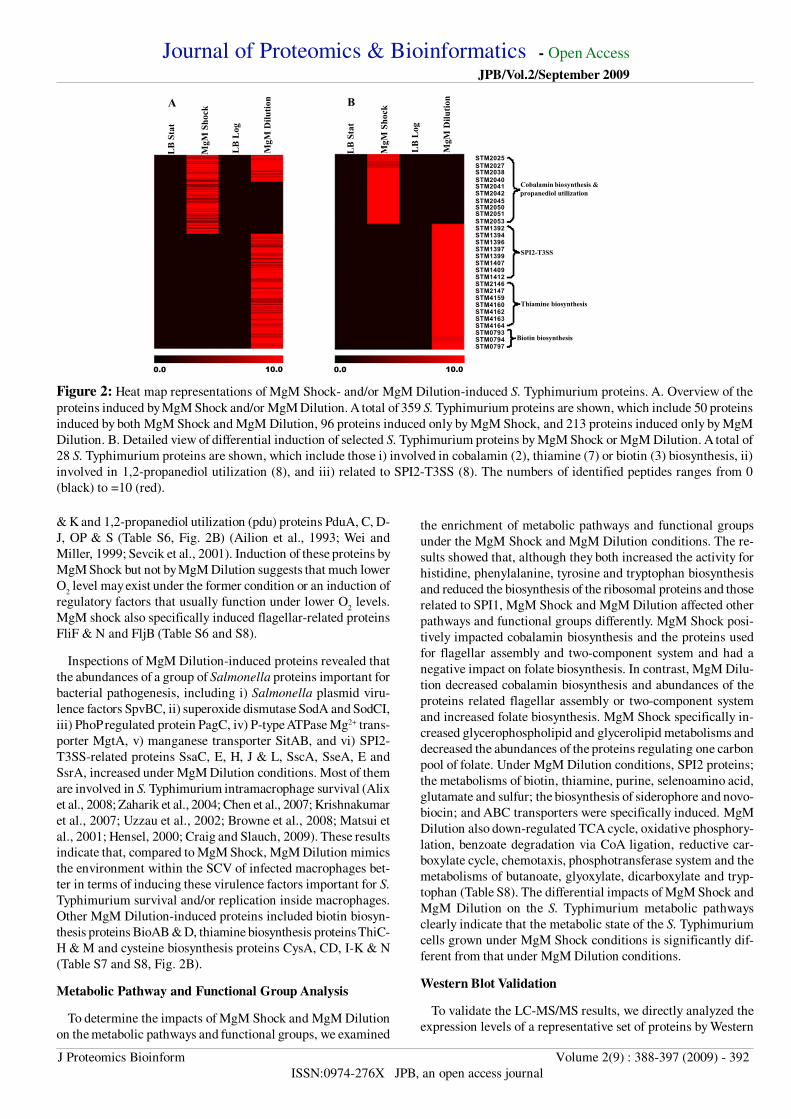
Journal of Proteomics amp Bioinformatics - Open Access
JPBVol2September 2009
J Proteomics Bioinform Volume 2(9) 388-397 (2009) - 392
ISSN0974-276X JPB an open access journal
amp K and 12-propanediol utilization (pdu) proteins PduA C D-
J OP amp S (Table S6 Fig 2B) (Ailion et al 1993 Wei and
Miller 1999 Sevcik et al 2001) Induction of these proteins by
MgM Shock but not by MgM Dilution suggests that much lower
O2 level may exist under the former condition or an induction of
regulatory factors that usually function under lower O2 levels
MgM shock also specifically induced flagellar-related proteins
FliF amp N and FljB (Table S6 and S8)
Inspections of MgM Dilution-induced proteins revealed that
the abundances of a group of Salmonella proteins important for
bacterial pathogenesis including i) Salmonella plasmid viru-
lence factors SpvBC ii) superoxide dismutase SodA and SodCI
iii) PhoP regulated protein PagC iv) P-type ATPase Mg2+ trans-
porter MgtA v) manganese transporter SitAB and vi) SPI2-
T3SS-related proteins SsaC E H J amp L SscA SseA E and
SsrA increased under MgM Dilution conditions Most of them
are involved in S Typhimurium intramacrophage survival (Alix
et al 2008 Zaharik et al 2004 Chen et al 2007 Krishnakumar
et al 2007 Uzzau et al 2002 Browne et al 2008 Matsui et
al 2001 Hensel 2000 Craig and Slauch 2009) These results
indicate that compared to MgM Shock MgM Dilution mimics
the environment within the SCV of infected macrophages bet-
ter in terms of inducing these virulence factors important for S
Typhimurium survival andor replication inside macrophages
Other MgM Dilution-induced proteins included biotin biosyn-
thesis proteins BioAB amp D thiamine biosynthesis proteins ThiC-
H amp M and cysteine biosynthesis proteins CysA CD I-K amp N
(Table S7 and S8 Fig 2B)
Metabolic Pathway and Functional Group Analysis
To determine the impacts of MgM Shock and MgM Dilution
on the metabolic pathways and functional groups we examined
the enrichment of metabolic pathways and functional groups
under the MgM Shock and MgM Dilution conditions The re-
sults showed that although they both increased the activity for
histidine phenylalanine tyrosine and tryptophan biosynthesis
and reduced the biosynthesis of the ribosomal proteins and those
related to SPI1 MgM Shock and MgM Dilution affected other
pathways and functional groups differently MgM Shock posi-
tively impacted cobalamin biosynthesis and the proteins used
for flagellar assembly and two-component system and had a
negative impact on folate biosynthesis In contrast MgM Dilu-
tion decreased cobalamin biosynthesis and abundances of the
proteins related flagellar assembly or two-component system
and increased folate biosynthesis MgM Shock specifically in-
creased glycerophospholipid and glycerolipid metabolisms and
decreased the abundances of the proteins regulating one carbon
pool of folate Under MgM Dilution conditions SPI2 proteins
the metabolisms of biotin thiamine purine selenoamino acid
glutamate and sulfur the biosynthesis of siderophore and novo-
biocin and ABC transporters were specifically induced MgM
Dilution also down-regulated TCA cycle oxidative phosphory-
lation benzoate degradation via CoA ligation reductive car-
boxylate cycle chemotaxis phosphotransferase system and the
metabolisms of butanoate glyoxylate dicarboxylate and tryp-
tophan (Table S8) The differential impacts of MgM Shock and
MgM Dilution on the S Typhimurium metabolic pathways
clearly indicate that the metabolic state of the S Typhimurium
cells grown under MgM Shock conditions is significantly dif-
ferent from that under MgM Dilution conditions
Western Blot Validation
To validate the LC-MSMS results we directly analyzed the
expression levels of a representative set of proteins by Western
Figure 2 Heat map representations of MgM Shock- andor MgM Dilution-induced S Typhimurium proteins A Overview of the
proteins induced by MgM Shock andor MgM Dilution A total of 359 S Typhimurium proteins are shown which include 50 proteins
induced by both MgM Shock and MgM Dilution 96 proteins induced only by MgM Shock and 213 proteins induced only by MgM
Dilution B Detailed view of differential induction of selected S Typhimurium proteins by MgM Shock or MgM Dilution A total of
28 S Typhimurium proteins are shown which include those i) involved in cobalamin (2) thiamine (7) or biotin (3) biosynthesis ii)
involved in 12-propanediol utilization (8) and iii) related to SPI2-T3SS (8) The numbers of identified peptides ranges from 0
(black) to =10 (red)
STM2025STM2027STM2038STM2040STM2041STM2042STM2045STM2050STM2051STM2053STM1392STM1394STM1396STM1397STM1399STM1407STM1409STM1412STM2146STM2147STM4159STM4160STM4162STM4163STM4164STM0793STM0794STM0797
Cobalamin biosynthesis amp
A
LB
Sta
t
MgM
Dilu
tion
MgM
Sho
ck
LB
Log
B
LB
Sta
t
MgM
Dilu
tion
MgM
Sho
ck
LB
Log
propanediol utilization
SPI2-T3SS
Thiamine biosynthesis
Biotin biosynthesis
1000010000
Journal of Proteomics amp Bioinformatics - Open Access JPBVol2September 2009
J Proteomics Bioinform Volume 2(9) 388-397 (2009) - 393
ISSN0974-276X JPB an open access journal
blot analysis These included SsaE SsaQ SseE CbiP ThiH
BioB and BioD that were tagged with 3 times FLAG epitope via
recombinant DNA techniques Although neither of them was
detected by LC-MSMS SsaQ and CbiP were tagged to aid in-
vestigations of the dynamic range of detection by LC-MSMS
used in this study The presence of these recombinant proteins
were measured by Western blot under three different conditions
which included MgM Shock MgM Dilution and infection of
macrophages
As shown in Fig 3A all three tagged SPI2-T3SS proteins were
detected under MgM Dilution condition while only SseE was
detected under MgM Shock condition Detection of SseE and
SsaQ by Western blot and detection of only SseE by LC-MS
MS under MgM Dilution condition suggest that the failure to
detect SsaQ by LC-MSMS during this study is most likely at-
tributed to the low abundance of SsaQ Consistent with this sug-
gestion the relative abundance of SsaQ detected under MgM-
Dilution condition was much lower than that of SseE (Fig 3B)
Together with that of LC-MSMS our results consistently dem-
onstrate that more SPI2-T3SS proteins are induced by MgM
Dilution than by MgM Shock The trace amount of CbiP was
found under MgM Shock condition while no CbiP was detected
under MgM Dilution condition In contrast to CbiP more ThiH
was detected under MgM Dilution condition than MgM Shock
condition (Fig 3C) While BioB could be found under both MgM
Shock and MgM Dilution conditions BioD was only detected
under MgM Dilution conditions (Fig 3D)
As shown in Figure 4A SsaQ SseE BioB and BioD were
induced when the S Typhimurium strains were used to infect
RAW 2647 macrophages Infection of the macrophages how-
ever did not induce SsaE CbiP or ThiH (data not shown) Both
SsaQ and BioD were detectable at 2 hr and peaked at 24 hr
following the macrophage infection Presence of SseE and BioB
could also be detected at 2 4 and 24 hpi The abundances of
SseE at 2 4 and 24 hpi were nearly the same while the abun-
Figure 3 Western blot confirmation of SsaE SsaQ and SseE
(panels A and B) CbiP and ThiH (panel C) and BioB and BioD
(panel D) The S Typhimurium strains with recombinant SsaE
SsaQ SseE CbiP ThiH BioB or BioD were cultured under
MgM Shock (lanes S) or MgM Dilution (lanes D) conditions
as described in Materials and Methods Relative abundance
between SseE (panel B lane 1) and SsaQ (panel B lane 2)
under MgM Dilution condition was also compared After sepa-
ration by SDS-PAGE the proteins were first probed with an
anti-FLAG antibody (ie OctA-Probe) and then stripped and
re-probed using anti-DnaK antibody The DnaK was used as
an internal control to ensure that similar amounts of proteins
were loaded in each lane Experiments were repeated three times
with similar results
Figure 4 Western blot identification of SsaQ SseE BioB and
BioD after S Typhimurium cells isolated from RAW 2647
macrophages The S Typhimurium strains with recombinant
SsaQ SseE BioB or BioD were used to infect RAW 2647
macrophages as described in Materials and Methods Relative
abundance between BioB (panel B lane 1) and BioD (panel B
lane 2) at 2 hr after infection of RAW 2647 macrophages was
also compared After separation by SDS-PAGE the proteins
were first probed with an anti-FLAG antibody and then stripped
and re-probed using anti-DnaK antibody Experiments were
repeated three times with similar results
Figure 5 Influence of deletion of bioB gene on S Typhimurium
replication in RAW 2647 macrophages A Deletion of bioB
gene Agarose gel showing the PCR products of bioB amplified
from WT (lane 1) and ∆bioB mutant (lane 2) The positions of
DNA standards (Stds) are indicated at left B Relative viability
of WT () and ∆bioB () cells in RAW 2647 macrophages
after compared to those at 0 hr infection respectively (n = V3 p
lt 001) At 0 hr infection the CFUwell values were 34 plusmn 04 times
105 for WT and 39 plusmn 04 times 105 for ∆bioB mutant (n = 3)
AS D
CS D
DS D
B1 2
BioB
DnaKBioDDnaK
CbiPDnaKThiHDnaK
SsaQ
SseEDnaK
SsaEDnaKSsaQDnaKSseEDnaK
A 0 2 4 24 hr
B 1 2
SsaQDnaKSseE
DnaK
BioBDnaKBioD
DnaK
BioB
BioD
DnaK
A B180
120
60
0 2 4 24Time of infection (hours)
Infe
ction
(of
0 ho
ur)
1 2Stds(kbp)
3
03
01
1
165
Journal of Proteomics amp Bioinformatics - Open Access
JPBVol2September 2009
J Proteomics Bioinform Volume 2(9) 388-397 (2009) - 394
ISSN0974-276X JPB an open access journal
macrophages suggest for the first time a biotin-limited environ-
ment within the SCV of RAW 2647 macrophages and a pos-
sible mechanism that co-regulates biotin synthesis and the SPI2-
T3SS secretion system
SPI2-T3SS Proteins
MgM has been routinely used to induce SPI2-T3SS related
proteins (Beuzon et al 1999 Mazurkiewicz et al 2008 Hansen-
Wester et al 2002) For instance detections of SseB-D are evi-
dent when S Typhimurium cells are grown in MgM to the Stat
phase (Beuzon et al 1999 Hansen-Wester et al 2002) The
reasons for differential induction of SPI2-T3SS proteins by the
culturing methods used in this study however remain obscure
but might be attributed to the different metabolic states of the S
Typhimurium cells grown under these conditions Nevertheless
to the best of our knowledge this is the first report that change
of culturing methods affects the SPI2-T3SS proteins in terms of
the extent and degree of their induction by this growth medium
Increase of SsaQ and SseE abundances following infection of
macrophages is consistent with the previous findings that both
SsaQ and SseE help S Typhimurium proliferate in RAW 2647
macrophages (Hensel et al 1998 Suvarnapunya et al 2003)
In addition SsaQ is required for S Typhimurium to cause sys-
temic infection in mice (Suvarnapunya et al 2003) Whether
SsaE is involved in intramacrophage survival of S Typhimurium
is still uncertain as it was undetected following macrophage
infection
Cobalamin and Thiamin Biosynthesis Proteins
Cobalamin is a cofactor for different types of enzymes in-
cluding isomerase metyltransferase and dehalogenase [for re-
view see (Brown 2006)] In S Typhimurium cobalamin serves
mainly as a cofactor for B12
-dependent diol dehydratase (an
isomerase) that catalyzes the conversion of 12-propanediol to
propionaldehyde the first step of pdu pathway For this reason
the expressions of pdu and cobalamin biosynthesis genes are
usually co-regulated in S Typhimurium (Bobik et al 1999)
Consistent with these previous observations our results showed
that the proteins used for cobalamin biosynthesis or pdu were
co-induced by MgM Shock Induction of PduB by MgM Shock
was confirmed previously (Adkins et al 2006) Also a water
soluble B-type vitamin thiamin is a cofactor for several impor-
tant enzymes such as pyruvate dehydrogenase a-ketoglutarate
dehydrogenase transketolase and pyruvate decarboxylase that
are involved in carbohydrate metabolism (Begley et al 1999)
The reasons for failure to detect CbiP or ThiH in the S
Typhimurium cells isolated from macrophages are currently
unknown Cobalamine thiamine and biotin are present in
DMEM medium as the vitamin supplements as well as the in-
herent components of added fetal bovine serum and can be taken
by RAW 2647 macrophages (Baker et al 1988) Detection of
CbiP and ThiH under MgM Shock and MgM Dilution condi-
tion respectively but not after macrophage infection indicates
that sufficient amount of cobalamine and thiamine may exist
within the SCV of RAW 2647 macrophages under the condi-
tions used in this study which could inhibit the induction of
CbiP and ThiH
Biotin Biosynthesis Proteins
dance of BioB peaked between 2 and 4 hpi At 2 hpi the abun-
dance of BioB was much higher than that of BioD (Fig 4B)
Induction of BioB and BioD in infected macrophages suggests
that biotin might be limited inside the SCV of RAW 2647 mac-
rophages under the condition tested
Replication of ∆bioB Mutant in RAW 2647 Macrophages
To further investigate whether biotin was limited in the SCV
the bioB gene was deleted and the resulting ∆bioB mutant was
used to measure its replication rates inside RAW 2647 mac-
rophages (Fig 5A) The ability of ∆bioB mutant to grow in MgM
medium was impaired Supplementing the MgM medium with
01 microM of biotin restored the ∆bioB mutantrsquos ability to grow
demonstrating the biotin auxotrophy of this mutant Elimina-
tion of BioB had no impact on the ability of S Typhimurium to
infect macrophages because the CFUwell were 34 plusmn 04 times 105
for wt and 39 plusmn 04 times 105 for ∆bioB mutant (n = 3) at 0 hpi
Although both WT and ∆bioB mutant began to replicate at 4
hpi the replication rates of ∆bioB mutant were lt72 of that for
WT (n = 3 Plt 001) (Fig 5B) These findings support the no-
tion that biotin is limited within the SCV of RAW 2647 mac-
rophages
Discussion
Although MgM had been used to mimic certain conditions
found inside the SCV the culturing methods varied from group
to group The impact of the different culturing method on the S
Typhimurium proteome had not been fully investigated with the
global proteomic methods previously In this study we used the
medium to grow S Typhimurium under two different culturing
methods (ie MgM Shock amp Dilution) and then used a global
proteomic method to comparatively analyze their effects on the
S Typhimurium proteome The results of proteomic as well as
confirmatory Western blot analyses consistently showed that
these two methods affected the S Typhimurium proteome dif-
ferently despite the same fresh medium being used for up to 4
hr Compared to MgM dilution MgM Shock induced a group
of proteins such as those related to cobalamin biosynthesis or
pdu whose induction is usually expected to occur only at low
O2 levels where induction for fermentation may be needed This
suggests that the O2 level in solution may be lower under MgM
Shock condition than MgM Dilution condition or there is the
presence of regulatory factors in one of the conditions that are
absent in the other MgM Dilution meanwhile induced a group
of Salmonella virulence factors including those related to SPI2-
T3SS that are required for intramacrophage survival of S
Typhimurium more effectively than MgM Shock In addition
MgM Dilution increased the abundances of the proteins related
to thiamine or biotin biosynthesis The results of metabolic path-
way enrichment analysis also demonstrated that the metabolic
state of the S Typhimurium cells grown under MgM Shock con-
dition differed significantly from that under MgM Dilution con-
dition Western blot analyses showed that the abundances of
SPI2-T3SS proteins SsaQ and SseE and biotin biosynthesis pro-
teins BioB and BioD increased following the infection of RAW
2647 macrophages Furthermore elimination of BioB lowered
the replication rates of S Typhimurium cells within the mac-
rophages Induction of BioB and BioD following macrophage
infection and the reduced ability of ∆bioB to replicate inside the
Journal of Proteomics amp Bioinformatics - Open Access
JPBVol2September 2009
J Proteomics Bioinform Volume 2(9) 388-397 (2009) - 395
ISSN0974-276X JPB an open access journal
approaches to Salmonella infections
Acknowledgements
This work was supported in part by the Laboratory Directed
Research and Development Program of US Department of
Energy (DOE) to LS and by the National Institute of Allergy
and Infectious Diseases NIHDHHS through interagency agree-
ments Y1-AI-4894-01 and Y1-AI-8401-01 This work used in-
strumentation and capabilities developed under support from
the National Center for Research Resources (Grant RR 018522
to RDS) and the DOErsquos Office of Biological and Environmen-
tal Research Significant portions of this work were performed
using EMSL a national scientific user facility sponsored by the
DOErsquos Office of Biological and Environmental Research lo-
cated at Pacific Northwest National Laboratory Pacific North-
west National Laboratory is operated for the DOE by Battelle
Memorial Institute under Contract DE-AC05-76RLO1830
References
1 Adkins JN Mottaz HM Norbeck AD Gustin JK Rue J et
al (2006) Analysis of the Salmonella typhimurium proteome
through environmental response toward infectious condi-
tions Mol Cell Proteomics 5 1450-1461
2 Adkins JN Varnum SM Auberry KJ Moore RJ Angell NH
et al (2002) Toward a human blood serum proteome analy-
sis by multidimensional separation coupled with mass spec-
trometry Mol Cell Proteomics 1 947-955
3 Ailion M Bobik TA Roth JR (1993) Two global regulatory
systems (Crp and Arc) control the cobalaminpropanediol
regulon of Salmonella typhimurium J Bacteriol 175 7200-
7208
4 Alix E Miki T Felix C Rang C Figueroa-Bossi N et al
(2008) Interplay between MgtC and PagC in Salmonella
enterica serovar Typhimurium Microb Pathog 45 236-240
5 Ansong C Yoon H Norbeck AD Gustin JK McDermott
JE et al (2008) Proteomics snalysis of the causative agent
of typhoid fever J Proteome Res 7 546-557
6 Baker H DeAngelis B Frank O (1988) Vitamins and other
metabolites in various sera commonly used for cell culturing
Experientia 44 1007-1010
7 Begley TP Downs DM Ealick SE McLafferty FW Van
Loon AP et al (1999) Thiamin biosynthesis in prokaryotes
Arch Microbiol 171 293-300
8 Beuzon CR Banks G Deiwick J Hensel M Holden DW
(1999) pH-dependent secretion of SseB a product of the
SPI-2 type III secretion system of Salmonella typhimurium
Mol Microbiol 33 806-816
9 Bobik TA Havemann GD Busch RJ Williams DS Aldrich
HC (1999) The propanediol utilization (pdu) operon of Sal-
monella enterica serovar Typhimurium LT2 includes genes
necessary for formation of polyhedral organelles involved
Biotin is a covalently bound cofactor for the enzymes that
catalyze carboxylation decarboxylation and transcarboxylation
reactions such as acetyl-CoA carboxylase pyruvate carboxy-
lase and glutaconyl decarboxylase (Streit and Entcheva 2003)
The findings that BioB and BioD increase their abundances af-
ter S Typhimurium infecting macrophages and that ∆bioB mu-
tant exhibits reduced replication rates inside macrophages con-
sistently suggest a biotin-limited condition inside the SCV of
RAW 2647 macrophages In agreement with this suggestion
transcriptomic analysis of the S Typhimurium strain SL1344
isolated from mouse J774-A1 macrophage showed a moderate
increase of expression of several genes involved in biotin bio-
synthesis at 8 hpi (Eriksson et al 2003) Likewise biotin bio-
synthesis genes were significantly up-regulated after infection
of epithelial cells by the strain SL 1344 The biotin limitation
inside the SCV of epithelial cells was considered as a possible
cause for the up-regulation of these genes (Hautefort et al 2008)
Differences however exist between our results and those pub-
lished previously in terms of replication of S Typhimurium
mutants deficient in biotin biosynthesis and induction of biotin
biosynthesis proteins inside host cells In previous studies de-
letion of biotin biosynthesis genes did not affect the bacterial
ability to replicate inside human HeLa epithelial cells (Hautefort
et al 2008) Our results showed that the replication rate of ∆bioB
mutant in mouse RAW 2647 macrophages decreased slightly
Previous studies found that neither BioB nor BioD was detected
in the S Typhi cells isolated from human THP-1 macrophages
(Ansong et al 2008) Our results clearly showed the induction
of S Typhimurium BioB and BioD following the infection of
RAW 2647 macrophages The different host cells (human HeLa
epithelial cell mouse RAW 2647 macrophage and human THP-
1 macrophage) bacterial cells (S Typhimurium strain 14028
and SL1344 and S Typhi) and media (DMEM and RPMI 1640)
used most likely contribute to the different results obtained from
these studies Alternatively co-regulation of pathogenesis pro-
teins and biotin pathways may be decoupled in S Typhi It
should be noted that BioH of the enteric pathogen Yersinia
enterocolitica was identified as a systemic infection factor (Gort
and Miller 2000) Future investigations should focus on whether
biotin biosynthesis proteins are also involved in the develop-
ment of S Typhimurium-mediated systemic infection in mice
Conclusions and Perspectives
This comparative analysis of the proteome of S Typhimurium
grown in MgM shows for the first time that MgM Shock and
MgM Dilution differentially affect the S Typhimurium proteome
as well as the S Typhimurium metabolic pathways and func-
tional groups Our results also show for the first time that the
infection of RAW 2647 macrophages increases BioB and BioD
abundances The general agreement between the MS and West-
ern blot results clearly demonstrates the repeatability and repro-
ducibility of the results as well as the robustness and accuracy
of the methodology used in this study In the future researchers
should consider the co-relationship between bacterial metabolic
states and induction SPI2-T3SS proteins and the roles of biotin
biosynthesis proteins in S Typhimurium pathogenesis This co-
regulation may be more informative of the in vivo metabolic
environment experienced by Salmonella Understanding these
relationships and functions should lead to improved therapeutic
raquo CrossRef raquo Pubmedraquo Google Scholar
raquo CrossRef raquo Pubmedraquo Google Scholar
raquo CrossRef raquo Pubmed raquo Google Scholar
raquo CrossRef raquo Pubmed raquo Google Scholar
raquo CrossRef raquo Pubmedraquo Google Scholar
raquo CrossRef raquo Pubmed raquo Google Scholar
raquo CrossRef raquo Pubmed raquo Google Scholar
raquo CrossRef raquo Pubmed raquo Google Scholar
Journal of Proteomics amp Bioinformatics - Open Access
JPBVol2September 2009
J Proteomics Bioinform Volume 2(9) 388-397 (2009) - 396
ISSN0974-276X JPB an open access journal
in coenzyme B(12)-dependent 1 2-propanediol degradation
J Bacteriol 181 5967-5975
10 Brown KL (2006) The enzymatic activation of coenzyme
B12 Dalton Trans 7 1123-1133
11 Browne SH Hasegawa P Okamoto S Fierer J Guiney DG
(2008) Identification of Salmonella SPI-2 secretion system
components required for SpvB-mediated cytotoxicity in
macrophages and virulence in mice FEMS Immunol Med
Microbiol 52 194-201
12 Chen X Kodama T Iida T Honda T (2007) Demonstration
and characterization of manganese superoxide dismutase of
Providencia alcalifaciens Microbiol Immunol 51 951-961
13 Craig M Slauch JM (2009) Phagocytic superoxide specifi-
cally damages an extracytoplasmic target to inhibit or kill
Salmonella PLoS One 4 e4975
14 Datsenko KA Wanner BL (2000) One-step inactivation of
chromosomal genes in Escherichia coli K-12 using PCR
products Proc Natl Acad Sci USA 97 6640-6645
15 Eng JK McCormack AL Yates JR (1994) An approach to
correlate tandem mass-spectral data of peptides with amino-
acid-sequences in a protein database Journal of the Ameri-
can Society for Mass Spectrometry 5 976-989
16 Eriksson S Lucchini S Thompson A Rhen M Hinton JC
(2003) Unravelling the biology of macrophage infection by
gene expression profiling of intracellular Salmonella
enterica Mol Microbiol 47 103-118
17 Fields PI Swanson RV Haidaris CG Heffron F (1986) Mu-
tants of Salmonella typhimurium that cannot survive within
the macrophage are avirulent Proc Natl Acad Sci USA 83
5189-5193
18 Gao J Opiteck GI Friedrichs MS Dongre AR Hefta SA
(2003) Changes in the protein expression of yeast as a func-
tion of carbon source J Proteome Res 2 643-649
19 Gort AS Miller VL (2000) Identification and characteriza-
tion of Yersinia enterocolitica genes induced during systemic
infection Infect Immun 68 6633-6642
20 Hansen-Wester I Stecher B Hensel M (2002) Type III se-
cretion of Salmonella enterica serovar Typhimurium trans-
located effectors and SseFG Infect Immun 70 1403-1409
21 Hautefort I Thompson A Eriksson-Ygberg S Parker ML
Lucchini S et al (2008) During infection of epithelial cells
Salmonella enterica serovar Typhimurium undergoes a time-
dependent transcriptional adaptation that results in simulta-
neous expression of three type 3 secretion systems Cell
Microbiol 10 958-984
22 Hensel M (2000) Salmonella pathogenicity island 2 Mol
Microbiol 36 1015-1023
23 Hensel M Shea JE Waterman SR Mundy R Nikolaus T
Banks G et a (1998) Genes encoding putative effector pro-
teins of the type III secretion system of Salmonella pathoge-
nicity island 2 are required for bacterial virulence and pro-
liferation in macrophages Mol Microbiol 30 163-174
24 Ishihama Y Oda Y Tabata T Sato T Nagasu T et al (2005)
Exponentially modified protein abundance index (emPAI)
for estimation of absolute protein amount in proteomics by
the number of sequenced peptides per protein Mol Cell
Proteomics 4 1265-1272
25 Jacobs JM Yang X Luft BJ Dunn JJ Camp DG 2nd et al
(2005) Proteomic analysis of Lyme disease global protein
comparison of three strains of Borrelia burgdorferi
Proteomics 5 1446-1453
26 Kanehisa M Goto S (2000) KEGG kyoto encyclopedia of
genes and genomes Nucleic Acids Res 28 27-30
27 Krishnakumar R Kim B Mollo EA Imlay JA Slauch JM
(2007) Structural properties of periplasmic SodCI that cor-
relate with virulence in Salmonella enterica serovar
Typhimurium J Bacteriol 189 4343-4352
28 Liu H Sadygov RG Yates JR 3rd (2004) A model for ran-
dom sampling and estimation of relative protein abundance
in shotgun proteomics Anal Chem 76 4193-4201
29 Livesay EA Tang K B Taylor K Buschbach MA Hopkins
DF et al (2008) Fully automated four-column capillary LC-
MS system for maximizing throughput in proteomic analyses
Anal Chem 80 294-302
30 Manes NP Gustin JK Rue J Mottaz HM Purvine SO et al
(2007) Targeted protein degradation by Salmonella under
phagosome-mimicking culture conditions investigated us-
ing comparative peptidomics Mol Cell Proteomics 6 717-
727
31 Matsui H Bacot CM Garlington WA Doyle TJ Roberts S
et al (2001) Virulence plasmid-borne spvB and spvC genes
can replace the 90-kilobase plasmid in conferring virulence
to Salmonella enterica serovar Typhimurium in subcutane-
ously inoculated mice J Bacteriol 183 4652-4658
32 Mazurkiewicz P Thomas J Thompson JA Liu M Arbibe
L et al (2008) SpvC is a Salmonella effector with
phosphothreonine lyase activity on host mitogen-activated
protein kinases Mol Microbiol 67 1371-1383
33 Qian WJ Liu T Monroe ME Strittmatter EF Jacobs JM et
al (2005a) Probability-based evaluation of peptide and pro-
tein identifications from tandem mass spectrometry and
SEQUEST analysis the human proteome J Proteome Res
4 53-62
34 Qian WJ Monroe ME Liu T Jacobs JM Anderson GA et
al (2005b) Quantitative proteome analysis of human plasma
following in vivo lipopolysaccharide administration using
raquo CrossRef raquo Pubmed raquo Google Scholar
raquo CrossRef raquo Pubmed
raquo CrossRef raquo Pubmed raquo Google Scholar
raquo CrossRef raquo Pubmed raquo Google Scholar
raquo CrossRef raquo Pubmedraquo Google Scholar
raquo CrossRef raquo Pubmed raquo Google Scholar
raquo CrossRef raquo Pubmed raquo Google Scholar
raquo CrossRef raquo Pubmedraquo Google Scholar
raquo CrossRef raquo Pubmed raquo Google Scholar
raquo CrossRef raquo Pubmed raquo Google Scholar
raquo CrossRef raquo Pubmedraquo Google Scholar
raquo CrossRef raquo Pubmed raquo Google Scholar
raquo CrossRef raquo Pubmed raquo Google Scholar
raquo CrossRef raquo Pubmed raquo Google Scholar
raquo CrossRef raquo Pubmed raquo Google Scholar
raquo CrossRef raquo Pubmed raquo Google Scholar
raquo CrossRef raquo Pubmed raquo Google Scholar
raquo CrossRef raquo Pubmed raquo Google Scholar
raquo CrossRef raquo Pubmedraquo Google Scholar
raquo CrossRef raquo Pubmed raquo Google Scholar
raquo CrossRef raquo Pubmed raquo Google Scholar
raquo CrossRef raquo Pubmed raquo Google Scholar
raquo CrossRef raquo Pubmed raquo Google Scholar
raquo CrossRef raquo Pubmed raquo Google Scholar
raquo CrossRef raquo Pubmed raquo Google Scholar
Journal of Proteomics amp Bioinformatics - Open Access
JPBVol2September 2009
J Proteomics Bioinform Volume 2(9) 388-397 (2009) - 397
ISSN0974-276X JPB an open access journal
16O18O labeling and the accurate mass and time tag ap-
proach Mol Cell Proteomics 4 700-709
35 Saeed AI Sharov V White J Li J Liang W et al (2003)
TM4 a free open-source system for microarray data man-
agement and analysis Biotechniques 34 374-378
36 Sevcik M Sebkova A Volf J Rychlik I (2001) Transcrip-
tion of arcA and rpoS during growth of Salmonella
typhimurium under aerobic and microaerobic conditions
Microbiology 147 701-708
37 Shi L Adkins JN Coleman JR Schepmoes AA Dohnkova
A et al (2006) Proteomic analysis of Salmonella enterica
serovar Typhimurium isolated from RAW 2647 macroph-
ages identification of a novel protein that contributes to the
replication of serovar Typhimurium inside macrophages J
Biol Chem 281 29131-29140
38 Shi L Chowdhury SM Smallwood HS Yoon H Mottaz-
Brewer HM et al (2009) Proteomic investigation of the time
course responses of RAW 2647 macrophages to infection
with Salmonella enterica Infect Immun 77 3227-3233
39 Streit WR Entcheva P (2003) Biotin in microbes the genes
involved in its biosynthesis its biochemical role and per-
spectives for biotechnological production Appl Microbiol
Biotechnol 61 21-31
40 Suvarnapunya AED Zurawski V Guy RL Stein MA (2003)
Molecular characterization of the prototrophic Salmonella
mutants defective for intraepithelial replication Infect Immun
71 2247-2252
41 Uzzau S Bossi L Figueroa-Bossi N (2002) Differential ac-
cumulation of Salmonella[Cu Zn] superoxide dismutases
SodCI and SodCII in intracellular bacteria correlation with
their relative contribution to pathogenicity Mol Microbiol
46 147-156
42 Uzzau S Figueroa-Bossi N Rubino S Bossi L (2001)
Epitope tagging of chromosomal genes in Salmonella Proc
Natl Acad Sci USA 98 15264-15269
43 VerBerkmoes NC Shah MB Lankford PK Pelletier DA
Strader MB et al (2006) Determination and comparison of
the baseline proteomes of the versatile microbe
Rhodopseudomonas palustris under its major metabolic
states J Proteome Res 5 287-298
44 Washburn MP Wolters D Yates JR 3rd (2001) Large-scale
analysis of the yeast proteome by multidimensional protein
identification technology Nat Biotechnol 19 242-247
45 Wei Y Miller CG (1999) Characterization of a group of
anaerobically induced fnr-dependent genes of Salmonella
typhimurium J Bacteriol 181 6092-6097
46 Zaharik ML Cullen VL Fung AM Libby SJ Kujat Choy
SL et al (2004) The Salmonella enterica serovar
typhimurium divalent cation transport systems MntH and
SitABCD are essential for virulence in an Nramp1G169 mu-
rine typhoid model Infect Immun 72 5522-5525
raquo CrossRef raquo Pubmed raquo Google Scholar
raquo Pubmed raquo Google Scholar
raquo CrossRef raquo Pubmed raquo Google Scholar
raquo CrossRef raquo Pubmed raquo Google Scholar
raquo CrossRef raquo Pubmed raquo Google Scholar
raquo CrossRef raquo Pubmed raquo Google Scholar
raquo CrossRef raquo Pubmed raquo Google Scholar
raquo CrossRef raquo Pubmed raquo Google Scholar
raquo CrossRef raquo Pubmedraquo Google Scholar
raquo CrossRef raquo Google Scholar
raquo CrossRef raquo Pubmed raquo Google Scholar
raquo CrossRef raquo Pubmedraquo Google Scholar
raquo Pubmed
- Title13
-
- Authors13
- Affiliations13
- Corresponding authors
- Dates13
- Citation
- Copyright
-
- Abstract
- Keywords
- Abbreviations
- Introduction
- Materials and Methods
-
- Reagents and Standard Procedures
- Bacterial Strains and Culture Conditions
- Lysis of S Typhimurium Cells and Tryptic Digestion
- Capillary LC-MSMS Analysis
- Data Analysis
- Functional Enrichment of Proteomic Data
- Genetic Manipulation of S Typhimurium Genes
- Cell Culture Macrophage Infection and Isolation of STyphimurium Cells
- Gentamicin Resistance Assay
-
- Results
-
- LC-MSMS Analysis
- Metabolic Pathway and Functional Group Analysis
- Western Blot Validation
- Replication of bioB Mutant in RAW 2647 Macrophages
-
- Discussion
- SPI2-T3SS Proteins
- Cobalamin and Thiamin Biosynthesis Proteins
- Biotin Biosynthesis Proteins
- Conclusions and Perspectives
- Acknowledgements
- Figures13
-
- Figure 113
- Figure 213
- Figure 313
- Figure 413
- Figure 513
-
- References
-

Journal of Proteomics amp Bioinformatics - Open Access
JPBVol2September 2009
J Proteomics Bioinform Volume 2(9) 388-397 (2009) - 395
ISSN0974-276X JPB an open access journal
approaches to Salmonella infections
Acknowledgements
This work was supported in part by the Laboratory Directed
Research and Development Program of US Department of
Energy (DOE) to LS and by the National Institute of Allergy
and Infectious Diseases NIHDHHS through interagency agree-
ments Y1-AI-4894-01 and Y1-AI-8401-01 This work used in-
strumentation and capabilities developed under support from
the National Center for Research Resources (Grant RR 018522
to RDS) and the DOErsquos Office of Biological and Environmen-
tal Research Significant portions of this work were performed
using EMSL a national scientific user facility sponsored by the
DOErsquos Office of Biological and Environmental Research lo-
cated at Pacific Northwest National Laboratory Pacific North-
west National Laboratory is operated for the DOE by Battelle
Memorial Institute under Contract DE-AC05-76RLO1830
References
1 Adkins JN Mottaz HM Norbeck AD Gustin JK Rue J et
al (2006) Analysis of the Salmonella typhimurium proteome
through environmental response toward infectious condi-
tions Mol Cell Proteomics 5 1450-1461
2 Adkins JN Varnum SM Auberry KJ Moore RJ Angell NH
et al (2002) Toward a human blood serum proteome analy-
sis by multidimensional separation coupled with mass spec-
trometry Mol Cell Proteomics 1 947-955
3 Ailion M Bobik TA Roth JR (1993) Two global regulatory
systems (Crp and Arc) control the cobalaminpropanediol
regulon of Salmonella typhimurium J Bacteriol 175 7200-
7208
4 Alix E Miki T Felix C Rang C Figueroa-Bossi N et al
(2008) Interplay between MgtC and PagC in Salmonella
enterica serovar Typhimurium Microb Pathog 45 236-240
5 Ansong C Yoon H Norbeck AD Gustin JK McDermott
JE et al (2008) Proteomics snalysis of the causative agent
of typhoid fever J Proteome Res 7 546-557
6 Baker H DeAngelis B Frank O (1988) Vitamins and other
metabolites in various sera commonly used for cell culturing
Experientia 44 1007-1010
7 Begley TP Downs DM Ealick SE McLafferty FW Van
Loon AP et al (1999) Thiamin biosynthesis in prokaryotes
Arch Microbiol 171 293-300
8 Beuzon CR Banks G Deiwick J Hensel M Holden DW
(1999) pH-dependent secretion of SseB a product of the
SPI-2 type III secretion system of Salmonella typhimurium
Mol Microbiol 33 806-816
9 Bobik TA Havemann GD Busch RJ Williams DS Aldrich
HC (1999) The propanediol utilization (pdu) operon of Sal-
monella enterica serovar Typhimurium LT2 includes genes
necessary for formation of polyhedral organelles involved
Biotin is a covalently bound cofactor for the enzymes that
catalyze carboxylation decarboxylation and transcarboxylation
reactions such as acetyl-CoA carboxylase pyruvate carboxy-
lase and glutaconyl decarboxylase (Streit and Entcheva 2003)
The findings that BioB and BioD increase their abundances af-
ter S Typhimurium infecting macrophages and that ∆bioB mu-
tant exhibits reduced replication rates inside macrophages con-
sistently suggest a biotin-limited condition inside the SCV of
RAW 2647 macrophages In agreement with this suggestion
transcriptomic analysis of the S Typhimurium strain SL1344
isolated from mouse J774-A1 macrophage showed a moderate
increase of expression of several genes involved in biotin bio-
synthesis at 8 hpi (Eriksson et al 2003) Likewise biotin bio-
synthesis genes were significantly up-regulated after infection
of epithelial cells by the strain SL 1344 The biotin limitation
inside the SCV of epithelial cells was considered as a possible
cause for the up-regulation of these genes (Hautefort et al 2008)
Differences however exist between our results and those pub-
lished previously in terms of replication of S Typhimurium
mutants deficient in biotin biosynthesis and induction of biotin
biosynthesis proteins inside host cells In previous studies de-
letion of biotin biosynthesis genes did not affect the bacterial
ability to replicate inside human HeLa epithelial cells (Hautefort
et al 2008) Our results showed that the replication rate of ∆bioB
mutant in mouse RAW 2647 macrophages decreased slightly
Previous studies found that neither BioB nor BioD was detected
in the S Typhi cells isolated from human THP-1 macrophages
(Ansong et al 2008) Our results clearly showed the induction
of S Typhimurium BioB and BioD following the infection of
RAW 2647 macrophages The different host cells (human HeLa
epithelial cell mouse RAW 2647 macrophage and human THP-
1 macrophage) bacterial cells (S Typhimurium strain 14028
and SL1344 and S Typhi) and media (DMEM and RPMI 1640)
used most likely contribute to the different results obtained from
these studies Alternatively co-regulation of pathogenesis pro-
teins and biotin pathways may be decoupled in S Typhi It
should be noted that BioH of the enteric pathogen Yersinia
enterocolitica was identified as a systemic infection factor (Gort
and Miller 2000) Future investigations should focus on whether
biotin biosynthesis proteins are also involved in the develop-
ment of S Typhimurium-mediated systemic infection in mice
Conclusions and Perspectives
This comparative analysis of the proteome of S Typhimurium
grown in MgM shows for the first time that MgM Shock and
MgM Dilution differentially affect the S Typhimurium proteome
as well as the S Typhimurium metabolic pathways and func-
tional groups Our results also show for the first time that the
infection of RAW 2647 macrophages increases BioB and BioD
abundances The general agreement between the MS and West-
ern blot results clearly demonstrates the repeatability and repro-
ducibility of the results as well as the robustness and accuracy
of the methodology used in this study In the future researchers
should consider the co-relationship between bacterial metabolic
states and induction SPI2-T3SS proteins and the roles of biotin
biosynthesis proteins in S Typhimurium pathogenesis This co-
regulation may be more informative of the in vivo metabolic
environment experienced by Salmonella Understanding these
relationships and functions should lead to improved therapeutic
raquo CrossRef raquo Pubmedraquo Google Scholar
raquo CrossRef raquo Pubmedraquo Google Scholar
raquo CrossRef raquo Pubmed raquo Google Scholar
raquo CrossRef raquo Pubmed raquo Google Scholar
raquo CrossRef raquo Pubmedraquo Google Scholar
raquo CrossRef raquo Pubmed raquo Google Scholar
raquo CrossRef raquo Pubmed raquo Google Scholar
raquo CrossRef raquo Pubmed raquo Google Scholar
Journal of Proteomics amp Bioinformatics - Open Access
JPBVol2September 2009
J Proteomics Bioinform Volume 2(9) 388-397 (2009) - 396
ISSN0974-276X JPB an open access journal
in coenzyme B(12)-dependent 1 2-propanediol degradation
J Bacteriol 181 5967-5975
10 Brown KL (2006) The enzymatic activation of coenzyme
B12 Dalton Trans 7 1123-1133
11 Browne SH Hasegawa P Okamoto S Fierer J Guiney DG
(2008) Identification of Salmonella SPI-2 secretion system
components required for SpvB-mediated cytotoxicity in
macrophages and virulence in mice FEMS Immunol Med
Microbiol 52 194-201
12 Chen X Kodama T Iida T Honda T (2007) Demonstration
and characterization of manganese superoxide dismutase of
Providencia alcalifaciens Microbiol Immunol 51 951-961
13 Craig M Slauch JM (2009) Phagocytic superoxide specifi-
cally damages an extracytoplasmic target to inhibit or kill
Salmonella PLoS One 4 e4975
14 Datsenko KA Wanner BL (2000) One-step inactivation of
chromosomal genes in Escherichia coli K-12 using PCR
products Proc Natl Acad Sci USA 97 6640-6645
15 Eng JK McCormack AL Yates JR (1994) An approach to
correlate tandem mass-spectral data of peptides with amino-
acid-sequences in a protein database Journal of the Ameri-
can Society for Mass Spectrometry 5 976-989
16 Eriksson S Lucchini S Thompson A Rhen M Hinton JC
(2003) Unravelling the biology of macrophage infection by
gene expression profiling of intracellular Salmonella
enterica Mol Microbiol 47 103-118
17 Fields PI Swanson RV Haidaris CG Heffron F (1986) Mu-
tants of Salmonella typhimurium that cannot survive within
the macrophage are avirulent Proc Natl Acad Sci USA 83
5189-5193
18 Gao J Opiteck GI Friedrichs MS Dongre AR Hefta SA
(2003) Changes in the protein expression of yeast as a func-
tion of carbon source J Proteome Res 2 643-649
19 Gort AS Miller VL (2000) Identification and characteriza-
tion of Yersinia enterocolitica genes induced during systemic
infection Infect Immun 68 6633-6642
20 Hansen-Wester I Stecher B Hensel M (2002) Type III se-
cretion of Salmonella enterica serovar Typhimurium trans-
located effectors and SseFG Infect Immun 70 1403-1409
21 Hautefort I Thompson A Eriksson-Ygberg S Parker ML
Lucchini S et al (2008) During infection of epithelial cells
Salmonella enterica serovar Typhimurium undergoes a time-
dependent transcriptional adaptation that results in simulta-
neous expression of three type 3 secretion systems Cell
Microbiol 10 958-984
22 Hensel M (2000) Salmonella pathogenicity island 2 Mol
Microbiol 36 1015-1023
23 Hensel M Shea JE Waterman SR Mundy R Nikolaus T
Banks G et a (1998) Genes encoding putative effector pro-
teins of the type III secretion system of Salmonella pathoge-
nicity island 2 are required for bacterial virulence and pro-
liferation in macrophages Mol Microbiol 30 163-174
24 Ishihama Y Oda Y Tabata T Sato T Nagasu T et al (2005)
Exponentially modified protein abundance index (emPAI)
for estimation of absolute protein amount in proteomics by
the number of sequenced peptides per protein Mol Cell
Proteomics 4 1265-1272
25 Jacobs JM Yang X Luft BJ Dunn JJ Camp DG 2nd et al
(2005) Proteomic analysis of Lyme disease global protein
comparison of three strains of Borrelia burgdorferi
Proteomics 5 1446-1453
26 Kanehisa M Goto S (2000) KEGG kyoto encyclopedia of
genes and genomes Nucleic Acids Res 28 27-30
27 Krishnakumar R Kim B Mollo EA Imlay JA Slauch JM
(2007) Structural properties of periplasmic SodCI that cor-
relate with virulence in Salmonella enterica serovar
Typhimurium J Bacteriol 189 4343-4352
28 Liu H Sadygov RG Yates JR 3rd (2004) A model for ran-
dom sampling and estimation of relative protein abundance
in shotgun proteomics Anal Chem 76 4193-4201
29 Livesay EA Tang K B Taylor K Buschbach MA Hopkins
DF et al (2008) Fully automated four-column capillary LC-
MS system for maximizing throughput in proteomic analyses
Anal Chem 80 294-302
30 Manes NP Gustin JK Rue J Mottaz HM Purvine SO et al
(2007) Targeted protein degradation by Salmonella under
phagosome-mimicking culture conditions investigated us-
ing comparative peptidomics Mol Cell Proteomics 6 717-
727
31 Matsui H Bacot CM Garlington WA Doyle TJ Roberts S
et al (2001) Virulence plasmid-borne spvB and spvC genes
can replace the 90-kilobase plasmid in conferring virulence
to Salmonella enterica serovar Typhimurium in subcutane-
ously inoculated mice J Bacteriol 183 4652-4658
32 Mazurkiewicz P Thomas J Thompson JA Liu M Arbibe
L et al (2008) SpvC is a Salmonella effector with
phosphothreonine lyase activity on host mitogen-activated
protein kinases Mol Microbiol 67 1371-1383
33 Qian WJ Liu T Monroe ME Strittmatter EF Jacobs JM et
al (2005a) Probability-based evaluation of peptide and pro-
tein identifications from tandem mass spectrometry and
SEQUEST analysis the human proteome J Proteome Res
4 53-62
34 Qian WJ Monroe ME Liu T Jacobs JM Anderson GA et
al (2005b) Quantitative proteome analysis of human plasma
following in vivo lipopolysaccharide administration using
raquo CrossRef raquo Pubmed raquo Google Scholar
raquo CrossRef raquo Pubmed
raquo CrossRef raquo Pubmed raquo Google Scholar
raquo CrossRef raquo Pubmed raquo Google Scholar
raquo CrossRef raquo Pubmedraquo Google Scholar
raquo CrossRef raquo Pubmed raquo Google Scholar
raquo CrossRef raquo Pubmed raquo Google Scholar
raquo CrossRef raquo Pubmedraquo Google Scholar
raquo CrossRef raquo Pubmed raquo Google Scholar
raquo CrossRef raquo Pubmed raquo Google Scholar
raquo CrossRef raquo Pubmedraquo Google Scholar
raquo CrossRef raquo Pubmed raquo Google Scholar
raquo CrossRef raquo Pubmed raquo Google Scholar
raquo CrossRef raquo Pubmed raquo Google Scholar
raquo CrossRef raquo Pubmed raquo Google Scholar
raquo CrossRef raquo Pubmed raquo Google Scholar
raquo CrossRef raquo Pubmed raquo Google Scholar
raquo CrossRef raquo Pubmed raquo Google Scholar
raquo CrossRef raquo Pubmedraquo Google Scholar
raquo CrossRef raquo Pubmed raquo Google Scholar
raquo CrossRef raquo Pubmed raquo Google Scholar
raquo CrossRef raquo Pubmed raquo Google Scholar
raquo CrossRef raquo Pubmed raquo Google Scholar
raquo CrossRef raquo Pubmed raquo Google Scholar
raquo CrossRef raquo Pubmed raquo Google Scholar
Journal of Proteomics amp Bioinformatics - Open Access
JPBVol2September 2009
J Proteomics Bioinform Volume 2(9) 388-397 (2009) - 397
ISSN0974-276X JPB an open access journal
16O18O labeling and the accurate mass and time tag ap-
proach Mol Cell Proteomics 4 700-709
35 Saeed AI Sharov V White J Li J Liang W et al (2003)
TM4 a free open-source system for microarray data man-
agement and analysis Biotechniques 34 374-378
36 Sevcik M Sebkova A Volf J Rychlik I (2001) Transcrip-
tion of arcA and rpoS during growth of Salmonella
typhimurium under aerobic and microaerobic conditions
Microbiology 147 701-708
37 Shi L Adkins JN Coleman JR Schepmoes AA Dohnkova
A et al (2006) Proteomic analysis of Salmonella enterica
serovar Typhimurium isolated from RAW 2647 macroph-
ages identification of a novel protein that contributes to the
replication of serovar Typhimurium inside macrophages J
Biol Chem 281 29131-29140
38 Shi L Chowdhury SM Smallwood HS Yoon H Mottaz-
Brewer HM et al (2009) Proteomic investigation of the time
course responses of RAW 2647 macrophages to infection
with Salmonella enterica Infect Immun 77 3227-3233
39 Streit WR Entcheva P (2003) Biotin in microbes the genes
involved in its biosynthesis its biochemical role and per-
spectives for biotechnological production Appl Microbiol
Biotechnol 61 21-31
40 Suvarnapunya AED Zurawski V Guy RL Stein MA (2003)
Molecular characterization of the prototrophic Salmonella
mutants defective for intraepithelial replication Infect Immun
71 2247-2252
41 Uzzau S Bossi L Figueroa-Bossi N (2002) Differential ac-
cumulation of Salmonella[Cu Zn] superoxide dismutases
SodCI and SodCII in intracellular bacteria correlation with
their relative contribution to pathogenicity Mol Microbiol
46 147-156
42 Uzzau S Figueroa-Bossi N Rubino S Bossi L (2001)
Epitope tagging of chromosomal genes in Salmonella Proc
Natl Acad Sci USA 98 15264-15269
43 VerBerkmoes NC Shah MB Lankford PK Pelletier DA
Strader MB et al (2006) Determination and comparison of
the baseline proteomes of the versatile microbe
Rhodopseudomonas palustris under its major metabolic
states J Proteome Res 5 287-298
44 Washburn MP Wolters D Yates JR 3rd (2001) Large-scale
analysis of the yeast proteome by multidimensional protein
identification technology Nat Biotechnol 19 242-247
45 Wei Y Miller CG (1999) Characterization of a group of
anaerobically induced fnr-dependent genes of Salmonella
typhimurium J Bacteriol 181 6092-6097
46 Zaharik ML Cullen VL Fung AM Libby SJ Kujat Choy
SL et al (2004) The Salmonella enterica serovar
typhimurium divalent cation transport systems MntH and
SitABCD are essential for virulence in an Nramp1G169 mu-
rine typhoid model Infect Immun 72 5522-5525
raquo CrossRef raquo Pubmed raquo Google Scholar
raquo Pubmed raquo Google Scholar
raquo CrossRef raquo Pubmed raquo Google Scholar
raquo CrossRef raquo Pubmed raquo Google Scholar
raquo CrossRef raquo Pubmed raquo Google Scholar
raquo CrossRef raquo Pubmed raquo Google Scholar
raquo CrossRef raquo Pubmed raquo Google Scholar
raquo CrossRef raquo Pubmed raquo Google Scholar
raquo CrossRef raquo Pubmedraquo Google Scholar
raquo CrossRef raquo Google Scholar
raquo CrossRef raquo Pubmed raquo Google Scholar
raquo CrossRef raquo Pubmedraquo Google Scholar
raquo Pubmed
- Title13
-
- Authors13
- Affiliations13
- Corresponding authors
- Dates13
- Citation
- Copyright
-
- Abstract
- Keywords
- Abbreviations
- Introduction
- Materials and Methods
-
- Reagents and Standard Procedures
- Bacterial Strains and Culture Conditions
- Lysis of S Typhimurium Cells and Tryptic Digestion
- Capillary LC-MSMS Analysis
- Data Analysis
- Functional Enrichment of Proteomic Data
- Genetic Manipulation of S Typhimurium Genes
- Cell Culture Macrophage Infection and Isolation of STyphimurium Cells
- Gentamicin Resistance Assay
-
- Results
-
- LC-MSMS Analysis
- Metabolic Pathway and Functional Group Analysis
- Western Blot Validation
- Replication of bioB Mutant in RAW 2647 Macrophages
-
- Discussion
- SPI2-T3SS Proteins
- Cobalamin and Thiamin Biosynthesis Proteins
- Biotin Biosynthesis Proteins
- Conclusions and Perspectives
- Acknowledgements
- Figures13
-
- Figure 113
- Figure 213
- Figure 313
- Figure 413
- Figure 513
-
- References
-
Journal of Proteomics amp Bioinformatics - Open Access
JPBVol2September 2009
J Proteomics Bioinform Volume 2(9) 388-397 (2009) - 396
ISSN0974-276X JPB an open access journal
in coenzyme B(12)-dependent 1 2-propanediol degradation
J Bacteriol 181 5967-5975
10 Brown KL (2006) The enzymatic activation of coenzyme
B12 Dalton Trans 7 1123-1133
11 Browne SH Hasegawa P Okamoto S Fierer J Guiney DG
(2008) Identification of Salmonella SPI-2 secretion system
components required for SpvB-mediated cytotoxicity in
macrophages and virulence in mice FEMS Immunol Med
Microbiol 52 194-201
12 Chen X Kodama T Iida T Honda T (2007) Demonstration
and characterization of manganese superoxide dismutase of
Providencia alcalifaciens Microbiol Immunol 51 951-961
13 Craig M Slauch JM (2009) Phagocytic superoxide specifi-
cally damages an extracytoplasmic target to inhibit or kill
Salmonella PLoS One 4 e4975
14 Datsenko KA Wanner BL (2000) One-step inactivation of
chromosomal genes in Escherichia coli K-12 using PCR
products Proc Natl Acad Sci USA 97 6640-6645
15 Eng JK McCormack AL Yates JR (1994) An approach to
correlate tandem mass-spectral data of peptides with amino-
acid-sequences in a protein database Journal of the Ameri-
can Society for Mass Spectrometry 5 976-989
16 Eriksson S Lucchini S Thompson A Rhen M Hinton JC
(2003) Unravelling the biology of macrophage infection by
gene expression profiling of intracellular Salmonella
enterica Mol Microbiol 47 103-118
17 Fields PI Swanson RV Haidaris CG Heffron F (1986) Mu-
tants of Salmonella typhimurium that cannot survive within
the macrophage are avirulent Proc Natl Acad Sci USA 83
5189-5193
18 Gao J Opiteck GI Friedrichs MS Dongre AR Hefta SA
(2003) Changes in the protein expression of yeast as a func-
tion of carbon source J Proteome Res 2 643-649
19 Gort AS Miller VL (2000) Identification and characteriza-
tion of Yersinia enterocolitica genes induced during systemic
infection Infect Immun 68 6633-6642
20 Hansen-Wester I Stecher B Hensel M (2002) Type III se-
cretion of Salmonella enterica serovar Typhimurium trans-
located effectors and SseFG Infect Immun 70 1403-1409
21 Hautefort I Thompson A Eriksson-Ygberg S Parker ML
Lucchini S et al (2008) During infection of epithelial cells
Salmonella enterica serovar Typhimurium undergoes a time-
dependent transcriptional adaptation that results in simulta-
neous expression of three type 3 secretion systems Cell
Microbiol 10 958-984
22 Hensel M (2000) Salmonella pathogenicity island 2 Mol
Microbiol 36 1015-1023
23 Hensel M Shea JE Waterman SR Mundy R Nikolaus T
Banks G et a (1998) Genes encoding putative effector pro-
teins of the type III secretion system of Salmonella pathoge-
nicity island 2 are required for bacterial virulence and pro-
liferation in macrophages Mol Microbiol 30 163-174
24 Ishihama Y Oda Y Tabata T Sato T Nagasu T et al (2005)
Exponentially modified protein abundance index (emPAI)
for estimation of absolute protein amount in proteomics by
the number of sequenced peptides per protein Mol Cell
Proteomics 4 1265-1272
25 Jacobs JM Yang X Luft BJ Dunn JJ Camp DG 2nd et al
(2005) Proteomic analysis of Lyme disease global protein
comparison of three strains of Borrelia burgdorferi
Proteomics 5 1446-1453
26 Kanehisa M Goto S (2000) KEGG kyoto encyclopedia of
genes and genomes Nucleic Acids Res 28 27-30
27 Krishnakumar R Kim B Mollo EA Imlay JA Slauch JM
(2007) Structural properties of periplasmic SodCI that cor-
relate with virulence in Salmonella enterica serovar
Typhimurium J Bacteriol 189 4343-4352
28 Liu H Sadygov RG Yates JR 3rd (2004) A model for ran-
dom sampling and estimation of relative protein abundance
in shotgun proteomics Anal Chem 76 4193-4201
29 Livesay EA Tang K B Taylor K Buschbach MA Hopkins
DF et al (2008) Fully automated four-column capillary LC-
MS system for maximizing throughput in proteomic analyses
Anal Chem 80 294-302
30 Manes NP Gustin JK Rue J Mottaz HM Purvine SO et al
(2007) Targeted protein degradation by Salmonella under
phagosome-mimicking culture conditions investigated us-
ing comparative peptidomics Mol Cell Proteomics 6 717-
727
31 Matsui H Bacot CM Garlington WA Doyle TJ Roberts S
et al (2001) Virulence plasmid-borne spvB and spvC genes
can replace the 90-kilobase plasmid in conferring virulence
to Salmonella enterica serovar Typhimurium in subcutane-
ously inoculated mice J Bacteriol 183 4652-4658
32 Mazurkiewicz P Thomas J Thompson JA Liu M Arbibe
L et al (2008) SpvC is a Salmonella effector with
phosphothreonine lyase activity on host mitogen-activated
protein kinases Mol Microbiol 67 1371-1383
33 Qian WJ Liu T Monroe ME Strittmatter EF Jacobs JM et
al (2005a) Probability-based evaluation of peptide and pro-
tein identifications from tandem mass spectrometry and
SEQUEST analysis the human proteome J Proteome Res
4 53-62
34 Qian WJ Monroe ME Liu T Jacobs JM Anderson GA et
al (2005b) Quantitative proteome analysis of human plasma
following in vivo lipopolysaccharide administration using
raquo CrossRef raquo Pubmed raquo Google Scholar
raquo CrossRef raquo Pubmed
raquo CrossRef raquo Pubmed raquo Google Scholar
raquo CrossRef raquo Pubmed raquo Google Scholar
raquo CrossRef raquo Pubmedraquo Google Scholar
raquo CrossRef raquo Pubmed raquo Google Scholar
raquo CrossRef raquo Pubmed raquo Google Scholar
raquo CrossRef raquo Pubmedraquo Google Scholar
raquo CrossRef raquo Pubmed raquo Google Scholar
raquo CrossRef raquo Pubmed raquo Google Scholar
raquo CrossRef raquo Pubmedraquo Google Scholar
raquo CrossRef raquo Pubmed raquo Google Scholar
raquo CrossRef raquo Pubmed raquo Google Scholar
raquo CrossRef raquo Pubmed raquo Google Scholar
raquo CrossRef raquo Pubmed raquo Google Scholar
raquo CrossRef raquo Pubmed raquo Google Scholar
raquo CrossRef raquo Pubmed raquo Google Scholar
raquo CrossRef raquo Pubmed raquo Google Scholar
raquo CrossRef raquo Pubmedraquo Google Scholar
raquo CrossRef raquo Pubmed raquo Google Scholar
raquo CrossRef raquo Pubmed raquo Google Scholar
raquo CrossRef raquo Pubmed raquo Google Scholar
raquo CrossRef raquo Pubmed raquo Google Scholar
raquo CrossRef raquo Pubmed raquo Google Scholar
raquo CrossRef raquo Pubmed raquo Google Scholar
Journal of Proteomics amp Bioinformatics - Open Access
JPBVol2September 2009
J Proteomics Bioinform Volume 2(9) 388-397 (2009) - 397
ISSN0974-276X JPB an open access journal
16O18O labeling and the accurate mass and time tag ap-
proach Mol Cell Proteomics 4 700-709
35 Saeed AI Sharov V White J Li J Liang W et al (2003)
TM4 a free open-source system for microarray data man-
agement and analysis Biotechniques 34 374-378
36 Sevcik M Sebkova A Volf J Rychlik I (2001) Transcrip-
tion of arcA and rpoS during growth of Salmonella
typhimurium under aerobic and microaerobic conditions
Microbiology 147 701-708
37 Shi L Adkins JN Coleman JR Schepmoes AA Dohnkova
A et al (2006) Proteomic analysis of Salmonella enterica
serovar Typhimurium isolated from RAW 2647 macroph-
ages identification of a novel protein that contributes to the
replication of serovar Typhimurium inside macrophages J
Biol Chem 281 29131-29140
38 Shi L Chowdhury SM Smallwood HS Yoon H Mottaz-
Brewer HM et al (2009) Proteomic investigation of the time
course responses of RAW 2647 macrophages to infection
with Salmonella enterica Infect Immun 77 3227-3233
39 Streit WR Entcheva P (2003) Biotin in microbes the genes
involved in its biosynthesis its biochemical role and per-
spectives for biotechnological production Appl Microbiol
Biotechnol 61 21-31
40 Suvarnapunya AED Zurawski V Guy RL Stein MA (2003)
Molecular characterization of the prototrophic Salmonella
mutants defective for intraepithelial replication Infect Immun
71 2247-2252
41 Uzzau S Bossi L Figueroa-Bossi N (2002) Differential ac-
cumulation of Salmonella[Cu Zn] superoxide dismutases
SodCI and SodCII in intracellular bacteria correlation with
their relative contribution to pathogenicity Mol Microbiol
46 147-156
42 Uzzau S Figueroa-Bossi N Rubino S Bossi L (2001)
Epitope tagging of chromosomal genes in Salmonella Proc
Natl Acad Sci USA 98 15264-15269
43 VerBerkmoes NC Shah MB Lankford PK Pelletier DA
Strader MB et al (2006) Determination and comparison of
the baseline proteomes of the versatile microbe
Rhodopseudomonas palustris under its major metabolic
states J Proteome Res 5 287-298
44 Washburn MP Wolters D Yates JR 3rd (2001) Large-scale
analysis of the yeast proteome by multidimensional protein
identification technology Nat Biotechnol 19 242-247
45 Wei Y Miller CG (1999) Characterization of a group of
anaerobically induced fnr-dependent genes of Salmonella
typhimurium J Bacteriol 181 6092-6097
46 Zaharik ML Cullen VL Fung AM Libby SJ Kujat Choy
SL et al (2004) The Salmonella enterica serovar
typhimurium divalent cation transport systems MntH and
SitABCD are essential for virulence in an Nramp1G169 mu-
rine typhoid model Infect Immun 72 5522-5525
raquo CrossRef raquo Pubmed raquo Google Scholar
raquo Pubmed raquo Google Scholar
raquo CrossRef raquo Pubmed raquo Google Scholar
raquo CrossRef raquo Pubmed raquo Google Scholar
raquo CrossRef raquo Pubmed raquo Google Scholar
raquo CrossRef raquo Pubmed raquo Google Scholar
raquo CrossRef raquo Pubmed raquo Google Scholar
raquo CrossRef raquo Pubmed raquo Google Scholar
raquo CrossRef raquo Pubmedraquo Google Scholar
raquo CrossRef raquo Google Scholar
raquo CrossRef raquo Pubmed raquo Google Scholar
raquo CrossRef raquo Pubmedraquo Google Scholar
raquo Pubmed
- Title13
-
- Authors13
- Affiliations13
- Corresponding authors
- Dates13
- Citation
- Copyright
-
- Abstract
- Keywords
- Abbreviations
- Introduction
- Materials and Methods
-
- Reagents and Standard Procedures
- Bacterial Strains and Culture Conditions
- Lysis of S Typhimurium Cells and Tryptic Digestion
- Capillary LC-MSMS Analysis
- Data Analysis
- Functional Enrichment of Proteomic Data
- Genetic Manipulation of S Typhimurium Genes
- Cell Culture Macrophage Infection and Isolation of STyphimurium Cells
- Gentamicin Resistance Assay
-
- Results
-
- LC-MSMS Analysis
- Metabolic Pathway and Functional Group Analysis
- Western Blot Validation
- Replication of bioB Mutant in RAW 2647 Macrophages
-
- Discussion
- SPI2-T3SS Proteins
- Cobalamin and Thiamin Biosynthesis Proteins
- Biotin Biosynthesis Proteins
- Conclusions and Perspectives
- Acknowledgements
- Figures13
-
- Figure 113
- Figure 213
- Figure 313
- Figure 413
- Figure 513
-
- References
-