journal of the mechanical behavior of biomedical...
TRANSCRIPT
Contents lists available at ScienceDirect
Journal of the Mechanical Behavior of Biomedical Materials
journal homepage: www.elsevier.com/locate/jmbbm
A comparative study of piscine defense: The scales of Arapaima gigas,Latimeria chalumnae and Atractosteus spatula
Vincent R. Shermana, Haocheng Quana, Wen Yangb, Robert O. Ritchiec, Marc A. Meyersa,d,⁎
a Department of Mechanical and Aerospace Engineering, Materials Science and Engineering Program, University of California San Diego, La Jolla, CA92093, USAb Department of Materials, ETH Zurich, 8093 Zurich, Switzerlandc Department of Materials Science and Engineering, University of California Berkeley, CA 94720, USAd Department of Nanoengineering, University of California San Diego, La Jolla, CA 92093, USA
A R T I C L E I N F O
Keywords:ScalesBioinspirationBouligandAlligator garCoelacanthArapaima
A B S T R A C T
We compare the characteristics of the armored scales of three large fish, namely the Arapaima gigas(arapaima), Latimeria chalumnae (coelacanth), and Atractosteus spatula (alligator gar), with specific focus ontheir unique structure-mechanical property relationships and their specialized ability to provide protection frompredatory pressures, with the ultimate goal of providing bio-inspiration for manmade materials. The arapaimahas flexible and overlapping cycloid scales which consist of a tough Bouligand-type arrangement of collagenlayers in the base and a hard external mineralized surface, protecting it from piranha, a predator with extremelysharp teeth. The coelacanth has overlapping elasmoid scales that consist of adjacent Bouligand-type pairs,forming a double-twisted Bouligand-type structure. The collagenous layers are connected by collagen fibrilstruts which significantly contribute to the energy dissipation, so that the scales have the capability to defendfrom predators such as sharks. The alligator gar has inflexible articulating ganoid scales made of a hard andhighly mineralized enamel-like outer surface and a tough dentine-like bony base, which resist powerful biteforces of self-predation and attack by alligators. The structural differences between the three scales correspondwith the attack of their predators, and show refined mechanisms which may be imitated and incorporated intosuperior bioinspired and biomimetic designs that are specialized to resist specific modes of predation.
1. Lessons from natural dermal armors
Nature has produced an extraordinary number of unique andspecialized materials over hundreds of millions and even billions ofyears of evolution. For thousands of years natural designs haveprovided inspiration for manmade structures, such as ancient armors.However, it is only in recent times that humans have come to realizethat studying, understanding, and mimicking these materials mayserve as an important route for the design and development of newspecialized synthetic materials. Despite being comprised of only alimited palette of constituents with relatively modest mechanicalproperties, biological materials can exhibit remarkable combinationsof strength, toughness and reliability that are crafted through ingeniousdesigns involving hierarchical assemblies and gradients in composi-tion, structure and properties. This has stimulated many studiesthroughout the world to seek to understand biological materials andthe mechanisms that are responsible for their functions, e.g., Sacks andSun (2003), Meyers et al. (2008), Ji and Gao, 2010 and Chen et al.
(2012). As the principles underlying the properties of biologicalmaterials become clarified, they can be applied to the development ofnew materials. Two recent examples include a bioinspired glass,produced by Chintapalli et al. (2014), which mimics natural designsto display exceptional toughness, and freeze-cast bioinspired aniso-tropic ceramic scaffolds produced by Porter et al. (2012) as a refine-ment of a synthesis method developed by Deville et al. (2006) andMunch et al. (2008). Unfortunately, there are not too many currentexamples of successful bioinspired structural materials and processingthem can be extremely complex (Wegst et al., 2015). However,advancements in manufacturing are opening new and exciting oppor-tunities, and the development of a bioinspired, synthetic flexible armoris a goal worth pursuing.
With regards to natural dermal armor, fish scales are a commonexample and have been the subject of much research, particularly overthe past decade. They are an intriguing topic because they haveprovided effective protection to fish for eons; some armored fish haveexisted prior to the dinosaurs, which came into existence 225 million
http://dx.doi.org/10.1016/j.jmbbm.2016.10.001Received 18 May 2016; Received in revised form 1 October 2016; Accepted 3 October 2016
⁎ Corresponding author at: Materials Science and Engineering Program, University of California, San Diego, CA 92093, USA.E-mail address: [email protected] (M.A. Meyers).
Journal of the mechanical behavior of biomedical materials xx (xxxx) xxxx–xxxx
1751-6161/ © 2016 Elsevier Ltd. All rights reserved.Available online xxxx
Please cite this article as: Sherman, V.R., Journal of the mechanical behavior of biomedical materials (2016),http://dx.doi.org/10.1016/j.jmbbm.2016.10.001
years ago. The fish scales have been traditionally classified into fourgroups: placoid, elasmoid, cosmoid and ganoid. Placoid scales aredenticles with a flattened rectangular base plate embedded in the fishbody, and spines which project from the posterior surface. They have acore with pulp which is surrounded by dentine and an outer vitroden-tine layer. Cosmoid scales are similar to placoid scales and likelyevolved from the fusion of them; they have dentine, vitrodentine and atissue complex known as cosmine with interconnected canals and flask-shaped cavities, but lack a pulp core. These rigid rhombic scales arenow, unlike the other scale classifications, entirely extinct. Ganoidscales are modified cosmoid scales which are also rhombic, rigid, andjointed articulating scales of two layers. A thin mineral surface layercalled ganoine replaces the vitrodentine, and lies atop a bony founda-tion which replaces the cosmine. Peg and socket joints often joinganoid scales. Elasmoid scales likely evolved from ganoid scales andare the most common among living vertebrates. They are thin andimbricate, resembling shingles on a roof, and consist of a bony surfaceand a fibrous layer beneath of collagen. There are two subcategories,ctenoid and cycloid, the difference between being that ctenoid scaleshave developed surface spines which are bony and grow from the bodyof the scale to the surface and the cycloid scales have a smooth surface(Sire and Huysseune, 2003; Helfman et al., 2009; Sire et al., 2009;Vickaryous and Sire, 2009).
Each of these scales has unique features and provides protectionwith a modest weight penalty. In order to learn from natural fish scales,modern tools and techniques, such as electron microscopy, nano-indentation, computer x-ray tomography and finite element analysis,provide insight into the features at the nano to macro level and reveal avariety of toughening mechanisms that make fish armors highlyeffective. Early studies on fish scales of this nature includePolypterus senegalus (Bruet et al., 2008; Song et al., 2011), Moronesaxatilis (Zhu et al., 2013), Arapaima gigas (Torres et al., 2008; Linet al., 2011) and Atractosteus spatula (Allison et al., 2013; Yang et al.,2013).
One principal function of fish scales is to resist penetration frompredators. In particular, the manner in which they resist pressure byteeth has been addressed by several researchers. Zhu et al. (2012)performed penetration tests on a Morone saxatilis (striped bass) fishscale using a steel stylet simulating a sharp tooth. They compared theforce-penetration response of whole fish scales and just the collagenlayer with that of the synthetic polymers and found marked differences.By analyzing the penetration sequence in the bony surface andcollagenous foundation, they classified it into three stages: stage Irepresents a linear relationship between force and penetration distancedue to flexing of the scale and penetrating into the surface bone layer.Stage II begins with a small force drop associated with the crackopening in the bone layer and radiating from the penetration pointwhich finally propagates into the collagen layer. In stage III the force-displacement curve plateaus as the stylet punctures the collagen layer.In similar vein, Vernerey and Barthelat (2010) described the load
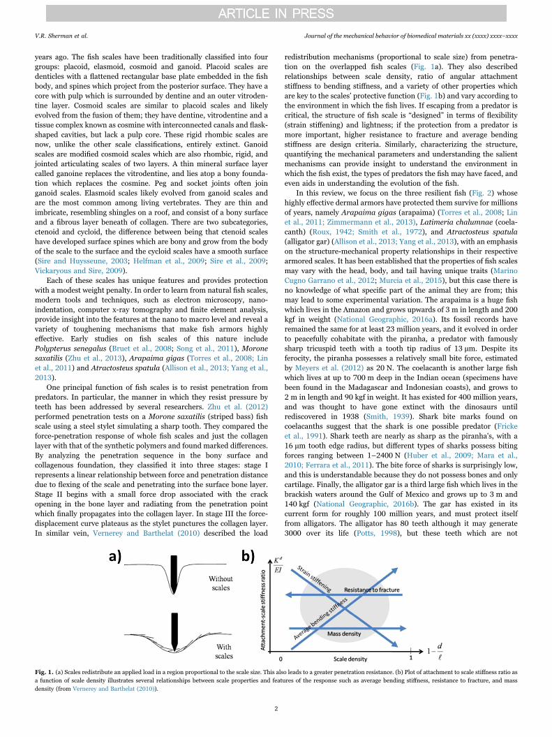
redistribution mechanisms (proportional to scale size) from penetra-tion on the overlapped fish scales (Fig. 1a). They also describedrelationships between scale density, ratio of angular attachmentstiffness to bending stiffness, and a variety of other properties whichare key to the scales’ protective function (Fig. 1b) and vary according tothe environment in which the fish lives. If escaping from a predator iscritical, the structure of fish scale is “designed” in terms of flexibility(strain stiffening) and lightness; if the protection from a predator ismore important, higher resistance to fracture and average bendingstiffness are design criteria. Similarly, characterizing the structure,quantifying the mechanical parameters and understanding the salientmechanisms can provide insight to understand the environment inwhich the fish exist, the types of predators the fish may have faced, andeven aids in understanding the evolution of the fish.
In this review, we focus on the three resilient fish (Fig. 2) whosehighly effective dermal armors have protected them survive for millionsof years, namely Arapaima gigas (arapaima) (Torres et al., 2008; Linet al., 2011; Zimmermann et al., 2013), Latimeria chalumnae (coela-canth) (Roux, 1942; Smith et al., 1972), and Atractosteus spatula(alligator gar) (Allison et al., 2013; Yang et al., 2013), with an emphasison the structure-mechanical property relationships in their respectivearmored scales. It has been established that the properties of fish scalesmay vary with the head, body, and tail having unique traits (MarinoCugno Garrano et al., 2012; Murcia et al., 2015), but this case there isno knowledge of what specific part of the animal they are from; thismay lead to some experimental variation. The arapaima is a huge fishwhich lives in the Amazon and grows upwards of 3 m in length and 200kgf in weight (National Geographic, 2016a). Its fossil records haveremained the same for at least 23 million years, and it evolved in orderto peacefully cohabitate with the piranha, a predator with famouslysharp tricuspid teeth with a tooth tip radius of 13 μm. Despite itsferocity, the piranha possesses a relatively small bite force, estimatedby Meyers et al. (2012) as 20 N. The coelacanth is another large fishwhich lives at up to 700 m deep in the Indian ocean (specimens havebeen found in the Madagascar and Indonesian coasts), and grows to2 m in length and 90 kgf in weight. It has existed for 400 million years,and was thought to have gone extinct with the dinosaurs untilrediscovered in 1938 (Smith, 1939). Shark bite marks found oncoelacanths suggest that the shark is one possible predator (Frickeet al., 1991). Shark teeth are nearly as sharp as the piranha's, with a16 μm tooth edge radius, but different types of sharks possess bitingforces ranging between 1–2400 N (Huber et al., 2009; Mara et al.,2010; Ferrara et al., 2011). The bite force of sharks is surprisingly low,and this is understandable because they do not possess bones and onlycartilage. Finally, the alligator gar is a third large fish which lives in thebrackish waters around the Gulf of Mexico and grows up to 3 m and140 kgf (National Geographic, 2016b). The gar has existed in itscurrent form for roughly 100 million years, and must protect itselffrom alligators. The alligator has 80 teeth although it may generate3000 over its life (Potts, 1998), but these teeth which are not
Fig. 1. (a) Scales redistribute an applied load in a region proportional to the scale size. This also leads to a greater penetration resistance. (b) Plot of attachment to scale stiffness ratio asa function of scale density illustrates several relationships between scale properties and features of the response such as average bending stiffness, resistance to fracture, and massdensity (from Vernerey and Barthelat (2010)).
V.R. Sherman et al. Journal of the mechanical behavior of biomedical materials xx (xxxx) xxxx–xxxx
2
particularly sharp, have a tip radius of the order of ~80–130 μm forjuveniles up to 3 mm for adults. This lack of sharpness, however, iscompensated by powerful jaws capable of bite forces of 10 N to 10 kN,depending on the size (Erickson et al., 2003; Erickson et al., 2004).
2. Arapaima gigas
2.1. Structure of arapaima scales
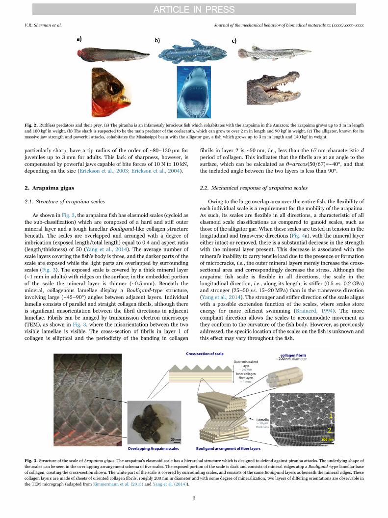
As shown in Fig. 3, the arapaima fish has elasmoid scales (cycloid asthe sub-classification) which are composed of a hard and stiff outermineral layer and a tough lamellar Bouligand-like collagen structurebeneath. The scales are overlapped and arranged with a degree ofimbrication (exposed length/total length) equal to 0.4 and aspect ratio(length/thickness) of 50 (Yang et al., 2014). The average number ofscale layers covering the fish's body is three, and the darker parts of thescale are exposed while the light parts are overlapped by surroundingscales (Fig. 3). The exposed scale is covered by a thick mineral layer(~1 mm in adults) with ridges on the surface; in the embedded portionof the scale the mineral layer is thinner (~0.5 mm). Beneath themineral, collagenous lamellae display a Bouligand-type structure,involving large (~45–90°) angles between adjacent layers. Individuallamella consists of parallel and straight collagen fibrils, although thereis significant misorientation between the fibril directions in adjacentlamellae. Fibrils can be imaged by transmission electron microscopy(TEM), as shown in Fig. 3, where the misorientation between the twovisible lamellae is visible. The cross-section of fibrils in layer 1 ofcollagen is elliptical and the periodicity of the banding in collagen
fibrils in layer 2 is ~50 nm, i.e., less than the 67 nm characteristic dperiod of collagen. This indicates that the fibrils are at an angle to thesurface, which can be calculated as θ=arccos(50/67)=~40°, and thatthe included angle between the two layers is less than 90°.
2.2. Mechanical response of arapaima scales
Owing to the large overlap area over the entire fish, the flexibility ofeach individual scale is a requirement for the mobility of the arapaima.As such, its scales are flexible in all directions, a characteristic of allelasmoid scale classifications as compared to ganoid scales, such asthose of the alligator gar. When these scales are tested in tension in thelongitudinal and transverse directions (Fig. 4a), with the mineral layereither intact or removed, there is a substantial decrease in the strengthwith the mineral layer present. This decrease is associated with themineral's inability to carry tensile load due to the presence or formationof microcracks, i.e., the outer mineral layers merely increase the cross-sectional area and correspondingly decrease the stress. Although thearapaima fish scale is flexible in all directions, the scale in thelongitudinal direction, i.e., along its length, is stiffer (0.5 vs. 0.2 GPa)and stronger (25–50 vs. 15–20 MPa) than in the transverse direction(Yang et al., 2014). The stronger and stiffer direction of the scale alignswith a possible exotendon function of the scales, where scales storeenergy for more efficient swimming (Brainerd, 1994). The morecompliant direction allows the scales to accommodate movement asthey conform to the curvature of the fish body. However, as previouslyaddressed, the specific location of the scales on the fish is unknown andthis effect may vary throughout the fish.
Fig. 2. Ruthless predators and their prey. (a) The piranha is an infamously ferocious fish which cohabitates with the arapaima in the Amazon; the arapaima grows up to 3 m in lengthand 180 kgf in weight. (b) The shark is suspected to be the main predator of the coelacanth, which can grow to over 2 m in length and 90 kgf in weight. (c) The alligator, known for itsmassive jaw strength and powerful attacks, cohabitates the Mississippi basin with the alligator gar, a fish which grows up to 3 m in length and 140 kgf in weight.
Fig. 3. Structure of the scale of Arapaima gigas. The arapaima's elasmoid scale has a hierarchal structure which is designed to defend against piranha attacks. The underlying shape ofthe scales can be seen in the overlapping arrangement schema of five scales. The exposed portion of the scale is dark and consists of mineral ridges atop a Bouligand -type lamellar baseof collagen, creating the cross-section shown. The white part of the scale is covered by surrounding scales, and consists of the same Bouligand layers as beneath the mineral ridges. Thesecollagen layers are made of sheets of oriented collagen fibrils, roughly 200 nm in diameter and with some degree of mineralization; two layers of differing orientations are observable inthe TEM micrograph (adapted from Zimmermann et al. (2013) and Yang et al. (2014)).
V.R. Sherman et al. Journal of the mechanical behavior of biomedical materials xx (xxxx) xxxx–xxxx
3
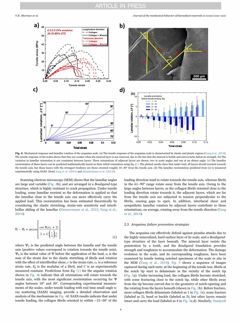
Scanning electron microscopy (SEM) shows that the lamellar anglesare large and variable (Fig. 4b), and are arranged in a Bouligand-typestructure, which is highly resistant to crack propagation. Under tensileloading, some lamellae reorient as the deformation is applied so thatthe lamellae close to the tensile axis can more effectively carry theapplied load. This reorientation has been estimated theoretically byconsidering the elastic stretching, strain-rate sensitivity and interfi-brillar sliding of the lamellae (Zimmermann et al., 2013; Yang et al.,2014):
⎡
⎣
⎢⎢⎢⎢ ⎛⎝⎜
⎞⎠⎟
⎤
⎦
⎥⎥⎥⎥Ψ Ψ Ψ ε
C− = arccos cos ( + 2)
2 +,t
εε
εE
1 00
tf0 (1)
where Ψ1 is the predicted angle between the lamella and the tensileaxis (positive values correspond to rotation towards the tensile axis),Ψ0 is the initial value of Ψ before the application of the load, εt is thesum of the strain due to the elastic stretching of fibrils and rotationwith the effect of interfibrillar shear, ε is the strain rate, ε0 is a referencestrain rate, Ef is the modulus of a fibril, and C is an experimentallymeasured constant. Predictions from Eq. (1) for the angular rotationshown in Fig. 4c indicate that all orientations will rotate towards thetensile axis, with the most significant reorientation occurring for Ψangles between 10° and 30°. Corresponding experimental measure-ments of the scales, under tensile loading with real time small angle x-ray scattering (SAXS) imaging, provide a detailed observation andanalysis of the mechanisms in Fig. 4d. SAXS results indicate that undertensile loading, the collagen fibrils oriented to within ~15–30° of the
loading direction tend to rotate towards the tensile axis, whereas fibrilsin the 61–90° range rotate away from the tensile axis. Owing to thelarge angles between layers, as the collagen fibrils oriented close to theloading direction rotate towards it, the adjacent layers, which are farfrom the tensile axis are subjected to tension perpendicular to thefibrils, causing gaps to open. In addition, interfacial shear andsympathetic lamellar rotation by adjacent layers contribute to theseorientations, on average, rotating away from the tensile direction (Yanget al., 2014).
2.3. Arapaima failure prevention strategies
The arapaima can effectively defend against piranha attacks due tothe highly mineralized, hard surface layer of its scale, and a Bouligand-type structure of the layer beneath. The mineral layer resists thepenetration by a tooth, and the Bouligand foundation providesstrength and toughness to accommodate the deformation. The damageevolution in the scale, and its corresponding toughness, have beenexamined by tensile testing notched specimens of the scale in situ inthe SEM (Yang et al., 2014). Fig. 5 shows a sequence of imagescaptured during such tests: at the beginning of the tensile test, fibrils atthe notch tip start to delaminate in the vicinity of the notch tip(Fig. 5a). Under increasing load, the collagen fibrils become stretched,with some fracturing close to the notch tip, while other fibrils awayfrom the tip become curved due to the geometry of notch-opening andthe rotating from the layers beneath (shown in Fig. 5b). Before fracture,more collagen fibrils delaminate (labeled as 1 in Fig. 5c), some fracture(labeled as 2), bend or buckle (labeled as 3), but other layers remainintact and carry the load (labeled as 4 in Fig. 5c,d). Similarly, Dastjerdi
Fig. 4. Mechanical response and lamellar rotation of the arapaima scale. (a) The tensile response of the arapaima scale is characterized by elastic and plastic regions (Yang et al., 2014).The tensile response of the scales shows that they are weaker when the mineral layer is not removed, due to the fact that the mineral is brittle and microcracks defeat its strength. (b) Thevariation in lamellar orientation is not consistent between layers. Three orientations of adjacent layers are shown; two at acute angles and one at an obtuse angle. (c) The lamellarreorientation of these layers can be predicted mathematically based on their initial orientation using Eq. (1). The plotted results show that under load, all layers should reorient towardsthe tensile axis, but those layers with the strongest tendency are those oriented roughly 10–30° from the tensile axis. (d) The lamellar reorientation predicted from (c) is measuredexperimentally using SAXS. (from Yang et al. (2014) and Zimmermann et al. (2013)).
V.R. Sherman et al. Journal of the mechanical behavior of biomedical materials xx (xxxx) xxxx–xxxx
4
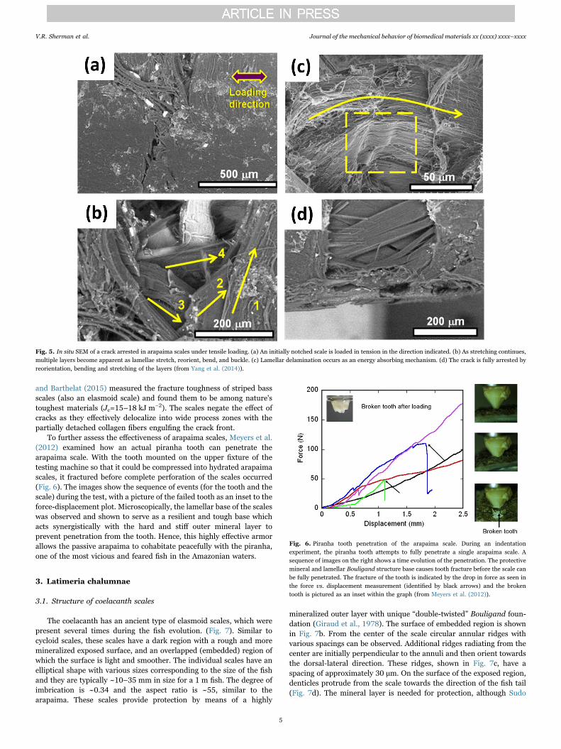
and Barthelat (2015) measured the fracture toughness of striped bassscales (also an elasmoid scale) and found them to be among nature'stoughest materials (Jc=15–18 kJ m−2). The scales negate the effect ofcracks as they effectively delocalize into wide process zones with thepartially detached collagen fibers engulfing the crack front.
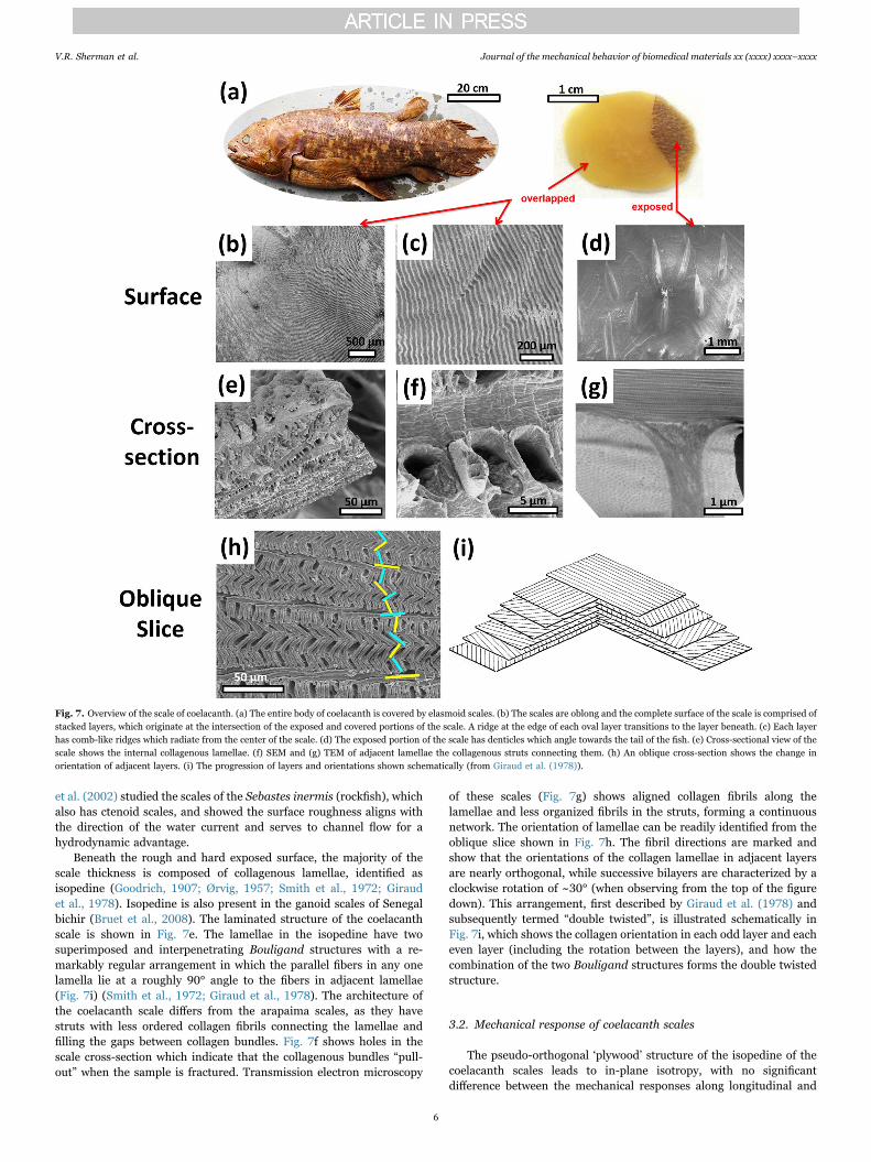
To further assess the effectiveness of arapaima scales, Meyers et al.(2012) examined how an actual piranha tooth can penetrate thearapaima scale. With the tooth mounted on the upper fixture of thetesting machine so that it could be compressed into hydrated arapaimascales, it fractured before complete perforation of the scales occurred(Fig. 6). The images show the sequence of events (for the tooth and thescale) during the test, with a picture of the failed tooth as an inset to theforce-displacement plot. Microscopically, the lamellar base of the scaleswas observed and shown to serve as a resilient and tough base whichacts synergistically with the hard and stiff outer mineral layer toprevent penetration from the tooth. Hence, this highly effective armorallows the passive arapaima to cohabitate peacefully with the piranha,one of the most vicious and feared fish in the Amazonian waters.
3. Latimeria chalumnae
3.1. Structure of coelacanth scales
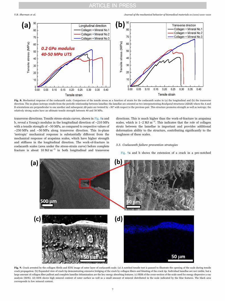
The coelacanth has an ancient type of elasmoid scales, which werepresent several times during the fish evolution. (Fig. 7). Similar tocycloid scales, these scales have a dark region with a rough and moremineralized exposed surface, and an overlapped (embedded) region ofwhich the surface is light and smoother. The individual scales have anelliptical shape with various sizes corresponding to the size of the fishand they are typically ~10–35 mm in size for a 1 m fish. The degree ofimbrication is ~0.34 and the aspect ratio is ~55, similar to thearapaima. These scales provide protection by means of a highly
mineralized outer layer with unique “double-twisted” Bouligand foun-dation (Giraud et al., 1978). The surface of embedded region is shownin Fig. 7b. From the center of the scale circular annular ridges withvarious spacings can be observed. Additional ridges radiating from thecenter are initially perpendicular to the annuli and then orient towardsthe dorsal-lateral direction. These ridges, shown in Fig. 7c, have aspacing of approximately 30 µm. On the surface of the exposed region,denticles protrude from the scale towards the direction of the fish tail(Fig. 7d). The mineral layer is needed for protection, although Sudo
Fig. 5. In situ SEM of a crack arrested in arapaima scales under tensile loading. (a) An initially notched scale is loaded in tension in the direction indicated. (b) As stretching continues,multiple layers become apparent as lamellae stretch, reorient, bend, and buckle. (c) Lamellar delamination occurs as an energy absorbing mechanism. (d) The crack is fully arrested byreorientation, bending and stretching of the layers (from Yang et al. (2014)).
Fig. 6. Piranha tooth penetration of the arapaima scale. During an indentationexperiment, the piranha tooth attempts to fully penetrate a single arapaima scale. Asequence of images on the right shows a time evolution of the penetration. The protectivemineral and lamellar Bouligand structure base causes tooth fracture before the scale canbe fully penetrated. The fracture of the tooth is indicated by the drop in force as seen inthe force vs. displacement measurement (identified by black arrows) and the brokentooth is pictured as an inset within the graph (from Meyers et al. (2012)).
V.R. Sherman et al. Journal of the mechanical behavior of biomedical materials xx (xxxx) xxxx–xxxx
5
et al. (2002) studied the scales of the Sebastes inermis (rockfish), whichalso has ctenoid scales, and showed the surface roughness aligns withthe direction of the water current and serves to channel flow for ahydrodynamic advantage.
Beneath the rough and hard exposed surface, the majority of thescale thickness is composed of collagenous lamellae, identified asisopedine (Goodrich, 1907; Ørvig, 1957; Smith et al., 1972; Giraudet al., 1978). Isopedine is also present in the ganoid scales of Senegalbichir (Bruet et al., 2008). The laminated structure of the coelacanthscale is shown in Fig. 7e. The lamellae in the isopedine have twosuperimposed and interpenetrating Bouligand structures with a re-markably regular arrangement in which the parallel fibers in any onelamella lie at a roughly 90° angle to the fibers in adjacent lamellae(Fig. 7i) (Smith et al., 1972; Giraud et al., 1978). The architecture ofthe coelacanth scale differs from the arapaima scales, as they havestruts with less ordered collagen fibrils connecting the lamellae andfilling the gaps between collagen bundles. Fig. 7f shows holes in thescale cross-section which indicate that the collagenous bundles “pull-out” when the sample is fractured. Transmission electron microscopy
of these scales (Fig. 7g) shows aligned collagen fibrils along thelamellae and less organized fibrils in the struts, forming a continuousnetwork. The orientation of lamellae can be readily identified from theoblique slice shown in Fig. 7h. The fibril directions are marked andshow that the orientations of the collagen lamellae in adjacent layersare nearly orthogonal, while successive bilayers are characterized by aclockwise rotation of ~30° (when observing from the top of the figuredown). This arrangement, first described by Giraud et al. (1978) andsubsequently termed “double twisted”, is illustrated schematically inFig. 7i, which shows the collagen orientation in each odd layer and eacheven layer (including the rotation between the layers), and how thecombination of the two Bouligand structures forms the double twistedstructure.
3.2. Mechanical response of coelacanth scales
The pseudo-orthogonal ‘plywood’ structure of the isopedine of thecoelacanth scales leads to in-plane isotropy, with no significantdifference between the mechanical responses along longitudinal and
Fig. 7. Overview of the scale of coelacanth. (a) The entire body of coelacanth is covered by elasmoid scales. (b) The scales are oblong and the complete surface of the scale is comprised ofstacked layers, which originate at the intersection of the exposed and covered portions of the scale. A ridge at the edge of each oval layer transitions to the layer beneath. (c) Each layerhas comb-like ridges which radiate from the center of the scale. (d) The exposed portion of the scale has denticles which angle towards the tail of the fish. (e) Cross-sectional view of thescale shows the internal collagenous lamellae. (f) SEM and (g) TEM of adjacent lamellae the collagenous struts connecting them. (h) An oblique cross-section shows the change inorientation of adjacent layers. (i) The progression of layers and orientations shown schematically (from Giraud et al. (1978)).
V.R. Sherman et al. Journal of the mechanical behavior of biomedical materials xx (xxxx) xxxx–xxxx
6
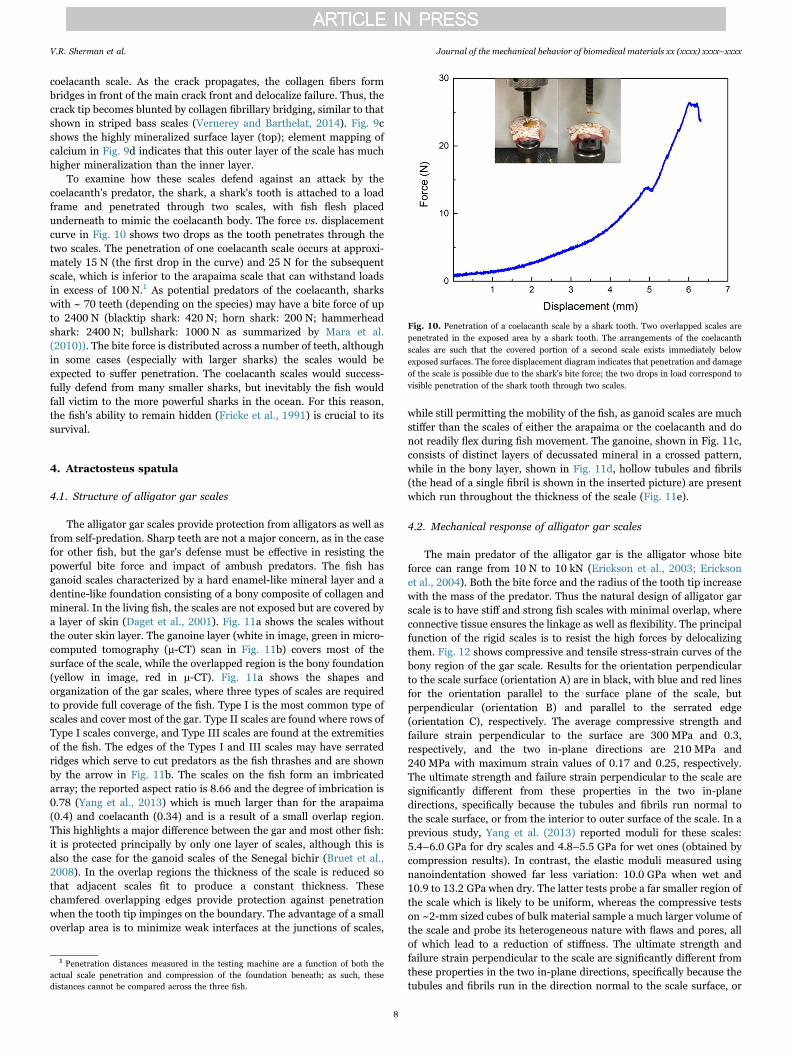
transverse directions. Tensile stress-strain curves, shown in Fig. 8a andb, reveal a Young's modulus in the longitudinal direction of ~210 MPawith a tensile strength of ~50 MPa, as compared to respective values of~250 MPa and ~50 MPa along transverse direction. This in-plane‘isotropic’ mechanical response is substantially different from themechanical response of arapaima scales, which have higher strengthand stiffness in the longitudinal direction. The work-of-fracture incoelacanth scales (area under the stress-strain curve) before completefracture is about 10 MJ m−3 in both longitudinal and transverse
directions. This is much higher than the work-of-fracture in arapaimascales, which is 1–2 MJ m−3. This indicates that the role of collagenstruts between the lamellae is important and provides additionaldeformation ability to the structure, contributing significantly to thetoughness of these scales.
3.3. Coelacanth failure prevention strategies
Fig. 9a and b shows the extension of a crack in a pre-notched
Fig. 8. Mechanical response of the coelacanth scale. Comparison of the tensile stress as a function of strain for the coelacanth scales in (a) the longitudinal and (b) the transversedirection. The in-plane isotropy results from the periodic relationship between lamellae; the lamellae are oriented as two interpenetrating Bouligand structures (ABAB) where the A andB orientations are perpendicular to one another and subsequent AB pairs are twisted by ~30° with respect to the previous pair. This structure promotes strength as well as isotropy; therelatively strong scales have an ultimate tensile strength between 40 and 50 MPa.
Fig. 9. Crack arrested by the collagen fibrils and EDX image of outer layer of coelacanth scale. (a) A notched tensile test is paused to illustrate the opening of the scale during tensilecrack propagation. (b) Expanded view of crack tip demonstrating extensive bridging of the crack by collagen fibers and blunting of the crack tip. Individual lamellae are not visible, but alarge amount of collagen fiber pullout and complete lamellar delamination are the key energy absorbing features. (c) SEM of the cross-section of the scale used for energy dispersive x-rayanalysis (EDX). (d) EDX shows high mineral content of outer surface as well as a small amount of mineral distributed in the scale indicated by the blue features. The black areacorresponds to low mineral content.
V.R. Sherman et al. Journal of the mechanical behavior of biomedical materials xx (xxxx) xxxx–xxxx
7
coelacanth scale. As the crack propagates, the collagen fibers formbridges in front of the main crack front and delocalize failure. Thus, thecrack tip becomes blunted by collagen fibrillary bridging, similar to thatshown in striped bass scales (Vernerey and Barthelat, 2014). Fig. 9cshows the highly mineralized surface layer (top); element mapping ofcalcium in Fig. 9d indicates that this outer layer of the scale has muchhigher mineralization than the inner layer.
To examine how these scales defend against an attack by thecoelacanth's predator, the shark, a shark's tooth is attached to a loadframe and penetrated through two scales, with fish flesh placedunderneath to mimic the coelacanth body. The force vs. displacementcurve in Fig. 10 shows two drops as the tooth penetrates through thetwo scales. The penetration of one coelacanth scale occurs at approxi-mately 15 N (the first drop in the curve) and 25 N for the subsequentscale, which is inferior to the arapaima scale that can withstand loadsin excess of 100 N.1 As potential predators of the coelacanth, sharkswith ~ 70 teeth (depending on the species) may have a bite force of upto 2400 N (blacktip shark: 420 N; horn shark: 200 N; hammerheadshark: 2400 N; bullshark: 1000 N as summarized by Mara et al.(2010)). The bite force is distributed across a number of teeth, althoughin some cases (especially with larger sharks) the scales would beexpected to suffer penetration. The coelacanth scales would success-fully defend from many smaller sharks, but inevitably the fish wouldfall victim to the more powerful sharks in the ocean. For this reason,the fish's ability to remain hidden (Fricke et al., 1991) is crucial to itssurvival.
4. Atractosteus spatula
4.1. Structure of alligator gar scales
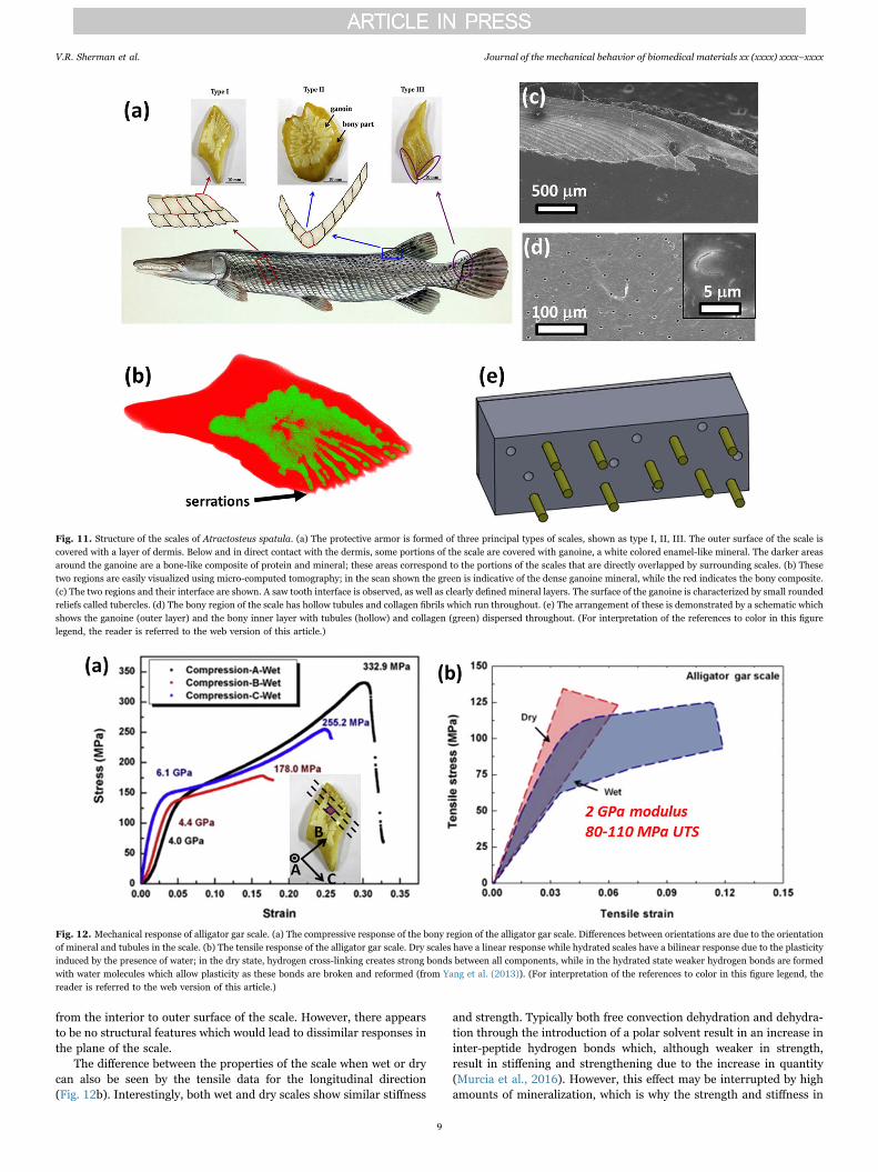
The alligator gar scales provide protection from alligators as well asfrom self-predation. Sharp teeth are not a major concern, as in the casefor other fish, but the gar's defense must be effective in resisting thepowerful bite force and impact of ambush predators. The fish hasganoid scales characterized by a hard enamel-like mineral layer and adentine-like foundation consisting of a bony composite of collagen andmineral. In the living fish, the scales are not exposed but are covered bya layer of skin (Daget et al., 2001). Fig. 11a shows the scales withoutthe outer skin layer. The ganoine layer (white in image, green in micro-computed tomography (μ-CT) scan in Fig. 11b) covers most of thesurface of the scale, while the overlapped region is the bony foundation(yellow in image, red in μ-CT). Fig. 11a shows the shapes andorganization of the gar scales, where three types of scales are requiredto provide full coverage of the fish. Type I is the most common type ofscales and cover most of the gar. Type II scales are found where rows ofType I scales converge, and Type III scales are found at the extremitiesof the fish. The edges of the Types I and III scales may have serratedridges which serve to cut predators as the fish thrashes and are shownby the arrow in Fig. 11b. The scales on the fish form an imbricatedarray; the reported aspect ratio is 8.66 and the degree of imbrication is0.78 (Yang et al., 2013) which is much larger than for the arapaima(0.4) and coelacanth (0.34) and is a result of a small overlap region.This highlights a major difference between the gar and most other fish:it is protected principally by only one layer of scales, although this isalso the case for the ganoid scales of the Senegal bichir (Bruet et al.,2008). In the overlap regions the thickness of the scale is reduced sothat adjacent scales fit to produce a constant thickness. Thesechamfered overlapping edges provide protection against penetrationwhen the tooth tip impinges on the boundary. The advantage of a smalloverlap area is to minimize weak interfaces at the junctions of scales,
while still permitting the mobility of the fish, as ganoid scales are muchstiffer than the scales of either the arapaima or the coelacanth and donot readily flex during fish movement. The ganoine, shown in Fig. 11c,consists of distinct layers of decussated mineral in a crossed pattern,while in the bony layer, shown in Fig. 11d, hollow tubules and fibrils(the head of a single fibril is shown in the inserted picture) are presentwhich run throughout the thickness of the scale (Fig. 11e).
4.2. Mechanical response of alligator gar scales
The main predator of the alligator gar is the alligator whose biteforce can range from 10 N to 10 kN (Erickson et al., 2003; Ericksonet al., 2004). Both the bite force and the radius of the tooth tip increasewith the mass of the predator. Thus the natural design of alligator garscale is to have stiff and strong fish scales with minimal overlap, whereconnective tissue ensures the linkage as well as flexibility. The principalfunction of the rigid scales is to resist the high forces by delocalizingthem. Fig. 12 shows compressive and tensile stress-strain curves of thebony region of the gar scale. Results for the orientation perpendicularto the scale surface (orientation A) are in black, with blue and red linesfor the orientation parallel to the surface plane of the scale, butperpendicular (orientation B) and parallel to the serrated edge(orientation C), respectively. The average compressive strength andfailure strain perpendicular to the surface are 300 MPa and 0.3,respectively, and the two in-plane directions are 210 MPa and240 MPa with maximum strain values of 0.17 and 0.25, respectively.The ultimate strength and failure strain perpendicular to the scale aresignificantly different from these properties in the two in-planedirections, specifically because the tubules and fibrils run normal tothe scale surface, or from the interior to outer surface of the scale. In aprevious study, Yang et al. (2013) reported moduli for these scales:5.4–6.0 GPa for dry scales and 4.8–5.5 GPa for wet ones (obtained bycompression results). In contrast, the elastic moduli measured usingnanoindentation showed far less variation: 10.0 GPa when wet and10.9 to 13.2 GPa when dry. The latter tests probe a far smaller region ofthe scale which is likely to be uniform, whereas the compressive testson ~2-mm sized cubes of bulk material sample a much larger volume ofthe scale and probe its heterogeneous nature with flaws and pores, allof which lead to a reduction of stiffness. The ultimate strength andfailure strain perpendicular to the scale are significantly different fromthese properties in the two in-plane directions, specifically because thetubules and fibrils run in the direction normal to the scale surface, or
Fig. 10. Penetration of a coelacanth scale by a shark tooth. Two overlapped scales arepenetrated in the exposed area by a shark tooth. The arrangements of the coelacanthscales are such that the covered portion of a second scale exists immediately belowexposed surfaces. The force displacement diagram indicates that penetration and damageof the scale is possible due to the shark's bite force; the two drops in load correspond tovisible penetration of the shark tooth through two scales.
1 Penetration distances measured in the testing machine are a function of both theactual scale penetration and compression of the foundation beneath; as such, thesedistances cannot be compared across the three fish.
V.R. Sherman et al. Journal of the mechanical behavior of biomedical materials xx (xxxx) xxxx–xxxx
8
from the interior to outer surface of the scale. However, there appearsto be no structural features which would lead to dissimilar responses inthe plane of the scale.
The difference between the properties of the scale when wet or drycan also be seen by the tensile data for the longitudinal direction(Fig. 12b). Interestingly, both wet and dry scales show similar stiffness
and strength. Typically both free convection dehydration and dehydra-tion through the introduction of a polar solvent result in an increase ininter-peptide hydrogen bonds which, although weaker in strength,result in stiffening and strengthening due to the increase in quantity(Murcia et al., 2016). However, this effect may be interrupted by highamounts of mineralization, which is why the strength and stiffness in
Fig. 11. Structure of the scales of Atractosteus spatula. (a) The protective armor is formed of three principal types of scales, shown as type I, II, III. The outer surface of the scale iscovered with a layer of dermis. Below and in direct contact with the dermis, some portions of the scale are covered with ganoine, a white colored enamel-like mineral. The darker areasaround the ganoine are a bone-like composite of protein and mineral; these areas correspond to the portions of the scales that are directly overlapped by surrounding scales. (b) Thesetwo regions are easily visualized using micro-computed tomography; in the scan shown the green is indicative of the dense ganoine mineral, while the red indicates the bony composite.(c) The two regions and their interface are shown. A saw tooth interface is observed, as well as clearly defined mineral layers. The surface of the ganoine is characterized by small roundedreliefs called tubercles. (d) The bony region of the scale has hollow tubules and collagen fibrils which run throughout. (e) The arrangement of these is demonstrated by a schematic whichshows the ganoine (outer layer) and the bony inner layer with tubules (hollow) and collagen (green) dispersed throughout. (For interpretation of the references to color in this figurelegend, the reader is referred to the web version of this article.)
Fig. 12. Mechanical response of alligator gar scale. (a) The compressive response of the bony region of the alligator gar scale. Differences between orientations are due to the orientationof mineral and tubules in the scale. (b) The tensile response of the alligator gar scale. Dry scales have a linear response while hydrated scales have a bilinear response due to the plasticityinduced by the presence of water; in the dry state, hydrogen cross-linking creates strong bonds between all components, while in the hydrated state weaker hydrogen bonds are formedwith water molecules which allow plasticity as these bonds are broken and reformed (from Yang et al. (2013)). (For interpretation of the references to color in this figure legend, thereader is referred to the web version of this article.)
V.R. Sherman et al. Journal of the mechanical behavior of biomedical materials xx (xxxx) xxxx–xxxx
9
the highly mineralized alligator gar scale are similar when wet andwhen dry. The critical difference is the post-yield behavior in the wetscales which display extensive plasticity, due to the breaking andreforming of hydrogen bonds between water molecules and collagenfibrils (Maciel et al., 1996). Such plasticity is clearly important to themechanical properties, especially the toughness of the gar scale. Intension, the dry scales do not exhibit significant plasticity. However,the compressive tests show a bi-linear response. This may be associatedwith a much greater difficulty in opening cracks in compression.Fig. 12b shows the curves in tension, with the absence of plasticity.Dry and wet fish scales are toughened by differing mechanisms whichare addressed in the following section.
4.3. Alligator gar failure prevention strategies
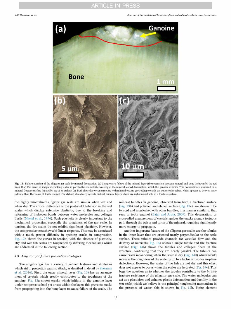
The alligator gar has a variety of refined features and strategieswhich aid in protection against attack, as desribed in detail by Shermanet al. (2016). First, the outer mineral layer (Fig. 13) has an arrange-ment of crystals which greatly contributes to the toughness of theganoine. Fig. 13a shows cracks which initiate in the ganoine layerunder compressive load yet arrest within the layer; this prevents cracksfrom propagating into the bony layer to cause failure of the scale. The
mineral bundles in ganoine, observed from both a fractured surface(Fig. 13b) and polished and etched surface (Fig. 13c), are shown to betwisted and interlocked with other bundles, in a manner similar to thatseen in tooth enamel (Bajaj and Arola, 2009). This decussation, orcross-plied arrangement of crystals, guides the cracks along a tortuouspath through the twists and turns of the mineral, requiring significantlymore energy to propagate.
Another important feature of the alligator gar scales are the tubulesin the inner layer that are oriented nearly perpendicular to the scalesurface. These tubules provide channels for vascular flow and thedelivery of nutrients. Fig. 14a shows a single tubule and the fracturesurface (Fig. 14b) shows the tubules and collagen fibers in thestructure, confirming that they are nearly parallel. The tubules cancause crack meandering when the scale is dry (Fig. 14d) which wouldincrease the toughness of the scale by up to a factor of two for in-planedeflections. However, the scales of the fish are not dry and this effectdoes not appear to occur when the scales are hydrated (Fig. 14c). Thisbegs the question as to whether the tubules contribute to the in vivofracture resistance of the alligator gar scale. The water molecules canact as a plasticizer and enhance plastic deformation and ductility in thewet scale, which we believe is the principal toughening mechanism inthe presence of water; this is shown in Fig. 12b. Finite element
Fig. 13. Failure aversion of the alligator gar scale by mineral decussation. (a) Compressive failure of the mineral layer (the separation between mineral and bone is shown by the redline). (b,c) The arrest of incipient cracking is due in part to the enamel-like weaving of the mineral, called decussation, which the ganoine exhibits. This decussation is observed on amineral fracture surface (b) and by use of an etchant (c). Both show the woven structure with mineral texture protruding towards the outer scale surface, which appears to be even moreextreme than the weave of tooth enamel. The etchant also clearly reveals distinct mineral layers which are indistinguishable in a fracture surface.
V.R. Sherman et al. Journal of the mechanical behavior of biomedical materials xx (xxxx) xxxx–xxxx
10
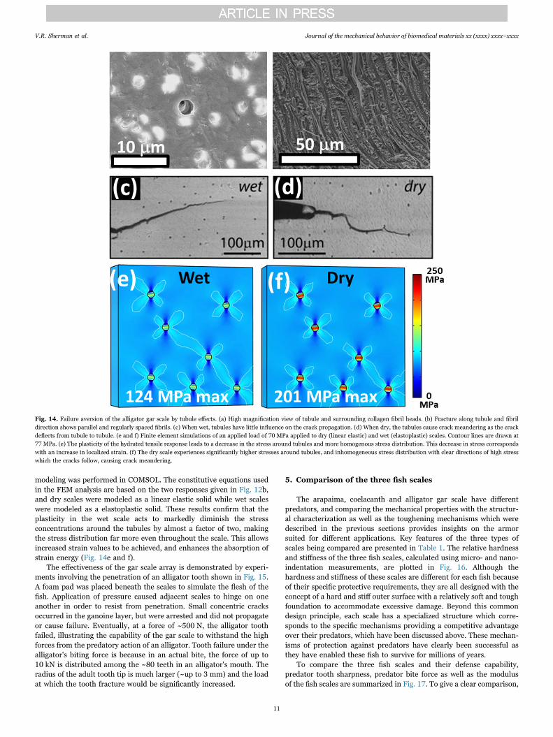
modeling was performed in COMSOL. The constitutive equations usedin the FEM analysis are based on the two responses given in Fig. 12b,and dry scales were modeled as a linear elastic solid while wet scaleswere modeled as a elastoplastic solid. These results confirm that theplasticity in the wet scale acts to markedly diminish the stressconcentrations around the tubules by almost a factor of two, makingthe stress distribution far more even throughout the scale. This allowsincreased strain values to be achieved, and enhances the absorption ofstrain energy (Fig. 14e and f).
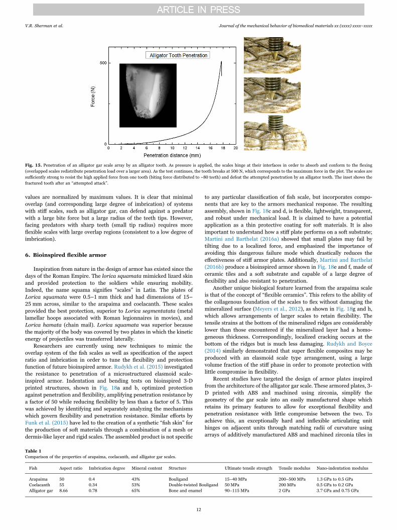
The effectiveness of the gar scale array is demonstrated by experi-ments involving the penetration of an alligator tooth shown in Fig. 15.A foam pad was placed beneath the scales to simulate the flesh of thefish. Application of pressure caused adjacent scales to hinge on oneanother in order to resist from penetration. Small concentric cracksoccurred in the ganoine layer, but were arrested and did not propagateor cause failure. Eventually, at a force of ~500 N, the alligator toothfailed, illustrating the capability of the gar scale to withstand the highforces from the predatory action of an alligator. Tooth failure under thealligator's biting force is because in an actual bite, the force of up to10 kN is distributed among the ~80 teeth in an alligator's mouth. Theradius of the adult tooth tip is much larger (~up to 3 mm) and the loadat which the tooth fracture would be significantly increased.
5. Comparison of the three fish scales
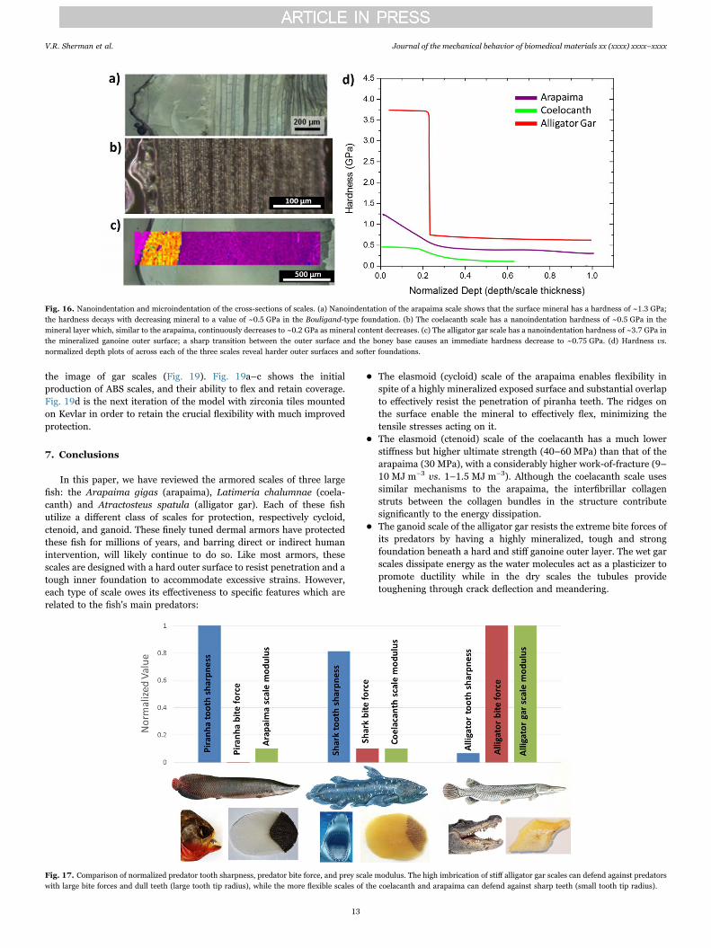
The arapaima, coelacanth and alligator gar scale have differentpredators, and comparing the mechanical properties with the structur-al characterization as well as the toughening mechanisms which weredescribed in the previous sections provides insights on the armorsuited for different applications. Key features of the three types ofscales being compared are presented in Table 1. The relative hardnessand stiffness of the three fish scales, calculated using micro- and nano-indentation measurements, are plotted in Fig. 16. Although thehardness and stiffness of these scales are different for each fish becauseof their specific protective requirements, they are all designed with theconcept of a hard and stiff outer surface with a relatively soft and toughfoundation to accommodate excessive damage. Beyond this commondesign principle, each scale has a specialized structure which corre-sponds to the specific mechanisms providing a competitive advantageover their predators, which have been discussed above. These mechan-isms of protection against predators have clearly been successful asthey have enabled these fish to survive for millions of years.
To compare the three fish scales and their defense capability,predator tooth sharpness, predator bite force as well as the modulusof the fish scales are summarized in Fig. 17. To give a clear comparison,
Fig. 14. Failure aversion of the alligator gar scale by tubule effects. (a) High magnification view of tubule and surrounding collagen fibril heads. (b) Fracture along tubule and fibrildirection shows parallel and regularly spaced fibrils. (c) When wet, tubules have little influence on the crack propagation. (d) When dry, the tubules cause crack meandering as the crackdeflects from tubule to tubule. (e and f) Finite element simulations of an applied load of 70 MPa applied to dry (linear elastic) and wet (elastoplastic) scales. Contour lines are drawn at77 MPa. (e) The plasticity of the hydrated tensile response leads to a decrease in the stress around tubules and more homogenous stress distribution. This decrease in stress correspondswith an increase in localized strain. (f) The dry scale experiences significantly higher stresses around tubules, and inhomogeneous stress distribution with clear directions of high stresswhich the cracks follow, causing crack meandering.
V.R. Sherman et al. Journal of the mechanical behavior of biomedical materials xx (xxxx) xxxx–xxxx
11
values are normalized by maximum values. It is clear that minimaloverlap (and corresponding large degree of imbrication) of systemswith stiff scales, such as alligator gar, can defend against a predatorwith a large bite force but a large radius of the teeth tips. However,facing predators with sharp teeth (small tip radius) requires moreflexible scales with large overlap regions (consistent to a low degree ofimbrication).
6. Bioinspired flexible armor
Inspiration from nature in the design of armor has existed since thedays of the Roman Empire. The lorica squamata mimicked lizard skinand provided protection to the soldiers while ensuring mobility.Indeed, the name squama signifies “scales” in Latin. The plates ofLorica squamata were 0.5–1 mm thick and had dimensions of 15–25 mm across, similar to the arapaima and coelacanth. These scalesprovided the best protection, superior to Lorica segmentatata (metallamellar hoops associated with Roman legionnaires in movies), andLorica hamata (chain mail). Lorica squamata was superior becausethe majority of the body was covered by two plates in which the kineticenergy of projectiles was transferred laterally.
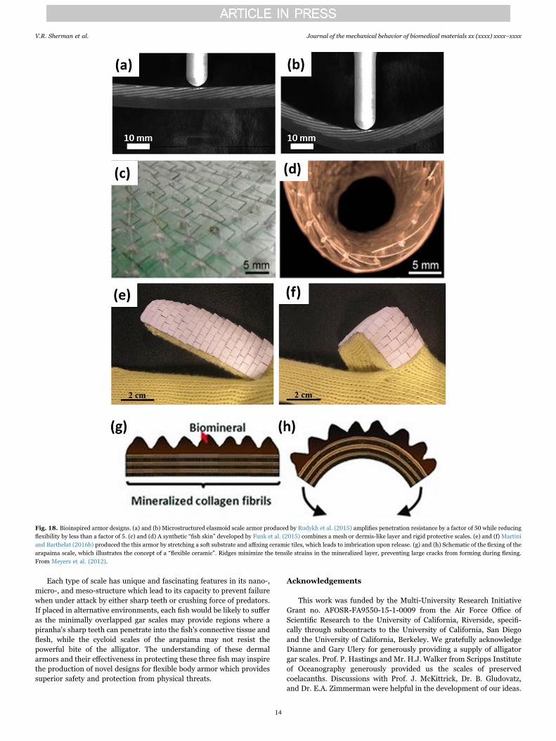
Researchers are currently using new techniques to mimic theoverlap system of the fish scales as well as specification of the aspectratio and imbrication in order to tune the flexibility and protectionfunction of future bioinspired armor. Rudykh et al. (2015) investigatedthe resistance to penetration of a microstructured elasmoid scale-inspired armor. Indentation and bending tests on bioinspired 3-Dprinted structures, shown in Fig. 18a and b, optimized protectionagainst penetration and flexibility, amplifying penetration resistance bya factor of 50 while reducing flexibility by less than a factor of 5. Thiswas achieved by identifying and separately analyzing the mechanismswhich govern flexibility and penetration resistance. Similar efforts byFunk et al. (2015) have led to the creation of a synthetic “fish skin” forthe production of soft materials through a combination of a mesh ordermis-like layer and rigid scales. The assembled product is not specific
to any particular classification of fish scale, but incorporates compo-nents that are key to the armors mechanical response. The resultingassembly, shown in Fig. 18c and d, is flexible, lightweight, transparent,and robust under mechanical load. It is claimed to have a potentialapplication as a thin protective coating for soft materials. It is alsoimportant to understand how a stiff plate performs on a soft substrate;Martini and Barthelat (2016a) showed that small plates may fail bytilting due to a localized force, and emphasized the importance ofavoiding this dangerous failure mode which drastically reduces theeffectiveness of stiff armor plates. Additionally, Martini and Barthelat(2016b) produce a bioinspired armor shown in Fig. 18e and f, made ofceramic tiles and a soft substrate and capable of a large degree offlexibility and also resistant to penetration.
Another unique biological feature learned from the arapaima scaleis that of the concept of “flexible ceramics”. This refers to the ability ofthe collagenous foundation of the scales to flex without damaging themineralized surface (Meyers et al., 2012), as shown in Fig. 18g and h,which allows arrangements of larger scales to retain flexibility. Thetensile strains at the bottom of the mineralized ridges are considerablylower than those encountered if the mineralized layer had a homo-geneous thickness. Correspondingly, localized cracking occurs at thebottom of the ridges but is much less damaging. Rudykh and Boyce(2014) similarly demonstrated that super flexible composites may beproduced with an elasmoid scale type arrangement, using a largevolume fraction of the stiff phase in order to promote protection withlittle compromise in flexibility.
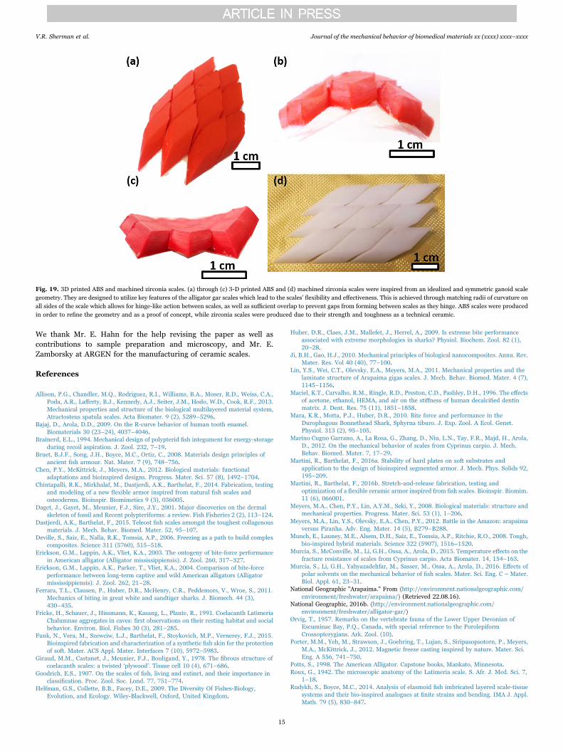
Recent studies have targeted the design of armor plates inspiredfrom the architecture of the alligator gar scale. These armored plates, 3-D printed with ABS and machined using zirconia, simplify thegeometry of the gar scale into an easily manufactured shape whichretains its primary features to allow for exceptional flexibility andpenetration resistance with little compromise between the two. Toachieve this, an exceptionally hard and inflexible articulating unithinges on adjacent units through matching radii of curvature usingarrays of additively manufactured ABS and machined zirconia tiles in
Fig. 15. Penetration of an alligator gar scale array by an alligator tooth. As pressure is applied, the scales hinge at their interfaces in order to absorb and conform to the flexing(overlapped scales redistribute penetration load over a larger area). As the test continues, the tooth breaks at 500 N, which corresponds to the maximum force in the plot. The scales aresufficiently strong to resist the high applied force from one tooth (biting force distributed to ~80 teeth) and defeat the attempted penetration by an alligator tooth. The inset shows thefractured tooth after an “attempted attack”.
Table 1Comparison of the properties of arapaima, coelacanth, and alligator gar scales.
Fish Aspect ratio Imbrication degree Mineral content Structure Ultimate tensile strength Tensile modulus Nano-indentation modulus
Arapaima 50 0.4 43% Bouligand 15–40 MPa 200–500 MPa 1.3 GPa to 0.5 GPaCoelacanth 55 0.34 53% Double-twisted Bouligand 50 MPa 200 MPa 0.5 GPa to 0.2 GPaAlligator gar 8.66 0.78 65% Bone and enamel 90–115 MPa 2 GPa 3.7 GPa and 0.75 GPa
V.R. Sherman et al. Journal of the mechanical behavior of biomedical materials xx (xxxx) xxxx–xxxx
12
the image of gar scales (Fig. 19). Fig. 19a–c shows the initialproduction of ABS scales, and their ability to flex and retain coverage.Fig. 19d is the next iteration of the model with zirconia tiles mountedon Kevlar in order to retain the crucial flexibility with much improvedprotection.
7. Conclusions
In this paper, we have reviewed the armored scales of three largefish: the Arapaima gigas (arapaima), Latimeria chalumnae (coela-canth) and Atractosteus spatula (alligator gar). Each of these fishutilize a different class of scales for protection, respectively cycloid,ctenoid, and ganoid. These finely tuned dermal armors have protectedthese fish for millions of years, and barring direct or indirect humanintervention, will likely continue to do so. Like most armors, thesescales are designed with a hard outer surface to resist penetration and atough inner foundation to accommodate excessive strains. However,each type of scale owes its effectiveness to specific features which arerelated to the fish's main predators:
• The elasmoid (cycloid) scale of the arapaima enables flexibility inspite of a highly mineralized exposed surface and substantial overlapto effectively resist the penetration of piranha teeth. The ridges onthe surface enable the mineral to effectively flex, minimizing thetensile stresses acting on it.
• The elasmoid (ctenoid) scale of the coelacanth has a much lowerstiffness but higher ultimate strength (40–60 MPa) than that of thearapaima (30 MPa), with a considerably higher work-of-fracture (9–10 MJ m−3 vs. 1–1.5 MJ m−3). Although the coelacanth scale usessimilar mechanisms to the arapaima, the interfibrillar collagenstruts between the collagen bundles in the structure contributesignificantly to the energy dissipation.
• The ganoid scale of the alligator gar resists the extreme bite forces ofits predators by having a highly mineralized, tough and strongfoundation beneath a hard and stiff ganoine outer layer. The wet garscales dissipate energy as the water molecules act as a plasticizer topromote ductility while in the dry scales the tubules providetoughening through crack deflection and meandering.
Fig. 16. Nanoindentation and microindentation of the cross-sections of scales. (a) Nanoindentation of the arapaima scale shows that the surface mineral has a hardness of ~1.3 GPa;the hardness decays with decreasing mineral to a value of ~0.5 GPa in the Bouligand-type foundation. (b) The coelacanth scale has a nanoindentation hardness of ~0.5 GPa in themineral layer which, similar to the arapaima, continuously decreases to ~0.2 GPa as mineral content decreases. (c) The alligator gar scale has a nanoindentation hardness of ~3.7 GPa inthe mineralized ganoine outer surface; a sharp transition between the outer surface and the boney base causes an immediate hardness decrease to ~0.75 GPa. (d) Hardness vs.normalized depth plots of across each of the three scales reveal harder outer surfaces and softer foundations.
Fig. 17. Comparison of normalized predator tooth sharpness, predator bite force, and prey scale modulus. The high imbrication of stiff alligator gar scales can defend against predatorswith large bite forces and dull teeth (large tooth tip radius), while the more flexible scales of the coelacanth and arapaima can defend against sharp teeth (small tooth tip radius).
V.R. Sherman et al. Journal of the mechanical behavior of biomedical materials xx (xxxx) xxxx–xxxx
13
Each type of scale has unique and fascinating features in its nano-,micro-, and meso-structure which lead to its capacity to prevent failurewhen under attack by either sharp teeth or crushing force of predators.If placed in alternative environments, each fish would be likely to sufferas the minimally overlapped gar scales may provide regions where apiranha's sharp teeth can penetrate into the fish's connective tissue andflesh, while the cycloid scales of the arapaima may not resist thepowerful bite of the alligator. The understanding of these dermalarmors and their effectiveness in protecting these three fish may inspirethe production of novel designs for flexible body armor which providessuperior safety and protection from physical threats.
Acknowledgements
This work was funded by the Multi-University Research InitiativeGrant no. AFOSR-FA9550-15-1-0009 from the Air Force Office ofScientific Research to the University of California, Riverside, specifi-cally through subcontracts to the University of California, San Diegoand the University of California, Berkeley. We gratefully acknowledgeDianne and Gary Ulery for generously providing a supply of alligatorgar scales. Prof. P. Hastings and Mr. H.J. Walker from Scripps Instituteof Oceanography generously provided us the scales of preservedcoelacanths. Discussions with Prof. J. McKittrick, Dr. B. Gludovatz,and Dr. E.A. Zimmerman were helpful in the development of our ideas.
Fig. 18. Bioinspired armor designs. (a) and (b) Microstructured elasmoid scale armor produced by Rudykh et al. (2015) amplifies penetration resistance by a factor of 50 while reducingflexibility by less than a factor of 5. (c) and (d) A synthetic “fish skin” developed by Funk et al. (2015) combines a mesh or dermis-like layer and rigid protective scales. (e) and (f) Martiniand Barthelat (2016b) produced the this armor by stretching a soft substrate and affixing ceramic tiles, which leads to imbrication upon release. (g) and (h) Schematic of the flexing of thearapaima scale, which illustrates the concept of a “flexible ceramic”. Ridges minimize the tensile strains in the mineralized layer, preventing large cracks from forming during flexing.From Meyers et al. (2012).
V.R. Sherman et al. Journal of the mechanical behavior of biomedical materials xx (xxxx) xxxx–xxxx
14
We thank Mr. E. Hahn for the help revising the paper as well ascontributions to sample preparation and microscopy, and Mr. E.Zamborsky at ARGEN for the manufacturing of ceramic scales.
References
Allison, P.G., Chandler, M.Q., Rodriguez, R.I., Williams, B.A., Moser, R.D., Weiss, C.A.,Poda, A.R., Lafferty, B.J., Kennedy, A.J., Seiter, J.M., Hodo, W.D., Cook, R.F., 2013.Mechanical properties and structure of the biological multilayered material system,Atractosteus spatula scales. Acta Biomater. 9 (2), 5289–5296.
Bajaj, D., Arola, D.D., 2009. On the R-curve behavior of human tooth enamel.Biomaterials 30 (23–24), 4037–4046.
Brainerd, E.L., 1994. Mechanical design of polypterid fish integument for energy-storageduring recoil aspiration. J. Zool. 232, 7–19.
Bruet, B.J.F., Song, J.H., Boyce, M.C., Ortiz, C., 2008. Materials design principles ofancient fish armour. Nat. Mater. 7 (9), 748–756.
Chen, P.Y., McKittrick, J., Meyers, M.A., 2012. Biological materials: functionaladaptations and bioinspired designs. Progress. Mater. Sci. 57 (8), 1492–1704.
Chintapalli, R.K., Mirkhalaf, M., Dastjerdi, A.K., Barthelat, F., 2014. Fabrication, testingand modeling of a new flexible armor inspired from natural fish scales andosteoderms. Bioinspir. Biomimetics 9 (3), 036005.
Daget, J., Gayet, M., Meunier, F.J., Sire, J.Y., 2001. Major discoveries on the dermalskeleton of fossil and Recent polypteriforms: a review. Fish Fisheries 2 (2), 113–124.
Dastjerdi, A.K., Barthelat, F., 2015. Teleost fish scales amongst the toughest collagenousmaterials. J. Mech. Behav. Biomed. Mater. 52, 95–107.
Deville, S., Saiz, E., Nalla, R.K., Tomsia, A.P., 2006. Freezing as a path to build complexcomposites. Science 311 (5760), 515–518.
Erickson, G.M., Lappin, A.K., Vliet, K.A., 2003. The ontogeny of bite-force performancein American alligator (Alligator mississippiensis). J. Zool. 260, 317–327.
Erickson, G.M., Lappin, A.K., Parker, T., Vliet, K.A., 2004. Comparison of bite-forceperformance between long-term captive and wild American alligators (Alligatormississippiensis). J. Zool. 262, 21–28.
Ferrara, T.L., Clausen, P., Huber, D.R., McHenry, C.R., Peddemors, V., Wroe, S., 2011.Mechanics of biting in great white and sandtiger sharks. J. Biomech. 44 (3),430–435.
Fricke, H., Schauer, J., Hissmann, K., Kasang, L., Plante, R., 1991. Coelacanth LatimeriaChalumnae aggregates in caves: first observations on their resting habitat and socialbehavior. Environ. Biol. Fishes 30 (3), 281–285.
Funk, N., Vera, M., Szewciw, L.J., Barthelat, F., Stoykovich, M.P., Vernerey, F.J., 2015.Bioinspired fabrication and characterization of a synthetic fish skin for the protectionof soft. Mater. ACS Appl. Mater. Interfaces 7 (10), 5972–5983.
Giraud, M.M., Castanet, J., Meunier, F.J., Bouligand, Y., 1978. The fibrous structure ofcoelacanth scales: a twisted ‘plywood’. Tissue cell 10 (4), 671–686.
Goodrich, E.S., 1907. On the scales of fish, living and extinct, and their importance inclassification. Proc. Zool. Soc. Lond. 77, 751–774.
Helfman, G.S., Collette, B.B., Facey, D.E., 2009. The Diversity Of Fishes-Biology,Evolution, and Ecology. Wiley-Blackwell, Oxford, United Kingdom.
Huber, D.R., Claes, J.M., Mallefet, J., Herrel, A., 2009. Is extreme bite performanceassociated with extreme morphologies in sharks? Physiol. Biochem. Zool. 82 (1),20–28.
Ji, B.H., Gao, H.J., 2010. Mechanical principles of biological nanocomposites. Annu. Rev.Mater. Res. Vol 40 (40), 77–100.
Lin, Y.S., Wei, C.T., Olevsky, E.A., Meyers, M.A., 2011. Mechanical properties and thelaminate structure of Arapaima gigas scales. J. Mech. Behav. Biomed. Mater. 4 (7),1145–1156.
Maciel, K.T., Carvalho, R.M., Ringle, R.D., Preston, C.D., Pashley, D.H., 1996. The effectsof acetone, ethanol, HEMA, and air on the stiffness of human decalcified dentinmatrix. J. Dent. Res. 75 (11), 1851–1858.
Mara, K.R., Motta, P.J., Huber, D.R., 2010. Bite force and performance in theDurophagous Bonnethead Shark, Sphyrna tiburo. J. Exp. Zool. A Ecol. Genet.Physiol. 313 (2), 95–105.
Marino Cugno Garrano, A., La Rosa, G., Zhang, D., Niu, L.N., Tay, F.R., Majd, H., Arola,D., 2012. On the mechanical behavior of scales from Cyprinus carpio. J. Mech.Behav. Biomed. Mater. 7, 17–29.
Martini, R., Barthelat, F., 2016a. Stability of hard plates on soft substrates andapplication to the design of bioinspired segmented armor. J. Mech. Phys. Solids 92,195–209.
Martini, R., Barthelat, F., 2016b. Stretch-and-release fabrication, testing andoptimization of a flexible ceramic armor inspired from fish scales. Bioinspir. Biomim.11 (6), 066001.
Meyers, M.A., Chen, P.Y., Lin, A.Y.M., Seki, Y., 2008. Biological materials: structure andmechanical properties. Progress. Mater. Sci. 53 (1), 1–206.
Meyers, M.A., Lin, Y.S., Olevsky, E.A., Chen, P.Y., 2012. Battle in the Amazon: arapaimaversus Piranha. Adv. Eng. Mater. 14 (5), B279–B288.
Munch, E., Launey, M.E., Alsem, D.H., Saiz, E., Tomsia, A.P., Ritchie, R.O., 2008. Tough,bio-inspired hybrid materials. Science 322 (5907), 1516–1520.
Murcia, S., McConville, M., Li, G.H., Ossa, A., Arola, D., 2015. Temperature effects on thefracture resistance of scales from Cyprinus carpio. Acta Biomater. 14, 154–163.
Murcia, S., Li, G.H., Yahyazadehfar, M., Sasser, M., Ossa, A., Arola, D., 2016. Effects ofpolar solvents on the mechanical behavior of fish scales. Mater. Sci. Eng. C – Mater.Biol. Appl. 61, 23–31.
National Geographic "Arapaima." From ⟨http://environment.nationalgeographic.com/environment/freshwater/arapaima/⟩ (Retrieved 22.08.16).
National Geographic, 2016b. ⟨http://environment.nationalgeographic.com/environment/freshwater/alligator-gar/⟩
Ørvig, T., 1957. Remarks on the vertebrate fauna of the Lower Upper Devonian ofEscuminac Bay, P.Q., Canada, with special reference to the PorolepiformCrossopterygians. Ark. Zool. (10).
Porter, M.M., Yeh, M., Strawson, J., Goehring, T., Lujan, S., Siripasopsotorn, P., Meyers,M.A., McKittrick, J., 2012. Magnetic freeze casting inspired by nature. Mater. Sci.Eng. A 556, 741–750.
Potts, S., 1998. The American Alligator. Capstone books, Mankato, Minnesota.Roux, G., 1942. The microscopic anatomy of the Latimeria scale. S. Afr. J. Med. Sci. 7,
1–18.Rudykh, S., Boyce, M.C., 2014. Analysis of elasmoid fish imbricated layered scale-tissue
systems and their bio-inspired analogues at finite strains and bending. IMA J. Appl.Math. 79 (5), 830–847.
Fig. 19. 3D printed ABS and machined zirconia scales. (a) through (c) 3-D printed ABS and (d) machined zirconia scales were inspired from an idealized and symmetric ganoid scalegeometry. They are designed to utilize key features of the alligator gar scales which lead to the scales’ flexibility and effectiveness. This is achieved through matching radii of curvature onall sides of the scale which allows for hinge-like action between scales, as well as sufficient overlap to prevent gaps from forming between scales as they hinge. ABS scales were producedin order to refine the geometry and as a proof of concept, while zirconia scales were produced due to their strength and toughness as a technical ceramic.
V.R. Sherman et al. Journal of the mechanical behavior of biomedical materials xx (xxxx) xxxx–xxxx
15
Rudykh, S., Ortiz, C., Boyce, M.C., 2015. Flexibility and protection by design: imbricatedhybrid microstructures of bio-inspired armor. Soft Matter 11 (13), 2547–2554.
Sacks, M.S., Sun, W., 2003. Multiaxial mechanical behavior of biological materials. Annu.Rev. Biomed. Eng. 5, 251–284.
Sherman, V.R., Yaraghi, N.A., Kisailus, D., Meyers, M.A., 2016. Microstructural andgeometric influences in the protective scales of Atractosteus spatula. J. R. Soc.Interface, In Press.
Sire, J.Y., Huysseune, A., 2003. Formation of dermal skeletal and dental tissues in fish: acomparative and evolutionary approach. Biol. Rev. 78 (2), 219–249.
Sire, J.Y., Donoghue, P.C.J., Vickaryous, M.K., 2009. Origin and evolution of theintegumentary skeleton in non-tetrapod vertebrates. J. Anat. 214 (4), 409–440.
Smith, J.L.B., 1939. A living fish of Mesozoic type. Nature 143, 455–456.Smith, M.M., Miller, W.A., Hobdell, M.H., 1972. Structure of scales of Latimeria-
Chalumnae. J. Zool. 167 (Aug), 501–509.Song, J., Ortiz, C., Boyce, M.C., 2011. Threat-protection mechanics of an armored fish. J.
Mech. Behav. Biomed. Mater. 4 (5), 699–712.Sudo, S., Tsuyuki, K., Ito, Y., Ikohagi, T., 2002. A study on the surface shape of fish scales.
JSME Int. J. Ser. C – Mech. Syst. Mach. Elem. Manuf. 45 (4), 1100–1105.Torres, F.G., Troncoso, O.P., Nakamatsu, J., Grande, C.J., Gomez, C.M., 2008.
Characterization of the nanocomposite laminate structure occurring in fish scalesfrom Arapaima Gigas. Mater. Sci. Eng. C 28 (8), 1276–1283.
Vernerey, F.J., Barthelat, F., 2010. On the mechanics of fishscale structures. Int. J SolidsStruct. 47 (17), 2268–2275.
Vernerey, F.J., Barthelat, F., 2014. Skin and scales of teleost fish: Simple structure buthigh performance and multiple functions. J. Mech. Phys. Solids 68, 66–76.
Vickaryous, M.K., Sire, J.Y., 2009. The integumentary skeleton of tetrapods: origin,evolution, and development. J. Anat. 214 (4), 441–464.
Wegst, U.G.K., Bai, H., Saiz, E., Tomsia, A.P., Ritchie, R.O., 2015. Bioinspired structuralmaterials. Nat. Mater. 14 (1), 23–36.
Yang, W., Gludovatz, B., Zimmermann, E.A., Bale, H.A., Ritchie, R.O., Meyers, M.A.,2013. Structure and fracture resistance of alligator gar (Atractosteus spatula)armored fish scales. Acta Biomater. 9 (4), 5876–5889.
Yang, W., Sherman, V.R., Gludovatz, B., Mackey, M., Zimmermann, E.A., Chang, E.H.,Schaible, E., Qin, Z., Buehler, M.J., Ritchie, R.O., Meyers, M.A., 2014. Protective roleof Arapaima gigas fish scales: structure and mechanical behavior. Acta Biomater. 10(8), 3599–3614.
Zhu, D.J., Szewciw, L., Vernerey, F., Barthelat, F., 2013. Puncture resistance of the scaledskin from striped bass: collective mechanisms and inspiration for new flexible armordesigns. J. Mech. Behav. Biomed. Mater. 24, 30–40.
Zhu, D.J., Ortega, C.F., Motamedi, R., Szewciw, L., Vernerey, F., Barthelat, F., 2012.Structure and mechanical performance of a “Modern” Fish Scale. Adv. Eng. Mater.14 (4), B185–B194.
Zimmermann, E.A., Gludovatz, B., Schaible, E., Dave, N.K.N., Yang, W., Meyers, M.A.,Ritchie, R.O., 2013. Mechanical adaptability of the Bouligand-type structure innatural dermal armour. Nat. Commun. 4, 2634.
V.R. Sherman et al. Journal of the mechanical behavior of biomedical materials xx (xxxx) xxxx–xxxx
16